User login
Maintenance rituximab extends progression-free but not overall survival in CLL
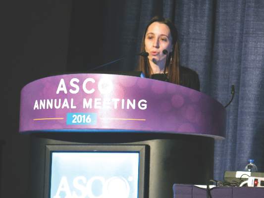
CHICAGO – After 2 years of maintenance immunotherapy with rituximab, elderly patients with chronic lymphocytic leukemia had better rates of progression-free survival than did patients in an observation group, based on results of the CLL 2007 SA trial from the French FILO (French Innovative Leukemia Organisation) Group.
The two groups did not significantly differ in overall survival, however, with 92.6% estimated 3-year overall survival in the rituximab group and 87.2% in the observation group. Further, the patients given rituximab had more adverse events, based on data presented by Dr. Caroline Dartigeas of the University Hospital in Tours, France, at the annual meeting of the American Society of Clinical Oncology.
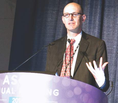
Given the cost and risk for events with rituximab, the findings raise the question of whether there are any meaningful benefits for maintenance rituximab after induction therapy, Dr. Jonathan W. Friedberg of the University of Rochester, N.Y., who was the discussant of the paper, remarked after the presentation. He asked whether there is any evidence that patients feel better if they’re in remission and, thus, their quality of life is improved.
The study included fit, treatment-naive patients aged 65 years and older with B-cell CLL who lacked del17p. Median patient age was 71.3 years, and two-thirds of the patients were men.
Patients received four cycles of induction therapy with fludarabine, cyclophosphamide, and rituximab on a shortened schedule chosen to reduce the risk of cumulative toxicity. At randomization, patients were stratified for immunoglobulin heavy chain variable (IGHV) status (54.8% of patients had unmutated status) and del11q (21.3% of patients had the deletion). Patients who had complete (25.7% of patients) or partial (62.8% of patients) responses were randomly allocated to either maintenance rituximab (202 patients given 500 mg/m2 twice per month for 2 years) or to observation (207 patients). Median follow-up from randomization was 43.6 months.
Median progression-free survival in the rituximab arm was 59.3 months (95% confidence interval, 49.6; not reached), compared with 49 months (95% CI, 40.9-60.5) in the observation group (hazard ratio, 0.597; 95% CI, 0.437-0.814; P = .0011), corresponding to 3-year progression-free survival of 83% and 64.2% in each arm, respectively.
Estimated overall survival at 3 years was 92.6% with rituximab maintenance and 87.2% in the observation group. Rituximab maintenance significantly improved progression-free survival in patients with and without del11q and in those with unmutated IGHV.
Serious adverse events for hematologic toxicity were seen in 6.9% of rituximab-treated patients and 1.9% of patients in the observation group (P = .027). Serious adverse events for infectious toxicity occurred in 18.8% of rituximab-treated patients and 10.1% of the observation group (P = .036). There were 69 deaths post randomization: 32 in the rituximab-treated group and 37 in the observation group. Secondary cancers, excluding basal cell carcinomas of the skin, occurred in 15.3% of the rituximab arm, including five cases of myelodysplastic syndrome, and in 11.1% of the observation group, including three cases of myelodysplastic syndrome.
Dr. Dartigeas is a consultant to Gilead Sciences and has provided expert testimony for Roche/Genentech. Genentech and Biogen jointly market rituximab (Rituxan).
On Twitter @maryjodales
CHICAGO – After 2 years of maintenance immunotherapy with rituximab, elderly patients with chronic lymphocytic leukemia had better rates of progression-free survival than did patients in an observation group, based on results of the CLL 2007 SA trial from the French FILO (French Innovative Leukemia Organisation) Group.
The two groups did not significantly differ in overall survival, however, with 92.6% estimated 3-year overall survival in the rituximab group and 87.2% in the observation group. Further, the patients given rituximab had more adverse events, based on data presented by Dr. Caroline Dartigeas of the University Hospital in Tours, France, at the annual meeting of the American Society of Clinical Oncology.
Given the cost and risk for events with rituximab, the findings raise the question of whether there are any meaningful benefits for maintenance rituximab after induction therapy, Dr. Jonathan W. Friedberg of the University of Rochester, N.Y., who was the discussant of the paper, remarked after the presentation. He asked whether there is any evidence that patients feel better if they’re in remission and, thus, their quality of life is improved.
The study included fit, treatment-naive patients aged 65 years and older with B-cell CLL who lacked del17p. Median patient age was 71.3 years, and two-thirds of the patients were men.
Patients received four cycles of induction therapy with fludarabine, cyclophosphamide, and rituximab on a shortened schedule chosen to reduce the risk of cumulative toxicity. At randomization, patients were stratified for immunoglobulin heavy chain variable (IGHV) status (54.8% of patients had unmutated status) and del11q (21.3% of patients had the deletion). Patients who had complete (25.7% of patients) or partial (62.8% of patients) responses were randomly allocated to either maintenance rituximab (202 patients given 500 mg/m2 twice per month for 2 years) or to observation (207 patients). Median follow-up from randomization was 43.6 months.
Median progression-free survival in the rituximab arm was 59.3 months (95% confidence interval, 49.6; not reached), compared with 49 months (95% CI, 40.9-60.5) in the observation group (hazard ratio, 0.597; 95% CI, 0.437-0.814; P = .0011), corresponding to 3-year progression-free survival of 83% and 64.2% in each arm, respectively.
Estimated overall survival at 3 years was 92.6% with rituximab maintenance and 87.2% in the observation group. Rituximab maintenance significantly improved progression-free survival in patients with and without del11q and in those with unmutated IGHV.
Serious adverse events for hematologic toxicity were seen in 6.9% of rituximab-treated patients and 1.9% of patients in the observation group (P = .027). Serious adverse events for infectious toxicity occurred in 18.8% of rituximab-treated patients and 10.1% of the observation group (P = .036). There were 69 deaths post randomization: 32 in the rituximab-treated group and 37 in the observation group. Secondary cancers, excluding basal cell carcinomas of the skin, occurred in 15.3% of the rituximab arm, including five cases of myelodysplastic syndrome, and in 11.1% of the observation group, including three cases of myelodysplastic syndrome.
Dr. Dartigeas is a consultant to Gilead Sciences and has provided expert testimony for Roche/Genentech. Genentech and Biogen jointly market rituximab (Rituxan).
On Twitter @maryjodales
AT 2016 ASCO ANNUAL MEETING
Key clinical point: Progression-free survival, but not overall survival, was improved after 2 years of maintenance immunotherapy with rituximab.
Major finding: Median progression-free survival in the rituximab arm was 59.3 months (95% CI, 49.6; not reached), compared with 49 months (95% CI, 40.9-60.5) in the observation group (HR, 0.597; 95% CI, 0.437-0.814; P = .0011), corresponding to a 3-year progression-free survival of 83% and 64.2% in each arm, respectively.
Data source: Maintenance rituximab (202 patients given 500 mg/m2 twice a month for 2 years) and observation (207 patients) in the CLL 2007 SA trial from the French FILO Group.
Disclosures: Dr. Dartigeas is a consultant to Gilead Sciences and has provided expert testimony for Roche/Genentech. Genentech and Biogen jointly market rituximab (Rituxan).

Rituximab plus chemo in kids with B-NHL could be practice changing
© ASCO/Matt Herp
CHICAGO—The first interim analysis of rituximab plus chemotherapy in children and adolescents with high-risk B-cell non-Hodgkin lymphoma (B-NHL) and acute leukemia has yielded results that “will change our clinical practice,” according to Veronique Minard-Colin, MD, PhD, one of the study investigators.
Patients who received rituximab had 13% better event-free survival (EFS) than those who did not. “The new standard of care will be rituximab plus chemotherapy,” she said, for these high-risk patients.
“And it is very unlikely that this outcome will change if the study continues,” she added.
Dr Minard-Colin, of Institut Gustave Roussy in Villejuif, France, presented the interim analysis of the phase 3 Intergroup trial Inter-B-NHL Ritux 2010 at the 2016 ASCO Annual Meeting as abstract 10507.
She explained that when the study was started in 2010, there was a clear need to demonstrate the effectiveness of rituximab in childhood B-NHL.
So the investigators conducted the trial, which took place in 292 sites in 12 countries.
The investigators enrolled 310 patients under the age of 18 years who had mature B-NHL, including Burkitt lymphoma, diffuse large B-cell lymphoma (DLBCL), high-grade B-NHL not otherwise specified, and B-cell acute leukemia (B-AL). The investigators excluded patients with primary mediastinal B-cell lymphoma.
They defined advanced stages as stage III with LDH levels more than twice normal, any stage IV disease, or B-AL.
They randomized patients to receive the French LMB chemotherapy regimen either with or without rituximab—6 doses at 375 mg/m2.
The randomization was stratified based on national group, histology, and therapeutic group. Group B patients were in stage III or IV, with no central nervous system symptoms; group C1 patients were stage IV/B-AL with cerebrospinal fluid (CSF) negative; and group C3 patients were CSF positive.
One hundred fifty-five patients were randomized to receive rituximab, and 155 were randomized to the control arm.
The primary endpoint was improvement in EFS. Secondary endpoints included complete remission (CR) rate, overall survival (OS), safety, immunity status, and long-term risks.
Investigators performed the first interim analysis after 27 events occurred.
Patient characteristics
Patients were a median age of 8.2 years, 45% were stage III with high LDH, and 85% had Burkitt lymphoma.
About half (51%) the patients were in group B, 39% in C1, and 10% in C3.
Toxicity
There were 6 deaths due to toxicity, 3 in each arm.
Dr Minard-Colin indicated that this number reflects the high toxicity of the LMB regimen and is “similar” to the previous rate of toxic deaths observed in the international LMB 96 study.
She added, “Importantly, 5 out of 6 toxic deaths occurred in group C. The only patient who died in group B died of surgical complications after extensive inappropriate surgery at diagnosis.”
Toxicity was similar between the 2 arms except for the high rate of febrile neutropenia after the third course of cytarabine and etoposide in the rituximab arm (50% vs 34%, P=0.012).
Of the 27 events, 20 occurred in the control arm and 7 in the rituximab arm.
The control arm had 17 lymphoma events, while the rituximab arm had 3.
Only 1 patient relapsed in the rituximab arm compared to 12 who relapsed in the control arm. And no patient died of lymphoma in the rituximab arm, while 2 died of lymphoma in the control arm.
One second malignancy, melanoma, occurred in the rituximab arm.
Dr Minard-Colin noted that all events occurred in the first year after randomization
Efficacy
Event-free survival at 1 year was 94.2% in the rituximab arm and 81.5% in the control arm.
However, the investigators could not definitely conclude superiority for the rituximab arm because the P value was higher than the significance level of 0.0014 required for this first interim analysis. The hazard ratio was 0.33 (90%CI: 0.16-0.69), P = 0.006.
The investigators performed additional analyses and found the probability of getting a positive study at final analysis was very high, from 99% – 100%.
This past November, following the recommendation of the independent monitoring committee, sponsors decided to halt the randomization and continue follow-up of all patients so as to have mature data.
And in March of this year, they reopened the study with single-arm rituximab for 120 additional patients to answer the secondary objectives.
Dr Minard-Colin emphasized that the results are consistent with the recently performed LMBA02 trial in adult Burkitt lymphoma, with a gain of 13% in EFS for the rituximab arm. The 3-year EFS was 62% in the control arm compared with 75% in the rituximab arm (HR 0.59).
So while rituximab in high-risk patients appears to be practice changing, “in the standard- risk patients,” she added, “the use of rituximab is questionable.”
Sponsors of the trial are Gustave Roussy Cancer, Children’s Oncology Group, and Roche.
Data analyses will be conducted annually hereafter. ![]()
Hispanic, black AYA more likely to die of their cancer

© ASCO/Zach Boyden-Holmes
CHICAGO—Hispanic white and non-Hispanic black adolescents and young adults (AYA) are more likely to die of their disease than the same-aged white patients, according to a study presented at the 2016 ASCO Annual Meeting.
If the chance of a young-adult white patient dying within 2 years of receiving a liver cancer diagnosis is a baseline of 1, the chance of a similar Hispanic white patient dying is 1.77 and a non-Hispanic black patient's chance of dying is 1.76.
And this holds true across cancer types, including germ cell tumors, soft tissue sarcomas, lymphomas, and leukemias.
"What this means is that black and Hispanic young adult patients are almost 75% more likely to die after being diagnosed with liver cancer than are white young adult patients," said co-investigator Meryl Colton, a medical student at the University of Colorado.
Using data from the Surveillance, Epidemiologic and End Results (SEER) database, Colton and Adam L. Green, MD, of Children’s Hospital Colorado in Aurora, compared adolescents and young adults between the ages of 15 and 29 to evaluate racial/ethnic disparities in this age population, which is at particular risk for disparities in socioeconomic status and delayed diagnosis.
Even after controlling for insurance status and stage at diagnosis, the researchers determined that there were disparities in death rates for Hispanic whites, non-Hispanic blacks, and Hispanic blacks.
This implies that there is an influence of race/ethnicity independent of financial resources.
For leukemia, the hazard ratio was 1.15 for Hispanic whites and 1.05 for non-Hispanic blacks compared with non-Hispanic whites.
And for lymphoma, the hazard ratio was 1.28 for Hispanic whites and 1.07 for non-Hispanic blacks compared with the control group.
Colton points to 3 possible reasons for the disparity—residual socioeconomic factors that could influence a patient’s diagnosis and/or care, the possibility for genetically distinct forms of the diseases to make cancers more dangerous in certain populations, or the medical system fails to offer equal diagnosis and treatment across racial/ethnic groups.
The researchers presented these findings as abstract 6557. They recommend further exploration to determine the mechanisms of these disparities. ![]()
VIDEO: CPX-351 ‘new standard of care’ for older patients with secondary AML
CHICAGO – The investigational drug CPX-351 (Vyxeos) may become the new standard of care for older patients with secondary acute myeloid leukemia (AML), based on data presented at the annual meeting of the American Society of Clinical Oncology.
CPX-351 significantly improved overall survival, event-free survival, and treatment response without an increase in 60-day mortality or in the frequency and severity of adverse events as compared to the standard 7+3 regimen of cytarabine and daunorubicin.
In a video interview, primary investigator Dr. Jeffrey Lancet of H. Lee Moffitt Cancer Center & Research Institute, Tampa, Fla., discusses the data to be presented to the Food and Drug Administration for approval of the drug, and why the liposomal formulation of cytarabine and daunorubicin achieved superior results in these difficult to treat patients.
The video associated with this article is no longer available on this site. Please view all of our videos on the MDedge YouTube channel
On Twitter @maryjodales
AT THE 2016 ASCO ANNUAL MEETING

EC broadens indication for ibrutinib

Photo from Janssen Biotech
The European Commission (EC) has broadened the indication for ibrutinib (Imbruvica) to include newly diagnosed patients with chronic lymphocytic leukemia (CLL).
The drug had already been approved in the European Union to treat adults with CLL who have received at least one prior therapy and adults with previously untreated CLL who have 17p deletion or TP53 mutation and are unsuitable for chemo-immunotherapy.
Ibrutinib is now approved for all patients with CLL.
The EC is following the recommendation of the European Medicines Agency’s Committee for Medicinal Products for Human Use (CHMP), which had previously sent its endorsement to the EC.
This approval also follows the decision by the US Food and Drug Administration in March to approve the expanded use of ibrutinib capsules for treatment-naïve patients with CLL.
Ibrutinib is also approved to treat adults with relapsed or refractory mantle cell lymphoma, adults with Waldenström’s macroglobulinemia who have received at least one prior therapy, and previously untreated adults with Waldenström’s macroglobulinemia who are unsuitable for chemo-immunotherapy.
RESONATE-2 trial
The expanded ibrutinib indication is based on data from the phase 3, randomized, open-label RESONATE-2 trial, published in NEJM in 2015.
Results from the RESONATE-2 study showed that ibrutinib significantly prolonged overall survival (OS) (HR=0.16, 95 percent CI 0.05 to 0.56; P=0.001).
Ninety-eight percent of patients were still alive after 2 years, compared to 85% percent for patients randomized to the chlorambucil arm.
Median progression-free survival (PFS) was not reached for patients receiving ibrutinib versus 18.9 months for those in the chlorambucil arm. This represented a statistically significant 84% reduction in the risk of death or progression in the ibrutinib arm (HR=0.16, 95 percent CI 0.09 to 0.28; P<0.001).
“Ibrutinib has shown remarkable improvements in overall survival, progression-free survival, and response rates compared with chlorambucil,” said Paolo Ghia, MD, PhD, one of the RESONATE-2 investigators.
The overall safety of ibrutinib in the treatment-naïve CLL patient population was consistent with previously reported studies.
The most common adverse events for ibrutinib of any grade occurring in 20% or more of the patients were diarrhea (42%), fatigue (30%), cough (22%), and nausea (22%).
“The RESONATE-2 data indicate that ibrutinib can provide a much-needed first line treatment alternative for many patients,” Dr Ghia affirmed.
Ibrutinib is co-developed by Cilag GmbH International, a member of the Janssen Pharmaceutical Companies, and Pharmacyclics LLC, an AbbVie company. Janssen affiliates market ibrutinib in all approved countries except the US. In the US, Janssen Biotech, Inc. and Pharmacyclics co-market it. ![]()
Ingredient in aspirin may help fight leukemia

Photo credit: Jill Watson
Researchers say they may have found a new use for an ancient anti-inflammatory drug, salicylate, which was first described by the Greek physician Hippocrates.
“Salicylic acid is one of the oldest drugs on the planet,” said senior author Eric Verdin, MD, of the Gladstone Institutes/UCSF, “dating back to the Egyptians and the Greeks, but we're still discovering new things about it.”
Its derivatives, acetylsalicylic acid, or aspirin, and diflunisal suppress 2 key proteins, CREB-binding protein (CBP) and p300, which control levels of proteins that cause inflammation or are involved in cell growth.
By inhibiting p300 and CBP, salicylic acid and diflunisal block the activation of these proteins and prevent cellular damage caused by inflammation. The research team believes that both p300 and CBP can be targeted by drugs, which would have important clinical implications.
Earlier research conducted by coauthor Stephen D. Nimer, MD, of the University of Miami Miller School of Medicine in Florida, and colleagues found a link between p300 and the leukemia-promoting protein AML1-ETO.
In the current study, published in eLife, researchers tested whether suppressing p300 with diflunisal would suppress leukemia growth in mice.
The research team inoculated SCID mice with Kasumi-1 cells, an AML cell line with the t(8;21) translocation, which represents one of the subtypes of CBF leukemia.
Three weeks later, they started treating the mice with diflunisal. Diflunisal reduced tumor size in a dose-dependent manner, and after 3 weeks of treatment, the tumors were significantly smaller than in the vehicle-treated control mice.
In fact, the researchers noted most tumors disappeared in mice treated with higher doses of diflunisal. The researchers also pointed out that diflunisal, an FDA-approved drug containing a salicylic acid substructure, inhibited CBP/p300 more potently than salicylate.
The researchers concluded that diflunisal and salicylate have promise as an oral therapy for AML patients with a t(8;21) translocation, and called it “an exciting potential application” for this new characterization of older drugs.
“Uncovering this pathway of inflammation that salicylic acid acts upon opens up a host of new clinical possibilities for these drugs,” Dr Verdin added.
The scientists are now pursuing a clinical trial to test whether salicylic acid can treat patients with leukemia as part of novel combination therapies. ![]()
AYAs still fare worse than kids with leukemia, lymphoma

patient and her father
Photo by Rhoda Baer
Adolescents and young adults (AYAs) are less likely than children to survive 8 relatively common types of cancer, according to a long-running study of cancer survival across Europe.
The study showed that AYAs had significantly worse survival rates than children if they were diagnosed with acute lymphoblastic leukemia (ALL), acute myeloid leukemia (AML), Hodgkin or non-Hodgkin lymphoma (NHL), and 4 types of solid tumor malignancies.
The study’s authors say that variations in survival between age groups are due to a number of factors, including delays in diagnosis and treatment, a lack of treatment guidelines and clinical trials specifically for AYAs, and differences in the biology of some cancers.
“The good news is that the number of children, adolescents, and young adults surviving for at least 5 years after diagnosis has risen steadily over time in Europe,” said author Annalisa Trama, PhD, of The National Institute of Cancer (Istituto Nazionale dei Tumori: Fondazione IRCCS) in Milan, Italy.
“Across all cancers, the level of improvement is similar in these age groups. This contrasts with earlier results that adolescents and young adults diagnosed up to the 1990s were lagging behind children in terms of survival.”
“However, we found that adolescents and young adults still tend to die earlier than children for several cancers common to these age groups, particularly blood cancers like leukemias and non-Hodgkin’s lymphoma.”
Dr Trama and her colleagues reported these findings in The Lancet Oncology.
The researchers compared survival between AYAs (ages 15 to 39), children (ages 0 to 14), and adults (ages 40 to 69) who were diagnosed from 2000 to 2007 and followed up to at least 2008.
The team analyzed data from population-based cancer registries covering all or part of 27 European countries* and estimated 5-year survival for 56,505 cancer cases in children; 312,483 in AYAs; and 3,567,383 in adults. The researchers also analyzed changes in survival over time from 1999 to 2007.
For AYAs, survival at 5 years from diagnosis for all cancers combined was 82% for 2005-2007, which is up from 79% for 1999-2001 (P<0.0001). In children, survival improved from 76% to 79% over the same time period (P<0.0001).
Survival improved significantly in children and AYAs for ALL (P<0.0001) and NHL (P<0.0001 in AYAs and P=0.023 in children). On the other hand, between 1999 and 2007, survival rates remained unchanged for AYAs with AML (around 50%).
Overall, AYAs had slightly better 5-year survival than children because they were diagnosed more often with cancers with fairly good prognoses—Hodgkin lymphoma, NHL, germ cell tumors, melanoma, thyroid cancer, and breast cancer.
However, the overall survival rates conceal differences between specific cancers. Survival was significantly worse for AYAs than for children when it came to 8 relatively common cancers affecting both age groups:
- ALL—55.6% for AYAs and 85.8% for children (P<0.0001)
- AML—49.8% and 60.5%, respectively (P<0.0001)
- Hodgkin lymphoma—92.9% and 95.1%, respectively (P<0.0001)
- NHL—77.4% and 83.0%, respectively (P<0.0001)
- Astrocytomas—46.4% and 61.9%, respectively (P<0.0001)
- Ewing’s sarcoma of bone—49.3% and 66.6%, respectively (P<0.0001)
- Rhabdomyosarcoma—37.8% and 66.6%, respectively (P<0.0001)
- Osteosarcoma—61.5% and 66.8%, respectively (P=0.011).
AYAs had a survival advantage over adults for almost all major cancers affecting both age groups, supporting the idea that younger patients with few other illnesses are likely to fare better than older patients.
There are only 2 types of cancer for which AYAs were at a survival disadvantage—breast (83.5% vs 87.0%) and prostate (79.9% vs 89.8%).
Dr Trama and her colleagues pointed out that this analysis pre-dates recent initiatives to improve outcomes for AYAs that have been implemented in several European countries.
“The European Network for Teenagers and Young Adults with Cancer is advocating collaboration between pediatric and adult oncologists, greater access to clinical trials and research to improve treatments for this specific age group, as well as developing adolescent and young adult-specific practice guidelines, encouraging healthier lifestyles and the greater involvement of patients and patients support groups,” Dr Trama said.
“This study will provide an important starting point from which to evaluate whether these initiatives will reduce the gulf in survival between European adolescents and young adults and children with cancer.” ![]()
*Finland, Iceland, Norway, Sweden, England, Ireland, Northern Ireland, Scotland, Wales, Austria, Belgium, France, Germany, Netherlands, Switzerland, Croatia, Italy, Malta, Portugal, Slovenia, Spain, Bulgaria, Estonia, Latvia, Lithuania, Poland, and Slovakia
Old theories spark new ideas about cancer cell metabolism
It has long been understood that cancer cells have a different metabolism from that of normal cells, but this was widely thought to be a by-product of the genetic alterations that are the suspected drivers of cancer development.
Though early proponents of tumor metabolism argued that cancer was fundamentally a metabolic disease, this idea was sidetracked as we entered the genome-sequencing era and focused on precision medicine. In the past decade, however, we’ve come full circle as many researchers have again begun to advocate for a revival of the idea that tumor metabolism might represent a root cause of cancer.
“Altered metabolism can be both a cause and an effect that is essential for cancer progression,” Dr. Chi Van Dang, director of the Abramson Cancer Center at the University of Pennsylvania, Philadelphia, said in an interview.
Cancer’s sweet tooth
The unique metabolism of cancer cells was first highlighted almost a century ago by the German physiologist Dr. Otto Warburg. He noticed that cancer cells change the way they derive energy from glucose. Typically, cells use glycolysis to convert glucose into pyruvate in the cytoplasm. The pyruvate is then fed into the mitochondria to produce energy through oxidative phosphorylation, which requires oxygen. When oxygen levels are low, cells are able to make energy through glycolysis, but this is much less efficient, yielding far less energy for each glucose molecule consumed.
Cancer cells, however, seem to prefer to use glycolysis, even when oxygen is plentiful, and in order to compensate for the fact that glycolysis produces significantly less energy, the cancer cells take up much more glucose than their normal counterparts.
Dr. Warburg’s rationale was that cancer cells use glycolysis because their mitochondria are damaged, and he even proposed that this defect was “the root cause of cancer,” that the genetic dysregulation of cancer cells could be traced to impaired respiration and energy metabolism. In fact, it turned out that mitochondria, for the most part, are intact in cancer cells, and, because the “Warburg effect,” as it has come to be known, didn’t fully explain how cancer develops, it was written off as an effect of cancer, rather than the cause.
Beyond the Warburg effect
As we have added to our understanding of how cancer develops, it has become increasingly clear that many of the drivers of tumorigenesis are intricately linked to cellular metabolism, which has renewed interest in Warburg’s hypothesis and in tumor metabolism.
Because the Warburg effect didn’t appear to be an adaptation to defective respiration, the question remained as to why cancer cells would use a less efficient method of energy production. The answer, at least in part, is that they aren’t just using it to make energy.
Cancer cells are growing and dividing very rapidly, and they need a quick and efficient means of producing all of the metabolic building blocks they need to do this. Using glycolysis is a trade-off between making less energy and making it more quickly. Furthermore, the conversion of glucose into pyruvate via glycolysis occurs through several intermediates that can be diverted into other biosynthetic pathways that produce some of those much needed building blocks required to produce essential cellular components.
Although the Warburg effect is still the best characterized metabolic distinction in cancer cells, it can now be added to a growing number of dysregulated metabolic processes.
“Cancer’s sweet tooth is not the entire picture,” said Dr. Dang, “because cells are made of more than just carbon, hydrogen, and oxygen.” Research has shown that there is a fundamental change in the metabolism of all four major classes of macromolecules – carbohydrates, proteins, lipids, and nucleic acids. Cancer-associated alterations have been shown to encompass all aspects of cellular metabolism, increasing the ability of tumors to acquire nutrients, but also dictating how those nutrients are used.
As the understanding of tumor metabolism is growing, so too is an appreciation of its heterogeneity. Not all tumor types and, indeed, not all cells within the same tumor, will display the same metabolic profile. The metabolic needs of a tumor can depend upon nutrient availability, the influence of its surrounding environment, and the distance to the nearest blood vessels.
The selfish cell
Many researchers have also begun to reexamine the idea that tumor metabolism is more than just a hallmark of cancer. On the one hand, Dr. Dang explained, “there are mutations in genes that affect mitochondrial function that leads to cancer and in metabolic enzymes that in turn trigger cancer development through altering the epigenome.” On the other hand, “cancer genes – oncogenes and tumor suppressor genes that drive cancer development – can alter cancer cell metabolism to support cancer cell growth.” Hence, one can argue for a role of tumor metabolism as both a cause of tumor growth and one of the many effects of the genetic mutability of cancer.
An example of a widely studied gene that has turned out to be vital to tumor metabolism is the MYC oncogene that encodes a transcription factor with a well-known role in cell cycle progression. It turns out, said Dr. Dang, that “MYC opens up the fuel line to drive cell growth; it drives genes that import nutrients into cells and metabolize nutrients to make the building blocks for the growing cell.”
Many of the growth factor signaling pathways that are commonly dysregulated in cancer don’t just regulate cell growth and proliferation, but are also intimately involved in dictating the response to nutrients. A prominent example is the PI3K/Akt pathway; among the most highly mutated pathways in human cancer, it acts as a master regulator of glucose uptake.
Some researchers propose that alterations in growth factor signaling pathways drive cancer formation because they allow the cell to flaunt the rules of the dinner table. Essentially, instead of acting like a cell that forms part of a multicellular organism, taking cues from the surrounding cells about when and how to respond to nutrients, cancer cells are able to ignore these signals and take up whatever nutrients they can find. This may also explain the observation that cancer development is closely linked to obesity and diabetes.
Targeting metabolism
These discoveries have led researchers to consider the possibility of targeting metabolism to prevent cancer cell growth. Some commonly used chemotherapy drugs have a metabolic mechanism of action; the antifolates methotrexate and pemetrexed, for example, target one-carbon metabolism.
Although no metabolism-based small-molecule drugs are approved yet, many are in preclinical development, and some are beginning to move forward into clinical trials. Dr. Dang highlighted the development of drugs targeting metabolic enzymes: “Drugs being developed against the mutant enzymes IDH1/2 have demonstrated clinical responses in acute myelocytic leukemia, and other drugs, such as CB-839 – an inhibitor of glutaminase – are undergoing clinical studies.”
Several existing drugs have also been repurposed in the fight against cancer, including some not previously thought of in this context, such as the antidiabetic drug metformin, which has shown significant promise. Dr. Dang concluded: “The future outlook is the generation of a new class of drugs that could, in combination with other drugs, result in better clinical treatments for cancers.”

Venetoclax achieves responses in CLL refractory to ibrutinib, idelalisib
Venetoclax monotherapy was active, and even elicited minimal residual disease negativity in a proportion of patients with chronic lymphocytic leukemia (CLL) that was resistant or refractory to ibrutinib or idelalisib, Dr. Jeffrey Alan Jones of the Ohio State University Comprehensive Cancer Center, Columbus, and colleagues wrote in an abstract to be presented at the annual meeting of the American Society of Clinical Oncology.
The findings from their ongoing phase II study are the first prospective results to demonstrate efficacy in this poor-prognosis population, the researchers wrote. Earlier results from the study were reported at the American Society of Hematology meeting.
The 54 patients in the study, 25 of them refractory to ibrutinib and 6 to idelalisib, received venetoclax 20 mg daily followed by a 5-week ramp up in dosage to 400 mg daily. Over half of the patients had received more than five prior therapies; 83% did not have IGHV mutations, 20% had absolute lymphocyte counts exceeding 100 x 109, 35% had 17p deletions, and 24% had at least one node that was 10 cm or larger.
Of the patients who had previously received ibrutinib, four discontinued venetoclax because of progressive disease and four others had respiratory failure, multiorgan failure, death of unknown cause, or consent withdrawal. Of those who previously received idelalisib, four halted therapy because of progressive disease and failure to respond. Of 48 evaluable patients, 38 previously received ibrutinib, and 10 previously received idelalisib.
The overall response rate was 61% (23 of 38 patients) for patients refractory to ibrutinib and 50% (5 of 10 patients) for those refractory to idelalisib. A complete response was seen in three patients, all refractory to ibrutinib.
Grade 3/4 adverse events were seen in over 10% of patients and included neutropenia (39%), thrombocytopenia (22%), anemia (20%), leukopenia (13%), and pneumonia (13%). Significant adverse events seen in at least two patients included pneumonia (9%), febrile neutropenia (7%), increased potassium, multiorgan failure, and septic shock (4% each).
The study is sponsored by AbbVie.
FROM ASCO 2016
Key clinical point: Venetoclax can achieve responses in patients who are refractory to ibrutinib.
Major finding: The overall response rate was 61% (23 of 38 patients) for patients refractory to ibrutinib and 50% (5 of 10 patients) for those refractory to idelalisib. A complete response was seen in three patients, all refractory to ibrutinib..
Data source: Data on 54 patients in an ongoing phase II study.
Disclosures: The study is sponsored by AbbVie.
Why patients don’t report possible cancer symptoms

Photo courtesy of NIH
Worrying about wasting their doctor’s time is stopping people from reporting symptoms that might be related to cancer, according to a small study published in the British Journal of General Practice.
The goal of the study was to determine why some people are more likely than others to worry about wasting a general practitioner’s (GP’s) time and delay reporting possible cancer symptoms.
“People worrying about wasting their doctor’s time is one of the challenges we need to tackle when thinking about trying to diagnose cancer earlier,” said study author Katriina Whitaker, PhD, of the University of Surrey in the UK.
“We need to get to the root of the problem and find out why people are feeling worried. Not a lot of work has been done on this so far. Our study draws attention to some reasons patients put off going to their GP to check out possible cancer symptoms.”
For this study, Dr Whitaker and her colleagues conducted interviews with subjects in London, South East England, and North West England.
The subjects were recruited from a sample of 2042 adults, age 50 and older, who completed a survey that included a list of “cancer alarm symptoms.”
Ultimately, the researchers interviewed 62 subjects who had reported symptoms at baseline, were still present at the 3-month follow-up, and had agreed to be contacted.
The interviews revealed a few reasons why subjects were hesitant to report symptoms to their GP.
Some subjects felt that long waiting times for appointments indicated GPs were very busy, so they shouldn’t bother making an appointment unless symptoms seemed very serious.
Other subjects felt that seeking help when their symptoms did not seem serious—ie, persistent, worsening, or life-threatening—was a waste of a doctor’s time.
Still other subjects were hesitant to seek help because their doctors had been dismissive about symptoms in the past.
On the other hand, subjects who reported positive interactions with GPs or good relationships with them were less worried about time-wasting.
And other subjects weren’t worried about wasting their doctor’s time because they think of GPs as fulfilling a service financed by taxpayers.
“We’ve all had times where we’ve wondered if we should go to see a GP, but getting unusual or persistent changes checked out is really important,” said Julie Sharp, head of health and patient information at Cancer Research UK, which funded this study.
“Worrying about wasting a GP’s time should not put people off. Doctors are there to help spot cancer symptoms early when treatment is more likely to be successful, and delaying a visit could save up bigger problems for later. So if you’ve noticed anything that isn’t normal for you, make an appointment to see your doctor.” ![]()