User login
VIDEO: Diabetes patients achieve lipid goals on alirocumab
BOSTON – The PCSK9 inhibitor alirocumab was superior to ezetimibe in meeting multiple lipid goals In patients with type 2 diabetes, according to results from a pooled analysis of randomized clinical trials.
“Alirocumab is an efficient therapy to get patients at target, which is our clinical daily business and the reason to treat patients,” said investigator Dirk Müller-Wieland, MD, an internist at University Hospital Aachen, Germany.
Dr. Müller-Wieland and his colleagues conducted a pooled analysis of 407 individuals with type 2 diabetes enrolled in one of three randomized trials who had hypercholesterolemia despite background lipid-lowering treatments. They found a total of 241 patients with diabetes who had received alirocumab in the trials, and 166 who had received ezetimibe.
With alirocumab on top of statins, 75.0% of patients met a combined LDL cholesterol, non–HDL cholesterol, and apolipoprotein B threshold after 24 weeks of treatment, compared with 56.7% of patients receiving ezetimibe along with their statins, a significant difference, it was reported at the annual meeting of the American Association of Clinical Endocrinologists.
The proportion of patients achieving LDL levels of less than 70 or 100 mg/dL (depending on cardiovascular risk) was significantly larger in the alirocumab group than in the ezetimibe group, at 80.8% versus 64.3%, Dr. Müller-Wieland reported.
In patients with extreme cardiovascular risk, the proportion of patients achieving LDL levels of less than 55 mg/dL was 66.0% in the alirocumab group, compared with 36.6% in the ezetimibe group, suggesting the PCSK9 inhibitor was “much more efficient than ezetimibe” in reaching that goal, Dr. Müller-Wieland said in a video interview.
For patients in the extreme cardiovascular risk category, as defined in recent guidelines, the AACE recommends a new LDL treatment goal of less than 55 mg/dL, Dr. Müller-Wieland noted.
Significant differences in favor of alirocumab were also reported for the proportion of patients achieving non-HDL and ApoB goals, the report showed.
Adverse events related to treatment occurred in a similar proportion of patients in the alirocumab and ezetimibe groups, according to the investigators.
SOURCE: Müller-Wieland D et al. AACE 2018. Abstract #402.
REPORTING FROM AACE 2018
Key clinical point:
Major finding: 75.0% of alirocumab-treated individuals met a combined LDL-C, non–HDL-C, and ApoB threshold, compared with 56.7% of ezetimibe-treated individuals (P = .0003).
Study details: A pooled analysis of 407 individuals with type 2 diabetes enrolled in one of three randomized trials of alirocumab. Of them, 241 had received alirocumab, and 166 received ezetimibe.
Disclosures: Dr. Müller-Wieland reported speakers bureau and consultant/advisory board fees from Amgen, Astrazeneca, Boehringer Ingelheim, Merck Sharp & Dohme, Novartis, Novo Nordisk, and Sanofi.
Source: Müller-Wieland D et al. AACE 2018. Abstract #402.
VIDEO: Pills alone not the answer for pain management
SANDESTIN, FLA. – More than ever, clinicians need to rely on a multimodal approach to pain management, Katherine Galluzzi, DO, said at the annual Congress of Clinical Rheumatology.
In the era of opioid addiction – in which she said physicians have sometimes been unfairly vilified – pharmaceutical options are limited not only by the threat of abuse but also by governmental regulation, explained Dr. Galluzzi, chair of geriatrics at the Philadelphia College of Osteopathic Medicine.
The underpinning of pain management in the future will need to be cognitive-behavioral therapy, such as changing behavior and meditation; physical approaches, such as exercise and acupuncture; and interventional treatments, such as nerve blocks and trigger-point injections. Pharmacotherapy can’t do it all, nor should it, she said.
“This is what we have, this is what we need to do,” Dr. Galluzzi said. “This impacts the quality of life, and patients need to begin providing self-care. It’s not going to come in the form of a pill. It has to be a commitment between the patient and the physician.”
The Centers for Medicare & Medicaid Services are proposing a new limit on opioid prescriptions for Medicare recipients – a maximum of 90 morphine mg equivalents per day for no more than 7 days. That will affect older people, who are most likely to be in need of pain management, she said. Those on hospice care and experiencing certain cancer pain will be exempt, she noted in an interview.
Concerns about addiction to drugs such as gabapentin and benzodiazepines might make these therapies less of an option in coming years, Dr. Galluzzi added.
Risk evaluation and mitigation strategy training is an important tool for helping physicians weigh the benefits and the risks of opioid prescriptions. Dr. Galluzzi particularly suggests enrolling in a 3-4 hour, in-person program, saying that it’s well worth the time.
“If you haven’t done a risk assessment and mitigation strategies course and you’re an opioid prescriber,” she said, “I highly recommend that you do that.”
EXPERT ANALYSIS FROM CCR 18
VIDEO: Characteristic flora define intestinal microbiome in scleroderma
SANDESTIN, FLA. – Scleroderma patients appear to have a characteristic microbiome composition, which is consistent in samples taken around the world.
These patients showed decreased populations of beneficial commensal flora and increased populations of proinflammatory species, Elizabeth Volkmann, MD, said at the annual Congress of Clinical Rheumatology.
Furthermore, specific species seem to correlate with specific gastrointestinal symptoms, said Dr. Volkmann of the University of California, Los Angeles. “Features also unexpectedly overlap with the consortium typical for Crohn’s disease, a disease with both inflammatory and fibrosing phenotype,” she said.
The video associated with this article is no longer available on this site. Please view all of our videos on the MDedge YouTube channel
Her recent exploration of this topic included 17 patients with scleroderma and GI symptoms and 17 matched healthy controls (BMJ Open Gastro. 2017;3:e000134). Everyone underwent a bowel prep and colonoscopy, during which cecum and sigmoid mucosal lavage samples were obtained. Those samples underwent RNA sequencing.
In addition to quantifying the species present, Dr. Volkmann sought to associate populations with symptoms. The primary assessment tool was the GIT 2.0, which measures distention/bloating; diarrhea; fecal soilage; constipation; emotional well-being; and social functioning.
Similar to the findings in inflammatory disease states, scleroderma patients had decreased levels of commensal Clostridia, a class of Firmicutes that is established in early infancy and very important in the maintenance of gut homeostasis. They also showed a decreased proportion of Faecalibacterium, a genus with anti-inflammatory activity; this finding has been observed in patients with Crohn’s disease.
Patients also showed relative increases in pathobionts. These are potentially pathological organisms that, under normal circumstances, live symbiotically. Janet Chow, PhD, who coined the term in a 2011 paper, said these species are typically proinflammatory (Curr Opin Immunol. 2011 Aug; 23[4]:473-80).
“Organisms proposed as pathobionts are associated with chronic inflammatory conditions – unlike opportunistic pathogens, which often cause acute infections and are typically acquired from the environment or other parts of the body. In addition, pathobionts are innocuous to the host under normal conditions,” wrote Dr. Chow of the California Institute of Technology, Pasadena.
In Dr. Volkmann’s study, Bifidobacterium and Lactobacillus, which are usually reduced in proinflammatory disorders, were relatively abundant in patients, compared with controls.
She noted specific associations with both symptoms. Parabacteroides and Enterobacteriaceae were associated with increased constipation. Prevotella was associated with increased diarrhea and increased distention/bloating.
Her results are consistent with a Swedish study (Arthritis Res Ther. 2016 Nov 1;18[1]:278) and three Italian studies conducted in Rome, Milan, and Piacenza.
“It’s fascinating that we seem to be identifying a consistent microbiome profile for scleroderma patients,” Dr. Volkmann said.
Dr. Volkmann had no relevant financial disclosures.
REPORTING FROM CCR 18
VIDEO: Skin exam crucial in rheumatic diseases, expert says
SANDESTIN, FLA. – Even when you know a patient’s serology and hear their symptoms and think you have a bead on their rheumatic disease, you might not. It’s vital to check the skin in patients with rheumatic disease to be sure the right disease is being treated and that they don’t actually have a more severe condition that might progress suddenly if left unchecked, said Alisa Femia, MD, assistant professor of dermatology at the annual Congress of Clinical Rheumatology.
In a session filled with pearls for rheumatologists on what to look for on their patients’ skin to help guide diagnosis and treatment, she told the story of a woman whom a rheumatologist colleague had correctly diagnosed with dermatomyositis. She was started on prednisone and mycophenolate mofetil, but her skin disease did not clear.
After examining her skin, Dr. Femia became immediately concerned.
“Despite prednisone, despite mycophenolate, here not only does she have Gottron’s papules, but she has erosions within her Gottron’s papules,” Dr. Femia said. The woman also had erosions within papules on her palms.
These were telltale signs of MDA5-associated dermatomyositis, which studies have found to be linked with interstitial lung disease (J Am Acad Dermatol. 2011 Jul;65[1]:25-34). Under her care, these patients ideally undergo lung monitoring every 3 months, Dr. Femia said.
“That is a form of dermatomyositis that you cannot miss,” she said.
The effects of discoid lupus are another reason to take special care in skin examination. Once the disease, which involves a scaling of the skin, is obvious, there can be permanent aesthetic effects that could have been avoided with earlier detection and treatment, Dr. Femia said.
Clinicians should also be on the lookout for volume loss, or contour change, in discoid lupus patients, because that’s a sign of lupus panniculitis, which involves deeper lesions mainly to fatty areas such as the cheeks or thighs. The disease can progress fast, with sudden, massive loss of body volume, so therapy should be escalated quickly, she said.
“We want to treat these patients aggressively in order to avoid this.”
SOURCE: Femia A. CCR 2018.
EXPERT ANALYSIS AT CCR 18
VIDEO: Researchers seek end to early corticosteroid use in AAV
SANDESTIN, FLA. – Clinicians have long wanted to avoid using corticosteroids in the treatment of ANCA-associated vasculitis (AAV). They’re drawing closer to getting their wish, said Christian Pagnoux, MD, of the department of internal medicine at Mount Sinai Hospital in Toronto.
The drugs have been a cornerstone in the treatments of these diseases – including granulomatosis with polyangiitis (GPA) and microscopic polyangiitis (MPA) – for decades, but they come at the price of osteoporosis, cardiovascular comorbidities, diabetes, increased infection risk, and other problems.
The video associated with this article is no longer available on this site. Please view all of our videos on the MDedge YouTube channel
The emergence of newer therapies such as rituximab and complement C5a-blocker avacopan could mean less of a reliance on corticosteroids, Dr. Pagnoux said. The ongoing ADVOCATE trial is assessing the efficacy of avacopan with rituximab or cyclophosphamide, with or without a tapered dose of prednisone for the first 21 weeks.
“Whether we can use a lighter, briefer, shorter corticosteroid regimen for induction is really a burning question,” Dr. Pagnoux said. Avacopan “may totally replace corticosteroids in the very near future,” he said.
Another trial taking an intense look at winnowing corticosteroids from GPA and MPA treatment is the eagerly awaited PEXIVAS trial, an international effort of 700 patients that is the largest ever in AAV, Dr. Pagnoux said.
The primary endpoint in the trial is assessing plasma exchange versus no plasma exchange, but the use of corticosteroids is being assessed as well.
“The PEXIVAS [trial] may give you some additional information,” Dr. Pagnoux said. “Patients were not only randomized to receive plasma exchange or no plasma exchange, but they were also randomized to receive the standard regimen of corticosteroids with a slow taper ... or a much faster regimen with a much faster tapering of the corticosteroids.” The fast taper involves a steep drop every week, so that, after just 1 month, doses have fallen from 60 mg to 10 mg.
Dr. Pagnoux said he can imagine the day when corticosteroids can be completely eliminated from induction treatment for GPA and MPA. But he added there are studies looking at the efficacy and safety of the drugs in maintenance treatment even once they’re eliminated from induction, but at far lower doses.
“The good news is that it would only be 5 mg per day, for example.”
SOURCE: Pagnoux C. CCR 2018.
EXPERT ANALYSIS AT CCR 18
VIDEO: Let clinical scenario, not imaging, guide sarcoidosis treatment
SANDESTIN, FLA. – Don’t be a slave to imaging when evaluating the patient with sarcoidosis.
“Sometimes, the worst-looking patients [on imaging] have the best prognosis,” Daniel Culver, DO, said at the annual Congress of Clinical Rheumatology. Patients with Löfgren’s syndrome are a very good example of this tenet, he said in an interview. Scans can look alarming, with multiple widespread granulomas. But Löfgren’s is generally a benign condition, despite its threatening mien.
The video associated with this article is no longer available on this site. Please view all of our videos on the MDedge YouTube channel
Instead of imaging, “Let two things drive your decision to treat: danger to an organ, and quality of life,” said Dr. Culver, a pulmonologist and director of the Sarcoidosis Center of Excellence at the Cleveland Clinic in Ohio; he is also president of the World Association for Sarcoidosis.
He agrees with a decision schema published in 2015 (Clin Chest Med. 2015;36[4]:751-67).
Six factors weigh in favor of treatment:
- Symptomatic disease.
- Impaired organ function.
- Disease endangering an organ.
- Progressive disease.
- Clear-cut disease activity.
- Low likelihood of remission.
These must be balanced – with patient input as the fulcrum – against five factors that favor conservative management:
- Minimal symptoms.
- Good organ function.
- Low risk of danger to organs.
- Inactive disease.
- Higher likelihood of remission.
The decision to embark on a treatment program, usually starting with a steroid-based regimen, can’t be taken lightly, Dr. Culver said. A 2017 study showed that steroids pose a cumulative risk of toxicities for sarcoidosis patients (Respir Med. 2017 Nov;132:9-14). Patients who started steroids faced more than a doubling in the risk of a toxic side effect by 96 months when compared with those who didn’t. But even short-term steroid use increased the risk of a toxicity, Dr. Culver said. The study noted that problems can begin to occur in as little as 1 month, at a cumulative dose as low as 1 g.
For patients who fall onto the “treat” side of the risk teeter-totter, Dr. Culver recommended starting with an initial course of prednisone at 20-30 mg daily for no more than 4 weeks. Responders can taper to less than 10 mg/day. Those who continue to do well can maintain low-dose prednisone for up to 12 months and then complete the taper. Patients who relapse can add an immune modulator (methotrexate, azathioprine, leflunomide, or mycophenolate).
Those who have an inadequate response to the initial prednisone course should then get an immune modulator. If they do well, that can be maintained; a second modulator can be brought on board if necessary.
For those who don’t respond at all to the initial prednisone course, it’s necessary to proceed immediately to an immunosuppressive regimen to prevent irreversible fibrosis.
Dr. Culver noted associations with multiple pharmaceutical companies, but said none were relevant to his talk.
SOURCE: Culver D. CCR 2018.
REPORTING FROM CCR 18

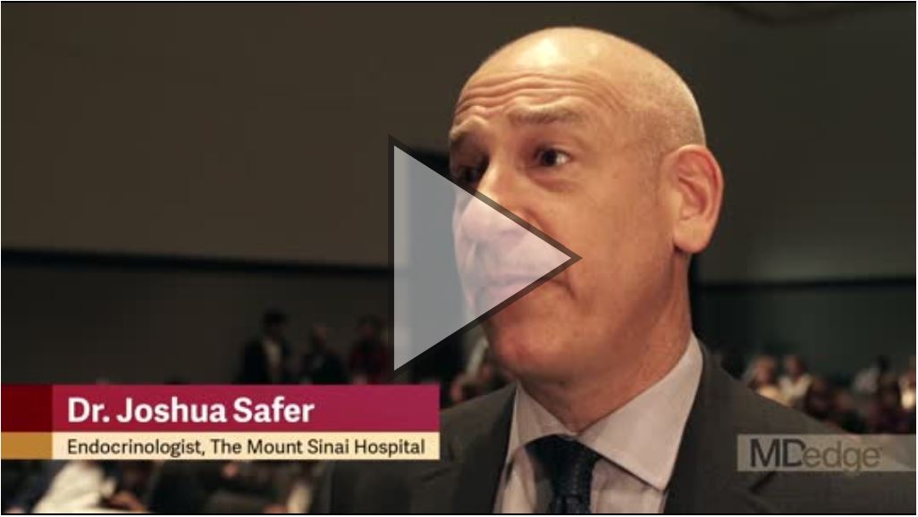
Transgender care mandates endocrinologists share their expertise
BOSTON – Endocrinologists need to be familiar with new practice guidelines and changes in the landscape of transgender health care, Joshua D. Safer, MD, executive director of the Mount Sinai Center for Transgender Medicine and Surgery, New York, said in a video interview at the annual meeting of the American Association of Clinical Endocrinologists.
“We endocrinologists ... need to be able to help (gender-dysphoric/gender-incongruent) individuals, even if it’s just an occasional patient, to do what is safe and to be expert (in transgender health care), just as we are with other hormone treatments,” he said in a discussion of aspects of the Endocrine Society clinical practice guideline on endocrine treatment of gender-dysphoric/gender-incongruent individuals.
The video associated with this article is no longer available on this site. Please view all of our videos on the MDedge YouTube channel
The new guidelines, published in November 2017, update 2009 guidance from the society. Among the big changes are the recognition that there may be “compelling reasons” to start cross-sex hormonal therapy prior to the old age cutoff of 16 years, which is “very late if you’re thinking about it from a biological perspective,” said Dr. Safer.
Another major change challenges the idea that a mental health professional is necessary to diagnose adults. Rather, any knowledgeable clinician could make the diagnosis, according to Dr. Safer.
The guidelines also recommend that endocrinologists provide education regarding onset and time course of physical changes induced by sex hormone treatments to transgender individuals undergoing treatment.
REPORTING FROM AACE 2018
VIDEO: Lyme disease spreading, but better testing may be coming
SANDESTIN, FLA. – Lyme disease is spreading in the United States, which makes it a high priority for rheumatologists, who will need to care for an increasing number of patients with posttreatment disorders affecting the joints, an expert said at the annual Congress of Clinical Rheumatology.
Sheila Arvikar, MD, an instructor in the rheumatology division at Harvard Medical School, Boston, said that the disease – the most common vector-borne illness in the United States – is no longer strictly confined to the U.S. Northeast and the upper Midwest, according to reports from the Centers for Disease Control and Prevention. Neighboring areas are increasingly affected, the reports have shown.
The video associated with this article is no longer available on this site. Please view all of our videos on the MDedge YouTube channel
That the disease may be spreading makes the need for awareness and better testing more acute, she said. Current testing is limited by a lack of sensitivity in early disease, and the standard two-tier combination of enzyme-linked immunosorbent assay and Western blot can be time consuming. But recent studies have found that whole cell sonicate ELISA combined with an ELISA for peptide C6 are equally or even more effective than the more cumbersome, two-tier version, Dr. Arvikar said.
A problem encountered by rheumatologists are patients who contracted Lyme disease but who continue to have joint pain and other symptoms despite treatment for the disease. This so-called posttreatment Lyme disease syndrome (PTLDS) can be similar to fibromyalgia or chronic fatigue syndrome, involving chronic symptoms but no chronic infection and no objective synovitis or inflammation.
There are no Food and Drug Administration–approved treatments for it, but options such as tricyclics, serotonin norepinephrine reuptake inhibitors, gabapentin, and pregabalin can be helpful, she said, along with exercise and cognitive-behavioral therapy. She also noted myriad alternative treatments marketed for PTLDS that have not been shown to be effective and can even be harmful, such as urine ingestion and treatment with bee venom.
“These patients are really desperate for anything to help with their symptoms, and there are lot of people out there who are preying on them with these therapies that aren’t really helpful. It’s important for us to be aware that these things are out there.”
Dr. Arvikar reported having no financial disclosures.
SOURCE: Arvikar S, CCR 2018.
EXPERT ANALYSIS AT CCR 18

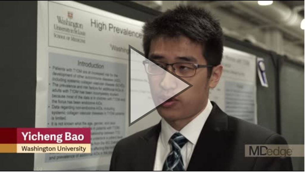
SCVD common in women with type 1 diabetes
BOSTON – Women with type 1 diabetes had a high prevalence of systemic collagen vascular diseases in a recent study, suggesting a global or progressive loss of immune tolerance, investigators reported at the annual meeting of the American Association of Clinical Endocrinologists.
“The median time of diagnosis for most of those autoimmune diseases was years after the diabetes diagnosis,” according to investigator Yicheng Bao, a medical student at University of Missouri-Kansas City.*
“I think there’s some loss of immune tolerance in these patients with type 1 diabetes that really deserves more study as these patients get older,” Mr. Bao said in a video interview.
The study from Mr. Bao and his colleagues was based on patient questionnaire responses and medical chart reviews for 1,167 adults with type 1 diabetes, including 628 women.
They found that SCVDs occurred in 9.2% of women, who had a significantly higher risk versus men (adjusted odds ratio, 2.57; 95% confidence interval, 1.98-3.34; P less than 0.0001).
Rheumatoid arthritis was the most commonly diagnosed SCVD, occurring in 4.3% of the women, followed by psoriasis at 2.6% and lupus at 1.8%. Others occurring in less than 1% of women included Sjögren’s, mixed connective tissue disease, granulomatosis with polyangiitis, juvenile RA, and scleroderma.
Older women were at higher risk of SCVD, with a mean age of 53.6 years versus 46.3 years for women with no SCVD (P = 0.006).
Looking at both men and women, investigators found that individuals with type 1 diabetes and an SCVD were more likely to have other autoimmune diseases, such as hypothyroidism, hyperthyroidism, and celiac disease (adjusted OR, 2.8; 95% CI, 1.71-4.60; P less than 0.0001).
Based on these findings, clinicians taking care of adults with type 1 diabetes need to be vigilant about checking for collagen vascular autoimmune diseases on review of systems, particularly in older women, Mr. Bao said.
“If the patient has a collagen vascular autoimmune disease with type 1 diabetes, they really need to be checking for these other autoimmune diseases,” he added.
Mr. Bao had no disclosures to report.
*This article was updated on May 18, 2018.
REPORTING FROM AACE 2018
Key clinical point: The high incidence of systemic collagen vascular diseases in women with type 1 diabetes suggests a potential progressive loss of immune tolerance.
Major finding: Systemic collagen vascular diseases occurred in 9.2% of women, who had a significantly higher risk versus men (adjusted odds ratio, 2.57; 95% confidence interval, 1.98-3.34; P less than 0.0001).
Study details: A nonrandomized study including retrospective chart review and responses to questionnaires for 1,212 individuals with type 1 diabetes.
Disclosures: Mr. Bao had no disclosures to report.
VIDEO: Dual studies seek answers in isolated skin vasculitis
SANDESTIN, FLA. – Patients with isolated skin vasculitis have always faced a frustrating clinical problem with no clear solution.
The video associated with this article is no longer available on this site. Please view all of our videos on the MDedge YouTube channel
ARAMIS (A Randomized Multicenter Study for Isolated Skin Vasculitis) and its linked genetic investigation, CUTIS (Clinical Transcriptomics in Systemic Vasculitis), may finally identify not only optimal treatments but also insight into the root causes and predictors of treatment response, Christian Pagnoux, MD, said at the annual Congress of Clinical Rheumatology.
“Isolated skin vasculitis is a much-understudied disease, with only one clinical trial to guide our treatment,” said Dr. Pagnoux of the Mount Sinai Hospital, Toronto. In 1995, a 3-month trial randomized 41 patients to skin emollients or to colchicine 0.5 mg/day. Colchicine wasn’t significantly better, but some who had attained remission on it relapsed after discontinuing the drug, which suggested there might be some benefit (Arch Dermatol. 1995;131[12]:1399-1402).
That hint of efficacy in just three patients 23 years ago forms the sole basis of the typical treatment for this disorder: colchicine, Dr. Pagnoux said. “We know that it doesn’t work, yet we continue to prescribe it. Patients deserve better.”
ARAMIS and CUTIS are the first attempts since then at solving this puzzle. ARAMIS is now recruiting about 90 patients in 10 North American medical centers. The three-armed crossover trial will randomize patients to colchicine 0.6 mg twice a day, dapsone 150 mg/day, or azathioprine 2 mg/kg per day for 6 months. Nonresponders can then be rerandomized to one of the other two study drugs for another 6 months. The primary endpoint is clinical response. Secondary endpoints include changes in physician and patient global assessment of response, Skindex29 score, health-related quality of life, and the Patient-Reported Outcomes Measurement Information System.
ARAMIS patients may also participate in CUTIS, the linked histopathologic and genetic investigation. More broad-ranging than ARAMIS, CUTIS is seeking 50 patients with several forms of idiopathic vasculitis, including cryoglobulinemic vasculitis, drug-induced vasculitis, eosinophilic granulomatosis with polyangiitis, IgA vasculitis, isolated cutaneous vasculitis, granulomatosis with polyangiitis, microscopic polyangiitis, polyarteritis nodosa, and urticarial vasculitis.
The study will examine histopathologic and transcriptomic characteristics in punch biopsies of the lesions. “We very much hope that gene expression profiling on these lesions will help define novel pathways and help us to classify and target therapies,” Dr. Pagnoux said.
To learn more about these studies and refer patients into them, visit the Rare Disease Network pages for ARAMIS and CUTIS.
Dr. Pagnoux had no financial disclosures relevant to either study.
SOURCE: Pagnoux C. CCR 2018
REPORTING FROM CCR 18