User login
Mitchel is a reporter for MDedge based in the Philadelphia area. He started with the company in 1992, when it was International Medical News Group (IMNG), and has since covered a range of medical specialties. Mitchel trained as a virologist at Roswell Park Memorial Institute in Buffalo, and then worked briefly as a researcher at Boston Children's Hospital before pivoting to journalism as a AAAS Mass Media Fellow in 1980. His first reporting job was with Science Digest magazine, and from the mid-1980s to early-1990s he was a reporter with Medical World News. @mitchelzoler
Hard road disproving that statins make you dumb
The impact of lipid-lowering drugs on patients’ mental states was on the minds of many attendees at the American College of Cardiology’s annual meeting in March.
The highest-profile report came from EBBINGHAUS (Evaluating PCSK9 Binding Antibody Influence on Cognitive Health in High Cardiovascular Risk Subjects), a substudy of the FOURIER (Further Cardiovascular Outcomes Research With PCSK9 Inhibition in Subjects With Elevated Risk) trial, the meeting’s blockbuster. For the first time, it proved that profoundly lowering low density lipoprotein cholesterol with the proprotein convertase subtilisin/kexin type 9 (PCSK9) inhibitor evolocumab led to a significant reduction in adverse clinical events. EBBINGHAUS focused on about 2,000 of the 27,000 FOURIER patients and subjected equal numbers of placebo and evolocumab patients to a battery of cognitive and memory tests over a median of 20 months. The results showed no hint of a decrement in brain function in the patients taking evolocumab, compared with either their baseline state or the controls who received placebo.
That perception wasn’t helped when, in 2012, the Food and Drug Administration required the labels of all statins to include a reference to postmarketing reports of cognitive side effects such as memory impairment and confusion. The current label for one statin says: “There have been rare postmarketing reports of cognitive impairment (e.g., memory loss, forgetfulness, amnesia, memory impairment, confusion) associated with statin use. These cognitive issues have been reported for all statins.”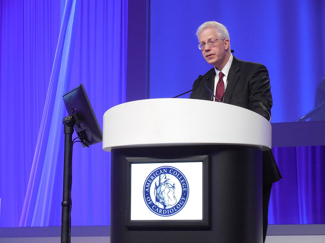
Following the FDA’s action, a series of analyses appeared that reviewed the evidence and found nothing to substantiate the concern. For example, a 2012 review done in direct response to the FDA labeling change looked at case reports, observational studies, and randomized trials and found “no convincing evidence for change in cognitive function” with statin use (J Am Coll Cardiol. 2012 Sept 4;60[10]:875-81). A 2015 meta-analysis that reviewed 14 studies with cognitive testing on more than 27,000 people randomized to either a statin or placebo also found no evidence for a statin effect on mental function (J Gen Intern Med. 2015 March;30[3]:348-58). “Given these results, it is questionable whether the FDA class warning about potential cognitive adverse effects of statins is still warranted,” the meta-analysis authors concluded.
Despite this, concerns about the impact of statins on cognition and memory linger for many patients, witness the anecdotal experiences of clinicians at the meeting. This led a team of researchers at the University of Connecticut and Hartford Hospital to try to directly address the controversy. They also reported their findings at the ACC meeting.
They ran their study as part of a larger trial, STOMP (Effect of Statins on Skeletal Muscle Function and Performance), which randomized 420 healthy and statin-naive individuals to 6 months of treatment with 80 mg atorvastatin or placebo (Circulation. 2013 Jan 2;127[1]:96-103). Their memory substudy included 66 people from the atorvastatin group and 84 placebo-treated controls who averaged 49 years old. Participants underwent a battery of eight memory, cognitive, attention, and executive function tests after 6 months on treatment and again 2 months after statin treatment stopped.
She and her associates took testing a step further and used an assessment never before applied to people taking statins. They ran functional MRIs on a subgroup of the participants while they took two additional memory tests at the end of 6 months on atorvastatin and again 2 months after atorvastatin stopped. They ran MRI scans during a figural memory task test on 42 placebo participants and 35 atorvastatin patients and during a Sternberg Task to test short-term memory on 68 people from the placebo group and 52 who received atorvastatin.
The functional MRI results showed some small but statistically significant changes during both tests in patterns of regional neural activation among those in the statin groups while on and off statins and also when compared with those who received placebo, but Dr. Taylor stressed that her group saw MRI differences between the statin and placebo subjects not only when people were on atorvastatin but also when they had been off the drug for 2 months. She also cautioned that “the clinical implications of the findings are unclear.”
Overall, the entire study’s results showed “no convincing evidence of measurable verbal or nonverbal memory dysfunction” linked with statin use, but Dr. Taylor also noted that the study was relatively small.
Proving the absence of a problem is always difficult. Adding Dr. Taylor’s new evidence to the case that statins really are safe when it comes to cognition and memory will undoubtedly fail to convince committed skeptics.
mzoler@frontlinemedcom.com
On Twitter @mitchelzoler
VIDEO: Internet-based intervention shows antihypertensive efficacy
WASHINGTON – Patients who regularly accessed 30 minute, Internet-based behavioral counseling videos cut their systolic blood pressure, compared with baseline over 1 year by an average 4 mm Hg more than control patients in a randomized, phase II study with 264 patients.
Electronic counseling (e-counseling) “enhanced the efficacy of usual care for hypertension,” Robert P. Nolan, Ph.D., said at the annual meeting of the American College of Cardiology.
“We hope to optimize the efficacy of medical treatments with a behavioral intervention,” said Dr. Nolan, who added that the magnitude of the added benefit from the e-counseling program was “like adding an additional antihypertensive medication.”
“We know antihypertensive treatments work, but compliance is a huge challenge” for health care providers, commented E. Magnus Ohman, MBBS, a professor and cardiologist at Duke University in Durham, N.C. “Having a new way to enhance compliance would be fantastic,” he said.
The Internet-based counseling program devised by Dr. Nolan and his associates consisted of a year-long series of 28 videos, each about 30 minutes long, that participants in the active arm accessed over the Internet. During the study, participants received a series of emailed messages that sent links to the videos on a set schedule over 12 months: During the first 4 months they received an emailed link weekly, during the next 4 months they received an emailed link to a new video every other week, and during the final 4 months of the intervention participants received emailed links once a month. Patients could access each video more than once if they wished, and they were free to share the links with any family members or friends who helped the patients with lifestyle management of their hypertension.
Patients in the e-counseling intervention arm received links to videos that focused on motivational messages and teaching cognitive behavioral skills. Patients in the control arm received emails with generic messages on blood pressure management and without links to videos.
The REACH (E-Counseling Promotes Blood Pressure Reduction and Therapeutic Lifestyle Change in Hypertension) study ran at four Canadian sites. It sent invitations to participate to 609 patients with stage 1 or 2 hypertension, with a blood pressure prior to treatment of 140/90-180/110 mm Hg. Among the invited patients, 264 elected to participate; 100 patients in the e-counseling arm and 97 control patients completed the 1-year program. Participants averaged 58 years of age, their average body mass index was 31 kg/m2, and 9% smoked. Their blood pressure at entry averaged 141/87 mm Hg, their average pulse pressure was about 54 mm Hg, and their average 10-year risk for a cardiovascular disease event, measured by the Framingham Risk Score, was about 16%. At entry, patients in the study received an average of 1.5 antihypertensive drugs each.
At the end of the 1-year program, systolic blood pressure fell by an average of 6 mm Hg from baseline among patients who completed the control program, and by an average of 10.1 mm Hg among the patients who completed the e-counseling arm, a statistically significant difference for one of the study’s primary endpoints. Change in pulse pressure from baseline showed an average 1.5–mm Hg drop in the control patients and an average 4.3–mm Hg decline in the e-counseling patients, another statistically significant difference for a second primary endpoint, reported Dr. Nolan, a clinical psychologist and director of the cardiac eHealth program at the University of Toronto.
A third primary endpoint was change in the Framingham Risk Score, which fell by an average of 1.9% after 12 months in the e-counseling patients and rose by an average of 0.2% among the controls.
The final primary endpoint was the change in diastolic blood pressure from baseline, which showed a better than 4–mm Hg incremental decline in the men who received e-counseling, compared with controls, but among women in the study, the drop in diastolic blood pressure from baseline was nearly the same – about 6 mm Hg – in both the controls and e-counseling patients.
“This tells us that we need to better tailor the [e-counseling] intervention to men and to women,” Dr. Nolan said in a video interview. He also envisions better tailoring of the e-counseling videos to various socioeconomic and ethnic groups. He plans to continue testing of a revised version of the e-counseling intervention in a larger, phase III study, but he also hopes that the intervention videos can soon be available at no charge for use in routine practice.
REACH received no commercial funding. Dr. Nolan had no disclosures.
The video associated with this article is no longer available on this site. Please view all of our videos on the MDedge YouTube channel
mzoler@frontlinemedcom.com
On Twitter @mitchelzoler
I love the REACH study. Hypertension is incredibly prevalent among the patients I see, and they often need three different antihypertensive drugs to control their blood pressure. Lifestyle interventions can be very effective at helping to lower blood pressure, but during the 10-minute visits I have with most of my patients, it’s hard for me to have much impact on their behavior.
What I especially like about the Internet-based counseling used in this study was its application of evidence-based approaches to change patient behavior. This was a well-designed and exciting trial.
Sandra J. Lewis, MD, chief of cardiology at Legacy Good Samaritan Hospital in Portland, Ore., made these comments during a press conference. She had no disclosures.
WASHINGTON – Patients who regularly accessed 30 minute, Internet-based behavioral counseling videos cut their systolic blood pressure, compared with baseline over 1 year by an average 4 mm Hg more than control patients in a randomized, phase II study with 264 patients.
Electronic counseling (e-counseling) “enhanced the efficacy of usual care for hypertension,” Robert P. Nolan, Ph.D., said at the annual meeting of the American College of Cardiology.
“We hope to optimize the efficacy of medical treatments with a behavioral intervention,” said Dr. Nolan, who added that the magnitude of the added benefit from the e-counseling program was “like adding an additional antihypertensive medication.”
“We know antihypertensive treatments work, but compliance is a huge challenge” for health care providers, commented E. Magnus Ohman, MBBS, a professor and cardiologist at Duke University in Durham, N.C. “Having a new way to enhance compliance would be fantastic,” he said.
The Internet-based counseling program devised by Dr. Nolan and his associates consisted of a year-long series of 28 videos, each about 30 minutes long, that participants in the active arm accessed over the Internet. During the study, participants received a series of emailed messages that sent links to the videos on a set schedule over 12 months: During the first 4 months they received an emailed link weekly, during the next 4 months they received an emailed link to a new video every other week, and during the final 4 months of the intervention participants received emailed links once a month. Patients could access each video more than once if they wished, and they were free to share the links with any family members or friends who helped the patients with lifestyle management of their hypertension.
Patients in the e-counseling intervention arm received links to videos that focused on motivational messages and teaching cognitive behavioral skills. Patients in the control arm received emails with generic messages on blood pressure management and without links to videos.
The REACH (E-Counseling Promotes Blood Pressure Reduction and Therapeutic Lifestyle Change in Hypertension) study ran at four Canadian sites. It sent invitations to participate to 609 patients with stage 1 or 2 hypertension, with a blood pressure prior to treatment of 140/90-180/110 mm Hg. Among the invited patients, 264 elected to participate; 100 patients in the e-counseling arm and 97 control patients completed the 1-year program. Participants averaged 58 years of age, their average body mass index was 31 kg/m2, and 9% smoked. Their blood pressure at entry averaged 141/87 mm Hg, their average pulse pressure was about 54 mm Hg, and their average 10-year risk for a cardiovascular disease event, measured by the Framingham Risk Score, was about 16%. At entry, patients in the study received an average of 1.5 antihypertensive drugs each.
At the end of the 1-year program, systolic blood pressure fell by an average of 6 mm Hg from baseline among patients who completed the control program, and by an average of 10.1 mm Hg among the patients who completed the e-counseling arm, a statistically significant difference for one of the study’s primary endpoints. Change in pulse pressure from baseline showed an average 1.5–mm Hg drop in the control patients and an average 4.3–mm Hg decline in the e-counseling patients, another statistically significant difference for a second primary endpoint, reported Dr. Nolan, a clinical psychologist and director of the cardiac eHealth program at the University of Toronto.
A third primary endpoint was change in the Framingham Risk Score, which fell by an average of 1.9% after 12 months in the e-counseling patients and rose by an average of 0.2% among the controls.
The final primary endpoint was the change in diastolic blood pressure from baseline, which showed a better than 4–mm Hg incremental decline in the men who received e-counseling, compared with controls, but among women in the study, the drop in diastolic blood pressure from baseline was nearly the same – about 6 mm Hg – in both the controls and e-counseling patients.
“This tells us that we need to better tailor the [e-counseling] intervention to men and to women,” Dr. Nolan said in a video interview. He also envisions better tailoring of the e-counseling videos to various socioeconomic and ethnic groups. He plans to continue testing of a revised version of the e-counseling intervention in a larger, phase III study, but he also hopes that the intervention videos can soon be available at no charge for use in routine practice.
REACH received no commercial funding. Dr. Nolan had no disclosures.
The video associated with this article is no longer available on this site. Please view all of our videos on the MDedge YouTube channel
mzoler@frontlinemedcom.com
On Twitter @mitchelzoler
AT ACC 17
Key clinical point:
Major finding: The Internet-based program reduced systolic blood pressure from baseline by an average additional 4.1 mm Hg, compared with controls.
Data source: REACH, a multicenter, randomized trial with 264 hypertensive patients.
Disclosures: REACH received no commercial funding. Dr. Nolan had no disclosures.

VIDEO: Postop troponin T spike flags high mortality risk
WASHINGTON – A rise in the blood level of troponin T immediately after patients underwent noncardiac surgery identified a high risk group with a 30-day mortality rate nearly fivefold higher than that of patients who did not have a postoperative troponin T spike, according to a prospective study of more than 21,000 patients.
For 93% of the patients who have these perioperative spikes in troponin T, a marker of myocardial ischemia, the increased level was the only indicator of a heart problem, P.J. Devereaux, MD, said at the annual meeting of the American College of Cardiology. The painkillers that patients receive following surgery generally mask the chest discomfort they might otherwise feel from their heart damage, he explained. The clinical condition is called myocardial injury after noncardiac surgery.
“We’re seeing more older patients with a high burden of vascular disease undergoing surgery, and surgery is a very significant stress, so a large proportion of these patients will have [myocardial injury after noncardiac surgery] and that affects 30-day survival,” said Dr. Devereaux, professor and director of cardiology at McMaster University in Hamilton, Ont.
Based on the new findings, he recommended performing a baseline assessment of troponin T levels in patients scheduled for noncardiac surgery if they are at least 65 years of age, or if they are age 45-64 years with known vascular disease, followed by repeat testing 1 and 2 days after surgery to check whether a spike in the measure had occurred. The high sensitivity troponin T (hsTnT) test he used in the study is relatively costly (and received Food and Drug Administration approval for U.S. marketing in January 2017), but Dr. Devereaux believed that, used in this way, the cost for testing would be reasonable, given its powerful ability to identify high-risk patients and relative to the cost of other screening tools routinely used in U.S. medical practice.
“It looks very cost-effective,” he said in a video interview.
If the baseline and two follow-up measures of hsTnT showed a postoperative level of at least 20 ng/L that rose above the baseline level by at least 5 ng/L, or if the postoperative level was at least 65 ng/L, Dr. Devereaux recommended starting daily treatment with aspirin and a statin to try to contain any perioperative myocardial damage the patient may have, and follow with comprehensive assessment of the patient by a cardiologist or other internal medicine physician.
“Given the risks associated with a rise in hsTnT in this study, Dr. Devereaux’s recommendations are very reasonable until we collect more data on this,” said Frank W. Sellke, MD, professor of surgery and chief of cardiothoracic surgery at Brown Medical School and the Lifespan Hospitals in Providence, R.I. “What was surprising was how few patients had symptoms” of myocardial ischemia. “You can’t do hsTnT measurements on every patient who goes in for a hernia operation; it’s not practical. But his findings are fairly compelling, and hopefully the cost of this testing will come down,” Dr. Sellke said in an interview.
In the multicenter study of 21,842 patients, 24% had a postoperative hsTnT level of 20 ng/L or greater, including 5% with a level of 65 ng/L or greater. The 30-day mortality rate was 3% among those with a perioperative level of 20-64 ng/L, 9% among patients with perioperative hsTnT levels of 65-999 ng/L, and 30% among the 54 patients (0.2% of the study group) with perioperative levels that reached 1,000 ng/L or greater. Dr. Devereaux reported.
This iteration of the Vascular Events In Noncardiac Surgery Patients Cohort Evaluation Study (VISION) enrolled patients who were at least 45 years of age and underwent noncardiac surgery at 23 centers in 13 countries, including the United States and Canada. All patients underwent hsTnT testing 6-12 hours after surgery and 1, 2, and 3 days after surgery, but only 40% also had a baseline measurement before their surgery began. Full 30-day follow-up occurred for 21,050. The patients’ average age was 63 years. The most common surgery was “low-risk,” in 35%, followed by “major” general surgery in 20%, and “major” orthopedic surgery in 16%. At 30 days, 266 patients (1.2%) had died.
These findings “help define a cutoff for hsTnT that will be clinically useful to change practice,” said Athena Poppas, MD, a cardiologist and director of the Cardiovascular Institute at Rhode Island Hospital in Providence. Dr. Poppas was a designated discussant for Dr. Devereaux’s report at the meeting.
In January 2017, the Canadian Cardiovascular Society issued guidelines for perioperative cardiac risk assessment and management for patients undergoing noncardiac surgery (Can J Cardiol. 2017 Jan;33[1]:17-32). Dr. Devereaux was a member of the writing panel for these guidelines. This is what the guidelines said about using troponin T measurements:
“We recommend obtaining daily troponin measurements for 48-72 hours after noncardiac surgery in patients with a baseline risk greater than 5% for cardiovascular death or nonfatal myocardial infarction at 30 days after surgery (i.e., patients with an elevated NT-proBNP/BNP measurement before surgery or, if there is no NT-proBNP/BNP measurement before surgery, in those who have an RCRI [revised cardiac risk index] score of 1 or greater, age 45-64 years with significant cardiovascular disease, or age 65 years or older).”
The VISION study is sponsored by Roche Diagnostics, which markets the high sensitivity troponin T assay used in the study. Dr. Devereaux has received research funding from Roche Diagnostics and from Abbott Diagnostics and Boehringer Ingelheim. Dr. Sellke and Dr. Poppas had no relevant disclosures.
The video associated with this article is no longer available on this site. Please view all of our videos on the MDedge YouTube channel
mzoler@frontlinemedcom.com
On Twitter @mitchelzoler
AT ACC 17
Key clinical point:
Major finding: A postoperative high sensitivity troponin T rise of 5 ng/L or more linked with a 4.7-fold increase in 30-day mortality.
Data source: VISION, a prospective, multicenter observational study of 21,842 patients undergoing noncardiac surgery.
Disclosures: The VISION study is sponsored by Roche Diagnostics, which markets the high sensitivity troponin T assay used in the study. Dr. Devereaux has received research funding from Roche Diagnostics and from Abbott Diagnostics and Boehringer Ingelheim.

Get With the Guidelines propels stroke thrombolytic therapy
HOUSTON – Thrombolytic therapy for U.S. patients experiencing an acute ischemic stroke is no longer the poster child for proven, but neglected, therapies.
Long bemoaned since its introduction in the mid-1990s as an effective but woefully underused treatment, thrombolytic therapy with tissue plasminogen activator (tPA) has seen robust growth in U.S. practice recently, largely due to the promotion and quality-improvement efforts of a voluntary, non-profit program, Get With the Guidelines (GWTG)-Stroke.
“We’ve seen a dramatic increase in tPA treatment in eligible patients,” said Dr. Smith, a neurologist and medical director of the Cognitive Neurosciences Clinic at the University of Calgary (Alta.).
“This represents remarkable and clinically meaningful improvements in stroke care, overcoming substantial barriers using a systems of care approach,” Gregg C. Fonarow, MD, professor of medicine and cochief of clinical cardiology at the University of California, Los Angeles and one of the long-time leaders of GWTG-Stroke, said in an interview. “This represents one of the most transformative improvements and success stories ever observed in stroke care.”
“These data show dramatic changes in the GWTG-Stroke hospitals,” agreed Lee H. Schwamm, MD, professor of neurology at Harvard Medical School and chief of stroke services at Massachusetts General Hospital in Boston and another leader of the GWTG-Stroke program. However, “we still have work to do,” cautioned Dr. Schwamm, who notes that 68% remains significantly short of the ideal 100%. The positive is that several GWTG-Stroke participating hospitals have surged to a better than 90% rate of administering tPA to eligible patients within a 60-minute door-to-needle window.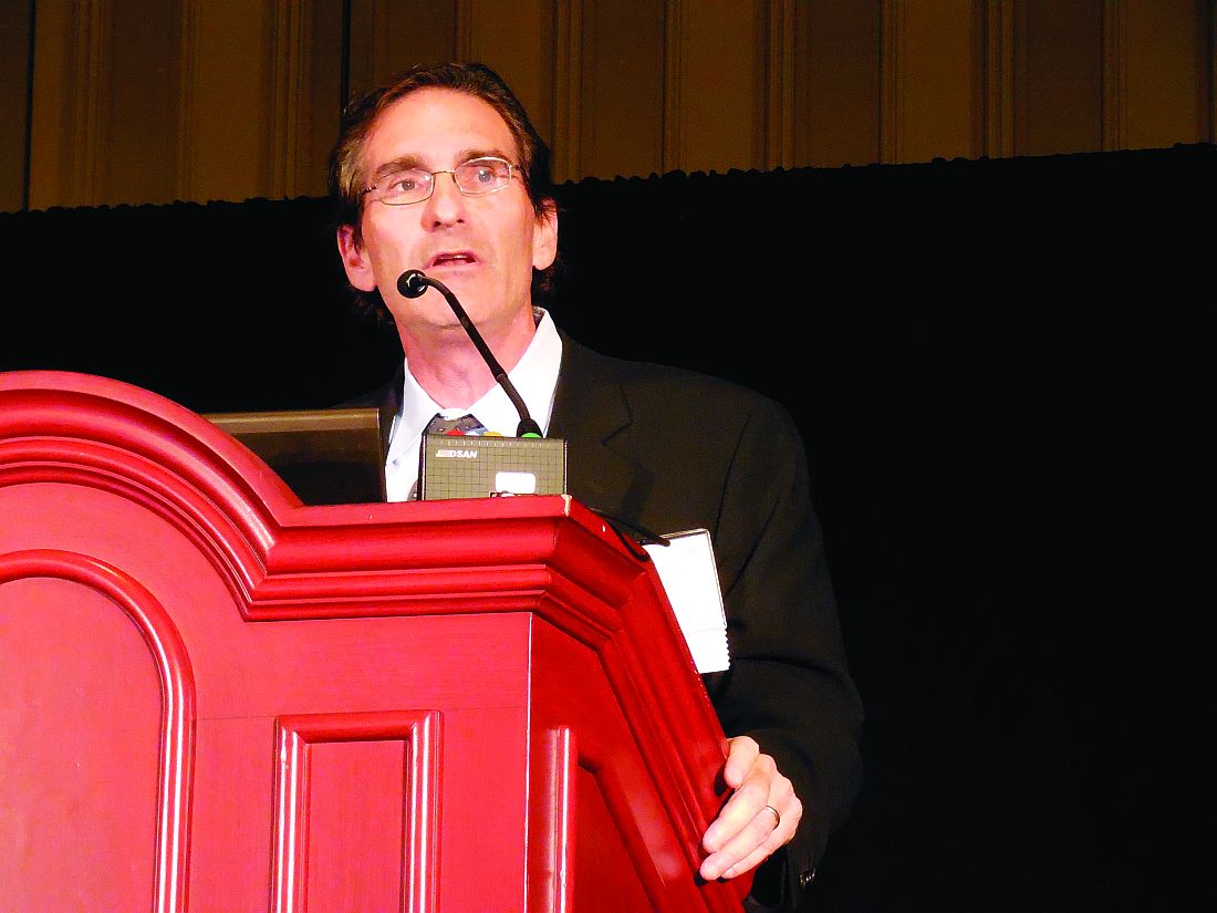
What is GWTG-Stroke?
GWTG was launched in 2000 by the American Heart Association as a hospital-based–quality improvement initiative focused on U.S. management of acute MI. In 2003, acute stroke became another focus of the program and inspired added participation of the American Stroke Association.
“Improvements in tPA use in eligible patients were seen soon after the introduction of GWTG-Stroke in 2003, but there was minimal improvement in the percentage of patients with door-to-needle times within 60 minutes despite national guidelines,” recalled Dr. Fonarow. This slow rate of advance led him, Dr. Schwamm, and others to create a more activist program within GWTG-Stroke, Target Stroke, that not only provided resources and collected data from participating hospitals but also set acute-treatment goals that challenged participating hospitals to up their game. Target Stroke phase I asked them to try to achieve a tPA door-to-needle time within 60 minutes in at least 50% of eligible patients. By mid-2013, the program reached this goal with 53% of patients treated in the allotted time frame (JAMA. 2014 Apr 23;311[16]:1632-40).
These time-based goals lead directly to meaningful improvements in patient outcomes. “We know that, for every 15 minute reduction in tPA door-to-needle time, there is a 5% reduction of in-hospital mortality and a substantial increase in the percentage of patients who are discharged home instead of to a rehabilitation hospital or nursing home,” Dr. Schwamm said.
Dr. Smith highlighted the 11 best practices steps that Target Stroke promotes to GWTG-Stroke hospitals as their road map to achieving the performance goals. These include things such as notification of a hospital by emergency medical services that a patient showing signs of an acute ischemic stroke is on the way, transfer of the patient after hospital arrival directly to a CT or MRI scanner to get the imaging that can confirm tPA eligibility, rapid reading of the scan, premixing of the tPA, a team-based approach to acute stroke management, and quick feedback to the team regarding their performance on each stroke patient they treat.
Dr. Schwamm noted that he, Dr. Fonarow, and other program leaders are already planning a phase III for Target Stroke that will branch out to even more facets of acute stroke care, such as incorporation of goals for using endovascular thrombectomy in appropriate patients and for using telemedicine to quicken and broaden the availability of expert neurologic consults for identifying tPA- and thrombectomy-eligible patients.
Growing the program
The success of GWTG-Stroke in transforming U.S. acute stroke care has not only relied on setting aggressive performance goals but also on advancing by boosting the number of U.S. hospitals participating of more U.S. hospitals. During the Target Stroke years since the start of 2010, the number of hospitals active in GWTG-Stroke jumped from 1427 to 1,950 by mid-2016, including a 12% year-over-year jump in 2016, compared with 2015. During October 2015-October 2016, participating hospitals treated more than 570,000 acute stroke patients, or roughly 71% of the estimated 800,000 U.S. patients who have an acute stroke each year.
These numbers seem on track to continue expanding. “With the recent evidence showing that participating in GWTG-Stroke improves care and outcomes there has been even greater interest by hospitals, recently, in joining,” said Dr. Fonarow. “While many hospitals currently participate, ideally all would join and be active.”
“Hospitals now feel that they can’t afford not to focus on stroke as a quality improvement program, so they join,” said Dr. Schwamm. He believes that essentially all of the roughly 200 certified U.S. Comprehensive Stroke Centers and of the roughly 1,000 certified U.S. Primary Stroke Centers are already members of GWTG-Stroke. “We’re now in all the high-value hospitals, based on their higher numbers of patients. It’s the Acute Stroke Ready hospitals where we now have the greatest potential to penetrate, hospitals that generally treat about 20-50 stroke patients a year,” he said.
Joining GWTG-Stroke holds major attraction for hospitals because the program gives them a tool for measuring their performance, data that hospitals often now need to prove the value of the care they deliver to insurers and to avoid penalization for readmissions. However, the barrier to hospitals, especially smaller hospitals, is that data collection can be expensive. It’s something that larger hospitals now routinely do, but smaller hospitals have often balked because of the expense. To address this, the GWTG-Stroke program is trying to develop a “lighter” version of their data collection tool that involves a smaller financial burden.
“I think we can sell a trimmed down version of GWTG-Stroke to smaller hospitals that is affordable and use it to recruit another 1,000 hospitals. That’s a realistic goal,” Dr. Schwamm said.
What is also notable about GWTG-Stroke is that hospitals sign up despite the somewhat ambiguous payback they receive.
“In the past, stroke was the third-leading U.S. cause of death, but now it’s fifth,” noted Steven R. Messe, MD, a stroke neurologist at the University of Pennsylvania in Philadelphia and a member of the GWTG-Stroke steering committee. “That’s an amazing accomplishment – to drop stroke from third place to fifth – and it’s due to advances in thrombolytic use and to improved systems of care and quality improvement measures.”
Get With the Guidelines-Stroke is a program of the American Heart Association and American Stroke Association and uses funding provided by several drug companies. Dr. Smith, Dr. Fonarow, Dr. Schwamm and Dr. Messe had no relevant commercial disclosures.
mzoler@frontlinemedcom.com
On Twitter @mitchelzoler
EXPERT ANALYSIS FROM THE INTERNATIONAL STROKE CONFERENCE
Evolocumab doesn’t make patients dumb
WASHINGTON – Concerns about possible adverse cognitive effects from the new lipid-lowering drug evolocumab and from aggressive lowering of LDL cholesterol were largely put to rest with results from a nearly 2,000-patient study showing that a median 20 months of treatment with evolocumab caused no cognitive or memory decline, either compared with baseline measures or compared with similar patients randomized to placebo treatment.

A subgroup analysis in the study also showed that, even among patients who achieved and sustained LDL cholesterol below 25 mg/dL, no hint arose of an adverse effect on memory or cognition, said Dr. Giugliano, a cardiologist at Brigham and Women’s Hospital in Boston.
“Every day in my office, patients tell me that statins make you dumb. Now I can comfortably tell my patients that we studied whether this drug [evolocumab] makes you dumb, and it doesn’t; nor did having an LDL cholesterol level below 25 mg/dL,” commented Sandra J. Lewis, MD, chief of cardiology at Legacy Good Samaritan Hospital in Portland, Ore.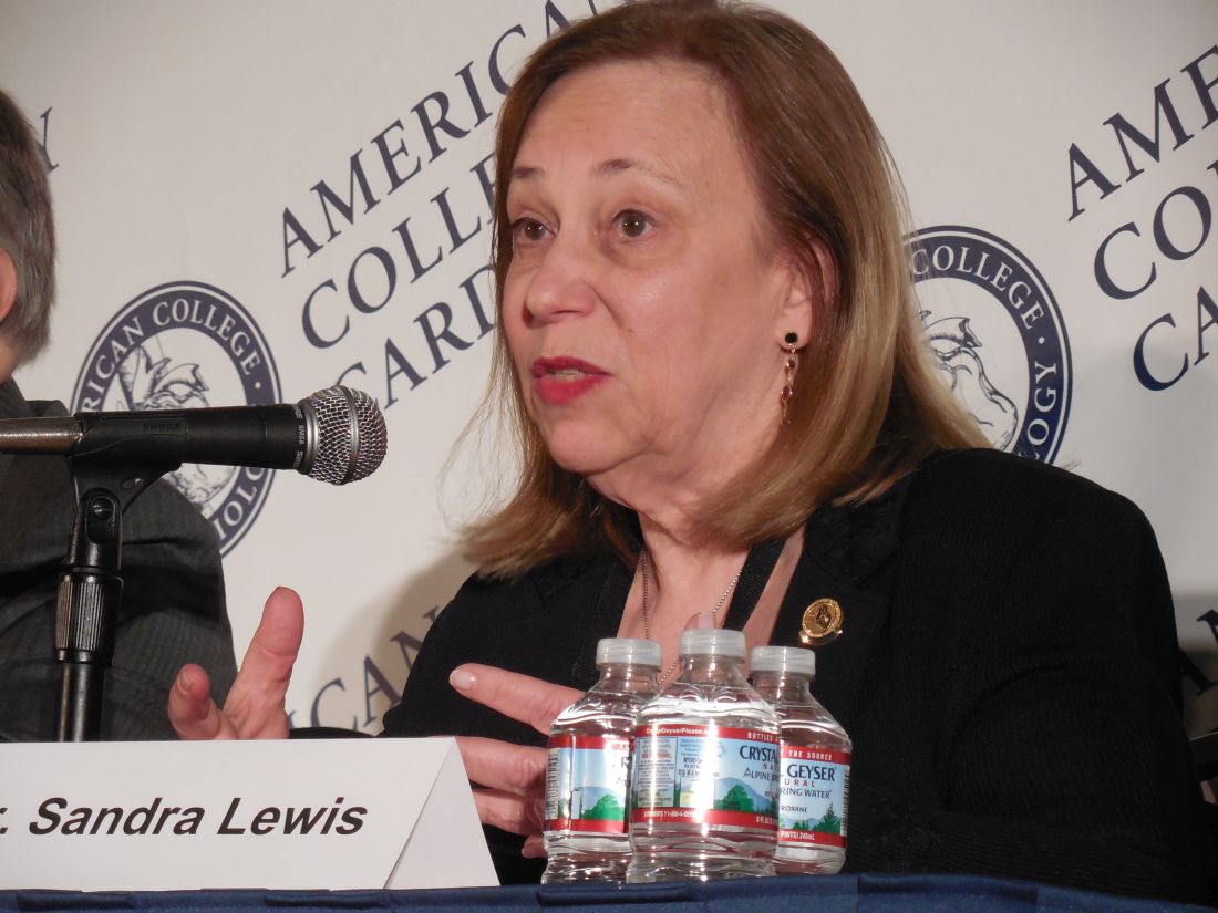
The primary assessment tool Dr. Giugliano and his associates used in EBBINGHAUS was the Cambridge Neuropsychological Test Automated Battery (CANTAB) Assessments, a computer-based test of spatial working memory strategy index of executive function, and 1,204 patients underwent CANTAB testing both before they received their first dose of their assigned agent and also at the end of treatment. The scores of the patients on evolocumab virtually superimposed on those of patients in the placebo group, both at baseline and at the end of treatment, easily meeting the study’s prespecified definition of noninferiority, Dr. Giugliano reported at the annual meeting of the American College of Cardiology.
The CANTAB scores also showed no change from baseline over time in the individual subgroups of both evolocumab-treated and placebo-treated patients. The total absence of a signal of mental deterioration in the placebo patients who remained on statin treatment throughout the study provided new evidence that medium-term treatment with a statin was not linked with any change in memory or cognition.
EBBINGHAUS also assessed participants using several other memory and cognition tests, had patients complete an end-of-study survey of their perceptions of their mental function, and questioned participating physicians about patients’ mental performance. None of these measures showed any signals of impairment. A full description of the range of assessments used in EBBINGHAUS appeared recently in a published article (Clin Cardiol. 2017 Feb;40[2]:59-65).
Dr. Giugliano conceded that what remains unknown at this point is the possible impact of further prolonged treatment with a PCSK9 drug or from extended maintenance of LDL cholesterol levels at very low levels, effects that could continue for decades in the routine treatment of selected patients. He noted that patients who participated in FOURIER and in EBBINGHAUS are undergoing continued follow-up to gain better insight into the longer-term effects of their treatment.
EBBINGHAUS was sponsored by Amgen, the company that markets evolocumab (Repatha). Dr. Giugliano has been a consultant to and received research support from Amgen and from several other drug companies. Dr. Bhatt has received research support from Amgen, from Sanofi Regeneron associated with research on another PCSK9 inhibitor, and from several other drug companies, Dr. Lewis had no disclosures.
mzoler@frontlinemedcom.com
On Twitter @mitchelzoler
WASHINGTON – Concerns about possible adverse cognitive effects from the new lipid-lowering drug evolocumab and from aggressive lowering of LDL cholesterol were largely put to rest with results from a nearly 2,000-patient study showing that a median 20 months of treatment with evolocumab caused no cognitive or memory decline, either compared with baseline measures or compared with similar patients randomized to placebo treatment.
A subgroup analysis in the study also showed that, even among patients who achieved and sustained LDL cholesterol below 25 mg/dL, no hint arose of an adverse effect on memory or cognition, said Dr. Giugliano, a cardiologist at Brigham and Women’s Hospital in Boston.
“Every day in my office, patients tell me that statins make you dumb. Now I can comfortably tell my patients that we studied whether this drug [evolocumab] makes you dumb, and it doesn’t; nor did having an LDL cholesterol level below 25 mg/dL,” commented Sandra J. Lewis, MD, chief of cardiology at Legacy Good Samaritan Hospital in Portland, Ore.
The primary assessment tool Dr. Giugliano and his associates used in EBBINGHAUS was the Cambridge Neuropsychological Test Automated Battery (CANTAB) Assessments, a computer-based test of spatial working memory strategy index of executive function, and 1,204 patients underwent CANTAB testing both before they received their first dose of their assigned agent and also at the end of treatment. The scores of the patients on evolocumab virtually superimposed on those of patients in the placebo group, both at baseline and at the end of treatment, easily meeting the study’s prespecified definition of noninferiority, Dr. Giugliano reported at the annual meeting of the American College of Cardiology.
The CANTAB scores also showed no change from baseline over time in the individual subgroups of both evolocumab-treated and placebo-treated patients. The total absence of a signal of mental deterioration in the placebo patients who remained on statin treatment throughout the study provided new evidence that medium-term treatment with a statin was not linked with any change in memory or cognition.
EBBINGHAUS also assessed participants using several other memory and cognition tests, had patients complete an end-of-study survey of their perceptions of their mental function, and questioned participating physicians about patients’ mental performance. None of these measures showed any signals of impairment. A full description of the range of assessments used in EBBINGHAUS appeared recently in a published article (Clin Cardiol. 2017 Feb;40[2]:59-65).
Dr. Giugliano conceded that what remains unknown at this point is the possible impact of further prolonged treatment with a PCSK9 drug or from extended maintenance of LDL cholesterol levels at very low levels, effects that could continue for decades in the routine treatment of selected patients. He noted that patients who participated in FOURIER and in EBBINGHAUS are undergoing continued follow-up to gain better insight into the longer-term effects of their treatment.
EBBINGHAUS was sponsored by Amgen, the company that markets evolocumab (Repatha). Dr. Giugliano has been a consultant to and received research support from Amgen and from several other drug companies. Dr. Bhatt has received research support from Amgen, from Sanofi Regeneron associated with research on another PCSK9 inhibitor, and from several other drug companies, Dr. Lewis had no disclosures.
mzoler@frontlinemedcom.com
On Twitter @mitchelzoler
AT ACC 17
Key clinical point:
Major finding: Patients on evolocumab for 20 months had noninferior memory and executive function test scores, compared with patients on placebo.
Data source: EBBINGHAUS, a multicenter randomized trial with 1,974 patients.
Disclosures: EBBINGHAUS was sponsored by Amgen, the company that markets evolocumab (Repatha). Dr. Giugliano has been a consultant to and received research support from Amgen and from several other drug companies.
BNP flags high atrial fibrillation prevalence in ESUS stroke
HOUSTON – Following an embolic stroke of undetermined source, a patient’s serum level of brain natriuretic peptide may identify patients at high risk of having covert atrial fibrillation that warrants assessment by prolonged arrhythmia monitoring, based on a post hoc analysis of data collected from 373 patients.
The analysis showed that in older patients with a recent embolic stroke of undetermined source (ESUS) who had a serum brain natriuretic peptide (BNP) level of 100 pg/mL or greater, the prevalence of atrial fibrillation (AF) detected by up to 30 days of Holter monitoring was 33%. In contrast the AF prevalence was 4% among similar ESUS patients with a lower BNP level, Rolf Wachter, MD, said at the International Stroke Conference sponsored by the American Heart Association.
“This evidence is pretty compelling,” commented Hooman Kamel, MD, a neurologist at Weill Cornell Medicine in New York. “We already use other lab tests that have less evidence supporting their use than this, and checking BNP is a simple lab test.”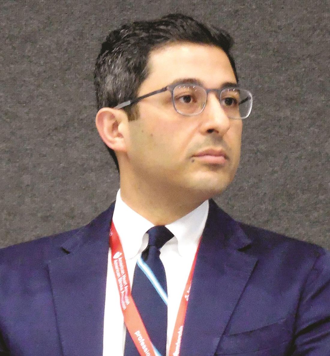
The results showed that monitoring identified AF in 5% of the control patients and in 14% of those who underwent extended monitoring, a statistically significant difference (Lancet Neurol. 2017 Apr;16[4]:282-90).
Dr. Wachter and his colleagues had baseline BNP measurements for 373 of the patients and analyzed the prevalence of AF based on these levels. The patients averaged 72 years old. Among those with BNP values of at least 100 pg/mL, prolonged arrhythmia monitoring found AF in 33%, while briefer, conventional monitoring found AF in about 4%. This meant that for every three patients receiving prolonged arrhythmia scrutiny in this subgroup, the assessment found one patient with AF. But among patients with a serum BNP of less than 100 pg/mL, prolonged arrhythmia assessment yielded a 10% rate of AF detection, compared with about 4% in the controls, meaning that for every 18 patients screened prolonged Holter monitoring detected one additional case of AF, he reported.
This relationship presumably exists because higher BNP levels identify patients with structural heart abnormalities, a subgroup at increased risk for AF, Dr. Wachter explained. He cautioned that, because this was a post hoc analysis, he would like to see the findings confirmed in prospective studies.
Dr. Wachter has been a speaker on behalf of Bayer, Boehringer Ingelheim, Bristol-Myers Squibb, Pfizer, and Daiichi Sankyo and has received research support from Boehringer Ingelheim. The FIND-AFRANDOMISED trial received funding from Boehringer Ingelheim.
mzoler@frontlinemedcom.com
On Twitter @mitchelzoler
AT THE INTERNATIONAL STROKE CONFERENCE
Key clinical point:
Major finding: Following ESUS, patients with a BNP of 100 pg/mL or greater had a 33% prevalence of AF.
Data source: A post hoc analysis of 373 German patients enrolled in the FIND-AFRANDOMISED trial.
Disclosures: Dr. Wachter has been a speaker on behalf of Bayer, Boehringer Ingelheim, Bristol-Myers Squibb, Pfizer, and Daiichi Sankyo and has received research support from Boehringer Ingelheim. The FIND-AFRANDOMISED trial received funding from Boehringer Ingelheim.
VIDEO: Evolocumab caused no cognitive issues in FOURIER substudy
WASHINGTON – Don’t give the cognitive effects of evolocumab a second thought, results from a preplanned substudy of the landmark FOURIER study that assessed the impact of this PCSK9 inhibitor on memory or other measures of cognition in a random subgroup of 1,973 patients enrolled in the main study suggest.
Based on these results “I wouldn’t be concerned about adverse cognitive effects” in patients treated with evolocumab (Repatha) or, for the time being, any other PCSK9 inhibitor, said Robert P. Giugliano, MD, in a video interview at the annual meeting of the American College of Cardiology”
Concerns about the cognitive effects of the drugs in this class of lipid-lowering drugs have lingered following suggestions of a possible problem that arose during earlier, much smaller studies of these agents as well as similar concerns that arose about statins that have been perpetuated through misleading posts on the Internet.
The battery of memory and cognition assessments used in EBBINGHAUS, the FOURIER substudy, should lay concerns about brain effects of evolocumab to rest, at least within the context of the median 26 months of treatment that patients in FOURIER received, said Dr. Giugliano, a cardiologist at Brigham and Women’s Hospital in Boston and lead investigator of the EBBINGHAUS substudy. Further analysis also showed that, even among patients who achieved LDL cholesterol levels of less than 25 mg/dL, treatment with evolocumab appeared to cause no deterioration of the measured mental functions.
The video associated with this article is no longer available on this site. Please view all of our videos on the MDedge YouTube channel
mzoler@frontlinemedcom.com
On Twitter @mitchelzoler
AT ACC 2017
Moderate exercise benefits hypertrophic cardiomyopathy patients
WASHINGTON – A regimen of moderately intense exercise for 16 weeks produced no adverse effects and led to a clinically meaningful improvement in exercise capacity in a multicenter, randomized trial involving 136 patients with hypertrophic cardiomyopathy.
While finding that regular exercise produced a statistically significant improvement, compared with control patients fulfilled the study’s primary endpoint, it was perhaps as important that patients with hypertrophic cardiomyopathy (HCM) randomized to the exercise arm experienced no ill effects, a finding that bucked conventional wisdom that, once diagnosed, HCM patients must adhere to a largely inactive lifestyle.
“I see patients with HCM who tell me that, when they were first diagnosed, they were told by their physician not to do anything at all,” said Dr. Saberi, a cardiologist at the University of Michigan in Arbor. Although she cautioned that the findings from this modestly sized trial cannot prove that exercise is safe for HMC patients – something that would require randomizing more than 2,000 patients and following them for about 3 years – the findings provide some level of reassurance that regular, moderately intense exercise as simple as walking can benefit patients and likely not cause any problems. “We saw no signal of harm,” she stressed in an interview. “I would recommend moderate-intensity, regular exercise. Physicians can feel comfortable using the exercise prescription” used in the study, she advised.
Concurrently with Dr. Saberi’s report at the meeting, the results also appeared in an article published online (JAMA. 2017 Mar 17. doi: 10.1001/jama.2017.2503).
The Study of Exercise Training in Hypertrophic Cardiomyopathy (RESET-HCM) ran during 2010-2015 at the University of Michigan and Stanford University, and enrolled 136 patients 18-80 years old diagnosed with HCM but without more severe manifestations such as a history of exercise-induced syncope or ventricular arrhythmia, a left ventricular ejection fraction less than 55%, recent clinical decompensation, or a history of a severe hypotensive response to exercise. Enrolled patients averaged about 50 years old, their baseline peak oxygen consumption, VO2, was about 22 mL/kg per min, and their maximal ventricular wall thickness was about 21 mm.
Following a comprehensive baseline heart function and imaging assessment, including a cardiopulmonary exercise test, the 67 patients in the intervention arm received an individualized exercise prescription based on their heart rate reserve. At a minimum, they started with three exercise sessions per week lasting at least 20 minutes on their own, a regimen designed to bring them to 60% of their heart rate reserve. Their prescription gradually increased to four to seven exercise periods weekly lasting up to 60 minutes and designed to bring them to 70% of their heart rate reserve. Although most patients accomplished this through walking, some engaged in activities that included cycling, swimming, jogging, or elliptical training. The researchers told the 69 control patients to continue doing their usual activities. Among the patients who entered the study, 57 in the exercise group and 56 controls remained for the full 16 weeks and had a complete final assessment.
After 16 weeks, the average change in peak VO2, was 1.35 mL/kg per min in the intervention group and 0.08 mL/kg per min in the control patients, a statistically significant difference for the primary endpoint and a 6% increase in exercise capacity, compared with baseline for the intervention group. Dr. Saberi noted that, in a prior study of patients with chronic heart failure, a 6% increase in peak VO2 linked with an 8% decrease in cardiovascular mortality of heart failure hospitalization.
This result is “a glimmer of hope for using exercise in HCM patients,” she said. “There are no other disease-modifying treatments for HCM, so this is the first ‘treatment’ to have any impact on the disease.”
The patients who exercised had no identified major adverse effects or changes in cardiac morphology after 16 weeks. Assessment of physical function quality of life using the SF-36v2 showed an average 8-point improvement in the patients who exercised, compared with the controls. In addition, “a lot of patients in the exercise group told us that they felt better,” Dr. Saberi said.
Because patients with HCM are often advised to limit their activity, the consequence is “we increasingly see HCM patients who are obese and have complications such as sleep apnea and atrial fibrillation. Patients are even fearful of going up and down stairs.” She stressed the simplicity and ease of the intervention she and her colleagues tested, which can involve nothing more than regular, daily walks of 20 minutes or more. Dr. Saberi advised tailoring the intensity and frequency of the exercise intervention to each patient and avoiding having the patient push beyond what they find comfortable.
A multicenter U.S. registry, the Lifestyle and Exercise in Hypertrophic Cardiomyopathy Study (LIVE-HCM), is now enrolling HCM patients that should eventually follow enough patients for a long enough period of time to definitely prove whether moderate exercise is safe for HCM patients, but the results will not be available for several more years, Dr. Saberi said. A recent analysis estimated that one in every 200 adults has HCM (J Am Coll Cardiol. 2015 Mar 31;65[12]:1249-54), which means the disease likely affects more than one million Americans.
mzoler@frontlinemedcom.com
On Twitter @mitchelzoler
AT ACC 17
Key clinical point:
Major finding: Average exercise capacity, peak VO2, increased by 1.27 mL/kg per min in exercising patients compared with nonexercising controls.
Data source: RESET-HCM, a multicenter, randomized trial with 136 hypertrophic cardiomyopathy patients.
Disclosures: Dr. Saberi had no disclosures.
VIDEO: Stroke thrombectomy count jumps after 2015 landmark reports
HOUSTON – Use of endovascular mechanical thrombectomy for treating selected patients with acute ischemic stroke surged in U.S. practice following publication of several studies in early 2015 that documented the treatment’s efficacy, in data collected by a large U.S. hospital registry.
During April-June 2016, 3.5% of all acute ischemic stroke patients seen at the nearly 2,000 U.S. hospitals enrolled in the Get With the Guidelines-Stroke program underwent treatment with endovascular thrombectomy, up from the 2% rate at the end of 2014,The new data he reported also showed substantial increases for other measures of thrombectomy use during a roughly 18-month period that followed a flurry of reports in late 2014 and early 2015 that presented clear evidence of the safety and efficacy of thrombectomy for selected ischemic stroke patients. The percentage of hospitals participating in the Get With the Guidelines-Stroke program that performed thrombectomies increased from about a quarter of enrolled hospitals at the end of 2014 to almost a third by mid 2016, and the average quarterly number of endovascular thrombectomy cases at hospitals offering the procedure rose from about 7 during the final 3 months of 2014 to about 12 during July-September 2016, reported Dr. Smith, a neurologist and medical director of the Cognitive Neurosciences Clinic at the University of Calgary (Alta.).
“Before 2015, we saw a slow increase in the use of intra-arterial therapy, but after studies showed it was effective, there was an acceleration in the proportion of hospitals providing this therapy, the number of cases treated at each hospital, and the number of ischemic stroke patients treated,” Dr. Smith said in a video interview. “This shows rapid uptake of endovascular thrombectomy, but we still have a ways to go.”
He estimated that roughly 10%-15% of all U.S. acute ischemic stroke patients are eligible for endovascular thrombectomy based on location of the occluding clot in a large cerebral artery and the time frame when patients appear at a thrombectomy hospital relative to their stroke onset. This suggests that by mid-2016, roughly 20%-33% of U.S. ischemic stroke patients eligible for thrombectomy actually received the treatment.
“I don’t think we should be satisfied until we treat every eligible patient as quickly as we can. We need to move toward 100%,” he said.
The analyses he reported came from data collected on more than 2.4 million ischemic stroke patients treated at more than 2,200 U.S. hospitals participating in the Get With the Guidelines-Stroke program during 2003-2016.
The 2016 data also showed that, while the median thrombectomy annual case volume from mid-2015 to mid-2016 was 32 patients per year at thrombectomy hospitals, about 5% of these centers performed 100 or more cases during this 1-year period, and about 10% performed 10 or fewer thrombectomy cases. “There may be a relationship between case volume and the skill of performing the procedure, and a potential need for a volume minimum for thrombectomy certification to ensure that centers and operators maintain their skills,” Dr. Smith said.
He contrasted the recent pace of thrombectomy uptake with the first few years of routine thrombolytic treatment for the same disease during the mid-1990s, when little uptake occurred. Dr. Smith attributed the more robust penetration of thrombectomy to several factors: the impressive benefit of the treatment, the concurrent reporting of several confirmatory studies, and the stronger acute stroke–care infrastructure now in place, compared with what was available to stroke patients a generation ago.
“It’s encouraging to see such early growth in thrombectomy when thrombolysis lagged for so many years,” Dr. Smith said.
Dr. Smith had no disclosures. Get With the Guidelines-Stroke is a program of the American Heart Association and American Stroke Association using funding provided by several drug companies.
Eric E. Smith, MD, said at the International Stroke Conference, sponsored by the American Heart Association.
The video associated with this article is no longer available on this site. Please view all of our videos on the MDedge YouTube channel
On Twitter @mitchelzoler
AT THE INTERNATIONAL STROKE CONFERENCE
Key clinical point:
Major finding: U.S. thrombectomy treatment jumped from 2% of all acute ischemic stroke patients in late 2014 to 3.5% in mid-2016.
Data source: Hospitalization records for more than 2.4 million ischemic stroke patients treated at more than 2,200 U.S. hospitals participating in the Get With the Guidelines-Stroke program during 2003-2016.
Disclosures: Dr. Smith had no disclosures. Get With the Guidelines-Stroke is a program of the American Heart Association and American Stroke Association using funding provided by several drug companies.

Mobile stroke units becoming more common despite cost effectiveness questions
HOUSTON – Mobile stroke units – specially equipped ambulances that bring a diagnostic CT scanner and therapeutic thrombolysis directly to patients in the field – have begun to proliferate across the United States, although they remain investigational, with no clear proof of their incremental clinical value or cost effectiveness.
The first U.S. mobile stroke unit (MSU) launched in Houston in early 2014 (following the world’s first in Berlin, which began running in early 2011), and by early 2017, at least eight other U.S. MSUs were in operation, most of them put into service during the prior 15 months. U.S. MSU locations now include Cleveland; Denver; Memphis; New York; Toledo, Ohio; Trenton, N.J., and Northwestern Medicine and Rush University Medical Center in the western Chicago region. A tenth MSU is slated to start operation at the University of California, Los Angeles later this year.
Early data collected at some of these sites show that initiating care of an acute ischemic stroke patient in an MSU shaves precious minutes off the time it takes to start thrombolytic therapy with tissue plasminogen activator (tPA) compared with that at a hospital, and findings from preliminary analyses suggest better functional outcomes for patients treated this way. However, leaders in the nascent field readily admit that the data needed to clearly prove the benefit patients receive from operating MSUs are still a few years off. This uncertainty about the added benefit to patients from MSUs couples with one clear fact: MSUs are expensive to start up, with a price tag of roughly $1 million to get a MSU on the road for the first time, and also expensive to operate, with one estimate for the annual cost of keeping an MSU on the street at about $500,000 per year for staffing, supplies, and other expenses.
“Every U.S. MSU I know of started with philanthropic gifts, but you need a business model” to keep the program running long-term, James C. Grotta, MD, said during a session focused on MSUs at the International Stroke Conference sponsored by the American Heart Association. “You can’t sustain an MSU with philanthropy,” said Dr. Grotta, professor of neurology at the University of Texas Health Science Center in Houston, director and founder of the Houston MSU, and acknowledged godfather of all U.S. MSUs.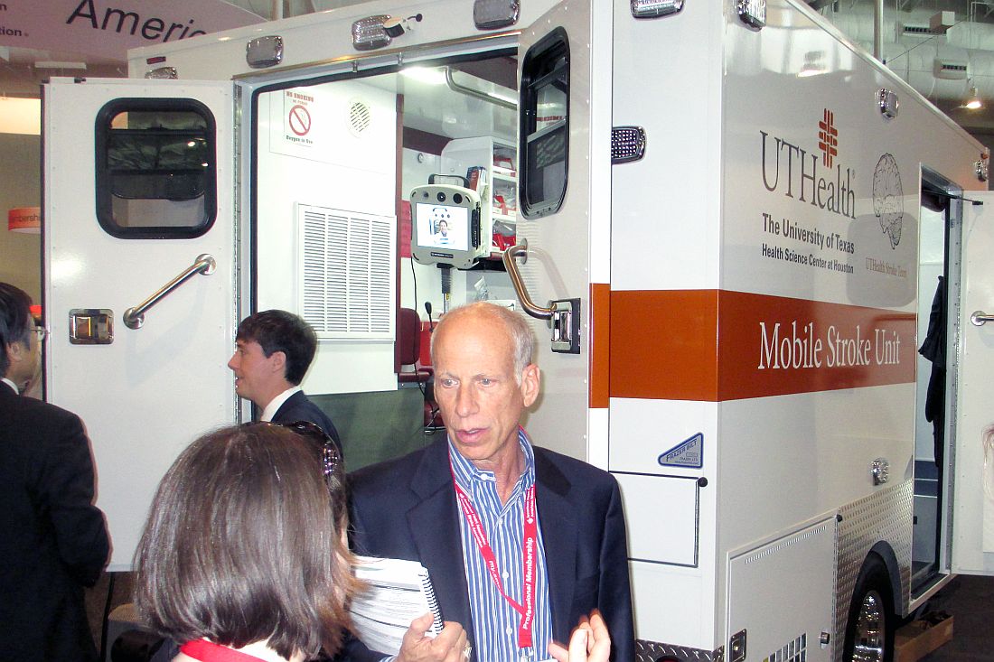
The concept behind MSUs is simple. Each one carries a CT scanner on board so that, once the vehicle’s staff identifies a patient with clinical signs of a significant–acute ischemic stroke in the field and confirms that the timing of the stroke onset suggests eligibility for tPA treatment, a CT scan can immediately be run on site to finalize tPA eligibility. The MSU staff can then begin infusing the drug in the ambulance as it speeds the patient to an appropriate hospital.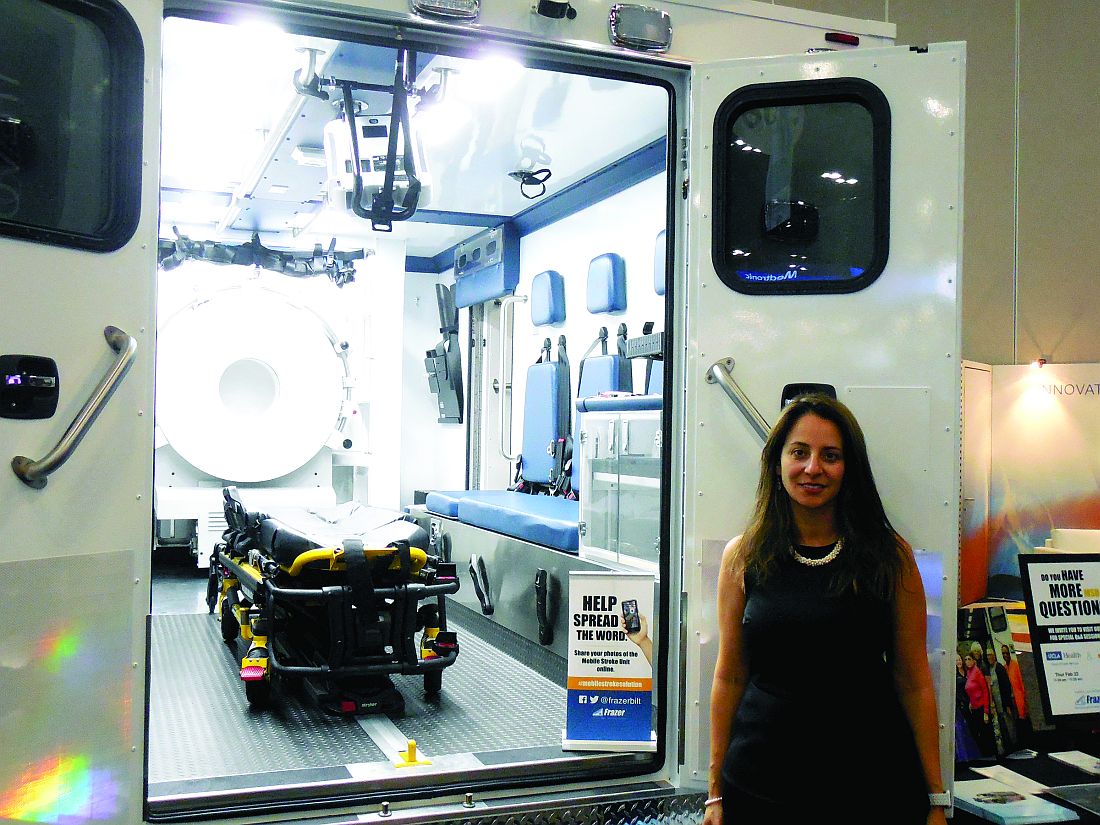
Another advantage to MSUs, in addition to quicker initiation of thrombolysis, is “getting patients to where they need to go faster and more directly,” said Dr. Nour.
“Instead of bringing patients first to a hospital that’s unable to do thrombectomy and where treatment gets slowed down, with an MSU you can give tPA on the street and go straight to a thrombectomy center,” agreed Jeffrey L. Saver, MD, professor of neurology and director of the stroke unit at UCLA. “The MSU offers the tantalizing possibility that you can give tPA with no time hit because you can give it on the way directly to a comprehensive stroke center,” Dr. Saver said during a session at the meeting.
Early data on effectiveness
Dr. Nour reported some of the best evidence for the incremental clinical benefit of MSUs based on the reduced time for starting a tPA infusion. She used data collected by the Berlin group and published in September 2016 that compared the treatment courses and outcomes of patients managed with an MSU with similar patients managed by conventional ambulance transport for whom CT scan assessment and the start of TPA treatment did not begin until the patient reached a hospital.
The German analysis showed that, in the observational Pre-Hospital Acute Neurological Therapy and Optimization of Medical Care in Stroke Patients–Study (PHANTOM-S), among 353 patients treated by conventional transport, the median time from stroke onset to thrombolysis was 112 minutes, compared with a median of 73 minutes among 305 patients managed with an MSU, a statistically significant difference. However, the study found no significant difference for its primary endpoint: the percentage of patients with a modified Rankin Scale score of 0-1 when measured 90 days after their respective strokes. This outcome occurred in 47% of the control patients managed conventionally and in 53% of those managed by an MSU, a difference that fell short of statistical significance (Lancet Neurol. 2016 Sept;15[10]:1035-43).
Dr. Nour attributed the lack of statistical significance for this primary endpoint to the relatively small number of patients enrolled in PHANTOM-S. “The study was underpowered,” she said.
Dr. Nour presented an analysis at the meeting that extrapolated the results out to 1,000 hypothetical patients and tallied the benefits that a larger number of patients could expect to receive if their outcomes paralleled those seen in the published results. It showed that, among 1,000 stroke patients treated with an MSU, 58 were expected to be free from disability 90 days later, and an additional 124 patients would have some improvement in their 90 day clinical outcome based on their modified Rankin Scale scores when compared with patients undergoing conventional hospitalization.
“If this finding was confirmed in a larger, controlled study, it would suggest that MSU-based thrombolysis has substantial clinical benefit,” she concluded.
Another recent report looked at the first 100 stroke patients treated by the Cleveland MSU during 2014. Researchers at the Cleveland Clinic and Case Western Reserve University said that 16 of those 100 patients received tPA, and the median time from their emergency call to thrombolytic treatment was 38.5 minutes faster than for 53 stroke patients treated during the same period at emergency departments operated by the Cleveland Clinic, a statistically significant difference. However, this report included no data on clinical outcomes (Neurology. 2017 March 8. doi: 10.1212/WNL.0000000000003786).
Running the financial numbers
Nailing down the incremental clinical benefit from MSUs is clearly a very important part of determining the value of this strategy, but another very practical concern is how much the service costs and whether it is financially sustainable.
“We did a cost-effectiveness analysis based on the PHANTOM-S data, and we were conservative by only looking at the benefit from early tPA treatment,” Heinrich J. Audebert, MD, professor of neurology at Charité Hospital in Berlin and head of the team running Berlin’s MSU, said during the MSU session at the meeting. “We did not take into account saving money by avoiding long-term stroke disability and just considered the cost of [immediate] care and the quality adjusted life years. We calculated a cost of $35,000 per quality adjusted life year, which is absolutely acceptable.”
He cautioned that this analysis was not based on actual outcomes but on the numbers needed to treat calculated from the PHANTOM-S results. “We need to now show this in controlled trials,” he admitted.
Income from transport reimbursement, currently $500 per trip, and reimbursements of $17,000 above costs for administering tPA and of roughly $40,000 above costs for performing thrombectomy are balancing these costs. Based on an estimated additional one thrombolysis case per month and one additional thrombectomy case per month, the MSU yields a potential incremental income to the hospital running the MSU of about $3.8 million over 5 years, enough to balance the operating cost, Dr. Grotta said.
A key part of controlling costs is having the neurologic consult done via a telemedicine link rather than by neurologist at the MSU. “Telemedicine reduces operational costs and improves efficiency,” noted M. Shazam Hussain, MD, interim director of the Cerebrovascular Center at the Cleveland Clinic. “Cost effectiveness is a very important part of the concept” of MSUs, he said at the session.
The Houston group reported results from a study that directly compared the diagnostic performance of an onboard neurologist with that of a telemedicine neurologist linked in remotely during MSU deployments for 174 patients. For these cases, the two neurologists each made an independent diagnosis that the researchers then compared. The two diagnoses concurred for 88% of the cases, Tzu-Ching Wu, MD, reported at the meeting. This rate of agreement matched the incidence of concordance between two neurologists who independently assessed the same patients at the hospital (Stroke. 2017 Jan;48[1]:222-4), said Dr. Wu, a vascular neurologist and director of the telemedicine program at the University of Texas Health Science Center in Houston.
“The results support using telemedicine as the primary means of assessment on the MSU,” said Dr. Wu. “This may enhance MSU efficiency and reduce costs.” His group’s next study of MSU telemedicine will compare the time needed to make a diagnostic decision using the two approaches, something not formally examined in the study he reported.
However, telemedicine assessment of CT results gathered in a MSU has one major limitation: the time needed to transmit the huge amount of information in a CT angiogram.
The MSU used by clinicians at the University of Tennessee, Memphis, incorporates an extremely powerful battery that enables “full CT scanner capability with a moving gantry,” said Andrei V. Alexandrov, MD, professor and chairman of neurology at the university. With this set up “we can do in-the-field multiphasic CT–angiography from the aortic arch up within 4 minutes. The challenge of doing this is simple. It’s 1.7 gigabytes of data,” which would take a prohibitively long time to transmit from a remote site, he explained. As a result, the complete set of images from the field CT angiogram are delivered on a memory stick to the attending hospital neurologist once the MSU returns.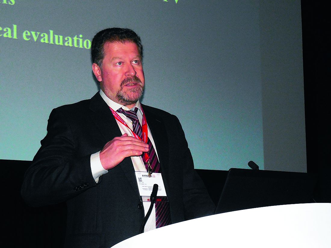
Waiting for more data
Despite these advances and the steady recent growth of MSUs, significant skepticism remains. “While mobile stroke units seem like a good idea and there is genuine hope that they will improve outcomes for selected stroke patients, there is not yet any evidence that this is the case,” wrote Bryan Bledsoe, DO, in a January 2017 editorial in the Journal of Emergency Medical Services. “They are expensive and financially non-sustainable. Without widespread deployment, they stand to benefit few, if any, patients. The money spent on these devices would be better spent on improving the current EMS system including paramedic education, the availability of stroke centers, and on the early recognition of ELVO [emergent large vessel occlusion] strokes,” wrote Dr. Bledsoe, professor of emergency medicine at the University of Nevada in Las Vegas.
Two other experts voiced concerns about MSUs in an editorial that accompanied a Cleveland Clinic report in March. “Even if MSUs meet an acceptable societal threshold for cost effectiveness, cost efficiency may prove a taller order to achieve return on investment for individual health systems and communities,” wrote Andrew M. Southerland, MD, and Ethan S. Brandler, MD (Neurology. 2017 March 8. doi: 10.1212/WNL.0000000000003833). They cited the Cleveland report, which noted that the group’s first 100 MSU-treated patients came from a total of 317 MSU deployments and included 217 trips that were canceled prior to the MSU’s arrival at the patient’s location. In Berlin’s initial experience, more than 2,000 MSU deployments led to 200 TPA treatments and 349 cancellations before arrival, noted Dr. Southerland, a neurologist at the University of Virginia in Charlottesville, and Dr. Brandler, an emergency medicine physician at Stony Brook (N.Y.) University.
“Hope remains that future trials may demonstrate the ultimate potential of mobile stroke units to improve long-term outcomes for more patients by treating them more quickly and effectively. In the meantime, ongoing efforts are needed to streamline MSU cost and efficiency,” they wrote.
Proponents of MSUs agree that what’s needed now are more data to prove efficacy and cost effectiveness, as well as better integration into EMS programs. The first opportunity for documenting the clinical impact of MSUs on larger numbers of U.S. patients may be from the BEnefits of Stroke Treatment Delivered using a Mobile Stroke Unit Compared to Standard Management by Emergency Medical Services (BEST-MSU) Study, funded by the Patient-Centered Outcomes Research Institute. This study is collecting data from the MSU programs in Denver Houston, and Memphis. Although currently designed to enroll 697 patients, Dr. Grotta said he hopes to kick that up to 1,000 patients.
“We are following the healthcare use and its cost for every enrolled MSU and conventional patient for 1 year,” Dr. Grotta explained in an interview. He hopes these results will provide the data needed to move MSUs from investigational status to routine and reimbursable care.
Dr. Southerland and Dr. Brandler suggested that “making MSUs multipurpose vehicles might also enhance cost-effectiveness,” an option that Dr. Grotta and his colleagues embrace. The MSUs on U.S. roads already also treat patients with intracranial hemorrhages using blood pressure reduction medications. Other neurologic diagnoses considered potential targets for MSU interventions include encephalopathy, seizures, central nervous system–tumors, and infections.
Stroke is a prime example of “a disease that is extremely time sensitive, and for the first time the field of stroke is ahead of the rest of the medical world in trying to speed treatment,” Dr. Grotta said. “We could add other diagnostic equipment monitored by telemedicine specialists. The MSU concept could be expanded to make it much more cost effective” and spur wider adoption by EMS, he suggested.
Dr. Grotta is a consultant to Frazer, a company that produces mobile stroke units, and to Stryker Corporation, and he has received research support from Genentech. Dr. Saver has been a consultant to and received research support from St. Jude. Dr. Audebert has received honoraria from Pfizer, Boehringer Ingelheim, Bristol-Myers Squibb, and Ever Pharma. He has been a consultant to ReNeuron, and he has served as an expert witness for Pfizer and for Lundbeck. Dr. Hussain has been a consultant to Pulsar. Dr. Alexandrov has been a speaker for Genentech. Dr. Nour, Dr. Wu, Dr. Bledsoe, Dr. Southerland, and Dr. Brandler had no disclosures.
mzoler@frontlinemedcom.com
On Twitter @mitchelzoler
EXPERT ANALYSIS FROM THE INTERNATIONAL STROKE CONFERENCE