User login
Mitchel is a reporter for MDedge based in the Philadelphia area. He started with the company in 1992, when it was International Medical News Group (IMNG), and has since covered a range of medical specialties. Mitchel trained as a virologist at Roswell Park Memorial Institute in Buffalo, and then worked briefly as a researcher at Boston Children's Hospital before pivoting to journalism as a AAAS Mass Media Fellow in 1980. His first reporting job was with Science Digest magazine, and from the mid-1980s to early-1990s he was a reporter with Medical World News. @mitchelzoler
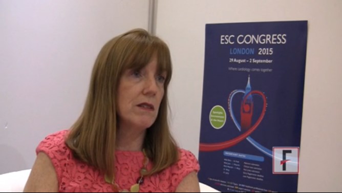
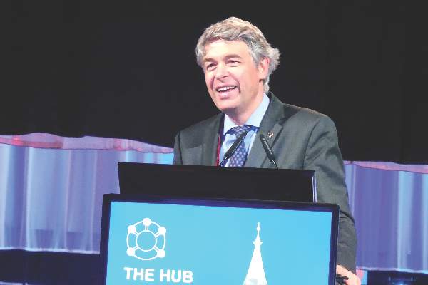
VIDEO: CardioMEMS early adopters must sort out data use
Use of CardioMEMS by U.S. clinicians to measure pulmonary artery pressures daily in more advanced heart failure patients is currently quite variable around the country, with some enthusiastic, early adopters of the technology while many other programs have so far opted not to invest in the technology
Among the early adopters of CardioMEMS many programs are “trying to sort through” how to use the data they collect from patients using the implanted device, Dr. Mary Norine Walsh said during an interview. “Each clinical teams needs to work out not only how the data will be interpreted but also who will act on it, and how will the patient get the information,” said Dr. Walsh, medical director of the heart failure and transplantation program at the St. Vincent Heart Center in Indianapolis. “We need to get the data to patients quickly,” she said.
Despite this uncertainty over how to best use and disseminate the pulmonary artery pressure data collected using CardioMEMS, Dr. Walsh voiced confidence that it will ultimately make a difference for how advanced heart failure patients are managed and their rehospitalization rates. But for the time being “how much CardioMEMS and other technologies can have an impact on avoidable readmissions is not yet clear” for real-world practice, she said.
Dr. Walsh had no disclosures.
The video associated with this article is no longer available on this site. Please view all of our videos on the MDedge YouTube channel
On Twitter @mitchelzoler
Empagliflozin’s triumph respins FDA’s diabetes drug mandate
What a difference a few weeks and the unexpected results from a home run–hitting trial made in the medical community’s take on the Food and Drug Administration’s demand to assess the cardiovascular safety of drugs for type 2 diabetes.
On August 31, the relatively ho-hum, noninferiority results from the two latest, large outcomes trials to assess the cardiovascular safety of new oral hypoglycemic drugs led to bashing of these trials by several cardiologists speaking from the main stage of the European Society of Cardiology’s (ESC) annual congress in London. On Sept. 17, less than 3 weeks later, the remarkably beneficial survival effect seen with another new oral hypoglycemic, empagliflozin (Jardiance) in a cardiovascular safety study and reported at the European Association for the Study of Diabetes (EASD) annual meeting in Stockholm and in a simultaneously-published article resulted in researchers calling the results “amazing” and audience members hailing the trial a “landmark.”
Is the biomedical field really so fickle, or did physicians just need proof of the serendipitous possibilities when running a large outcomes clinical trial using a potent drug with underexplored potential?
The back story to this attitudinal change began in 2008, when the FDA issued guidance that called on companies to collect evidence for the cardiovascular safety of new drugs that treat type 2 diabetes, a stand that launched a fleet of big studies. Reports of the results from the first two trials conducted to meet this mandate came out 2 years ago from studies of new dipeptidyl peptidase 4 (DPP-4) inhibitors, the SAVOR-TIMI-53 trial of saxagliptin (Onglyza), and the EXAMINE trial of alogliptin (Nesina). Results from the two studies were notable not only for generally showing cardiovascular safety but also for showing a small but apparently real uptick in the rate of hospitalization for heart failure associated with saxagliptin treatment.
Findings from the next two trials in the series came out in reports in June at the American Diabetes Association annual meeting in Boston, with follow-up reports on the same two studies presented at the ESC meeting on August 31. Results from the TECOS study of yet a third new DDP-4 inhibitor, sitagliptin (Januvia), showed no cardiovascular safety signals with notably no suggestion of causing any sort of heart failure problem. The fourth study, ELIXA, was the first of the FDA mandates to report on a drug from a different class, lixisenatide (Lyxumia), a glucagonlike–peptide 1 receptor agonist, and it too provided a clean outcome with no excess of cardiovascular events, compared with the control arm.
It was this steady drumbeat of neutral results showing no evidence of cardiovascular harm – aside from the problem of heart-failure exacerbation with saxagliptin – that triggered criticism of the FDA’s mandate and its consequences at ESC, specifically in remarks by Dr. Philippe Gabriel Steg, the ESC’s designated discussant for the ELIXA report.
“Are these trials a waste of resources?” asked Dr. Steg in his comments. He questioned how representative and generalizable the studies are, by enrolling patients at very high cardiovascular risk, usually patients with a recent acute coronary syndrome event. He also critiqued the trials’ relatively short follow-up, on the order of 2-3 years, saying that this is generally too brief to demonstrate a potential benefit. Most of all, he questioned launching a series of safety trials that have enrolled a total of roughly 150,000 patients in randomized, controlled trials designed to test noninferiority for cardiovascular safety against standard-treatment control arms, studies that he posited divert money and resources from investigations focused on finding new treatments and could be accomplished in a different way for a lot less money.
Dr. Steg further complained that the series of neutral results have fostered misleading beliefs about their implications. The noninferiority results “have been mistakenly interpreted as lack of efficacy,” resulting in “greater skepticism among nonspecialists about treatment of diabetes and the need to control glycemia,” he said. He also said that the noninferiority design shortchanged enrolled patients, inconveniencing them by the demands of the trial when the best they could expect was to fare no worse than control patients.
Even more striking, Dr. Steg’s critique received immediate support from the next two speakers at the meeting, Dr. Jaakko Tuomilehto, designated discussant for the TECOS sidy, and then Dr. Thomas M. MacDonald, who followed at the podium to report results from an entirely different study. “I fully agree. I think it’s a waste of resources,” said Dr. Tuomilehto.
Was it reasonable for these trialists to anticipate anything more from studies designed to confirm cardiovascular safety? “Some think the results [from these four trials] have been disappointing, others think it’s what you would expect,” commented Dr. Bernard Zinman a few weeks later in September at the EASD meeting.
I spoke with Dr. Steg soon after he lambasted the FDA-mandated cardiovascular safety trials at ESC, and he further explained to me that while he didn’t dispute the importance of better evaluating the cardiovascular safety of these oral hypoglycemic drugs, he believed this could be more efficiently assessed with a more comprehensive approach to postmarketing surveillance. He also highlighted the importance of examining cardiovascular safety in type 2 diabetes patients who better resemble real-world patients instead of in the extreme high-risk patients who have enrolled in the trials. Finally, Dr. Steg told me that he wasn’t nearly as skeptical of these trials when the FDA first announced its guidance 7 years ago, but grew increasingly dismayed as he saw how the laudable goal of collecting data on cardiovascular safety led to such profligate and questionable studies.
Attitudes shifted dramatically fewer than 3 weeks later at EASD when the empagliflozin results came out from the EMPA-REG OUTCOME study. Randomizing 7,028 patients, the study showed that treatment with either of two dosages of empagliflozin, a blocker of the sodium glucose cotransporter 2 (SGLT-2) protein in the kidney, on top of standard oral hypoglycemic and other standard lipid-lowering and antihypertensive therapies produced a “wonderful and quite profound” 38% relative risk reduction in the rate of cardiovascular death, a 32% drop in all-cause death, and a 35% cut in heart failure hospitalizations, noted trial discussant Dr. Hertzel C. Gerstein. The “unexpected” results “will open new research,” Dr. Gerstein added when speaking at EASD. “The claims that people have made that large, randomized clinical trials in patients with diabetes are not needed appear unfounded. Here is a perfect case; we need to do these trials to find lifesaving therapies.”
The empagliflozin results were so dramatic and surprising that you can’t help wondering what would have happened if the FDA had not issued its mandate for cardiovascular safety studies in 2008. Would a trial like EMPA-REG OUTCOME ever have been done? Would this effect, which the researchers suggested was likely a class effect for all SGLT-2 inhibitors, ever have been found?
The empagliflozin findings also raise doubts about the unwavering reliance cardiovascular studies have had on a primary combined outcome that marries patient survival with the incidence of nonfatal ischemic events, strokes, and MIs. The EMPA-REG OUTCOME showed a major survival benefit while simultaneously having no discernible impact on nonfatal ischemic events.
The question of what aside from the FDA’s mandate might have gotten a trial like EMPA-REG OUTCOME off the ground is especially important because the findings hint that SGLT-2 inhibitors may have treatment implications that go beyond patients with type 2 diabetes. The rapid onset of the mortality benefit seemed to point to the diuretic effect of the drug as a major mediating factor, said several at the EASD session. Might SGLT-2 inhibitors be the eagerly sought new option for managing fluid congestion in the organs of patients with acute heart failure? Aside from survival it was heart failure hospitalization where the treatment showed benefit. Better fluid management is currently a desperate need as heart failure physicians increasingly recognize that diuretic treatment alone often is inadequate for purging excess fluid from affected organs in patients with acute heart failure episodes.
Some critics of the FDA mandate weren’t willing to accept that it made EMPA-REG OUTCOME possible. A New York Times news article published on September 18 quoted Dr. Robert E. Ratner from the American Diabetes Association as saying that studies like EMPA-REG OUTCOME get launched through the initiative of drug companies acting on their own, without FDA prodding. Perhaps Dr. Ratner is correct, but which studies out there now reflect this? It certainly doesn’t seem like companies have been motivated to organize trials that assess the cardiovascular impact of oral hypoglycemic drugs outside of meeting the FDA’s requirements.
A comment that nicely summed up the game-changing impact of the empagliflozin study came from a member of the EASD audience in Stockholm, the last attendee to pose a comment from the floor as the session wrapped up on Sept. 17. “I hope everyone in the audience understands what has happened, how important this is, a real landmark study,” commented Dr. Klas Malmberg, a cardiologist and diabetes researcher at the Karolinska Institute in Stockholm.
This unexpected finding from a large outcomes study looks like it may substantially alter the way type 2 diabetes and perhaps other diseases will get treated in the future. The finding also singlehandedly morphed the FDA’s safety study mandate from “a waste of resources” to the heroic driver behind a landmark study.
On Twitter @mitchelzoler
ESC: Obstructive sleep apnea often complicates heart failure
LONDON – The majority of patients with severe heart failure had sleep-disordered breathing and, in most affected patients, this manifested as obstructive sleep apnea, in an analysis of more than 1,000 German heart failure patients enrolled in a multicenter registry.
“The vast majority of heart failure patients with sleep-disordered breathing [SDB] have obstructive sleep apnea, which differs from previous results,” said Dr. Olaf Oldenburg at the annual congress of the European Society of Cardiology. Possible reasons why this German registry had different findings, compared with prior reports, were its inclusion of heart failure patients with milder symptoms, inclusion of patients with preserved ejection fraction, and inclusion of more women, suggested Dr. Oldenburg, director of the sleep laboratory at the Heart and Diabetes Center of Ruhr University of Bochum in Bad Oeynhausen, Germany.
His finding that nearly two-thirds of the heart failure patients with SDB in this registry had obstructive sleep apnea and that one-third had moderate or severe obstructive sleep apnea was notable because this remains a form of sleep-disordered breathing that can be treated, he said. “There is still enough evidence to treat obstructive sleep apnea” in heart failure patients when it has a moderate or severe presentation, which is defined as causing 15 or more apnea-hypopnea events/hour during sleep. “Obstructive sleep apnea is definitely not a compensatory mechanism in heart failure,” Dr. Oldenburg said.
He highlighted the ongoing need to treat more severe obstructive sleep apnea in heart failure patients because this form of SDB sharply contrasts with the results of the SERVE-HF trial, also reported at the congress, which showed that in patients with advanced heart failure and central sleep apnea nocturnal treatment with adaptive servo ventilation failed to provide benefit and also appeared to boost patient mortality (N Engl J Med. 2015 Sep 17;373[12]:1095-105).
Following that report “we need to think about which heart failure patients to treat” with nocturnal ventilation, and differentiate between heart failure patients with obstructive sleep apnea and those with central sleep apnea, Dr. Oldenburg said.
The data he reported came from the SchlaHF-XT (Sleep Disordered Breathing in Heart Failure) registry, which enrolled patients with heart failure and reduced or preserved ejection fraction and any New York Heart Association functional class treated either at German hospitals or in physician offices. He reported data for 1,186 fully assessed and classified patients, who averaged 68 years old and two-thirds of whom were men. Slightly more than half had heart failure with reduced ejection fraction, and about half had New York Heart Association class II heart failure, a quarter had class III heart failure, with the remaining patients divided roughly equally between class I and IV.
Screening for SDB showed that 24% had no SDB, 37% had mild SBD, 21% had moderate SDB, and 19% had severe SDB (percentages total 101% because of rounding). Among those with SDB, 64% had obstructive sleep apnea, 22% had central sleep apnea, and the remaining 14% had either a mixed form of sleep apnea or were not classifiable.
The analysis also showed that moderate and severe SDB, the forms that require treatment, occurred more often among patients with heart failure and reduced ejection fraction, 43%, compared with patients with heart failure and preserved ejection fraction, who had a 36% prevalence of SDB requiring treatment. Moderate or severe central sleep apnea occurred in 15% of patients with reduced ejection fraction and in 9% of patients with preserved ejection fraction.
A second report at the congress by Dr. Oldenburg showed that the duration of time when a patient’s oxygen saturation fell below 90% was a better gauge of the severity of SDB than was the traditional measure of the apnea-hypopnea index (AHI), the average number of apnea-hypopnea episodes a patient has during an hour of sleep. For this analysis, he used data collected on 963 patients with chronic, stable heart failure with reduced ejection fraction who underwent a comprehensive sleep study with pulse oximetry measurements during 2002-2013.
The results showed that while the measured AHI significantly linked with the 5-year mortality rate of these patients, the relationship became statistically insignificant after researchers adjusted for age, sex, body mass index, heart failure severity, ejection fraction, medications, and other clinical variables.
In contrast, the average time a patient spent with an oxygen saturation level below 90% overnight strong linked with 5-year mortality even after adjusting for all these covariables. The analysis showed that each hour of sleep a heart failure patient spent with an oxygen saturation level below 90% linked with a relative 16% reduction in 5-year survival. Patients in the quartile with the greatest amount of time spent with a oxygen saturation level below 90% had a 50% 5-year mortality rate, while those in the quartile with the least amount of time spent with severely depressed oxygen saturation had a 30% 5-year mortality rate.
Based on this finding “you need to look at the effect of SDB and not just the apnea-hypopnea index” when assessing SDB in patients with heart failure, Dr. Oldenburg said.
On Twitter @mitchelzoler
AT THE ESC CONGRESS 2015
Key clinical point: Significant sleep disordered breathing is common among heart failure patients, most often manifesting as obstructive sleep apnea.
Major finding: In a real-world population of heart failure patients 64% of those with sleep disordered breathing had obstructive sleep apnea.
Data source: The SchlaHF-XT registry with 1,1816 heart failure patients enrolled at several German hospitals and private practices.
Disclosures: The SchlaHF-XT registry was sponsored by ResMed. Dr. Oldenburg said that he has received research support from several companies.

ESC: Registry confirms liver dysfunction’s heart-failure importance
LONDON – Hepatic dysfunction, which is beginning to be appreciated as an important complication of patients with acute heart failure, boosted the relative rate of death in patients with acute heart failure by 57% in an analysis of more than 5,000 patients followed for a year in a registry maintained by the European Society of Cardiology (ESC).
The registry data also showed that at entry into the registry the acute heart failure patients had an 8% prevalence of hepatic dysfunction.
These observations on the prevalence and impact of liver dysfunction in acute heart failure patients highlight a new recognition that acute heart failure can trigger morbidity and mortality through its hepatic effects in a manner similar to the better-appreciated effect that acute heart failure has on renal and pulmonary function, Dr. Alexandre Mebazaa said at the annual congress of the European Society of Cardiology.

“Everybody knows about the kidney, but not many know about the liver,” said Dr. Mebazaa in an interview. Recent studies on mechanisms of organ damage during acute heart failure episodes indicates that the liver, kidneys, and lungs are all damaged by fluid congestion in these organs, he said. “Fluid overload leads to organ dysfunction,” and this complication seems relatively resistant to the diuretic treatment that acute heart failure patients typically receive when they are hospitalized for decompensation episodes. “New approaches are needed to remove fluid from the organs. Diuretics remove fluid from the blood, but not from the organs,” said Dr. Mebazaa, a professor of anesthesiology and critical care medicine at Lariboisière Hospital in Paris.
The data Dr. Mebazaa reported at the congress were the first 1-year follow-up data from the ESC’s Heart Failure Long-Term Registry, begun in 2012 and involving centers from more than 30 countries in Europe as well as in North Africa and the Middle East. The panel of ESC members who oversee this registry “selected centers dedicated to the database,” he said.
The registry followed 5,039 patients with acute heart failure for at least 1 year, as well as 7,401 patients diagnosed with chronic heart failure. Another striking, though not surprising finding of the 1-year follow-up data was the disparity in mortality and hospitalization rates between these two subgroups.
After 1 year, the mortality rate was 24% among the acute heart failure patients, compared with 6% among the chronic heart failure patients. Dr. Mebazaa called the acute heart failure mortality rate “terrible.”
Hospitalization rates in the two subgroups over the 1-year follow-up ran to 19% among the acute heart failure patients ands 10% for chronic heart failure patients. The combined rate of death or hospitalization over 1 year was 36% in the acute heart failure patients and 15% in those with chronic heart failure.
These data “show that we really need new treatments for acute heart failure to reduce rehospitalizations and deaths,” Dr. Mebazaa said.
A multivariate adjusted analysis identified several baseline factors that significantly linked with mortality among the acute heart-failure patients during the 1-year follow-up. In addition to hepatic dysfunction linking with a 57% increased rate of death other factors included age, which linked with a 24% increased rate for every 5 years of older age; New York Heart Association class III or IV severity, which linked with 50% higher mortality; renal dysfunction, which linked with a 52% higher mortality rate; and aortic stenosis, linked with a 54% increased mortality rate.
The finding that hepatic dysfunction was an independent mortality risk in acute heart failure confirmed a finding that Dr. Mebazaa and his associates reported in 2013 in a post hoc analysis of 1,134 patients with acute heart failure who had been enrolled in a drug-intervention trial (Eur Heart J. 2013 Sep 15;34:742-9). The registry results confirm for the first time that finding in an independent and much larger group of patients, Dr. Mebazaa said.
On Twitter @mitchelzoler
LONDON – Hepatic dysfunction, which is beginning to be appreciated as an important complication of patients with acute heart failure, boosted the relative rate of death in patients with acute heart failure by 57% in an analysis of more than 5,000 patients followed for a year in a registry maintained by the European Society of Cardiology (ESC).
The registry data also showed that at entry into the registry the acute heart failure patients had an 8% prevalence of hepatic dysfunction.
These observations on the prevalence and impact of liver dysfunction in acute heart failure patients highlight a new recognition that acute heart failure can trigger morbidity and mortality through its hepatic effects in a manner similar to the better-appreciated effect that acute heart failure has on renal and pulmonary function, Dr. Alexandre Mebazaa said at the annual congress of the European Society of Cardiology.
“Everybody knows about the kidney, but not many know about the liver,” said Dr. Mebazaa in an interview. Recent studies on mechanisms of organ damage during acute heart failure episodes indicates that the liver, kidneys, and lungs are all damaged by fluid congestion in these organs, he said. “Fluid overload leads to organ dysfunction,” and this complication seems relatively resistant to the diuretic treatment that acute heart failure patients typically receive when they are hospitalized for decompensation episodes. “New approaches are needed to remove fluid from the organs. Diuretics remove fluid from the blood, but not from the organs,” said Dr. Mebazaa, a professor of anesthesiology and critical care medicine at Lariboisière Hospital in Paris.
The data Dr. Mebazaa reported at the congress were the first 1-year follow-up data from the ESC’s Heart Failure Long-Term Registry, begun in 2012 and involving centers from more than 30 countries in Europe as well as in North Africa and the Middle East. The panel of ESC members who oversee this registry “selected centers dedicated to the database,” he said.
The registry followed 5,039 patients with acute heart failure for at least 1 year, as well as 7,401 patients diagnosed with chronic heart failure. Another striking, though not surprising finding of the 1-year follow-up data was the disparity in mortality and hospitalization rates between these two subgroups.
After 1 year, the mortality rate was 24% among the acute heart failure patients, compared with 6% among the chronic heart failure patients. Dr. Mebazaa called the acute heart failure mortality rate “terrible.”
Hospitalization rates in the two subgroups over the 1-year follow-up ran to 19% among the acute heart failure patients ands 10% for chronic heart failure patients. The combined rate of death or hospitalization over 1 year was 36% in the acute heart failure patients and 15% in those with chronic heart failure.
These data “show that we really need new treatments for acute heart failure to reduce rehospitalizations and deaths,” Dr. Mebazaa said.
A multivariate adjusted analysis identified several baseline factors that significantly linked with mortality among the acute heart-failure patients during the 1-year follow-up. In addition to hepatic dysfunction linking with a 57% increased rate of death other factors included age, which linked with a 24% increased rate for every 5 years of older age; New York Heart Association class III or IV severity, which linked with 50% higher mortality; renal dysfunction, which linked with a 52% higher mortality rate; and aortic stenosis, linked with a 54% increased mortality rate.
The finding that hepatic dysfunction was an independent mortality risk in acute heart failure confirmed a finding that Dr. Mebazaa and his associates reported in 2013 in a post hoc analysis of 1,134 patients with acute heart failure who had been enrolled in a drug-intervention trial (Eur Heart J. 2013 Sep 15;34:742-9). The registry results confirm for the first time that finding in an independent and much larger group of patients, Dr. Mebazaa said.
On Twitter @mitchelzoler
AT THE ESC CONGRESS 2015
Key clinical point: Liver dysfunction was a statistically significant, independent predictor of 1-year mortality in acute heart-failure patients.
Major finding: Acute heart-failure patients had a 57% relative increased rate of 1-year death, compared with patients without liver dysfunction.
Data source: One-year follow-up of 5,039 patients with acute heart failure enrolled in a multinational registry maintained by the European Society of Cardiology.
Disclosures: Dr. Mebazaa has received speaking honoraria and consulting fees from 11 drug or diagnostic companies.
First MRI-compatible Implantable Defibrillator Approved
The Food and Drug Administration approved the first MRI-compatible implantable cardioverter defibrillator on Sept. 14, starting a new era of convenience and flexibility when performing MRI scans on patients who carry this type of cardiac implant.
Eventually most, if not all, implantable cardioverter defibrillators (ICDs) will have MRI compatibility, electrophysiologists predicted, a change that’s already been occurring for pacemakers following FDA approval of the first MRI-compatible pacemaker in 2011.

“This is a major step forward and sets a new standard” for ICDs, commented Dr. Rod S. Passman, professor of medicine and electrophysiologist at Northwestern University, Chicago, who has not been involved in developing MRI-compatible ICDs. “If a patient has an ICD, they should be able to go to any community emergency room and get what could be a life-saving MRI. MRI scans of patients with ICDs should not be limited to experienced academic centers. Ultimately all [cardiac] devices will be MRI compatible,” Dr. Passman said in an interview.
“There is no downside” to the newly approved, MRI-compatible ICD, said Dr. Michael R. Gold, an electrophysiologist at the Medical University of South Carolina, Charleston, who led the pivotal study that showed the device’s safety and efficacy during and after MRI scanning. Dr. Gold first reported results from the Evera MRI Study at the Heart Rhythm Society annual meeting in May and in a concurrently published report (J Am Soc Cardiol. 2015;65[24]:2581-8). The study enrolled 275 patients at 42 centers. Medtronic, the company that makes the newly approved ICD, plans to begin U.S. sales the week of Sept. 20, a company spokesperson said.
“I’m not sure I’d use it in all patients” who need an ICD once it’s on the market, admitted Dr. Gold. For example, some patients already have ICD leads in place that are not MRI compatible, so placing an ICD capable of MRI exposure in such patients would be moot, he noted. In other cases, the patient might best receive an ICD model made by a different manufacturer because of other device features.
“Every physician will need to choose the ICD that is best for each patient, so I don’t think we’ll see immediate, wholesale adoption [of the MRI-compatible ICD], but I expect the field will move in this direction,” Dr. Passman said.

Dr. Gold agreed that, as time goes by, the ICD models sold for U.S. patients increasingly will be MRI compatible, although that might take several years to happen.
For example, the transition to pacemakers that are MRI compatible has been gradual and incomplete, even though the first of these came onto the U.S. market in 2011.
“Only two pacemaker manufacturers sell MRI-compatible devices in the United States,” noted Dr. Gold, professor of medicine and director of the division of adult cardiology at the university. Several other manufacturers produce MRI-compatible pacemakers, but so far they have not sought FDA approval for these and they only sell them outside the United States, he noted.
“Many physicians don’t think about a patient’s long-term needs [for MRI] and may instead focus on the device they are most comfortable with” or a device with other attractive features, Dr. Passman said.
The possibility of performing MRI on a patient with an ICD is not totally new. A relatively small number of sophisticated U.S. centers have been performing MRIs on patients with conventional ICDs or pacemakers for several years, especially in circumstances when the MRI was considered vitally needed. Some of these centers have participated in the MagnaSafe registry, which reported results documenting the safety and efficacy of the procedure at a cardiology meeting in 2013.
“If you take certain precautions, the risk from MRI appears to be quite low in the most experienced hands, although even in experienced hands adverse events have been reported,” said Dr. Passman. Performing an MRI on a patient with a conventional ICD or pacemaker is also a relatively labor-intensive process that requires temporarily reprogramming the device, closely monitoring the patient during the MRI scan, and then checking out the device thoroughly after the scan to make sure it is functioning correctly.
In addition to the extra labor and uncertainty about outcome, running an MRI scan on a conventional cardiac device is generally not reimbursed by insurers and creates medicolegal exposure, Dr. Gold noted. “Even though it can be done, it often is not done, and it clearly compromises patient care,” he said.
Although the Medtronic unit was the first to get FDA approval, a competitor model seems on track to also hit the U.S. market soon. Two different ICD models made by Biotronik showed safety and efficacy in a study with 153 patients (Heart Rhythm. 2015 doi. org/10.1016/j.hrthm.2015.06.002). Biotronik has submitted an application to the FDA to market these ICDs and associated leads as MRI compatible, and an agency decision is pending, a company spokeswoman said.
The Evera MRI Study was sponsored by Medtronic, which produces and will market the Evera ICD. Dr. Gold has been a consultant to and has received research and speaker funding from Medtronic as well as from Boston Scientific and St. Jude. Dr. Passman has received research support from Medtronic but not for work on ICDs.
The Food and Drug Administration approved the first MRI-compatible implantable cardioverter defibrillator on Sept. 14, starting a new era of convenience and flexibility when performing MRI scans on patients who carry this type of cardiac implant.
Eventually most, if not all, implantable cardioverter defibrillators (ICDs) will have MRI compatibility, electrophysiologists predicted, a change that’s already been occurring for pacemakers following FDA approval of the first MRI-compatible pacemaker in 2011.
“This is a major step forward and sets a new standard” for ICDs, commented Dr. Rod S. Passman, professor of medicine and electrophysiologist at Northwestern University, Chicago, who has not been involved in developing MRI-compatible ICDs. “If a patient has an ICD, they should be able to go to any community emergency room and get what could be a life-saving MRI. MRI scans of patients with ICDs should not be limited to experienced academic centers. Ultimately all [cardiac] devices will be MRI compatible,” Dr. Passman said in an interview.
“There is no downside” to the newly approved, MRI-compatible ICD, said Dr. Michael R. Gold, an electrophysiologist at the Medical University of South Carolina, Charleston, who led the pivotal study that showed the device’s safety and efficacy during and after MRI scanning. Dr. Gold first reported results from the Evera MRI Study at the Heart Rhythm Society annual meeting in May and in a concurrently published report (J Am Soc Cardiol. 2015;65[24]:2581-8). The study enrolled 275 patients at 42 centers. Medtronic, the company that makes the newly approved ICD, plans to begin U.S. sales the week of Sept. 20, a company spokesperson said.
“I’m not sure I’d use it in all patients” who need an ICD once it’s on the market, admitted Dr. Gold. For example, some patients already have ICD leads in place that are not MRI compatible, so placing an ICD capable of MRI exposure in such patients would be moot, he noted. In other cases, the patient might best receive an ICD model made by a different manufacturer because of other device features.
“Every physician will need to choose the ICD that is best for each patient, so I don’t think we’ll see immediate, wholesale adoption [of the MRI-compatible ICD], but I expect the field will move in this direction,” Dr. Passman said.
Dr. Gold agreed that, as time goes by, the ICD models sold for U.S. patients increasingly will be MRI compatible, although that might take several years to happen.
For example, the transition to pacemakers that are MRI compatible has been gradual and incomplete, even though the first of these came onto the U.S. market in 2011.
“Only two pacemaker manufacturers sell MRI-compatible devices in the United States,” noted Dr. Gold, professor of medicine and director of the division of adult cardiology at the university. Several other manufacturers produce MRI-compatible pacemakers, but so far they have not sought FDA approval for these and they only sell them outside the United States, he noted.
“Many physicians don’t think about a patient’s long-term needs [for MRI] and may instead focus on the device they are most comfortable with” or a device with other attractive features, Dr. Passman said.
The possibility of performing MRI on a patient with an ICD is not totally new. A relatively small number of sophisticated U.S. centers have been performing MRIs on patients with conventional ICDs or pacemakers for several years, especially in circumstances when the MRI was considered vitally needed. Some of these centers have participated in the MagnaSafe registry, which reported results documenting the safety and efficacy of the procedure at a cardiology meeting in 2013.
“If you take certain precautions, the risk from MRI appears to be quite low in the most experienced hands, although even in experienced hands adverse events have been reported,” said Dr. Passman. Performing an MRI on a patient with a conventional ICD or pacemaker is also a relatively labor-intensive process that requires temporarily reprogramming the device, closely monitoring the patient during the MRI scan, and then checking out the device thoroughly after the scan to make sure it is functioning correctly.
In addition to the extra labor and uncertainty about outcome, running an MRI scan on a conventional cardiac device is generally not reimbursed by insurers and creates medicolegal exposure, Dr. Gold noted. “Even though it can be done, it often is not done, and it clearly compromises patient care,” he said.
Although the Medtronic unit was the first to get FDA approval, a competitor model seems on track to also hit the U.S. market soon. Two different ICD models made by Biotronik showed safety and efficacy in a study with 153 patients (Heart Rhythm. 2015 doi. org/10.1016/j.hrthm.2015.06.002). Biotronik has submitted an application to the FDA to market these ICDs and associated leads as MRI compatible, and an agency decision is pending, a company spokeswoman said.
The Evera MRI Study was sponsored by Medtronic, which produces and will market the Evera ICD. Dr. Gold has been a consultant to and has received research and speaker funding from Medtronic as well as from Boston Scientific and St. Jude. Dr. Passman has received research support from Medtronic but not for work on ICDs.
First MRI-compatible implantable defibrillator approved
The Food and Drug Administration approved the first MRI-compatible implantable cardioverter defibrillator on Sept. 14, starting a new era of convenience and flexibility when performing MRI scans on patients who carry this type of cardiac implant.
Eventually most, if not all, implantable cardioverter defibrillators (ICDs) will have MRI compatibility, electrophysiologists predicted, a change that’s already been occurring for pacemakers following FDA approval of the first MRI-compatible pacemaker in 2011.

“This is a major step forward and sets a new standard” for ICDs, commented Dr. Rod S. Passman, professor of medicine and electrophysiologist at Northwestern University, Chicago, who has not been involved in developing MRI-compatible ICDs. “If a patient has an ICD, they should be able to go to any community emergency room and get what could be a life-saving MRI. MRI scans of patients with ICDs should not be limited to experienced academic centers. Ultimately all [cardiac] devices will be MRI compatible,” Dr. Passman said in an interview.
“There is no downside” to the newly approved, MRI-compatible ICD, said Dr. Michael R. Gold, an electrophysiologist at the Medical University of South Carolina, Charleston, who led the pivotal study that showed the device’s safety and efficacy during and after MRI scanning. Dr. Gold first reported results from the Evera MRI Study at the Heart Rhythm Society annual meeting in May and in a concurrently published report (J Am Soc Cardiol. 2015;65[24]:2581-8). The study enrolled 275 patients at 42 centers. Medtronic, the company that makes the newly approved ICD, plans to begin U.S. sales the week of Sept. 20, a company spokesperson said.
“I’m not sure I’d use it in all patients” who need an ICD once it’s on the market, admitted Dr. Gold. For example, some patients already have ICD leads in place that are not MRI compatible, so placing an ICD capable of MRI exposure in such patients would be moot, he noted. In other cases, the patient might best receive an ICD model made by a different manufacturer because of other device features.
“Every physician will need to choose the ICD that is best for each patient, so I don’t think we’ll see immediate, wholesale adoption [of the MRI-compatible ICD], but I expect the field will move in this direction,” Dr. Passman said.
Dr. Gold agreed that, as time goes by, the ICD models sold for U.S. patients increasingly will be MRI compatible, although that might take several years to happen.
For example, the transition to pacemakers that are MRI compatible has been gradual and incomplete, even though the first of these came onto the U.S. market in 2011.
“Only two pacemaker manufacturers sell MRI-compatible devices in the United States,” noted Dr. Gold, professor of medicine and director of the division of adult cardiology at the university. Several other manufacturers produce MRI-compatible pacemakers, but so far they have not sought FDA approval for these and they only sell them outside the United States, he noted.
“Many physicians don’t think about a patient’s long-term needs [for MRI] and may instead focus on the device they are most comfortable with” or a device with other attractive features, Dr. Passman said.
The possibility of performing MRI on a patient with an ICD is not totally new. A relatively small number of sophisticated U.S. centers have been performing MRIs on patients with conventional ICDs or pacemakers for several years, especially in circumstances when the MRI was considered vitally needed. Some of these centers have participated in the MagnaSafe registry, which reported results documenting the safety and efficacy of the procedure at a cardiology meeting in 2013.
“If you take certain precautions, the risk from MRI appears to be quite low in the most experienced hands, although even in experienced hands adverse events have been reported,” said Dr. Passman. Performing an MRI on a patient with a conventional ICD or pacemaker is also a relatively labor-intensive process that requires temporarily reprogramming the device, closely monitoring the patient during the MRI scan, and then checking out the device thoroughly after the scan to make sure it is functioning correctly.
In addition to the extra labor and uncertainty about outcome, running an MRI scan on a conventional cardiac device is generally not reimbursed by insurers and creates medicolegal exposure, Dr. Gold noted. “Even though it can be done, it often is not done, and it clearly compromises patient care,” he said.
Although the Medtronic unit was the first to get FDA approval, a competitor model seems on track to also hit the U.S. market soon. Two different ICD models made by Biotronik showed safety and efficacy in a study with 153 patients (Heart Rhythm. 2015 doi. org/10.1016/j.hrthm.2015.06.002). Biotronik has submitted an application to the FDA to market these ICDs and associated leads as MRI compatible, and an agency decision is pending, a company spokeswoman said.
The Evera MRI Study was sponsored by Medtronic, which produces and will market the Evera ICD. Dr. Gold has been a consultant to and has received research and speaker funding from Medtronic as well as from Boston Scientific and St. Jude. Dr. Passman has received research support from Medtronic but not for work on ICDs.
On Twitter @mitchelzoler
The Food and Drug Administration approved the first MRI-compatible implantable cardioverter defibrillator on Sept. 14, starting a new era of convenience and flexibility when performing MRI scans on patients who carry this type of cardiac implant.
Eventually most, if not all, implantable cardioverter defibrillators (ICDs) will have MRI compatibility, electrophysiologists predicted, a change that’s already been occurring for pacemakers following FDA approval of the first MRI-compatible pacemaker in 2011.
“This is a major step forward and sets a new standard” for ICDs, commented Dr. Rod S. Passman, professor of medicine and electrophysiologist at Northwestern University, Chicago, who has not been involved in developing MRI-compatible ICDs. “If a patient has an ICD, they should be able to go to any community emergency room and get what could be a life-saving MRI. MRI scans of patients with ICDs should not be limited to experienced academic centers. Ultimately all [cardiac] devices will be MRI compatible,” Dr. Passman said in an interview.
“There is no downside” to the newly approved, MRI-compatible ICD, said Dr. Michael R. Gold, an electrophysiologist at the Medical University of South Carolina, Charleston, who led the pivotal study that showed the device’s safety and efficacy during and after MRI scanning. Dr. Gold first reported results from the Evera MRI Study at the Heart Rhythm Society annual meeting in May and in a concurrently published report (J Am Soc Cardiol. 2015;65[24]:2581-8). The study enrolled 275 patients at 42 centers. Medtronic, the company that makes the newly approved ICD, plans to begin U.S. sales the week of Sept. 20, a company spokesperson said.
“I’m not sure I’d use it in all patients” who need an ICD once it’s on the market, admitted Dr. Gold. For example, some patients already have ICD leads in place that are not MRI compatible, so placing an ICD capable of MRI exposure in such patients would be moot, he noted. In other cases, the patient might best receive an ICD model made by a different manufacturer because of other device features.
“Every physician will need to choose the ICD that is best for each patient, so I don’t think we’ll see immediate, wholesale adoption [of the MRI-compatible ICD], but I expect the field will move in this direction,” Dr. Passman said.
Dr. Gold agreed that, as time goes by, the ICD models sold for U.S. patients increasingly will be MRI compatible, although that might take several years to happen.
For example, the transition to pacemakers that are MRI compatible has been gradual and incomplete, even though the first of these came onto the U.S. market in 2011.
“Only two pacemaker manufacturers sell MRI-compatible devices in the United States,” noted Dr. Gold, professor of medicine and director of the division of adult cardiology at the university. Several other manufacturers produce MRI-compatible pacemakers, but so far they have not sought FDA approval for these and they only sell them outside the United States, he noted.
“Many physicians don’t think about a patient’s long-term needs [for MRI] and may instead focus on the device they are most comfortable with” or a device with other attractive features, Dr. Passman said.
The possibility of performing MRI on a patient with an ICD is not totally new. A relatively small number of sophisticated U.S. centers have been performing MRIs on patients with conventional ICDs or pacemakers for several years, especially in circumstances when the MRI was considered vitally needed. Some of these centers have participated in the MagnaSafe registry, which reported results documenting the safety and efficacy of the procedure at a cardiology meeting in 2013.
“If you take certain precautions, the risk from MRI appears to be quite low in the most experienced hands, although even in experienced hands adverse events have been reported,” said Dr. Passman. Performing an MRI on a patient with a conventional ICD or pacemaker is also a relatively labor-intensive process that requires temporarily reprogramming the device, closely monitoring the patient during the MRI scan, and then checking out the device thoroughly after the scan to make sure it is functioning correctly.
In addition to the extra labor and uncertainty about outcome, running an MRI scan on a conventional cardiac device is generally not reimbursed by insurers and creates medicolegal exposure, Dr. Gold noted. “Even though it can be done, it often is not done, and it clearly compromises patient care,” he said.
Although the Medtronic unit was the first to get FDA approval, a competitor model seems on track to also hit the U.S. market soon. Two different ICD models made by Biotronik showed safety and efficacy in a study with 153 patients (Heart Rhythm. 2015 doi. org/10.1016/j.hrthm.2015.06.002). Biotronik has submitted an application to the FDA to market these ICDs and associated leads as MRI compatible, and an agency decision is pending, a company spokeswoman said.
The Evera MRI Study was sponsored by Medtronic, which produces and will market the Evera ICD. Dr. Gold has been a consultant to and has received research and speaker funding from Medtronic as well as from Boston Scientific and St. Jude. Dr. Passman has received research support from Medtronic but not for work on ICDs.
On Twitter @mitchelzoler

VIDEO: Patients use CardioMEMS feedback to improve self-management
Some of the highly selected heart failure patients who have received the CardioMEMS device at Brigham and Women’s Hospital in Boston have shown clinically meaningful behavior changes in response to the feedback they receive on fluctuations in their pulmonary artery pressures. This feedback has prompted patients to change things like their salt and fluid intake so that they have fewer spikes in their pulmonary-artery diastolic pressure, an important step toward reducing their need for hospitalization because of acute decompensation episodes and possibly improving their long-term outcomes, Dr. Eldrin F. Lewis said in an interview.
“In our clinical practice we notice that when patients get feedback on their pulmonary-artery pressure they often change their behavior. In some patients their pulmonary-artery pressures normalized and stayed in the normal range more consistently,” said Dr. Lewis, a heart failure cardiologist at Brigham and Women’s. This apparent effect of daily monitoring of pulmonary artery pressure using CardioMEMS on patient behavior had not previously been assessed in the device’s clinical trials.
Those trials documented that diligent monitoring of pulmonary artery pressures and tweaking therapy to optimize those pressures led to significant reduction in heart failure hospitalizations, but Dr. Lewis and other heart failure specialists speculate that patients may reap other benefits.
“If we can dramatically reduce heart failure hospitalization rates in these patients, that should eventually translate into improved survival, and I think that by keeping fluid levels down it should also improve exercise capacity and quality of life,” he said.
The CardioMEMS monitoring system is marketed by St. Jude Medical. Dr. Lewis has no disclosures that involve St. Jude. He has received research grants from Amgen, Novartis, and Sanofi.
The video associated with this article is no longer available on this site. Please view all of our videos on the MDedge YouTube channel
On Twitter @mitchelzoler
VIDEO: Adverse ventilation effect means rethinking Cheyne-Stokes respiration
LONDON – The management of Cheyne-Stokes respiration in patients with heart failure with reduced ejection fraction needs to be reconsidered following the troubling outcome of a major trial that tested adaptive servo-ventilation as treatment for this symptom, Dr. Lars Køber commented during an interview at the annual congress of the European Society of Cardiology.
Cheyne-Stokes respiration, a form of central sleep apnea, differs from obstructive sleep apnea in that heart failure patients do not seem to derive symptomatic benefit from adaptive servo-ventilation treatment, but “physicians have thought they could treat this sleep apnea [with ventilation] and it would change prognosis,” said Dr. Køber. “Treatment of sleep apnea is possible, so physicians had started doing it.” But instead of helping patients, the trial results strongly suggested that patients were harmed by treatment, which was significantly linked with increased rates of both all-cause and cardiovascular mortality (N Engl J Med. 2015 Sep 1. doi: 10.1056/NEJMoa1506459).
In our video interview, Dr. Køber, professor of cardiology at Rigshospitalet and the University of Copenhagen, discusses the results and what the findings imply for future treatment.
The video associated with this article is no longer available on this site. Please view all of our videos on the MDedge YouTube channel
On Twitter @mitchelzoler
AT THE ESC CONGRESS 2015

Heart Failure’s Surprises Keep Coming
Heart failure has historically been one of the most poorly understood and hard to treat cardiovascular diseases, and many reports at the annual congress of the European Society of Cardiology in London hammered home how many mysteries remain about heart failure and how many tricks it still keeps up its sleeves.
Probably the most high-profile example was the stunning result of the SERVE-HF trial, reported as a hot-line talk and published concurrently (N Engl J Med. 2015 Sep 1. doi: 10.1056/NEJMoa1506459). The trial randomized more than 1,300 patients with heart failure with reduced ejection fraction and central sleep apnea presenting as Cheyne-Stokes respiration to nocturnal treatment with adaptive servo-ventilation. The idea was simple: These heart failure patients have trouble breathing while asleep, so help them with a ventilator.

“We had a technology that could alleviate central sleep apnea, and we just wanted to prove how large the benefit was,” said Dr. Martin R. Cowie, the trial’s lead investigator. But in results that “surprised us completely,” said Dr. Cowie, ventilation not only failed to produce a significant benefit for the trial’s primary, combined endpoint, it also showed a significant deleterious effect for the secondary endpoints of death by any cause and for cardiovascular death. In short, treating these patients with a ventilator was killing them. “We don’t understand what’s going on here,” Dr. Cowie said. He characterized the outcome as a “game changer” because many physicians had already begun using this form of ventilation on patients as it made such perfect sense.
“It’s unbelievable. We thought it was a slam dunk,” commented Dr. Frank Rushitka when he summed up the congress’ heart failure program at the end of the meeting.
Dr. Athena Poppas, who delivered the invited commentary on SERVE-HF, highlighted the “complexity” of heart failure and how “poorly understood” it is. “We’ve seen so many times where we saw something that we thought was bad [and treated] without fully understanding the mechanisms,” she said in an interview.
A great example was the misbegotten flirtation a couple of decades ago with inotropic therapy. It was another no-brainer: drugs that stimulate the heart’s pumping function will help patients with impaired cardiac output. But then trial results showed that chronic inotropic treatment actually hastened patients’ demise. “Patients felt better but they died faster,” Dr. Poppas noted.
Heart failure’s surprises keep coming. During the congress, heart failure expert Dr. Marriel L. Jessup said, “We used to think the reason why patients with acute heart failure were diuretic resistant was because of poor cardiac output. Now we know that they have a lot of venous congestion that impacts the kidneys and liver.”
Another researcher, Dr. John C. Burnett Jr. added, “It’s not just plumbing” that harms the kidneys and liver, but also “deleterious molecules” released because of venous congestion that causes organ damage. The identify of those molecules is only now being explored. And recognition that liver damage is an important part of the heart failure syndrome occurred only recently.
Heart failure continues to reveal its mysteries slowly and reluctantly. Clinicians who believe they know all they need about how to safely and effectively treat it are fooling themselves and run the risk of hurting their patients.
Heart failure has historically been one of the most poorly understood and hard to treat cardiovascular diseases, and many reports at the annual congress of the European Society of Cardiology in London hammered home how many mysteries remain about heart failure and how many tricks it still keeps up its sleeves.
Probably the most high-profile example was the stunning result of the SERVE-HF trial, reported as a hot-line talk and published concurrently (N Engl J Med. 2015 Sep 1. doi: 10.1056/NEJMoa1506459). The trial randomized more than 1,300 patients with heart failure with reduced ejection fraction and central sleep apnea presenting as Cheyne-Stokes respiration to nocturnal treatment with adaptive servo-ventilation. The idea was simple: These heart failure patients have trouble breathing while asleep, so help them with a ventilator.
“We had a technology that could alleviate central sleep apnea, and we just wanted to prove how large the benefit was,” said Dr. Martin R. Cowie, the trial’s lead investigator. But in results that “surprised us completely,” said Dr. Cowie, ventilation not only failed to produce a significant benefit for the trial’s primary, combined endpoint, it also showed a significant deleterious effect for the secondary endpoints of death by any cause and for cardiovascular death. In short, treating these patients with a ventilator was killing them. “We don’t understand what’s going on here,” Dr. Cowie said. He characterized the outcome as a “game changer” because many physicians had already begun using this form of ventilation on patients as it made such perfect sense.
“It’s unbelievable. We thought it was a slam dunk,” commented Dr. Frank Rushitka when he summed up the congress’ heart failure program at the end of the meeting.
Dr. Athena Poppas, who delivered the invited commentary on SERVE-HF, highlighted the “complexity” of heart failure and how “poorly understood” it is. “We’ve seen so many times where we saw something that we thought was bad [and treated] without fully understanding the mechanisms,” she said in an interview.
A great example was the misbegotten flirtation a couple of decades ago with inotropic therapy. It was another no-brainer: drugs that stimulate the heart’s pumping function will help patients with impaired cardiac output. But then trial results showed that chronic inotropic treatment actually hastened patients’ demise. “Patients felt better but they died faster,” Dr. Poppas noted.
Heart failure’s surprises keep coming. During the congress, heart failure expert Dr. Marriel L. Jessup said, “We used to think the reason why patients with acute heart failure were diuretic resistant was because of poor cardiac output. Now we know that they have a lot of venous congestion that impacts the kidneys and liver.”
Another researcher, Dr. John C. Burnett Jr. added, “It’s not just plumbing” that harms the kidneys and liver, but also “deleterious molecules” released because of venous congestion that causes organ damage. The identify of those molecules is only now being explored. And recognition that liver damage is an important part of the heart failure syndrome occurred only recently.
Heart failure continues to reveal its mysteries slowly and reluctantly. Clinicians who believe they know all they need about how to safely and effectively treat it are fooling themselves and run the risk of hurting their patients.

Heart failure’s surprises keep coming
Heart failure has historically been one of the most poorly understood and hard to treat cardiovascular diseases, and many reports at the annual congress of the European Society of Cardiology in London hammered home how many mysteries remain about heart failure and how many tricks it still keeps up its sleeves.
Probably the most high-profile example was the stunning result of the SERVE-HF trial, reported as a hot-line talk and published concurrently (N Engl J Med. 2015 Sep 1. doi: 10.1056/NEJMoa1506459). The trial randomized more than 1,300 patients with heart failure with reduced ejection fraction and central sleep apnea presenting as Cheyne-Stokes respiration to nocturnal treatment with adaptive servo-ventilation. The idea was simple: These heart failure patients have trouble breathing while asleep, so help them with a ventilator.

“We had a technology that could alleviate central sleep apnea, and we just wanted to prove how large the benefit was,” said Dr. Martin R. Cowie, the trial’s lead investigator. But in results that “surprised us completely,” said Dr. Cowie, ventilation not only failed to produce a significant benefit for the trial’s primary, combined endpoint, it also showed a significant deleterious effect for the secondary endpoints of death by any cause and for cardiovascular death. In short, treating these patients with a ventilator was killing them. “We don’t understand what’s going on here,” Dr. Cowie said. He characterized the outcome as a “game changer” because many physicians had already begun using this form of ventilation on patients as it made such perfect sense.
“It’s unbelievable. We thought it was a slam dunk,” commented Dr. Frank Rushitka when he summed up the congress’ heart failure program at the end of the meeting.
Dr. Athena Poppas, who delivered the invited commentary on SERVE-HF, highlighted the “complexity” of heart failure and how “poorly understood” it is. “We’ve seen so many times where we saw something that we thought was bad [and treated] without fully understanding the mechanisms,” she said in an interview.
A great example was the misbegotten flirtation a couple of decades ago with inotropic therapy. It was another no-brainer: drugs that stimulate the heart’s pumping function will help patients with impaired cardiac output. But then trial results showed that chronic inotropic treatment actually hastened patients’ demise. “Patients felt better but they died faster,” Dr. Poppas noted.
Heart failure’s surprises keep coming. During the congress, heart failure expert Dr. Marriel L. Jessup said, “We used to think the reason why patients with acute heart failure were diuretic resistant was because of poor cardiac output. Now we know that they have a lot of venous congestion that impacts the kidneys and liver.”
Another researcher, Dr. John C. Burnett Jr. added, “It’s not just plumbing” that harms the kidneys and liver, but also “deleterious molecules” released because of venous congestion that causes organ damage. The identity of those molecules is only now being explored. And recognition that liver damage is an important part of the heart failure syndrome occurred only recently.
Heart failure continues to reveal its mysteries slowly and reluctantly. Clinicians who believe they know all they need about how to safely and effectively treat it are fooling themselves and run the risk of hurting their patients.
On Twitter @mitchelzoler
Heart failure has historically been one of the most poorly understood and hard to treat cardiovascular diseases, and many reports at the annual congress of the European Society of Cardiology in London hammered home how many mysteries remain about heart failure and how many tricks it still keeps up its sleeves.
Probably the most high-profile example was the stunning result of the SERVE-HF trial, reported as a hot-line talk and published concurrently (N Engl J Med. 2015 Sep 1. doi: 10.1056/NEJMoa1506459). The trial randomized more than 1,300 patients with heart failure with reduced ejection fraction and central sleep apnea presenting as Cheyne-Stokes respiration to nocturnal treatment with adaptive servo-ventilation. The idea was simple: These heart failure patients have trouble breathing while asleep, so help them with a ventilator.
“We had a technology that could alleviate central sleep apnea, and we just wanted to prove how large the benefit was,” said Dr. Martin R. Cowie, the trial’s lead investigator. But in results that “surprised us completely,” said Dr. Cowie, ventilation not only failed to produce a significant benefit for the trial’s primary, combined endpoint, it also showed a significant deleterious effect for the secondary endpoints of death by any cause and for cardiovascular death. In short, treating these patients with a ventilator was killing them. “We don’t understand what’s going on here,” Dr. Cowie said. He characterized the outcome as a “game changer” because many physicians had already begun using this form of ventilation on patients as it made such perfect sense.
“It’s unbelievable. We thought it was a slam dunk,” commented Dr. Frank Rushitka when he summed up the congress’ heart failure program at the end of the meeting.
Dr. Athena Poppas, who delivered the invited commentary on SERVE-HF, highlighted the “complexity” of heart failure and how “poorly understood” it is. “We’ve seen so many times where we saw something that we thought was bad [and treated] without fully understanding the mechanisms,” she said in an interview.
A great example was the misbegotten flirtation a couple of decades ago with inotropic therapy. It was another no-brainer: drugs that stimulate the heart’s pumping function will help patients with impaired cardiac output. But then trial results showed that chronic inotropic treatment actually hastened patients’ demise. “Patients felt better but they died faster,” Dr. Poppas noted.
Heart failure’s surprises keep coming. During the congress, heart failure expert Dr. Marriel L. Jessup said, “We used to think the reason why patients with acute heart failure were diuretic resistant was because of poor cardiac output. Now we know that they have a lot of venous congestion that impacts the kidneys and liver.”
Another researcher, Dr. John C. Burnett Jr. added, “It’s not just plumbing” that harms the kidneys and liver, but also “deleterious molecules” released because of venous congestion that causes organ damage. The identity of those molecules is only now being explored. And recognition that liver damage is an important part of the heart failure syndrome occurred only recently.
Heart failure continues to reveal its mysteries slowly and reluctantly. Clinicians who believe they know all they need about how to safely and effectively treat it are fooling themselves and run the risk of hurting their patients.
On Twitter @mitchelzoler