User login
American Heart Association (AHA): Scientific Sessions 2016
Which maternal beta-blockers boost SGA risk?
NEW ORLEANS – The use of labetalol or atenolol in pregnancy is associated with significantly increased risk of having a small-for-gestational-age (SGA) baby; metoprolol and propranolol are not.
And none of these four beta-blockers are associated with increased risk of congenital cardiac anomalies, Angie Ng, MD, reported at the American Heart Association scientific sessions.
Overall, the average birth weight for babies whose mothers were on a beta-blocker was 2,996 g, significantly less than the 3,353 g in 374,391 controls who weren’t exposed to beta-blockers during pregnancy. But beta-blockers are not a monolithic class of drugs; their pharmacokinetics and physical properties differ. And so did their associated incidence of SGA, according to Dr. Ng of Kaiser Permanente Los Angeles.
The rate of SGA below the 10th percentile was 17.6% in the 3,357 women on labetalol during pregnancy and the same in the 638 women on atenolol. In contrast, the SGA rates in women on metoprolol or propranolol – 10.8% and 10.3%, respectively – weren’t significantly different from the 8.7% incidence in controls.
To deal with the possibility of confounding by indication, Dr. Ng and her coinvestigators performed a multivariate analysis adjusted for maternal age, white race, body mass index, gestational age, diabetes, hypertension, arrhythmias, dyslipidemia, and renal insufficiency. The resultant adjusted risk of having an SGA baby was 2.9-fold greater in women on labetalol and 2.4-fold greater in those on atenolol than in controls. Women on the other two beta-blockers faced no increased risk.
The incidence of congenital cardiac anomalies was 5.1% in women exposed to beta-blockers in pregnancy and 1.9% in controls who weren’t. The most commonly diagnosed anomalies – patent ductus arteriosus, atrial septal defect, and ventricular septal defect – were two- to threefold more frequent in the setting of maternal beta-blocker exposure. However, in a multivariate analysis the use of any beta-blocker was no longer associated with significantly elevated risk of congenital cardiac anomalies.
“This suggests that the initial association we see in the unadjusted analysis is likely due to confounders and not due to the beta-blocker exposure,” Dr. Ng said.
Labetalol and atenolol were prescribed during pregnancy most often for hypertension, while metoprolol and propranolol were typically prescribed to control arrhythmias.
Previous reports by other investigators have yielded conflicting results as to whether maternal beta-blocker therapy is associated with increased risk of SGA. A major limitation of those studies was that they examined beta-blockers as a class rather than assessing the impact of specific agents, according to Dr. Ng.
She reported having no financial conflicts of interest regarding her study, which was conducted free of commercial support.
AT THE AHA SCIENTIFIC SESSIONS
Key clinical point:
Major finding: Women on labetalol or atenolol during pregnancy had a 17.6% incidence of small-for-gestational-age babies, a rate more than twice that in women not exposed to a beta-blocker during pregnancy.
Data source: This was a retrospective study of fetal outcomes in nearly 380,000 pregnant women, 4,847 of whom were on beta-blocker therapy during their pregnancy.
Disclosures: The presenter reported having no financial conflicts of interest regarding her study, which was conducted free of commercial support.
Regenerative medicine is likely game changer for cardiovascular disease
NEW ORLEANS – Regenerative medicine has much to offer the cardiovascular field, although there is still a way to go before it is ready for routine clinical application, according to Andre Terzic, MD, PhD, director of the Mayo Clinic Center for Regenerative Medicine and a professor in Cardiovascular Diseases Research at the Mayo Clinic, in Rochester, Minn.
Trials testing a variety of stem cell approaches in patients with conditions such as acute myocardial infarction, peripheral arterial disease, and dilated cardiomyopathy are providing important lessons about this novel strategy and showing its potential, he said in a state of the science talk at the American Heart Association scientific sessions.
Impact on therapeutic approaches and goals
“This is a true paradigm shift in how we are approaching patients from a more traditional fighting of disease, whether it’s in the vascular or cardiac arena, to ultimately really where regenerative medicine is driving: rebuilding vascular and heart health,” Dr. Terzic maintained.
Leaders in the field have put forth an associated value proposition, estimating that a decade from now, regenerative medicine will account for about 10% of all health care delivered globally, according to Dr. Terzic. And research presented at the meeting is a major step in that direction.
The potential applications in cardiovascular medicine are numerous, he proposed. Regenerative medicine might be used in prophylactic strategies, for protection against chronic disease, and in bridging strategies, to delay transplantation in patients with end organ damage. It might also be used in combination approaches, to augment the efficacy of primary therapy, and even to generate new organs for patients who have run out of options.
The specific therapeutic goals depend on the stage of disease. For example, the goal is to boost cardioprotection and limit inflammation in acute myocardial infarction; to augment myocardial regenerative reserve and improve survival in subacute disease; and to enhance the pro-reparative environment and restore structure and function in the setting of chronic cardiomyopathy.
Bringing regenerative medicine to the clinic
Ultimately, the health care field is striving to bring the science of regenerative medicine into the clinic, and turn the research pipeline into a clinical service line, Dr. Terzic said.
As might be expected, the whole process begins with identification of an unmet need in patients, which in turn generates a specific practice advancement goal. In the cardiovascular arena, the process has largely been driven by smaller collaborative groups and champions in the field, but is increasingly garnering new support from the business world and national networks.
“The importance at the moment is the experiences that are being built allow for a true iterative process in harnessing this new knowledge and essentially developing even more refined and more effective solutions as we look into the future,” Dr. Terzic commented.
“The cardiovascular field is clearly not alone, but is working in tandem with many other fields that are evolving rapidly in this area,” he added. “And I think the collective experience will be one of the main drivers as we build this shared vision for a practice not only for regenerative solutions, but for practice-integrated regenerative medicine, ultimately, as one of the models of care in the decade to come.”
A blueprint for moving forward
Dr. Terzic and his colleagues at the Mayo Clinic have developed a framework for advancing the field that is being used at their institution but could also be used elsewhere (Eur Heart J. 2016;37:1089-90).
“What we have historically been doing for the last decade or so is really building this discovery knowledge, and then moving it into translation and ultimately application,” he explained. “We call this a blueprint, a blueprint that has helped us all collectively get where we are today.”
For each of these three domains, investigators strive to build out the concept, starting with the specific regenerative technology or product, proceeding to its manufacture or production, and eventually devising ways to deliver it to patients through a clinical-grade supply chain available at the point of care.
“There is a new evolving concept of truly building a regenerative medicine care model,” summarized Dr. Terzic, who disclosed no relevant conflicts of interest. “Today, we are not fully there. We are really speaking about technological and translational readiness. But ultimately, the goal is to ensure clinical readiness.”
Key clinical point:
Major finding: Trials testing a variety of stem cell approaches in patients with conditions such as acute MI, PAD, and dilated cardiomyopathy are providing important lessons.
Data source: Expert analysis of current trials by Dr. Terzic.
Disclosures: Dr. Terzic disclosed that he had no relevant conflicts of interest.
Options narrow for acute decompensated heart failure
Acute decompensated heart failure is a condition that clinicians want to prevent rather than treat.
The idea that patients hospitalized with acute decompensated heart failure can have a substantial change in their prognosis from an acute intervention given in the hospital seemed to finally hit a brick wall in November at the American Heart Association scientific sessions.
Treatment of acute heart failure patients with the vasodilating natriuretic peptide ularitide failed to cut long-term cardiovascular mortality or improve several other acute and mid-term outcomes in the TRUE-AHF trial. In a second report, ATHENA-HF, acute treatment with high-dose spironolactone during acute heart failure hospitalizations failed to improve a marker of heart failure severity, N-terminal pro B-type natriuretic peptide.
What alternative interventions are left? Dr. Yancy, as well as Milton Packer, MD, the cardiologist who led TRUE-HF, had somewhat similar answers.
In his discussion of TRUE-AHF, Dr. Yancy cited two possibilities: greater use of the relatively new drug formulation sacubitril plus valsartan (Entresto), which showed strong benefit treating patients with chronic heart failure with reduced ejection fraction (HFrEF); and expanded use of pulmonary-artery pressure (PAP) monitoring using an implanted device that gives clinicians early warning when a heart failure patient’s fluid volume moves into the danger zone that precedes by days or weeks the acute decompensation that produces dyspnea and drives a patient to the hospital.
Increasing experience with PAP monitoring “continues to endorse the notion that having early warning [of fluid overload] is important,” Dr. Yancy said in an interview. “The point of acuity in acute decompensated heart failure predates hospital admission, and this monitoring system allows us to catch this before it causes an emergency department visit. The data are very persuasive.”
“PAP usually rises 2-4 weeks before a heart failure hospitalization. Perhaps we need to focus more on long-term treatment rather than treatment in the hospital.” That’s especially true for patients with heart failure with preserved ejection fraction (HFpEF) because right now no treatment is clearly proven to improve HFpEF outcomes (although several experts make a persuasive case for spironolactone).
“Tight regulation of volume status is our best chance to improve outcomes in HFpEF,” said Dr. Stevenson, who helped pioneer the idea of PAP monitoring for heart failure patients. “Maintaining good volume is very important for HFpEF patients.”
But success with PAP monitoring requires more than gathering the pressure data. Producing benefit for patients “is predicated on having an infrastructure to accommodate the influx of PAP data, being nimble enough to respond to the data and being very precise about which patients you use this in,” cautioned Dr. Yancy. “The tool is not beneficial if the infrastructure is not there,”
The idea is still so new (the first implanted PAP monitor received U.S. approval in 2014) that at his institution, Northwestern Medicine in Chicago, about a dozen patients now have a monitor, he said.
“We’re looking to use it in patients with HFpEF, whom we can’t offer anything else. I’m intrigued by what I see,” in this first wave of Northwestern patients. Dr. Yancy’s anecdotal experience with PAP monitoring so far “helps endorse what the trial results suggested” about providing incremental benefit to heart failure patients.
“The solution is to prevent hospitalization in the first place with the medications we already have, but they’re not used. The medications we already have are enough, but they need to be used,” Dr. Packer told me in an interview during the meeting. He speculated that perhaps 10%-15% of HFrEF patients currently receive the full guideline-directed regimen of heart failure drugs. “That’s unbelievable,” he exclaimed. If clinicians diligently treated advanced HFrEF patients with these four agents, “you’d see a 60%, 70% drop in hospitalizations,” he suggested.
Dr. Packer put some of the blame for underuse of guideline-directed medications on the low financial incentive physicians have to vigorously apply this strategy.
He backed PAP monitoring as a good additional step for selected patients with unstable heart failure. “But as a general approach to managing patients with class III HFrEF, first put them on optimal medical therapy; then we can talk about an invasive procedure to place a PAP monitoring device.”
mzoler@frontlinemedcom.com
On Twitter @mitchelzoler
Entresto cuts LV mass in hypertensive patients
NEW ORLEANS – Hypertensive patients without heart failure treated with the heart failure formulation sacubitril plus valsartan had a significantly greater drop in their left ventricular mass than did patients treated with olmesartan in a randomized trial with 114 patients.
Treatment with sacubitril plus valsartan for both 3 and 12 months led to roughly twice as much reduction in left ventricular (LV) mass as did treatment with olmesartan, and this difference persisted after adjustment for between-group differences in blood pressure reduction, Roland E. Schmieder, MD, reported at the American Heart Association Scientific Sessions.
During clinical development of sacubitril plus valsartan (Entresto), the company that owns this compound, Novartis, ran a few small studies using the formulation to treat hypertension, but eventually those studies stopped, he said in an interview. “I hope this pushes development of other neprilysin inhibitor formulations for their blood pressure effects. I think this finding helps us understand why sacubitril plus valsartan was so effective for treating heart failure.” (N Engl J Med. 2014 Sep 11;371[11]:933-1004.)
The study enrolled patients with mild or moderate hypertension; the average blood pressure of enrolled patients was 155/92 mm Hg. They averaged 60 years of age, two-thirds were men, and their average LV mass index at baseline was 72 g/m2. The study excluded patients with heart failure. Patients randomized to receive sacubitril plus valsartan began on a dose of 200 mg/day, and after 2 weeks this rose to 400 mg/day, the maximum recommended dosage in the labeling. Patients randomized to olmesartan began on 20 mg/day, and after 2 weeks their dosage increased to 40 mg/day. Patients in both arms were also eligible to receive amlodipine for additional blood pressure lowering if deemed necessary by the treating physician.
The sacubitril plus valsartan group patients had an average cut in systolic blood pressure of about 26 mm Hg, both 3 and 12 months after the start of treatment. Patients in the olmesartan arm had decreases of 23 mm Hg and 21 mm Hg, respectively, at the two follow-up times. These between-group differences were statistically significant, Dr. Schmieder said.
Measurement of LV mass index using MRI scans showed an average reduction of LV mass index, compared with baseline of 6.4 g/m2 and 6.8 g/m2 after 3 and 12 months of treatment with sacubitril plus valsartan, and average reductions from baseline of 2.3 g/m2 and 3.5 g/m2 at the two follow-up examinations for patients treated with olmesartan. These statistically significant differences remained after adjustment for degree of blood pressure reduction at 3 and 12 months.
Additional measurements showed no between-group differences in aortic distensibility, but central pulse pressure also showed a significantly greater reduction with sacubitril plus valsartan, compared with olmesartan.
The trial was investigator initiated and received funding from Novartis. Dr. Schmieder has received honoraria from Novartis and also from AstraZeneca, Boehringer Ingelheim, Daiichi Sankyo, and Servier.
mzoler@frontlinemedcom.com
On Twitter @mitchelzoler
This study produced very interesting and convincing data. The results suggested that treating hypertension with sacubitril plus valsartan produced a significant, incremental improvement in left ventricular mass beyond the formulation’s blood pressure effect. This could potentially have importance when treating patients with hypertension and left ventricular hypertrophy.
Olmesartan was a fair comparator to use. It arguably is the most potent angiotensin receptor blocker for reducing blood pressure. However, in routine practice we generally combine an angiotensin receptor blocker with a diuretic to get maximum blood pressure lowering. In addition, it is not new to show that blood pressure lowering reduces left ventricular size.
These data are too limited and the cost for prescribing sacubitril plus valsartan is so high, compared with most other antihypertensive drugs, that I would like to see additional study results documenting this effect before I’d be willing to prescribe sacubitril plus valsartan to patients with hypertension but no heart failure.
Dan J. Fintel, MD , a cardiologist and professor of medicine at Northwestern University in Chicago, made these comments in an interview. He has been a speaker on behalf of AstraZeneca, BMS, Daiichi Sankyo, Janssen, Merck, and Pfizer.
NEW ORLEANS – Hypertensive patients without heart failure treated with the heart failure formulation sacubitril plus valsartan had a significantly greater drop in their left ventricular mass than did patients treated with olmesartan in a randomized trial with 114 patients.
Treatment with sacubitril plus valsartan for both 3 and 12 months led to roughly twice as much reduction in left ventricular (LV) mass as did treatment with olmesartan, and this difference persisted after adjustment for between-group differences in blood pressure reduction, Roland E. Schmieder, MD, reported at the American Heart Association Scientific Sessions.
During clinical development of sacubitril plus valsartan (Entresto), the company that owns this compound, Novartis, ran a few small studies using the formulation to treat hypertension, but eventually those studies stopped, he said in an interview. “I hope this pushes development of other neprilysin inhibitor formulations for their blood pressure effects. I think this finding helps us understand why sacubitril plus valsartan was so effective for treating heart failure.” (N Engl J Med. 2014 Sep 11;371[11]:933-1004.)
The study enrolled patients with mild or moderate hypertension; the average blood pressure of enrolled patients was 155/92 mm Hg. They averaged 60 years of age, two-thirds were men, and their average LV mass index at baseline was 72 g/m2. The study excluded patients with heart failure. Patients randomized to receive sacubitril plus valsartan began on a dose of 200 mg/day, and after 2 weeks this rose to 400 mg/day, the maximum recommended dosage in the labeling. Patients randomized to olmesartan began on 20 mg/day, and after 2 weeks their dosage increased to 40 mg/day. Patients in both arms were also eligible to receive amlodipine for additional blood pressure lowering if deemed necessary by the treating physician.
The sacubitril plus valsartan group patients had an average cut in systolic blood pressure of about 26 mm Hg, both 3 and 12 months after the start of treatment. Patients in the olmesartan arm had decreases of 23 mm Hg and 21 mm Hg, respectively, at the two follow-up times. These between-group differences were statistically significant, Dr. Schmieder said.
Measurement of LV mass index using MRI scans showed an average reduction of LV mass index, compared with baseline of 6.4 g/m2 and 6.8 g/m2 after 3 and 12 months of treatment with sacubitril plus valsartan, and average reductions from baseline of 2.3 g/m2 and 3.5 g/m2 at the two follow-up examinations for patients treated with olmesartan. These statistically significant differences remained after adjustment for degree of blood pressure reduction at 3 and 12 months.
Additional measurements showed no between-group differences in aortic distensibility, but central pulse pressure also showed a significantly greater reduction with sacubitril plus valsartan, compared with olmesartan.
The trial was investigator initiated and received funding from Novartis. Dr. Schmieder has received honoraria from Novartis and also from AstraZeneca, Boehringer Ingelheim, Daiichi Sankyo, and Servier.
mzoler@frontlinemedcom.com
On Twitter @mitchelzoler
AT THE AHA SCIENTIFIC SESSIONS
Key clinical point: .
Major finding: After 1 year, average left ventricular mass index fell 6.8 g/m2 from baseline with sacubitril/valsartan and 3.5 g/m2 with olmesartan.
Data source: A multicenter, randomized trial with 114 patients with mild or moderate hypertension.
Disclosures: The trial was investigator initiated and received funding from Novartis, the company that markets sacubitril plus valsartan (Entresto). Dr. Schmieder has received honoraria from Novartis and also from AstraZeneca, Boehringer Ingelheim, Daiichi Sankyo, and Servier.
TAVR concerns hinder use in younger, lower-risk patients
NEW ORLEANS – Despite increasing use of transcatheter aortic valve replacement for patients with severe aortic stenosis at intermediate risk for surgery, the procedure is meeting selected resistance because of ongoing concerns about the procedure’s limitations.
As more data emerge from randomized trials and registries, cardiologists and cardiothoracic surgeons see the choice between transcatheter aortic valve replacement (TAVR) and surgical aortic valve replacement (SAVR) for patients at intermediate surgical risk as something to individualize based on factors that include age, the type of TAVR access possible (transfemoral or an alternative route), and of course, patient preference. An added variable is the constant stream of new data that keeps TAVR in flux, with improved and smaller valves and delivery systems coming onto the market that eclipse the experience and lessons learned from older TAVR systems.
In intermediate-risk patients, usually defined as those with a Society of Thoracic Surgeons (STS) mortality risk score of 4%-8%, “I think you can go either way,” said Frank W. Sellke, MD, at the American Heart Association scientific sessions.
“If a patient is 90 years old and can’t expect to live more than a couple of years, you use TAVR; but if the patient is 55 years old and can expect to live 30 years, I would recommend SAVR,” said Dr. Sellke, professor and chief of cardiothoracic surgery at Brown University in Providence, R.I.
He rattled off four things about TAVR that keep it from being for everyone: periprocedural vascular complications, higher rates of paravalvular leak than with surgery, leaflet thrombosis (a phenomenon with unclear clinical consequences), and undocumented long-term durability.
Dr. Sellke made these comments while discussing the report at the meeting on registry data collected from nearly 6,000 intermediate-risk patients who underwent TAVR or SAVR in Germany during January 2011 through December 2013 and assembled in the German Aortic Valve Registry. During that 3-year period, German TAVR patients could receive either the SAPIEN XT valve or the CoreValve, two TAVR systems that now have been superseded in both Europe and the United States by next-generation systems, SAPIEN 3 and Evolut R.
Limitations of a transthoracic approach
Another factor limiting TAVR is the endovascular approach. The best TAVR results by far have come from using a transfemoral approach for endovascular valve placement, but experts estimate that today at least 10% of patients considered for TAVR have a vascular anatomy that makes the transfemoral TAVR impossible. In the past, when such patients underwent TAVR, it was via either a transapical or transaortic approach (collectively called transthoracic), although additional endovascular entry sites are now being tested.
The limitations of transthoracic TAVR were underscored by results from a prespecified quality-of-life analysis done as part of the PARTNER 2 trial that compared the SAPIEN XT system with SAVR in intermediate-risk patients (N Engl J Med. 2016 April 27;374[17]:1609-20).
In contrast, the patients in the study who had their TAVR done by a transthoracic route had a statistically nonsignificant incremental gain in their KCCQ score, compared with randomized SAVR patients, after 1 month, and their incremental rise in KCCQ score was not clinically meaningful.
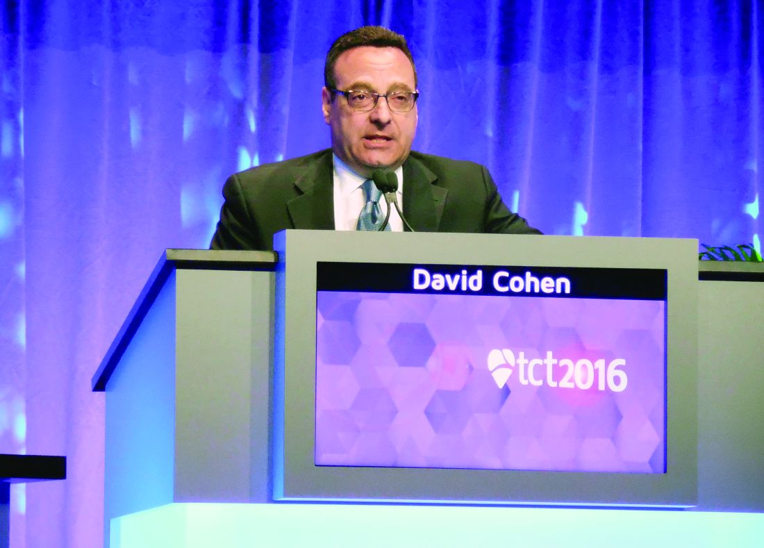
The investigators measured KCCQ scores at both 1 and 2 years after treatment, and they were similar regardless of whether patients had undergone TAVR or SAVR in both the transfemoral and transthoracic subgroups. All the quality-of-life benefit from TAVR compared with SAVR occurred only during the first month (and possibly for a few additional months beyond that) and only in TAVR patients treated by the transfemoral route. Dr. Cohen stressed that the SAVR patients in both the transfemoral and transthoracic subgroups had very similar outcomes, showing that patient differences could not explain why the transfemoral patients received a much greater incremental benefit, compared with the SAVR patients, at 1 month than did the transthoracic patients.
“A transthoracic TAVR approach may not be preferable to SAVR, at least in the short to intermediate term,” said Dr. Cohen. “There is no benefit from TAVR, compared with SAVR, if you can’t do it transfemorally, although emerging evidence has suggested that other nontransfemoral approaches that stay out of the chest may provide benefit similar to transfemoral TAVR. The message is, stay out of the chest,” he concluded.
Concerns about durability
The durability of TAVR valves is another concern that has recently been influencing patients as they decide between TAVR and SAVR, said Dr. Smith. “A lot of patients don’t want TAVR because of their concerns about its durability.” These patients usually cite evidence reported in May 2016 at the annual congress of the European Association of Percutaneous Cardiovascular Interventions in Paris by Danny Dvir, MD, on longer-term follow-up of 378 patients who underwent TAVR at either of two pioneering centers. A retrospective review suggested a valve degeneration rate of about 50% after 8 years, Dr. Dvir reported.
This report “has gotten a lot of penetration over the Internet, and a lot of patients don’t like the uncertainty” about TAVR durability that this report produced. “A lot of the time now, patients come in with a fixed idea of whether they want TAVR or SAVR,” Dr. Smith said.
Dr. Smith essentially agreed with Dr. Sellke on the current role for TAVR relative to SAVR in lower-risk patients.
“If the patient is clearly intermediate risk, with an STS mortality risk of more than 6% and is at least 80 years old, then they’ll have TAVR 99% of the time. But if it’s a 75 year old with an STS score of 3.2% and otherwise healthy, the best choice is not as clear.”
Another cardiothoracic surgeon with lots of TAVR experience, Michael J. Reardon, MD, has much more enthusiasm for TAVR. “In my practice, I use TAVR exclusively in patients at least 80 years old. I don‘t care how healthy they look,” said Dr. Reardon, professor of cardiovascular surgery at Houston Methodist. He acknowledged that broader use of TAVR for intermediate-risk patients is getting push back from other cardiothoracic surgeons.
Dr. Sellke is one such surgeon, and he called uncertain TAVR valve durability the deciding factor. “We need longer-term data on [TAVR] valve longevity before we routinely put them into intermediate- or low-risk patients,” he said during a panel discussion at the meeting.
Dr. Reardon highlighted that newer TAVR systems have been reducing problems such as paravalvular leaks and the need for pacemakers following placement of self-expanding TAVR valves. Despite these technical improvements, the final frontier for TAVR for lower-risk patients is valve durability, Dr. Reardon said in an interview.
“I’m convinced the durability is there, and that any 80-year-old patient who is anatomically suited for transfemoral TAVR should get it no matter now healthy they look. If their likely survival is 15 years or less, then they are reasonable candidates for TAVR.”
Dr. Sellke had no relevant disclosures. PARTNER 2 was funded by Edwards, the company that markets Sapient TAVR systems. Dr. Cohen had no relevant disclosures. Dr. Smith has been an investigator in the PARTNER studies. Dr. Reardon has been a consultant to Medtronic, the company that markets the CoreValve and Evolut R TAVR systems.
mzoler@frontlinemedcom.com
On Twitter @mitchelzoler
EXPERT ANALYSIS FROM THE AHA SCIENTIFIC SESSIONS
Stem cell therapy for STEMI falls short at 2 years
NEW ORLEANS – Longer follow-up has not altered initial negative findings of the randomized phase II TIME trial, which tested the efficacy of early intracoronary stem cell therapy, given on day 3 or 7 after an ST-segment acute MI (STEMI). However, loss of the sickest patients from follow-up may have influenced the findings.
“When TIME (Transplantation in Myocardial Infarction Evaluation) was developed several years ago, the optimal timing for cell delivery following myocardial infarction was not known and had not been directly tested in a prospective clinical trial,” explained lead investigator Jay H. Traverse, MD, director of research at the Minneapolis Heart Institute Foundation and a senior consulting cardiologist at the Abbott Northwestern Hospital, Minneapolis.
With the new, additional follow-up out to 2 years in 85 patients, both groups saw further gains in LV ejection fraction and further reductions in LV infarct size, as well as stable LV end-diastolic volume index, but still with no significant differences between them.
Of note, however, 10 of the patients lost to follow-up were lost because they received implantable cardioverter defibrillators (ICDs) and, given technologic limitations at the time, could no longer undergo cardiac MRI, Dr. Traverse noted. “These 10 patients are important as we look at the long-term follow-up of TIME because these patients were more likely to have the most severe LV dysfunction and most remodeled left ventricles. This [subset] represents a limitation to long-term follow-up studies in this population.”
Challenges to research
Two challenging issues seen in many trials of cell therapy for cardiovascular disease are their underpowered nature and the changing natural history of the disease, according to session comoderator Timothy D. Henry, MD, head of cardiology at Cedars-Sinai in Los Angeles.
“One of the things with the TIME trial that’s striking, really, is that even though these are high-risk patients, there was almost no mortality in 2 years,” he commented. “Second of all... there were no differences in ejection fraction, but that’s because you are missing 30% of people. And your missing 30% of people are the sick people, so of course if you are just going to follow normal people for 2 years, you’re going to have normal results.”
“What you are seeing is a little bit illusory because all the sick patients got ICDs and we could no longer image them at that time,” Dr. Traverse agreed. “That will be less of an issue in some of our future trials that we have started now, like CONCERT and SENECA, where we are now able to do MRI analysis of people with devices. So that will certainly help.”
Nonetheless, all trials in similar patient populations are plagued by substantial rates of dropout over time and faced with improving outcomes generally, because of better medical therapy, he acknowledged. “You can see as far as the natural history, even taking into account the ICD changes that would have lowered volumes, people are pretty stable on medical therapy. Their ejection fractions and volumes out to 2 years were really quite stable. We have certainly impacted that.”
That issue raises the question of whether investigators should be using other endpoints going forward, according to session panelist Doris A. Taylor, PhD, director of Regenerative Medicine Research at the Texas Heart Institute in Houston.
“One of the things I’ve seen over and over in this field is constantly evolving questions about which endpoints we should follow and what constitutes positive effects,” she commented. “So given the natural history, what are the endpoints we should be using?”
“Patients don’t care what their ejection fraction is per se. But they want to know if they have to get an ICD or if they will be hospitalized for heart failure, and their family is affected if they die,” Dr. Traverse replied. “We definitely need these hard endpoints. The problem is that you need so many patients with these hard endpoints that [the trials] are just financially not very doable. So that’s a big issue.”
Trial details
Patients enrolled in TIME had a first anterior STEMI, underwent reperfusion by angioplasty and stenting, and had LV dysfunction with an ejection fraction of 45% or lower. They all received either autologous bone marrow mononuclear cells or placebo on day 3 or day 7 after their MI by intracoronary infusion.
Of the initial 120 randomized patients, 10 patients were lost to follow-up because of receipt of an ICD, 3 had died (1 each from cardiovascular causes, pancreatitis, and hemorrhagic stroke), 7 were lost to follow-up for other reasons, and 15 had acquired other contraindications to MRI, according to Dr. Traverse.
At 2 years, the remaining patients in both the cell therapy and placebo groups had roughly 5% absolute increases in LV ejection fraction from baseline and roughly 45% reductions in infarct size from baseline, with no significant differences between groups.
When all patients were combined, about half were determined to have had microvascular obstruction at baseline. This finding was an adverse prognostic factor, associated with poorer recovery of LV function over time, greater adverse LV remodeling, and a higher likelihood of receiving an ICD, Dr. Traverse reported.
He has received a research grant from the National Heart, Lung, and Blood Institute, which sponsored the TIME trial.
AT THE AHA SCIENTIFIC SESSIONS
Key clinical point:
Major finding: At 2 years, differences in LV ejection fraction, infarct size, and end-diastolic volume index between the cell therapy and placebo groups remained nonsignificant.
Data source: TIME, a randomized, phase II trial of intracoronary delivery of autologous bone marrow mononuclear cells in 120 patients with ST-segment acute MI and LV dysfunction.
Disclosures: Dr. Traverse has received a research grant from the National Heart, Lung, and Blood Institute.
Dementia prevalence increased in heart failure patients
NEW ORLEANS – Elderly patients with heart failure had a significantly increased prevalence of both dementia and mild cognitive impairment, compared with similar people without heart failure, in an analysis of data collected from more than 6,000 U.S. residents enrolled in a long-term observational study.
Patients diagnosed with either heart failure with reduced ejection fraction or heart failure with preserved ejection fraction had an 89% increased prevalence of dementia and a 41% increased prevalence of mild cognitive impairment (MCI), compared with people from the same cohort who did not develop heart failure, in an analysis that adjusted for several demographic and clinical variables, Lucy S. Witt, MD, reported at the American Heart Association scientific sessions. She speculated that the link between heart failure and dementia and MCI might result from impaired cerebral perfusion in heart failure patients or from effects from heart failure medications.
The analysis used data collected for the Atherosclerosis Risk in Communities (ARIC) study, which began in 1987 and enrolled a randomly selected representative cohort of nearly 16,000 women and men aged 45-64 years old who resided in any of four U.S. communities. She specifically focused on the data collected from 6,431 of the participants who returned for a fifth follow-up examination during 2011-2013, including 5,490 people without heart failure, whose average age was 76 years, and 941 participants with heart failure, whose average age was 78 years.
Dementia prevalence at the fifth follow-up visit occurred at an adjusted rate of 5.6% among those without heart failure and 7.0% in those with heart failure. The examinations also found MCI in an adjusted 21.5% of those without heart failure and in 26.2% of those with heart failure, Dr. Witt reported. Adjustments included age, sex, location, education, hypertension, diabetes, depression, alcohol and tobacco use, cerebral vascular disease, marital status, and several other factors.
The relative risk for having dementia among the heart failure patients was roughly similar, regardless of whether ARIC participants with heart failure had a reduced or preserved left ventricular ejection fraction, she said.
ARIC is funded by the National Heart, Lung, and Blood Institute. Dr. Witt had no disclosures.
mzoler@frontlinemedcom.com
On Twitter @mitchelzoler
AT THE AHA SCIENTIFIC SESSIONS
Key clinical point:
Major finding: Patients with heart failure had an adjusted 86% increased rate of dementia, compared with similar people without heart failure.
Data source: Analysis of 6,431 ARIC participants who returned for a fifth follow-up examination during 2011-2013.
Disclosures: The Atherosclerosis Risk in Commmunities (ARIC) study is funded by the National Heart, Lung, and Blood Institute. Dr. Witt had no disclosures.
Observational hospital stays for HF linked to worse outcomes
NEW ORLEANS – The Centers for Medicare & Medicaid Services policy providing financial incentives for hospitals to readmit patients for heart failure for an observational stay rather than as an inpatient is antithetical to the patients’ best interests, according to data presented at the American Heart Association scientific sessions.
“We showed that if you get admitted under observation, the risk of you coming back is much higher than if you’re under an inpatient stay,” said Ahmad Masri, MBBS, of the University of Pittsburgh.
“Since CMS instituted this rule in 2013, there has been a surge in utilization of observational status versus inpatient status,” Dr. Masri noted.
That might make sense if the patients selected for in-hospital observation were less ill at the time than the heart failure patients admitted as inpatients, but that wasn’t the case in his large, retrospective study.
Dr. Masri reported on 21,339 patients with a total of 52,493 admissions for a primary diagnosis of heart failure during 2008-2015 in an 18-hospital health care system. After excluding admissions which involved cardiac surgery or in-hospital mortality, the total was 50,654 admissions.
Of these admissions, 5% were for in-hospital observation; 17% were inpatient admissions with discharge in less than 2 days. The two groups were similar in terms of age, comorbid conditions, and use of guideline-directed medications, although 36% of patients admitted under observation had a left ventricular ejection fraction below 40%, compared with 30% of those with an inpatient admission for less than 2 days.
The majority of patients in both groups were readmitted for heart failure within 1 year; however, the readmission rate was 23% lower in the group with an inpatient stay of less than 2 days, in an analysis adjusted for age, sex, ejection fraction, hypertension, diabetes, pneumonia, chronic obstructive pulmonary disease, liver disease, and renal failure.
Similarly, the group with an inpatient stay of less than 2 days’ duration was 24% less likely to have a cardiac readmission within 1 year than the group admitted for a penalty-free observational stay. The short inpatient stay group’s 1-year all-cause readmission rate was also 24% lower. All of these differences were statistically significant and clinically meaningful.
Yet 1-year all-cause mortality in the two groups was no different.
“This suggests that the difference between these two groups is more of an administrative distinction than a reflection of patient status at time of admission. It looks like it’s just random,” according to Dr. Masri. “There is a real need for a patient-centered, streamlined approach in evaluating and treating patients with heart failure, with a revised treatment-based algorithm and admission rules that guide physicians and shape health care policy.”
He reported having no financial conflicts of interest regarding this study.
AT THE AHA SCIENTIFIC SESSIONS
Key clinical point:
Major finding: The 1-year rates of readmission for heart failure, cardiac readmission, and all-cause readmission were each 23%-24% lower in heart failure patients admitted for an inpatient stay of less than 2 days’ duration than if they were designated as being admitted under observation.
Data source: A retrospective analysis of more than 50,000 hospital admissions with a primary diagnosis of heart failure in 21,339 patients during 2008-2015.
Disclosures: The presenter reported having no financial conflicts of interest regarding the study.
VIDEO: MILANO-PILOT marks end of the road for HDL mimetic
NEW ORLEANS – The future doesn’t look bright at the moment for use of agents that mimic HDL cholesterol to reverse coronary disease, based on disappointing results of the randomized, phase II MILANO-PILOT trial.
Among 120 patients with recent acute coronary syndrome, a mimetic known as MDCO-216 was no better than placebo at reducing coronary plaque measured by intravascular ultrasound (IVUS), according to data presented at the American Heart Association scientific sessions.
The median reduction in percent atheroma volume, the trial’s primary endpoint, was 0.5% with the mimetic and 0.8% with placebo, reported Stephen J. Nicholls, MD, of the South Australian Health and Medical Research Institute, University of Adelaide (Australia). Findings were similar for secondary outcomes assessing other measures of disease regression.
These data bring to an end a story that began in the 1980s, with discovery that a family in a town in northern Italy had a naturally occurring apolipoprotein A-I variant that mimics the actions of HDL cholesterol, subsequently named ApoA-IMilano. Despite having low levels of HDL, family members have a reduced risk of coronary disease.
In a small randomized trial, patients with acute coronary syndrome given an early recombinant form of this apolipoprotein saw a reduction in IVUS-measured coronary atherosclerosis (JAMA. 2003;290:2292-300). This prompted development of the refined recombinant form, MDCO-216, tested in the new trial.
“MDCO-216 did not produce a significant effect on coronary disease progression as measured by IVUS,” said Dr. Nicholls, who discussed the findings in a video interview. “These results occurred on a background of contemporary therapy in the post-ACS setting, which in 2016 is very effective for many patients.”
“The findings from this pilot study do not provide the evidence required to proceed with further development,” he concluded.
The video associated with this article is no longer available on this site. Please view all of our videos on the MDedge YouTube channel
Hope hinges on wild type
“This trial was extremely well done, world-class investigators carried out this trial, it was three times the size of the previous IVUS trial, and I think it is convincingly negative,” said discussant Daniel J. Rader, MD, associate director of the Institute for Translational Medicine and Therapeutics and director of the Preventive Cardiovascular Program at Penn Medicine, Philadelphia. “The sponsor has elected to stop the development of this product, and I think we can probably write the obituary for ApoA-IMilano, even though it was never studied in cardiovascular outcomes.”
“At the end of the day, my message is that this has been a wild ride with ApoA-IMilano, and it’s sad to see that, at least when it comes to coronary atherosclerosis, this trial does not support that ApoA-IMilano has an effect,” Dr. Rader concluded. “But I do think we need to see through the other products with wild-type ApoA-I given the marked difference in structure before we can conclude that this general approach of infusing a reconstituted ApoA-I particle is not going to work.”
Trial details
Patients from 22 centers globally were randomized in MILANO-PILOT. All patients had experienced acute coronary syndrome in the past 14 days and had maximum stenosis of 20%-50% of a target vessel on coronary angiography.
Results showed – as expected based on past experience – that HDL cholesterol levels increased by 8.0% with placebo and decreased by 7.8% with MDCO-216 (P less than .001), Dr. Nicholls reported. Apolipoprotein A-I levels increased by 5.6% in the former and decreased by 5.3% in the latter (P less than .001). Changes in a variety of other lipid measures did not differ significantly between groups.
“These study patients were extraordinarily well treated. We achieved LDL cholesterol levels in both treatment groups that were less than 70 mg/dL,” he pointed out.
In addition to the lack of difference in the reduction in percent atheroma volume, there was also no significant difference between MDCO-216 and placebo in the secondary endpoints of median change in total atheroma volume, either in the entire vessel length imaged (–4.7 vs. –6.9 mm3) or in the most diseased 10-mm segment (–2.4 vs. –2.4 mm3).
The percentage of patients having any degree of disease regression was 55.8% with MDCO-216 and 67.2% with placebo, another nonsignificant difference.
“The majority of patients in this study demonstrated some degree of regression, and I believe that really highlights the impact of contemporary treatment guidelines have on coronary atherosclerosis in the early post-ACS setting in the contemporary era,” Dr. Nicholls maintained.
Exploratory analyses did not indicate any clear differential impact of MDCO-216 versus placebo according to whether patients had already been taking statins at baseline.
MDCO-216 was generally safe and well tolerated. “We saw no differences between the groups in terms of a range of biochemical adverse events, serious adverse events, and infusion site reactions,” reported Dr. Nicholls, who disclosed that he received a research grant from The Medicines Company, which sponsored the trial.
AT THE AHA SCIENTIFIC SESSIONS
Key clinical point:
Major finding: The percent atheroma volume decreased by 0.5% with the mimetic and 0.8% with placebo, a nonsignificant difference.
Data source: A randomized phase II trial among 120 patients with recent ACS and coronary stenosis (MILANO-PILOT trial).
Disclosures: Dr. Nicholls has received a research grant from The Medicines Company, which sponsored the study.
New PAD guidelines expected to boost awareness and treatment
NEW ORLEANS – The latest revision of U.S. guidelines for diagnosing and managing lower-extremity peripheral artery disease is seen by several experts as primarily a renewed call to action by American physicians to more diligently identify at-risk people in their practices, diagnose the disease with ankle-brachial index measurement, and appropriately treat patients who have the disease.
The new lower-extremity peripheral artery disease (PAD) guidelines “give us a platform to get something done,” said Heather L. Gornik, MD, vice chair of the guidelines panel and medical director of the noninvasive vascular lab at the Cleveland Clinic. Improved PAD identification and care “starts with the fundamentals of recognizing who is at risk for PAD and then doing some clinical investigation to find it, because it’s there. You just need to ask patients if they have leg symptoms, have them take off their socks and examine their feet,” Dr. Gornik said in an interview following a program devoted to the revised guidelines at the American Heart Association scientific sessions.
The new guidelines are “a clarion call to action,” commented Alan T. Hirsch, MD, professor of medicine, epidemiology, and community health and director of the vascular medicine program at the University of Minnesota in Minneapolis. PAD “is the single most morbid and fatal of all cardiovascular diseases, so why in 2016 are we challenged to have every cardiologist trained in basic PAD competency?” asked Dr. Hirsch, who chaired the 2005 guidelines panel.
The AHA wants to “elevate awareness of PAD among the public and health professionals, and we want to better understand how we can be a catalyst for change,” said Terri Wiggins, the organization’s vice president for vascular health programs. AHA publications now stress that clinicians need to have patients “take off their socks and look at the patient’s feet,” she said.
The problem with the way many U.S. physicians handle PAD goes beyond a failure to properly screen and diagnose the disease. Analysis of data collected by the National Ambulatory Medical Care Survey and the National Hospital Ambulatory Medical Care Survey during 2006-2013 showed that, among an average of 3.9 million patients seen each year with PAD, just 38% received treatment with an antiplatelet drug, 35% received a statin, and among those patients who smoked, 36% received counseling for smoking cessation, Jeffrey S. Berger, MD, reported in a separate talk at the meeting. These rates were roughly constant throughout the 8-year period he examined.
“These data are eye-opening. They highlight a clear opportunity to improve the care of patients with PAD with guideline-directed therapy. PAD is problematic because only 10%-15% of patients have typical symptoms, so many physicians have a false perception that these patients are not at high risk,” Dr. Berger said in an interview. “What the AHA is doing is great” for raising awareness, he added.
The revised 2016 PAD guidelines made several changes, compared with what had been on the books from the 2005 guidelines and 2011 update, including classifying vorapaxar (Zontivity) treatment a class IIb recommendation with “uncertain” incremental benefit when used as an add-on agent on top of standard antiplatelet therapy, endorsement of annual influenza vaccination as something every PAD patient should receive and a class I recommendation, and acknowledgment that selected patients with critical limb ischemia are candidates for an “endovascular first” approach to revascularization,
Perhaps just as important was inclusion for the first time in the new guidelines of three advocacy priorities for initiatives by various professional societies with an interest in PAD: easy availability of ankle-brachial index measurement as the initial test to establish a diagnosis of PAD in patients with physical examination findings suggestive of the disease; access to supervised exercise programs for patients diagnosed with PAD; and incorporation of patient-centered outcomes into the regulatory approval process of new medical therapies and revascularization technologies for treating PAD.
The new guidelines “ form the basis for the AHA establishing a Get With The Guidelines program for PAD so that clinicians can be held accountable for delivering these treatments,” said Naomi M. Hamburg, MD, chief of vascular biology at Boston University and a member of the guidelines panel.
Dr. Gornik has an ownership interest in Summit Doppler Systems and Zin Medical and has received research support from AstraZeneca and Theravasc. Dr. Hamburg has been a consultant to Acceleron and has received research support from Everest Genomics, Hershey’s, Unex, and Welch’s. Dr. Hirsch, Ms. Wiggins, and Dr. Berger had no disclosures.
mzoler@frontlinemedcom.com
On Twitter @mitchelzoler
EXPERT ANALYSIS FROM THE AHA SCIENTIFIC SESSIONS


