User login
VIDEO: Laser innovations target tattoos, acne, body fat
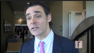
PHOENIX – Advances in laser technology allow dermatologists to remove tattoos in half the time but at twice the cost. Cold therapy for sebaceous glands theoretically could cure acne. Nonsurgical body sculpting and contouring techniques are making it easier to take away love handles and other unwanted body fat.
These are some of the highlights from the annual meeting of the American Society for Laser Medicine and Surgery. Dr. Jeffrey S. Dover, immediate past president of the ASLMS, explains the details of these developments and how to make the most of them in practice. Dr. Dover is a dermatologist practicing in Chestnut Hill, Mass.
The video associated with this article is no longer available on this site. Please view all of our videos on the MDedge YouTube channel
On Twitter @sherryboschert
AT LASER 2014
Pulsed Dye Laser for the Treatment of Macular Amyloidosis: A Case Report
VIDEO: Battlefield lessons improve treatment of traumatic scars
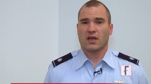
DENVER – Military dermatologists have played an active role in helping wounded warriors with their scars, and those physicians’ practices, including the use of ablative fractional resurfacing, are now entering the civilian world to treat injuries from fire, car crashes, or blasts.
Earlier this year, a group of dermatologists published a consensus report to highlight best practices for laser treatment of traumatic scars. The groups, which included several military dermatologists, concluded that "laser treatment, particularly ablative fractional resurfacing, deserves a prominent role in future scar treatment paradigms, with the possible inclusion of early intervention for contracture avoidance and assistance with wound healing."
In a video interview at the American Academy of Dermatology’s annual meeting, Lt. Col. Chad M. Hivnor, USAF, MC, FS, USA, a staff dermatologist for the San Antonio (Tex.) Military Health System, discussed the use of lasers in treating traumatic scars, talked about the psychology of scars, and shared a few of his own practice pearls.
The video associated with this article is no longer available on this site. Please view all of our videos on the MDedge YouTube channel
On Twitter @naseemmiller
AT THE AAD ANNUAL MEETING
‘Culture of Safety’ best defense against sharps injury
DENVER – Of all the procedures and behaviors that place dermatologists at risk for occupational exposure, needlestick injuries rank at the top.
According to 1999 data from the National Institute for Occupational Safety and Health, one in five health care workers sustains a needlestick injury each year, putting them at risk for acquiring pathogens such as HIV and hepatitis B and C viruses. "Conjunctival transmission of blood-borne pathogens can occur, and there are at least two cases of documented transmission of HIV via splashes," Dr. Joseph F. Sobanko said at the annual meeting of the American Academy of Dermatology. "Operating with exposed, nonintact skin also poses a risk of transmission of bloodborne pathogens."

Dr. Sobanko, a Mohs and reconstructive surgeon and director of dermatologic surgery education at the Hospital of the University of Pennsylvania, Philadelphia, emphasized that all health care employees are at risk of sharps injury. "Those physicians with the least experience are the most likely to receive an occupational exposure," he said. "When trainees and students receive injury, it is a risk factor for receiving future injuries, perhaps because of improper training early on."
A pilot study of sharps-related injuries in California health care facilities found that 49% occurred among nurses, followed by physicians (10%), phlebotomists (8%), and housekeeping and laundry personnel (7%), while the remainder occurred among a variety of other health care workers (Infect. Control Hosp. Epidemiol. 2003;24:113-21).
A separate survey study of needlestick injuries among 699 recent medical school graduates enrolled in a surgery residency at one of 17 medical centers in the United States found that 59% had a stick as a student, most commonly from suturing (42%), passing the needle (17%), and loading the needle (12%) (Academic Med. 2009;84:1815-21).
"Other studies have found that needlestick injuries commonly occur during device use and after device use during disposal," Dr. Sobanko noted.
Most occupational exposures are self-inflicted, and most sharps injuries tend to affect the left hand and digits, he continued. "When suturing, the injury that is self inflicted often happens on the nondominant hand," he said. "However, when not suturing but passing instruments (which shouldn’t be done), health care professionals are more likely to be injured on the dominant hand, because they accept the instrument with this hand. This is why ‘surgical neutral zones’ should be created to transfer instruments and eliminate this particular form of injury."
Occupational exposure is especially high in dermatology. One survey of 452 dermatologists queried in November of 2009 found a 90% injury rate (J. Am. Acad. Dermatol. 2011;65:648-50), while a separate, more recent survey of 336 dermatologists found an 85% injury rate 40% of the injuries had occurred within the year before the survey (Dermatol. Surg. 2013;39:1813-21). More than two-thirds of those same respondents (64%) reported having ever had a sharps injury that went unreported.
Procedures and behaviors that place dermatologists at highest risk for occupational exposure include drawing up solution, setting up a tray, injection, excision, biopsy, obtaining hemostasis, suturing, and disposal of sharps.
"Shortcuts, lack of focus, and improper training lead to avoidable accidents," Dr. Sobanko said. "Fostering a culture of safety can help reduce the risk of future injuries."
His recommended technique for uncapping a needle, for example, involves anchoring the top hand to the bottom hand, as in a golfer’s grip. Gentle extension releases the cap. His recommended technique for drawing solution involves resting the syringe on the hypothenar eminence of the left hand while holding the barrel with the thumb and index finger. This allows for safe placement of the bottle onto the needle. "This technique eliminates the risk of recoil injury if a cap is simply just pulled off a syringe at chest level, analogous to stretching a rubber band," he explained.
To avoid injuries while injecting, Dr. Sobanko advises ensuring that the hand or finger stabilizing the skin stays behind the path of the needle.
Dermatologists can keep themselves safe during office procedures by using protective sharps, eye protection, and gloves and by transferring instruments by implementing a neutral zone on the surgical tray. One review of seven studies of needle protective devices demonstrated an average 71% reduction in needlestick injuries (J. Hosp. Infect. 2003;53:237-42).
Dr. Sobanko described a safe needle device as one that is "easy to use and requires minimal effort to activate by the user. If activation is required, it must be a single-handed technique. The safety feature should click or be clear that it has activated, and the safety feature should remain protective throughout disposal."
Mental preparedness and motor repetition also play a role in protecting yourself. Dr. Sobanko’s five strategies for mental preparedness involve not rushing, knowing the pertinent anatomy for each case, having a proper tray set-up, having a proper preoperative plan, and not operating until an assistant is available.
Dr. Sobanko disclosed that he is a coeditor with Dr. Jacob Levitt of the forthcoming Springer book, "Atlas of Safe Practices in Office-Based Surgery."
DENVER – Of all the procedures and behaviors that place dermatologists at risk for occupational exposure, needlestick injuries rank at the top.
According to 1999 data from the National Institute for Occupational Safety and Health, one in five health care workers sustains a needlestick injury each year, putting them at risk for acquiring pathogens such as HIV and hepatitis B and C viruses. "Conjunctival transmission of blood-borne pathogens can occur, and there are at least two cases of documented transmission of HIV via splashes," Dr. Joseph F. Sobanko said at the annual meeting of the American Academy of Dermatology. "Operating with exposed, nonintact skin also poses a risk of transmission of bloodborne pathogens."
Dr. Sobanko, a Mohs and reconstructive surgeon and director of dermatologic surgery education at the Hospital of the University of Pennsylvania, Philadelphia, emphasized that all health care employees are at risk of sharps injury. "Those physicians with the least experience are the most likely to receive an occupational exposure," he said. "When trainees and students receive injury, it is a risk factor for receiving future injuries, perhaps because of improper training early on."
A pilot study of sharps-related injuries in California health care facilities found that 49% occurred among nurses, followed by physicians (10%), phlebotomists (8%), and housekeeping and laundry personnel (7%), while the remainder occurred among a variety of other health care workers (Infect. Control Hosp. Epidemiol. 2003;24:113-21).
A separate survey study of needlestick injuries among 699 recent medical school graduates enrolled in a surgery residency at one of 17 medical centers in the United States found that 59% had a stick as a student, most commonly from suturing (42%), passing the needle (17%), and loading the needle (12%) (Academic Med. 2009;84:1815-21).
"Other studies have found that needlestick injuries commonly occur during device use and after device use during disposal," Dr. Sobanko noted.
Most occupational exposures are self-inflicted, and most sharps injuries tend to affect the left hand and digits, he continued. "When suturing, the injury that is self inflicted often happens on the nondominant hand," he said. "However, when not suturing but passing instruments (which shouldn’t be done), health care professionals are more likely to be injured on the dominant hand, because they accept the instrument with this hand. This is why ‘surgical neutral zones’ should be created to transfer instruments and eliminate this particular form of injury."
Occupational exposure is especially high in dermatology. One survey of 452 dermatologists queried in November of 2009 found a 90% injury rate (J. Am. Acad. Dermatol. 2011;65:648-50), while a separate, more recent survey of 336 dermatologists found an 85% injury rate 40% of the injuries had occurred within the year before the survey (Dermatol. Surg. 2013;39:1813-21). More than two-thirds of those same respondents (64%) reported having ever had a sharps injury that went unreported.
Procedures and behaviors that place dermatologists at highest risk for occupational exposure include drawing up solution, setting up a tray, injection, excision, biopsy, obtaining hemostasis, suturing, and disposal of sharps.
"Shortcuts, lack of focus, and improper training lead to avoidable accidents," Dr. Sobanko said. "Fostering a culture of safety can help reduce the risk of future injuries."
His recommended technique for uncapping a needle, for example, involves anchoring the top hand to the bottom hand, as in a golfer’s grip. Gentle extension releases the cap. His recommended technique for drawing solution involves resting the syringe on the hypothenar eminence of the left hand while holding the barrel with the thumb and index finger. This allows for safe placement of the bottle onto the needle. "This technique eliminates the risk of recoil injury if a cap is simply just pulled off a syringe at chest level, analogous to stretching a rubber band," he explained.
To avoid injuries while injecting, Dr. Sobanko advises ensuring that the hand or finger stabilizing the skin stays behind the path of the needle.
Dermatologists can keep themselves safe during office procedures by using protective sharps, eye protection, and gloves and by transferring instruments by implementing a neutral zone on the surgical tray. One review of seven studies of needle protective devices demonstrated an average 71% reduction in needlestick injuries (J. Hosp. Infect. 2003;53:237-42).
Dr. Sobanko described a safe needle device as one that is "easy to use and requires minimal effort to activate by the user. If activation is required, it must be a single-handed technique. The safety feature should click or be clear that it has activated, and the safety feature should remain protective throughout disposal."
Mental preparedness and motor repetition also play a role in protecting yourself. Dr. Sobanko’s five strategies for mental preparedness involve not rushing, knowing the pertinent anatomy for each case, having a proper tray set-up, having a proper preoperative plan, and not operating until an assistant is available.
Dr. Sobanko disclosed that he is a coeditor with Dr. Jacob Levitt of the forthcoming Springer book, "Atlas of Safe Practices in Office-Based Surgery."
AT THE AAD ANNUAL MEETING

AAD 2014 sessions offer something for everyone
The American Academy’s 2014 annual meeting in Denver will feature new CME sessions and updates on the latest dermatology research.
This year’s program features expert commentary on key issues in medical dermatology, including "Melanoma Multidisciplinary Care 2014: What You Need to Know" on Sunday, March 23, from 1 p.m. to 3 p.m. in Room 705/707 and "Dermatologic Manifestations of New Oncology Drugs," also on Sunday, March 23, from 1 p.m. to 3 p.m. in the Mile High Ballroom 3B. Looking for the latest in aesthetic dermatology? Check out the "Advanced Botulinum Toxin" live demonstration session on Saturday, March 22, from 2 p.m. to 5 p.m. in the Bellco Theater.
There will be expert sessions on pregnancy dermatoses, cutaneous T-cell lymphoma, pediatric dermatology, skin of color, and the latest on treatments for hair and nail conditions. The full scientific session list is available online.
A series of practice management lectures includes topics such as "How to Have an Unforgettably Positive Office Visit" on Saturday, March 22, from 10:00 a.m. to 12:00 p.m. in Room 709/7111 and "Hot Buttons: Recognizing What Sets You Off and Managing Your Triggers" on Sunday, March 23, from 1:00 p.m. to 3:00 p.m. in Room 702.
There is also a mobile device app that meeting attendees can download that contains session schedules, exhibitor and attendee lists, and more.
Can’t attend the meeting? Visit www.eDermatologyNews.com for live conference coverage.
On Twitter @Sknews
How an expert uses Voluma
WAIKOLOA, HAWAII – A key point to understand about Juvederm Voluma XC, the recently approved filler for age-related midface volume deficit, is that it’s a pillar or lift product, according to Dr. Sue Ellen Cox.
"Voluma loves to lift. It works great when placed on bone, such as the malar bone. With a supraperiosteal vertical puncture, you’ll see the skin lift right in front of your eyes," said Dr. Cox, a dermatologist at the University of North Carolina at Chapel Hill and principal investigator in the pivotal clinical trial that led to Food and Drug Administration approval of Voluma.

This characteristic of the highly cohesive 20-mg/mL hyaluronic acid filler has important implications for the product’s optimal use and achieving maximal patient satisfaction. For one, Voluma absolutely should not be used for patients with thin skin. For these patients, a more effective option is a product containing monophasic monodensified hyaluronic acids, such as Juvederm Ultra or Ultra Plus, Dr. Cox said at the Hawaii Dermatology Seminar sponsored by Global Academy for Medical Education/Skin Disease Education Foundation.
As a rule of thumb, approximately 40% of Voluma is needed compared with the amount of monophasic monodensified hyaluronic acid fillers dermatologists are accustomed to working with, she said.
It’s crucial to inject Voluma extremely slowly, Dr. Cox emphasized. She advised scheduling 30 minutes for a patient’s first volumizing session. It’s also important to avoid using a large bolus, and be sure not to overcorrect. Voluma loves water and will draw it from tissue, Dr. Cox noted. Therefore it’s important to use the exact correction. Remember that at 9 months post treatment, 50% or more of the original correction will remain, so the 9-month mark is a good time to schedule a touch-up, she added.
Another pearl: Inject struts or pillars from the periostium; then blend and mold them, Dr. Cox continued.
She urged her colleagues to be conservative in using Voluma around the eyes. In her experience, too much Voluma in this area causes the product to migrate anteriorly on the cheek, which could result in an unwelcome doughy appearance.
To achieve improvement in the submalar area, it’s best to utilize tangential microdroplets of Voluma after reconstitution with saline so the filler doesn’t affect the nerve and cause a lip drop, according to Dr. Cox.
Should it become necessary to dissolve Voluma, use twice as much hyaluronidase (Hylenex).
Dr. Cox reported serving as a consultant to Allergan and Medicis and serving as principal investigator in trials funded by those companies, as well as in studies funded by Revance and Kythera.
SDEF and this news organization are owned by the same parent company.
WAIKOLOA, HAWAII – A key point to understand about Juvederm Voluma XC, the recently approved filler for age-related midface volume deficit, is that it’s a pillar or lift product, according to Dr. Sue Ellen Cox.
"Voluma loves to lift. It works great when placed on bone, such as the malar bone. With a supraperiosteal vertical puncture, you’ll see the skin lift right in front of your eyes," said Dr. Cox, a dermatologist at the University of North Carolina at Chapel Hill and principal investigator in the pivotal clinical trial that led to Food and Drug Administration approval of Voluma.
This characteristic of the highly cohesive 20-mg/mL hyaluronic acid filler has important implications for the product’s optimal use and achieving maximal patient satisfaction. For one, Voluma absolutely should not be used for patients with thin skin. For these patients, a more effective option is a product containing monophasic monodensified hyaluronic acids, such as Juvederm Ultra or Ultra Plus, Dr. Cox said at the Hawaii Dermatology Seminar sponsored by Global Academy for Medical Education/Skin Disease Education Foundation.
As a rule of thumb, approximately 40% of Voluma is needed compared with the amount of monophasic monodensified hyaluronic acid fillers dermatologists are accustomed to working with, she said.
It’s crucial to inject Voluma extremely slowly, Dr. Cox emphasized. She advised scheduling 30 minutes for a patient’s first volumizing session. It’s also important to avoid using a large bolus, and be sure not to overcorrect. Voluma loves water and will draw it from tissue, Dr. Cox noted. Therefore it’s important to use the exact correction. Remember that at 9 months post treatment, 50% or more of the original correction will remain, so the 9-month mark is a good time to schedule a touch-up, she added.
Another pearl: Inject struts or pillars from the periostium; then blend and mold them, Dr. Cox continued.
She urged her colleagues to be conservative in using Voluma around the eyes. In her experience, too much Voluma in this area causes the product to migrate anteriorly on the cheek, which could result in an unwelcome doughy appearance.
To achieve improvement in the submalar area, it’s best to utilize tangential microdroplets of Voluma after reconstitution with saline so the filler doesn’t affect the nerve and cause a lip drop, according to Dr. Cox.
Should it become necessary to dissolve Voluma, use twice as much hyaluronidase (Hylenex).
Dr. Cox reported serving as a consultant to Allergan and Medicis and serving as principal investigator in trials funded by those companies, as well as in studies funded by Revance and Kythera.
SDEF and this news organization are owned by the same parent company.
EXPERT ANALYSIS FROM SDEF HAWAII DERMATOLOGY SEMINAR

Midface filler Voluma provides long-lasting patient satisfaction
WAIKOLOA, HAWAII – Juvederm Voluma XC continues to show significant results in extended follow-up data from the pivotal phase III trial that earned the product marketing approval from the Food and Drug Administration late last year as the first filler indicated specifically for treating age-related midface volume deficit.
One of the notable new findings: At 6 months post treatment, 73% of Voluma-treated study participants rated themselves as looking younger than at baseline – and by an average of 5 years less than their mean baseline chronologic age of 56 years. Moreover, at 24 months, 55% of patients said they felt that they still looked younger by an average of 3 years, Dr. Sue Ellen Cox reported at the Hawaii Dermatology Seminar sponsored by Global Academy for Medical Education/Skin Disease Education Foundation.

"The improvement was really profound. What was also profound was how long it lasted. I’m now 3 years out seeing these patients and they still have retention of their product. So I am a believer," said Dr. Cox, a dermatologist at the University of North Carolina at Chapel Hill, who was principal investigator in the pivotal phase III trial.
Dr. Cox shared highlights of the extended follow-up data, along with her personal observations regarding how to use Voluma most effectively.
Voluma XC is a highly cohesive volumizing hyaluronic acid filler formulated at a concentration of 20 mg/mL. It fills what has been widely regarded as a major unmet need in aesthetic dermatology, said Dr. Cox
"It’s a wonderful filler we’re all really going to enjoy using. I think it’s going to prove to be everything we want it to be," she said.
The pivotal data reviewed by the FDA came from a 15-center, randomized, single-blind clinical trial including 235 Voluma-treated patients and 47 no-treatment controls. All patients had moderate or severe baseline midface volume deficits as reflected by scores of 3-5 on a standardized 0-5 scoring system. The active treatment group received one treatment with the option of a touch-up session a month later.
The primary study endpoint prespecified by the FDA was an improvement of at least 1 point between baseline and 6 months on the Mid-Facial Volume Deficit Scale (MFVDS). This endpoint was achieved in 86% of the Voluma group and 39% of controls. Moreover, 51% of the active treatment group had an improvement of 2 points or greater compared with 11% of controls. And 26% of the Voluma group, but none of the controls, showed a 2.5-point improvement or better.
The durability of the treatment response was noteworthy, Dr. Cox added. At 2 years, 67% of patients in the Voluma group maintained a clinically significant improvement based upon MFVDS scores.
Every 3 months for the 2 years of follow-up, patients were asked how they felt about their appearance. As Dr. Cox noted, this is the true litmus test for any aesthetic dermatology procedure. At 6 months, 90% of patients pronounced themselves satisfied with the improvement in their facial appearance. At 12 months, 82% said they were satisfied; and at 2 years post treatment, 76% of patients remained satisfied with their facial appearance.
At baseline, 67% of patients rated their midface appearance as making them look "very much" older; at 6 months post treatment, only 12% of patients felt that way. Similarly, at baseline 55%-66% of patients characterized their midface appearance as variously "very much" unattractive, sad, and/or tired; at 6 months post treatment, only 9%-11% of subjects did so.
Treatment of the nasolabial folds and tear ducts was not permitted in the pivotal trial. Yet by investigator assessment at 6 months’ follow-up 32% of the active treatment group had a clinically meaningful improvement of at least 1 point on the 5-point Nasolabial Fold Photo Severity Scale, compared with 8% of controls, said Dr. Cox. Moreover, 54% of Voluma-treated patients rated themselves as moderately, very much, or completely satisfied with the appearance of their tear trough area, a marked improvement over the 17% rate at baseline. These findings underscore the point that effectively reinflating the midface and reestablishing optimal proportion provides ancillary benefits that may render treatment of the tear troughs and nasolabial folds unnecessary, she said.
Common treatment side effects consisted of mild to moderate injection site tenderness, swelling, bruising, lumps and bumps, and pain. All cases of side effects resolved within 30 days, and most resolved within 2 weeks.
Dr. Cox reported acting as a consultant to Allergan and Medicis and serving as principal investigator in trials funded by those companies, as well as in studies funded by Revance and Kythera.
SDEF and this news organization are owned by the same parent company.
WAIKOLOA, HAWAII – Juvederm Voluma XC continues to show significant results in extended follow-up data from the pivotal phase III trial that earned the product marketing approval from the Food and Drug Administration late last year as the first filler indicated specifically for treating age-related midface volume deficit.
One of the notable new findings: At 6 months post treatment, 73% of Voluma-treated study participants rated themselves as looking younger than at baseline – and by an average of 5 years less than their mean baseline chronologic age of 56 years. Moreover, at 24 months, 55% of patients said they felt that they still looked younger by an average of 3 years, Dr. Sue Ellen Cox reported at the Hawaii Dermatology Seminar sponsored by Global Academy for Medical Education/Skin Disease Education Foundation.
"The improvement was really profound. What was also profound was how long it lasted. I’m now 3 years out seeing these patients and they still have retention of their product. So I am a believer," said Dr. Cox, a dermatologist at the University of North Carolina at Chapel Hill, who was principal investigator in the pivotal phase III trial.
Dr. Cox shared highlights of the extended follow-up data, along with her personal observations regarding how to use Voluma most effectively.
Voluma XC is a highly cohesive volumizing hyaluronic acid filler formulated at a concentration of 20 mg/mL. It fills what has been widely regarded as a major unmet need in aesthetic dermatology, said Dr. Cox
"It’s a wonderful filler we’re all really going to enjoy using. I think it’s going to prove to be everything we want it to be," she said.
The pivotal data reviewed by the FDA came from a 15-center, randomized, single-blind clinical trial including 235 Voluma-treated patients and 47 no-treatment controls. All patients had moderate or severe baseline midface volume deficits as reflected by scores of 3-5 on a standardized 0-5 scoring system. The active treatment group received one treatment with the option of a touch-up session a month later.
The primary study endpoint prespecified by the FDA was an improvement of at least 1 point between baseline and 6 months on the Mid-Facial Volume Deficit Scale (MFVDS). This endpoint was achieved in 86% of the Voluma group and 39% of controls. Moreover, 51% of the active treatment group had an improvement of 2 points or greater compared with 11% of controls. And 26% of the Voluma group, but none of the controls, showed a 2.5-point improvement or better.
The durability of the treatment response was noteworthy, Dr. Cox added. At 2 years, 67% of patients in the Voluma group maintained a clinically significant improvement based upon MFVDS scores.
Every 3 months for the 2 years of follow-up, patients were asked how they felt about their appearance. As Dr. Cox noted, this is the true litmus test for any aesthetic dermatology procedure. At 6 months, 90% of patients pronounced themselves satisfied with the improvement in their facial appearance. At 12 months, 82% said they were satisfied; and at 2 years post treatment, 76% of patients remained satisfied with their facial appearance.
At baseline, 67% of patients rated their midface appearance as making them look "very much" older; at 6 months post treatment, only 12% of patients felt that way. Similarly, at baseline 55%-66% of patients characterized their midface appearance as variously "very much" unattractive, sad, and/or tired; at 6 months post treatment, only 9%-11% of subjects did so.
Treatment of the nasolabial folds and tear ducts was not permitted in the pivotal trial. Yet by investigator assessment at 6 months’ follow-up 32% of the active treatment group had a clinically meaningful improvement of at least 1 point on the 5-point Nasolabial Fold Photo Severity Scale, compared with 8% of controls, said Dr. Cox. Moreover, 54% of Voluma-treated patients rated themselves as moderately, very much, or completely satisfied with the appearance of their tear trough area, a marked improvement over the 17% rate at baseline. These findings underscore the point that effectively reinflating the midface and reestablishing optimal proportion provides ancillary benefits that may render treatment of the tear troughs and nasolabial folds unnecessary, she said.
Common treatment side effects consisted of mild to moderate injection site tenderness, swelling, bruising, lumps and bumps, and pain. All cases of side effects resolved within 30 days, and most resolved within 2 weeks.
Dr. Cox reported acting as a consultant to Allergan and Medicis and serving as principal investigator in trials funded by those companies, as well as in studies funded by Revance and Kythera.
SDEF and this news organization are owned by the same parent company.
EXPERT ANALYSIS FROM SDEF HAWAII DERMATOLOGY SEMINAR

VIDEO: Photodynamic therapy pearls can improve results
CHAMPIONSGATE, FLA. – Do you use photodynamic therapy to treat actinic keratoses? Dr. Joel Cohen has some tips for you. At the Orlando Dermatology Aesthetic and Clinical Conference, Dr. Cohen spoke to us about current strategies for making the most of PDT, and which treatment plans work best for certain patients. What’s next for PDT? Dr. Cohen of the department of dermatology at the University of Colorado, Denver, highlights promising preliminary studies involving different types of combination therapies that can yield cosmetic and medical benefits.
The video associated with this article is no longer available on this site. Please view all of our videos on the MDedge YouTube channel
EXPERT ANALYSIS FROM ODAC
VIDEO: Generational dermatology teaches patients to think long term
CHAMPIONSGATE, FLA. – "Aging doesn’t happen overnight," according to Dr. Wendy Roberts, medical director of Desert Dermatology in Rancho Mirage, Calif.
In a video interview at the Orlando Dermatology Aesthetic and Clinical Conference, Dr. Roberts explained the concept of "generational dermatology" and how dermatologists are uniquely qualified to educate patients about taking a long-term, preventative approach to skin care.
The video associated with this article is no longer available on this site. Please view all of our videos on the MDedge YouTube channel
EXPERT ANALYSIS FROM THE ODAC CONFERENCE

VIDEO: Try ‘restaurant menu’ approach to laser treatment of scars
CHAMPIONSGATE, FLA. – Patients seeking treatment for scars can benefit from a "restaurant menu" approach that involves using a series of techniques in a single visit, according to Dr. Jill Waibel, medical director of the Miami Dermatology and Laser Institute. At the Orlando Dermatology Aesthetic and Clinical Conference, Dr. Waibel spoke to us about one of her favorite strategies for scar treatment: a multiprocedure, multilaser protocol in a single visit that includes an "appetizer," such as a pulsed dye laser; followed by the "main course" of scar treatment, a fractional ablative device; and then "dessert" of adjunctive and topical therapies.
CHAMPIONSGATE, FLA. – Patients seeking treatment for scars can benefit from a "restaurant menu" approach that involves using a series of techniques in a single visit, according to Dr. Jill Waibel, medical director of the Miami Dermatology and Laser Institute. At the Orlando Dermatology Aesthetic and Clinical Conference, Dr. Waibel spoke to us about one of her favorite strategies for scar treatment: a multiprocedure, multilaser protocol in a single visit that includes an "appetizer," such as a pulsed dye laser; followed by the "main course" of scar treatment, a fractional ablative device; and then "dessert" of adjunctive and topical therapies.
CHAMPIONSGATE, FLA. – Patients seeking treatment for scars can benefit from a "restaurant menu" approach that involves using a series of techniques in a single visit, according to Dr. Jill Waibel, medical director of the Miami Dermatology and Laser Institute. At the Orlando Dermatology Aesthetic and Clinical Conference, Dr. Waibel spoke to us about one of her favorite strategies for scar treatment: a multiprocedure, multilaser protocol in a single visit that includes an "appetizer," such as a pulsed dye laser; followed by the "main course" of scar treatment, a fractional ablative device; and then "dessert" of adjunctive and topical therapies.
EXPERT ANALYSIS FROM THE ODAC CONFERENCE