User login
FOUND IN TRANSLATION Minimal nomenclature and maximum sensitivity complicate MRD measures
In hematologic malignancies, there is a deep and direct connection between each individual patient, that patient’s symptoms, the visible cells that cause the disease, and the direct measurements and assessments of those cells. The totality of these factors helps to determine the diagnosis and treatment plan. As a butterfly needle often is sufficient for obtaining a diagnostic tumor biopsy, it is not surprising that these same diagnostic techniques are now standardly being used to monitor disease response.
The techniques differ in their limits of detection, however. With sequencing depths able to reliably detect variant allele frequencies of less than 10%, even when patients’ overt leukemia may no longer be detectable, clinicians may be left to ponder what to do with persistent “preleukemic” or “rising clones.”1-3
These patients, now more appropriately stratified for risk of recurrence, are in desperate need of better care algorithms. Standard MRD assessment by flow cytometric analysis is able to detect less than 1 x 10⁻⁴ cells. While it can be applied to most patients, its sensitivity will likely be surpassed by new and emerging genomic assays. Real time quantitative polymerase chain reaction (RT-qPCR) and next generation sequencing (NGS) require a leukemia-specific abnormality but have the potential for far greater sensitivity with deeper sequencing techniques.
Long-term follow up data in acute promyelocytic leukemia (APL) provides the illustrative example where morphologic remission is not durable in the setting of a persistent PML-RARa transcript and therapeutic goals for PCR negativity irrespective of morphology are standard. Pathologic fusion proteins are ideal for marker-driven therapy, but are found in only about 50% of patients, mainly those with APL and Philadelphia chromosome-positive leukemias.
With driver mutations identified in the majority of patients, we can be hopeful that NGS negativity may be a useful clinical endpoint. In work presented at ASH 2016 by Bartlomiej M Getta, MBBS, of Memorial Sloan Kettering Cancer Center, New York, and his colleagues, patients with concordant MRD positivity by flow cytometry and NGS had inferior outcomes, even after allogeneic transplant, compared to patients with MRD positivity on one assay but not both.5 Nonetheless, caution should be taken in early adoption of NGS as a independent marker of MRD status for two main reasons: 1) False positives and lack of standardization make current interpretation difficult. 2) The presence of “preleukemic” clones remains enigmatic – and no matter the nomenclature used, can a DNMT3A or IDH-mutant clone really be deemed “clonal hematopoiesis of indeterminate potential” when a patient has already had clonal transformation?
Conversely, not all patients reported in the work by Klco2 and Getta ultimately relapse. Thus, while it would be preferred to clear all mutant clones, as a therapeutic goal this likely would subject many patients to unnecessary toxicity. One half of the patients reported by Getta were disease free at a year with concordant flow and NGS positive MRD while patients with NGS positivity alone had outcomes equivalent to those of MRD-negative patients, highlighting that certain persistent clones in NGS-only, MRD-positive patients might be amenable to immunotherapy, either with checkpoint inhibitors or allogeneic transplant. Insight into which clones remain quiescent and which are more sinister will require more investigation, but there does seem to be an additive role to NGS-positivity, whereby all MRD is not created equal and the precision and clinical utility of MRD status will likely take on nuanced nomenclature.
References
1. Jan, M. et al. Clonal evolution of preleukemic hematopoietic stem cells precedes human acute myeloid leukemia. Science Translational Medicine 4, 149ra118, doi: 10.1126/scitranslmed.3004315 (2012).
2. Klco, J. M. et al. Association Between Mutation Clearance After Induction Therapy and Outcomes in Acute Myeloid Leukemia. JAMA 2015;314:811-22. doi: 10.1001/jama.2015.9643.
3. Wong, T. N. et al. Rapid expansion of preexisting nonleukemic hematopoietic clones frequently follows induction therapy for de novo AML. Blood 2016;127:893-7. doi: 10.1182/blood-2015-10-677021 (2016).
4. Lane, A. A. et al. Results from Ongoing Phase II Trial of SL-401 As Consolidation Therapy in Patients with Acute Myeloid Leukemia (AML) in Remission with High Relapse Risk Including Minimal Residual Disease (MRD), Abstract 215, ASH 2016.
5. Getta, B. M. et al. Multicolor Flow Cytometry and Multi-Gene Next Generation Sequencing Are Complimentary and Highly Predictive for Relapse in Acute Myeloid Leukemia Following Allogeneic Hematopoietic Stem Cell Transplant, Abstract 834, ASH 2016.
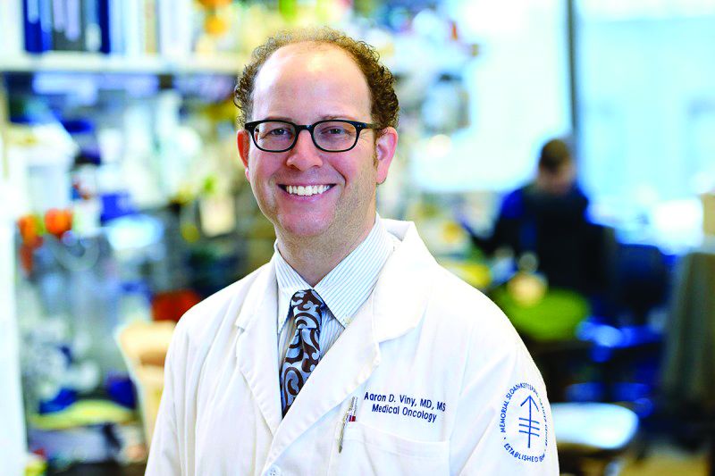
Dr. Viny is with the Memorial Sloan-Kettering Cancer Center, New York, where he is a clinical instructor, on the staff of the leukemia service, and a clinical researcher in The Ross Levine Lab. Contact Dr. Viny at vinya@mskcc.org.
Intermittent fasting fights ALL, not AML, in mice

Photo by Steve Berger
Intermittent fasting inhibits the development and progression of acute lymphoblastic leukemia (ALL), according to preclinical research published in Nature Medicine.
Fasting had an inhibitory effect in mouse models of T-cell and B-cell ALL but not acute myeloid leukemia (AML).
“This study using mouse models indicates that the effects of fasting on blood cancers are type-dependent and provides a platform for identifying new targets for leukemia treatments,” said study author Chengcheng “Alec” Zhang, PhD, of UT Southwestern Medical Center in Dallas, Texas.
“We also identified a mechanism responsible for the differing response to the fasting treatment.”
For this study, Dr Zhang and his colleagues created mouse models of acute leukemia—N-Myc B-ALL, activated Notch1 T-ALL, MLL-AF9 AML, and AML driven by the AML1-Eto9a oncogene—and tested the effects of various dietary restriction plans.
The team used green or yellow florescent proteins to mark and trace the leukemia cells so they could determine if the cells’ levels rose or fell in response to the fasting treatment.
“Strikingly, we found that, in models of ALL, a regimen consisting of 6 cycles of 1 day of fasting followed by 1 day of feeding completely inhibited cancer development,” Dr Zhang said.
At the end of 7 weeks, fasted mice with B-ALL had virtually no detectible cancerous cells—an average of 0.48%—compared to an average of 67.68% of cells found to be cancerous in the test areas of the non-fasted B-ALL mice.
Dr Zhang noted that, compared to B-ALL mice that ate normally, the mice on alternate-day fasting had dramatic reductions in the percentage of ALL cells in the bone marrow and spleen, as well as reduced numbers of white blood cells.
In addition, the spleens and lymph nodes in the fasted mice with B-ALL were similar in size to those of normal mice.
“Although initially cancerous, the few fluorescent cells that remained in the fasted mice after 7 weeks appeared to behave like normal cells,” Dr Zhang said. “Mice in the [B-ALL] model group that ate normally died within 59 days, while 75% of the fasted mice survived more than 120 days without signs of leukemia.”
Dr Zhang and his colleagues said they observed similar results in the T-ALL model but not the AML models. There was no decrease in leukemia cells among fasted mice with AML. And fasting actually shortened survival time in these mice.
Identifying the mechanism
Fasting is known to reduce the level of leptin, a cell signaling molecule created by fat tissue. In addition, previous studies have shown weakened activity by leptin receptors in humans with ALL. For those reasons, the researchers studied both leptin levels and leptin receptors in the mouse models.
The team found that mice with ALL showed reduced leptin receptor activity that increased with intermittent fasting.
“We found that fasting decreased the levels of leptin circulating in the bloodstream as well as decreased the leptin levels in the bone marrow,” Dr Zhang said. “These effects became more pronounced with repeated cycles of fasting. After fasting, the rate at which the leptin levels recovered seemed to correspond to the rate at which the cancerous ALL cells were cleared from the blood.”
The researchers also found that AML was associated with higher levels of leptin receptors that were unaffected by fasting, which could help explain why the fasting treatment was ineffective against this type of leukemia.
It also suggests a mechanism—the leptin receptor pathway—by which fasting exerts its effects in ALL, Dr Zhang said.
“It will be important to determine whether ALL cells can become resistant to the effects of fasting,” he noted. “It also will be interesting to investigate whether we can find alternative ways that mimic fasting to block ALL development.”
Given that this study did not involve drug treatment, researchers are discussing with clinicians whether the tested regimen might be able to move forward quickly to clinical trials. ![]()
Congenital CMV linked to increased risk of ALL

Photo by Vera Kratochvil
Newborns with congenital cytomegalovirus (CMV) infection may have an increased risk of developing acute lymphoblastic leukemia (ALL), according to a study published in Blood.
The data also suggest the risk may be particularly high among Hispanic children.
Researchers said this is the first study to suggest that congenital CMV infection is a risk factor for childhood ALL and is more prominent in Hispanic children.
To conduct this study, the researchers first identified all known infections present in the bone marrow of 127 children diagnosed with ALL and 38 children diagnosed with acute myeloid leukemia (AML).
The team found CMV infection was prevalent in children with ALL but rare in those with AML.
Next, the researchers looked for CMV in newborn blood samples from 268 children who went on to develop ALL. The team compared the samples with samples from 270 healthy children.
“Our goal in tracking CMV back from the time of diagnosis to the womb was to establish that this infection occurred well before initiation of disease,” said lead study author Stephen Francis, PhD, of the University of Nevada and University of California, San Francisco.
He and his colleagues found that children who went on to develop ALL were nearly 4 times more likely than control subjects to be CMV-positive at birth. The odds ratio was 3.71 (P=0.0016).
The odds ratio was 5.9 in Hispanic children and 2.1 in non-Hispanic whites. The researchers said this finding is particularly interesting because of the high rate of ALL observed in Hispanics.
“If it’s true that in utero CMV is one of the initiating events in the development of childhood leukemia, then control of the virus has the potential to be a prevention target,” Dr Francis said. “That’s the real take-home message.”
While this research is in the early stages, the researchers hope these results will inspire more studies that will validate these findings and lead to the development of a CMV vaccine.
“This is the first step, but if we do end up finding a causal link to the most common childhood cancer, we hope that will light a fire in terms of stopping mother-to-child transmission of CMV,” Dr Francis said. ![]()
Platform could optimize treatment of ALL, other diseases

A digital health technology platform may prove useful for optimizing treatment of acute lymphoblastic leukemia (ALL), according to research published in SLAS Technology.
The platform is based on phenotypic personalized medicine (PPM), in which a patient’s response to treatment can be visually represented in the shape of a parabola.
The graph plots the drug dose along the horizontal axis and the patient’s response on the vertical axis.
Researchers said PPM has the ability to accurately identify a person’s optimal drug and dose combinations throughout an entire course of treatment.
“Phenotypic personalized medicine is like turbocharged artificial intelligence,” said study author Dean Ho, PhD, of the University of California, Los Angeles.
“It personalizes combination therapy to optimize efficacy and safety. The ability for our technology to continuously pinpoint the proper dosages of multiple drugs from such a large pool of possible combinations overcomes a challenge that is substantially more difficult than finding a needle in a haystack.”
In this study, Dr Ho and his colleagues used PPM to (retrospectively) individualize drug ratios/dosages in 2 pediatric patients with standard-risk ALL.
The researchers looked at the patients’ records to examine the administration of 4-drug maintenance regimens (dexamethasone, vincristine, mercaptopurine, and methotrexate).
The team said the drug doses served as the inputs, and maintaining absolute neutrophil counts and platelet counts within target ranges served as the outputs for optimization.
Using PPM, the researchers generated individualized 3-dimensional maps to determine the optimal drug ratios.
The team found their technology-suggested drug dosages were as much as 40% lower than clinical chemotherapy dosages, but they still maintained target neutrophil/platelet levels.
The parabolas showed that markedly different dosages of each drug were required to maintain normal cell counts for each patient.
The researchers said their results demonstrate a clear need to personalize ALL treatment and will serve as a foundation for a pending clinical trial to optimize multidrug chemotherapy.
“PPM has the ability to personalize combination therapy for a wide spectrum of diseases, making it a broadly applicable technology,” said study author Chih-Ming Ho, PhD, of the University of California, Los Angeles.
“The fact that we don’t need any information pertaining to a disease’s biological process in order to optimize and personalize treatment is a revolutionary advance. We’re at the interface of digital health and cancer treatment.”
The research team is planning to recruit patients for a prospective trial within the next year. The technology is approved for additional infectious disease and oncology studies. ![]()
How old is too old to be on a kids’ protocol for ALL?

Photo by Bill Branson
SAN DIEGO—In recent years, pediatric or pediatric-inspired protocols have become the preferred treatment approach for younger adults with acute lymphoblastic leukemia (ALL).
These protocols include higher doses of steroids, vincristine, methotrexate, and L-asparaginase.
However, the upper age limit for this strategy has not been defined.
With the GRAALL-2005 study, investigators set out to determine how old is too old to be treated on pediatric protocols.
Their results suggest 55 is likely the upper age limit for patients with Ph-negative ALL.
The investigators also evaluated a hyper-fractionated (hyper-C) versus standard dose (standard-C) of cyclophosphamide during induction and late intensification.
They found that hyper-C did not provide an event-free survival (EFS) benefit in the overall study population, but patients age 55 and older did appear to benefit from hyper-C.
Françoise Huguet, MD, of the Institut Universitaire du Cancer de Toulouse in Toulouse, France, presented these findings at the 2016 ASH Annual Meeting (abstract 762).
GRAALL investigators had previously evaluated a pediatric-inspired protocol for adult patients in the GRAALL-2003 study, which validated the approach.
Study design
Patients with newly diagnosed, Ph-negative ALL were eligible to enroll if they were 18 to 59 years of age.
Treatment comprised a steroid pre-phase, a 5-drug induction, two 3-block dose-dense consolidation phases, a late intensification, a third consolidation phase, CNS irradiation, and a 2-year maintenance phase.
Patients could proceed to allogeneic transplant in first complete remission (CR) if eligible.
During induction and late intensification, patients received cyclophosphamide at 750 mg/m2 on day 1 and were then randomized to hyper-C (300 mg/m2/every 12 hours on days 15 to 17) or standard-C (750 mg/m2 on day 15).
The primary endpoint was EFS.
Patient population
Investigators randomized 787 evaluable patients—398 in the standard-C arm and 389 in the hyper-C arm.
Their median age was 36 years, 67% of patients had B-cell precursor ALL, and 33% had T-ALL.
Most had high-risk ALL, 72% of them receiving standard-C and 66% receiving hyper-C.
About a third of the patients in each arm proceeded to allogeneic stem cell transplant in first CR.
Results
The CR rate after induction therapy was 90.2% in the standard-C arm and 93.6% in the hyper-C arm, for an overall CR rate of 92%.
Most patients—87.5% in the standard-C arm and 91.8% in the hyper-C arm—achieved a response in 1 course of therapy.
Sixty percent of patients tested in the standard-C arm and 66% of those tested in the hyper-C arm were minimal residual disease negative at less than 10-4.
There were 26 (6.5%) deaths in the standard-C arm and 18 (4.6%) in the hyper-C arm.
The 5-year EFS rate was 52% overall, and hyper-C treatment had no impact on EFS (hazard ratio=0.89 [range, 0.7-1.1]; P=0.26).
Impact of age
Investigators conducted a post-hoc subgroup analysis of 5 age groups—18-24 years (n=200), 25-34 (n=172), 35-44 (n=171), 45-54 (n=151), and 55+ (n=93).
Overall, the CR rate tended to decrease with age. The rates were 98.5% (18-24), 95.3% (25-34), 87.7% (35-44), 89.4% (45-54), and 79.6% (55+).
Induction death rates increased from 0.5% in the youngest group to 18.3% in the oldest, but the rate of cumulative incidences of failure at 5 years was similar among all the age groups.
The cumulative incidence of treatment-related mortality, without censoring for transplant, ranged from 7.6% in the youngest group to 39.7% in the oldest.
And the 5-year EFS for the youngest patients was 60%, while, for the oldest, it was 26%.
“Above 50 years, the increase in age became highly significant,” Dr Huguet emphasized. “There were fewer CRs and lower survival.”
Treatment compliance
In terms of treatment compliance and median dose received in the induction course, patients aged 55-59 received significantly less L-asparaginase than those aged 18-54 (P<0.001).
During all 3 consolidation phases, patients aged 55-59 received significantly lower median doses of all medications—cytarabine, methotrexate, cyclophosphamide—than patients aged 18-54.
And in late intensification, patients aged 55-59 received significantly lower median doses of vincristine, prednisone, daunorubicin, and hyper-C than all other patients. The median doses of L-asparaginase and standard-C received were lower in the older patients but not significantly so.
EFS by age and randomization
The 5-year EFS for patients aged 18-54 was 57% with hyper-C, compared with 55% in the standard-C arm (P=0.66).
However, for older patients, there was a significant advantage for those receiving hyper-C. The 5-year EFS was 38% with hyper-C, compared to 12% with standard-C (P=0.007).
Dr Huguet explained that inferior compliance in patients 55 and older “might explain why a benefit associated with early hyper-C reinforcement became apparent in these older patients only.”
Dr Huguet concluded that the results “suggest that 55 years is likely to be the upper age limit to tolerate a pediatric-like therapy for younger adults with Ph-negative ALL.”
She added that patients over 54 might benefit from alternative front-line strategies.
Accordingly, investigators are planning to use new agents, such as blinatumomab or inotuzumab ozogamicin, in the next European Working Group on Adult ALL studies. ![]()
Good response from CAR T cells with ‘safety switch’ for advanced ALL
SAN DIEGO – Anti-CD19 chimeric antigen receptor (CAR) T cells engineered with a “safety switch” yielded high rates of complete response and an acceptable toxicity profile in chemotherapy-resistant B cell acute lymphoblastic leukemia, according to a multicenter phase I/II trial.
Importantly, high tumor burden did not increase the risk of cytokine release syndrome, said Lung-Ji Chang, PhD, of Shenzhen (China) Genoimmune Medical Institute and the University of Florida in Gainesville. “This reliable, standardized CAR T-cell preparation protocol has now served more than 30 major medical centers in China,” he said at the annual meeting of the American Society of Hematology.
Anti-CD19 CAR T cells have shown dramatic potential for treating B-cell malignancies, but toxicities have been a concern. One potentially serious adverse reaction is cytokine release syndrome, in which patients develop marked rises in blood levels of several types of cytokines. Another problem is that anti-CD19 CAR T cells can trigger loss of CD19 B cells, ultimately leading to humoral deficiencies, Dr. Chang noted. Consequently, researchers have searched for ways to continue controlling the activity of CAR T cells even after infusing them into patients.
As part of that effort, Dr. Chang and his associates developed a standardized protocol for engineering next-generation anti-CD19 CAR T cells based on the established concept of a “safety switch.” After collecting T cells from patients with chemotherapy-resistant ALL, they used a lentiviral vector to transform them into CAR T cells with fusion proteins consisting of a proapoptotic molecule called caspase-9 that is linked to modified human FK506-binding proteins, or FKBP. The addition of iCaspase9-FKBP enables clinicians to induce CAR T cell apoptosis by treating patients with a synthetic dimerizer called AP1903.
Apoptosis occurs about 45 minutes after this drug is given, according to Dr. Chang. This “safety switch” also enables clinicians to eliminate anti-CD19 CAR T cells after tumor cells are eradicated so that patients can recover their humoral immunity. He and his associates further modified these anti-CD19 CAR T cells by introducing four intracellular signaling domains that are associated with T-cell activation, survival, and longevity, he said.
A total of 22 treatment centers helped test this approach in a phase I/II trial of 110 leukemia patients, about half of whom were children with a median age of 9 years. The median age of adults was 37 years, and the oldest patient was 70. Cancer types included Philadelphia chromosome–positive ALL, Philadelphia chromosome–negative ALL, and chronic myeloid leukemia with blast crisis. About a third of patients had bone marrow samples with at least 50% blasts, and a similar proportion had already undergone hematopoietic stem cell transplantation.
Cytokine release syndrome affected 86% of patients with low or no tumor burden, but only 53% of patients with bone marrow blasts exceeding 5%, Dr. Chang reported. He emphasized that patients with high tumor burden were no more likely to develop moderate or severe cytokine release syndrome than were patients with little or no tumor burden (P = .3). Furthermore, among 17 patients with more than 80% bone marrow involvement, only three developed grade 3-4 cytokine release syndrome, while eight developed grade 1 cytokine release syndrome.
A total of 96 patients (87%) had a complete response to this CAR T cell regimen, including 51 children and 45 adults, Dr. Chang reported. Median overall survival was 222 days (range, 23-1,041 days), and 60% of patients lived at least 400 days after treatment. Patients survived a median of 115 days without relapsing (range, 0-455 days), and 55% ultimately relapsed. Age did not appear to predict relapse, he noted.
Kaplan-Meier curves revealed no major differences in rates of overall survival (OS) between adults and children at 400-day data cutoff, Dr. Chang said. However, patients with more than 50% blast cells in their bone marrow had significantly lower rates of survival (P = .02) than did patients with less advanced ALL. A lower T-cell dose predicted lower survival in children (P = .04), but not in adults. Dr. Chang and his colleagues now dose patients of all ages with 106 cells per kilogram, he said.
Survival was significantly more likely when CAR T cell recipients went on to allogeneic hematopoietic stem cell transplantation (P = .0002) than otherwise. Based on the findings, Dr. Chang particularly recommends this approach for highly chemotherapy-resistant disease with a high tumor burden. Among patients who relapsed, repeating CAR T cell therapy led to better survival than administering combination chemotherapy-tyrosine kinase inhibitor therapy (P = .01).
These safety and efficacy results suggest that CAR T cell immunotherapy can benefit patients if they have very high-burden leukemia, Dr. Chang concluded. Patients outcomes remained consistent across centers due to a “highly standardized CAR T cell preparation profile,” he said.
Dr. Chang did not report funding sources. He reported having no relevant conflicts of interest.
AT ASH 2016
Key clinical point: Safety-engineered anti-CD19 autologous chimeric antigen receptor (CAR) T cells achieved good efficacy and adequate safety results in a multicenter study of children and adults with acute lymphoblastic leukemia.
Major finding: A total of 96 patients (87%) had a complete response, and median overall survival was 222 days. High tumor burden did not increase the risk of cytokine release syndrome.
Data source: A multicenter phase I/II study of 110 children and adults with ALL.
Disclosures: The researchers had no relevant financial disclosures.
Group estimates global cancer cases, deaths in 2015

receiving chemotherapy
Photo by Rhoda Baer
Researchers have estimated the global incidence of 32 cancer types and deaths related to these malignancies in 2015.
The group’s data, published in JAMA Oncology, suggest there were 17.5 million cancer cases and 8.7 million cancer deaths last year.
There were 78,000 cases of Hodgkin lymphoma and 24,000 deaths from the disease, as well as 666,000 cases of non-Hodgkin lymphoma (NHL) and 231,000 NHL deaths.
There were 154,000 cases of multiple myeloma and 101,000 deaths from the disease.
And there were 606,000 cases of leukemia, with 353,000 leukemia deaths. This included 161,000 cases of acute lymphoid leukemia (110,000 deaths), 191,000 cases of chronic lymphoid leukemia (61,000 deaths), 190,000 cases of acute myeloid leukemia (147,000 deaths), and 64,000 cases of chronic myeloid leukemia (35,000 deaths).
The data also show that, between 2005 and 2015, cancer cases increased by 33%, mostly due to population aging and growth, plus changes in age-specific cancer rates.
Globally, the odds of developing cancer during a lifetime were 1 in 3 for men and 1 in 4 for women in 2015.
Prostate cancer was the most common cancer in men (1.6 million cases), although tracheal, bronchus, and lung cancer was the leading cause of cancer deaths for men.
Breast cancer was the most common cancer for women (2.4 million cases) and the leading cause of cancer deaths in women.
The most common childhood cancers were leukemia, “other neoplasms,” NHL, and brain and nervous system cancers. ![]()
Anti-CD22 CAR T-cells shift ALL into complete remission
SAN DIEGO – When one CAR stops one working, try another: chimeric antigen receptor (CAR) T-cell therapy for children and young adults with acute lymphoblastic leukemia is driving forward with a novel anti-CD22 target that in an early dose-finding trial has induced complete remissions in some patients with relapsed or refractory disease, including patients previously treated with anti-CD19 CAR-T therapy.
In the first-in-humans trial, CAR T-cell therapy directed against CD22 was shown to be safe and was associated with minimal residual disease (MRD)-negative complete remissions in eight of 10 children and young adults with relapsed/refractory B-precursor acute lymphoblastic leukemia treated at the highest dose level.
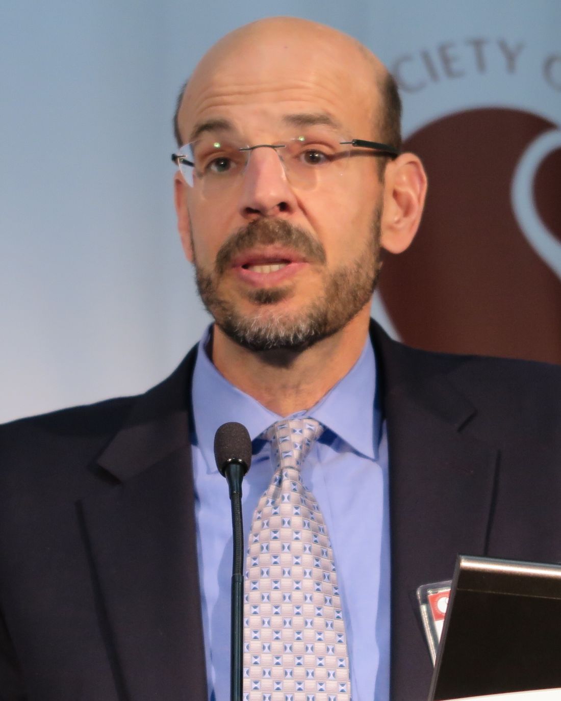
“This is the first successful salvage CAR therapy for CD19-negative B-[lineage] ALL,” said co-principal investigator Terry J. Fry, MD, from the Center for Cancer Research at the National Cancer Institute in Bethesda, Md.
Preliminary experience with anti-CD22 immunotherapy suggests that it is comparable in potency to anti-CD19 CAR, and investigators are exploring the possibility that the two chimeric antigen targets could be combined for greater efficacy, he said during a briefing at the annual meeting of the American Society of Hematology.
Tough target
As reported previously from the 2013 ASH annual meeting, anti-CD19 CAR T cells induced complete responses in 10 of 16 children and young adults with relapsed/refractory ALL, and in a second study, CD19-targeted T cells induced complete molecular responses in 12 of 16 adults with B-lineage ALL refractory to chemotherapy.
In current phase 2 trials, anti-CD19 CAR-T therapy is associated with complete remission rates of 80% to 90% of those treated.
However, “we’re learning now that one of the limitations of this approach is the loss of CD19 expression occurring in a substantial number of patients, although it has not been systematically analyzed,” Dr. Fry said.
CD22, an antigen restricted to B-lineage cells, is a promising alternative to CD19 as a target, but finding just the right anti-CD22 CAR was tricky, Dr. Fry said in an interview. The investigators found that many candidate antigens bound well to T cells but had no efficacy, and it took several years of trying before they identified the current version of the antigen
In the phase I trial, the investigators enrolled 16 children and young adults (ages 7 to 22 years) with relapsed/refractory CD22-positive hematologic malignancies. All patients had previously undergone at least one allogeneic stem cell transplant, 11 had previously received anti-CD19 CAR-T cell therapy, and 9 were CD19-negative or had reduced CD19 expression on ALL cells.
The patients underwent peripheral blood mononuclear cells (PBMCs) collected through autologous leukapheresis. The cells were then enriched and expanded, and transduced with a lentiviral vector containing an anti-CD22 CAR for 7 to 10 days, allowing the cells to identify and bind to CD22 expressed on ALL blasts.
The patients then underwent lymphodepletion with fludarabine, and cyclophosphamide, and received infusions of the transduced T-cells at one of three dose levels, starting at 3 x 10⁵ transduced T-cells per recipient weight in kilograms (DL-1), 1 x 10⁶/kg (DL-2), and 3 x 10⁶/kg (DL-3).
The complete remission rate at DL-2 and -3 combined was 80%, with the cytokine-release syndrome (CRS) at a maximum of grade 2.
As noted before, three of the remissions were comparatively durable, with one lasting more than a year.
There were no dose-limiting toxicities at DL-2, and grade 4 hypoxia at DL-3 was seen in one patient.There was one death from sepsis and multi-organ failure in one patient in an expansion cohort. There have been no cases of severe neurotoxicity thus far.
In five patients who experienced relapse, one treated at DL-1 had a loss of CAR cells, and four had changes in CD22 expression, primarily a decrease in site density that may cause the CD22 expression to fall below the threshold for CAR activity, Dr. Fry said.
“At least in our eyes, this may not be best used as a salvage therapy, but we’re beginning to think about how this should be included with CD19 in the upfront CAR treatment,” he said.
The study was funded by the National Institutes of Health with support from Lentigen and Juno Therapeutics. Dr. Fry reported no relevant disclosures.
FROM ASH 2016
Key clinical point: CAR T-cell therapy with an anti-CD22 antigen induced complete, MRD-negative remissions in children/young adults with acute lymphoblastic leukemia.
Major finding: The complete remission rate among patients treated at the two highest dose levels was 80%.
Data source: Phase 1 dose-finding trial in 16 children/young adults with relapsed/refractory ALL or diffuse large B-cell lymphoma.
Disclosures The study was funded by the National Institutes of Health with support from Lentigen and Juno Therapeutics. Dr. Fry reported no relevant disclosures views
VIDEO: Anti-CD22 CAR for R/R ALL impresses in early trial
SAN DIEGO – In a first-in-humans trial, chimeric antigen receptor (CAR) T-cell therapy directed against CD22 was shown to be safe and was associated with minimal residual disease (MRD)–negative complete remissions in 8 of 10 children and young adults with relapsed/refractory B-precursor acute lymphoblastic leukemia treated at the highest dose levels. One patient remains in remission more than 1 year of treatment, one had a 6-month remission, and one had a remission lasting 3 months.
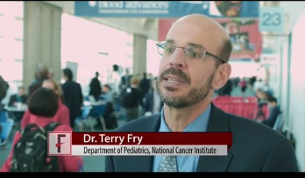
In a video interview, co-principal investigator Terry J. Fry, MD, of the Center for Cancer Research at the National Cancer Institute in Bethesda, Md., discusses the rationale behind using an alternative antigen target in salvage therapy for ALL, and the potential for combining antigen targets to treat patients with relapsed/refractory ALL.
The video associated with this article is no longer available on this site. Please view all of our videos on the MDedge YouTube channel
AT ASH 2016
FDA grants full approval for ponatinib
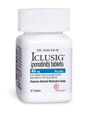
Photo from Business Wire
The US Food and Drug Administration (FDA) has granted full approval for the kinase inhibitor ponatinib (Iclusig®) and updated the drug’s label.
Ponatinib now has full approval as a treatment for adults with chronic myeloid leukemia (CML) or Philadelphia chromosome-positive acute lymphoblastic leukemia (Ph+ ALL) when no other tyrosine kinase inhibitor is indicated.
Ponatinib is also approved to treat adults with T315I-positive CML or T315I-positive Ph+ ALL.
Ponatinib was initially approved in December 2012 under the FDA’s accelerated approval program.
This program allows the FDA to approve a drug to treat a serious or life-threatening disease based on clinical data showing the drug has an effect on a surrogate endpoint reasonably likely to predict clinical benefit to patients.
The company developing the drug must conduct post-approval research to determine if the drug provides a clinical benefit. If so, the drug can be granted full approval.
The full approval and label update for ponatinib is based on 48-month follow-up data (as of August 2015) from the phase 2 PACE trial, which enrolled heavily pretreated patients with resistant or intolerant CML or Ph+ ALL. These data were presented at the 2016 ASCO Annual Meeting.
“The longer follow up of the PACE study confirms the clinical benefit of ponatinib in this setting,” said Jorge Cortes, MD, a professor at The University of Texas MD Anderson Cancer Center in Houston and a leading investigator in the PACE trial.
“We had learned from the initial report of the high response rate with ponatinib among CML patients with resistance or intolerance to prior therapies. The 4-year follow-up and updated safety profile demonstrate durability of responses in this heavily pretreated population. These results solidify ponatinib as an important and valuable treatment option for refractory patients with CML where no other TKI therapy is appropriate, including those who have the T315I mutation.”
Past problems with ponatinib
Previous follow-up data from the PACE trial, collected in 2013, suggested ponatinib can increase the risk of thrombotic events. When these data came to light, officials in the US and European Union, where ponatinib had already been approved, began to investigate the drug.
Ponatinib was pulled from the US market for a little over 2 months, and trials of the drug were placed on partial hold while the FDA evaluated the drug’s safety. Ponatinib went back on the market in January 2014, with new safety measures in place.
Ponatinib was not pulled from the market in the European Union, but the European Medicine’s Agency released recommendations for safer use of the drug. The Committee for Medicinal Products for Human Use reviewed data on ponatinib and decided its benefits outweigh its risks. ![]()

