User login
Study explains how a mutation spurs AML development
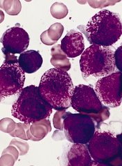
A set of faulty genetic instructions keeps hematopoietic stem/progenitor cells (HSPCs) from maturing and contributes to the development of acute myeloid leukemia (AML), according to research published in Cancer Cell.
Researchers found that a mutation in the gene DNMT3A removes a “brake” on the activity of stemness genes, which leads to the creation of immature precursor cells that can become AML cells.
Specifically, the DNMT3A mutational hotspot at Arg882 (DNMT3AR882H) cooperates with an NRAS mutation (NRASG12D) to transform HSPCs and induce AML development.
“Due to a large-scale cancer sequencing project, the DNMT3A gene is now appreciated to be one of the top 3 most frequently mutated genes in human acute myeloid leukemia, and yet the role of its mutation in the disease has remained far from clear,” said G. Greg Wang, PhD, of the University of North Carolina Lineberger Comprehensive Cancer Center in Chapel Hill.
“Our findings not only provide a deeper understanding of how this prevalent mutation contributes to the development of AML, but it also offers useful information on how to develop new strategies to treat AML patients.”
In an attempt to understand how the DNMT3A mutation helps drive AML, Dr Wang and his colleagues created one of the first laboratory AML models for studying somatic mutations in DNMT3A.
The DNMT3A gene codes for a protein that binds to specific sections of DNA with a chemical tag that can influence the activity and expression of the underlying genes in cells.
The researchers found that DNMT3AR882H caused AML cells to have a different pattern of chemical tags that affect how the genetic code is interpreted and how the cell develops.
In cancerous cells with DNMT3AR882H, a set of gene enhancers for several stemness genes—including Meis1, Mn1, and the Hoxa gene cluster—were left unchecked. Therefore, HSPCs were left with a constant “on” switch, allowing the cells to “forget” to mature.
“In acute myeloid leukemia, the expression of these stemness genes are aberrantly maintained at a higher level,” Dr Wang said. “As a result, cells ‘forget’ to proceed to normal differentiation and maturation, generating immature precursor blood cells and a prelude to full-blown cancer.”
The researchers also found that, while the DNMT3A mutation is required for AML development, the mutation itself is not sufficient to cause cancer alone. DNMT3AR882H cooperates with another mutation, NRASG12D.
“We found the RAS mutation stimulates these immature blood cells to be hyper-proliferate,” said study author Rui Lu, PhD, of the University of North Carolina Lineberger Comprehensive Cancer Center.
“However, these cells cannot maintain their stem cell properties. While the DNMT3A mutation itself does not have hyper-proliferative effects, [it] does promote stemness properties and generates leukemia stem/initiating cells together with the RAS mutation.”
The researchers also reported testing a potential treatment in cells with the DNMT3A mutation. They found that AML cells with DNMT3AR882H were sensitive to inhibitors of DOT1L, a cellular enzyme involved in modulation of gene expression activities.
As DOT1L inhibitors are currently under clinical investigation, this finding suggests a potential strategy for treating DNMT3A-mutated AML. ![]()
A new approach to treat AML?

Preclinical research suggests that activating the STING pathway may be a feasible approach for treating acute myeloid leukemia (AML).
The STING protein has been shown to play a crucial role in the immune system’s ability to “sense” cancer by recognizing and responding to DNA from tumor cells.
In past studies, researchers injected compounds that activate the STING pathway directly into solid tumors in mice, and this produced potent anti-tumor immune responses.
In a new study, researchers injected substances that mimic tumor-cell DNA into the bloodstream and found they could stimulate STING to provoke a life-extending immune response in mice with AML.
“Delivery of these substances into the blood led to massive immune responses,” said study author Justin Kline, MD, of the University of Chicago in Illinois.
“I’ve worked extensively with animal models of this disease, and have never seen responses like this.”
This research, published in Cell Reports, is the first demonstration that activating the STING pathway could be effective in hematologic malignancies.
STING (short for STimulator of INterferon Genes) plays a role in detecting threats, such as viral infections or cancer. STING is activated when DNA turns up in the wrong place, inside the cell but outside the nucleus.
When it encounters such misplaced DNA, STING induces the production of interferon-beta and other chemical signals that recruit certain components of the immune system to manage the threat, such as leukemia-specific killer T cells.
In this study, the researchers found that mice with established AML were rarely able to launch an effective immune response against the disease.
But when the team exposed the mice to DMXAA (5,6-dimethylxanthenone-4-acetic acid), a molecule that activates STING, the immune system responded aggressively, culminating in the activation of highly potent, cancer-cell killing T cells.
This response prolonged survival and, in some cases, cured the mice of their leukemia. About 60% of DMXAA-treated mice survived long-term. They were even able to protect themselves when “re-challenged” with AML cells.
Because of significant differences between mice and humans, DMXAA does not activate the human STING pathway, but researchers have found that several cyclic dinucleotides—signaling molecules produced by bacteria—have a comparable effect in stimulating the STING pathway.
This leads to an immune response that begins with the production of type I interferons and proceeds to later, more powerful stages, ultimately including leukemia-specific T cells.
“Our results provide strong rationale for the clinical translation of STING agonists as immune therapy for leukemia and possibly other hematologic
malignancies,” said study author Emily Curran, MD, of the University of Chicago.
However, Dr Kline noted that this approach is “not without risk.” He said it can induce “a lot of inflammation, fever, even shock.” Such a stimulated immune system can be “too effective,” especially when the therapy is given through the blood stream, rather than injected into a solid tumor.
“I think drug makers will want to focus on intra-tumoral injection studies before they are ready to bet on systemic infusion,” Dr Kline said. “But this is an important first step.” ![]()
Study provides clues to AML survival after chemotherapy

Preclinical research suggests that some leukemia cells harvest energy resources from normal cells during chemotherapy, and this helps the leukemia cells survive and thrive after treatment.
Investigators found that acute myeloid leukemia (AML) cells are capable of stealing mitochondria from stromal cells, and these stolen mitochondria give an energy boost to surviving AML cells, which helps fuel the leukemia’s resurgence after chemotherapy.
“There are multiple mechanisms for resistance to chemotherapy, and it will be important to target them all in order to eliminate all leukemic cells,” said Jean-François Peyron, PhD, of the Centre Méditerranéen de Médecine Moléculaire (C3M) in Nice, France.
“Targeting this protective mitochondrial transfer could represent a new strategy to improve the efficacy of the current treatments for acute myeloid leukemia.”
Dr Peyron and his colleagues described their discovery of the mitochondrial transfer in Blood.
The team conducted their experiments using cell cultures and mouse models of AML. They found that nearly all AML cells died when exposed to chemotherapy drugs, but some survived. And these cells issued a “mayday” signal that “tricked” nearby non-cancerous cells into yielding their mitochondria to the AML cells, thus strengthening the leukemia cells.
“Mitochondria produce the energy that is vital for cell functions,” explained study author Emmanuel Griessinger, PhD, also of C3M.
“Through the uptake of mitochondria, chemotherapy-injured acute myeloid leukemia cells recover new energy to survive.”
The AML cells were found to increase their mitochondria mass by an average of 14%. This increase led to a 1.5-fold increase in energy production and significantly better survival rates. That is, the leukemia cells that had a high level of mitochondria were also more resistant to the chemotherapy.
The investigators observed the phenomenon in several types of leukemia cells, most notably leukemia-initiating cells. The team said this finding may explain why it can be difficult to treat AML and other cancers.
They also believe these findings offer new hope for developing better treatments for AML. If researchers can find a way to interfere with the transfer of mitochondria, that could reduce the risk of relapse.
The study may also shed light on other cancer types. Similar mechanisms may be at play in other hematologic malignancies and even solid tumors, according to the investigators.
For now, the team’s next step for this research is to identify the mechanism underlying the transfer of mitochondria in AML. ![]()
FDA reports shortage of doxorubicin for injection, initiates importation
A critical shortage of doxorubicin hydrochloride 50 mg powder for injection has been reported to the Food and Drug Administration.
Doxorubicin is approved to treat acute lymphoblastic leukemia, acute myeloid leukemia, breast cancer, gastric cancer, ovarian cancer, neuroblastoma, and other cancer types.
To increase availability, the pharmaceutical company Hospira (a Pfizer company) is coordinating with the FDA to import the drug from Ahmedabad, India, where it is manufactured by Zydus Hospira Oncology Private Ltd. at an FDA-inspected facility that is in compliance with current good manufacturing practice requirements.
“It is important to note that there are substantive differences in the format and content of the labeling between the U.S.-approved doxorubicin hydrochloride for injection, USP and the Hospira Limited’s doxorubicin hydrochloride 50 mg powder for injection,” Hospira reported in a letter to health care providers.
To place an order or to get questions answered, contact Hospira directly by calling customer care at 1-877-946-7747 (Mondays-Fridays, 7 a.m.-6 p.m. Central time).
For clinical inquiries, contact Hospira Medical Communications at 1-800-615-0187 or email medcom@hospira.com.
According to the letter, adverse events or quality problems associated with use of this product should be reported by calling Hospira Global Complaint Management by phone, 1-800-441-4100; by sending an email to ProductComplaintsPP@hospira.com; or by submitting a report online to Medwatch.
On Twitter @jessnicolecraig
Acute myeloid leukemia genomic classification and prognosis
Acute myeloid leukemia (AML) consists of at least 11 disease classes that represent distinct paths in the evolution of AML and have prognostic implications, based on an analysis of somatic driver mutations in 1,540 patients.
In total, 5,234 driver mutations were identified in 76 genes or regions, with 96% of patients having at least one mutation and 86% having two or more mutations. However, nearly one-half of the cohort did not fall into one of the molecular groups defined by the World Health Organization in 2008.
“The characterization of many new leukemia genes, multiple driver mutations per patient, and complex co-mutation patterns prompted us to reevaluate genomic classification of AML from the beginning,” wrote Elli Papaemmanuil, Ph.D., a molecular geneticist at Memorial Sloan Kettering, New York, and of the Cancer Genome Project, Wellcome Trust Sanger Institute, and her colleagues (N Engl J Med. 2016 Jun 9; 374:2209-21).
The team developed a Bayesian statistical model to define 11 mutually exclusive subtypes based on patterns of co-mutations. The schema unambiguously classified 1,236 of 1,540 patients (80%) into a single subgroup and 56 (4%) into two or more groups. A subset of patients (166, 11%) remained unclassified, possibly due to mutations in genes not sequenced in the study.
NPM1-mutated AML was the largest class (27% of the cohort), followed by the chromatin-spliceosome group (18% of the cohort) that included mutations in genes regulating RNA splicing (SRSF2, SF3B1, U2AF1, and ZRSR2), chromatin (ASXL1, STAG2, BCOR, MLLPTD, EZH2, and PHF6), or transcription (RUNX1). Another subgroup consisted of mutations in TP53, as well as complex karyotype alterations, cytogenetically visible copy-number alterations (aneuploidies), or a combination. While broader than previous classifications, such as “monosomal karyotype AML” and “complex karyotype AML,” this group emerged from correlated chromosomal abnormalities and was mutually exclusive of other class-defining lesions. In general, patients in this group were older and had fewer RAS pathway mutations.
The groups had considerable differences in clinical presentation and overall survival, according to the report. The TP53-aneuploidy subgroup had poor outcomes, as previously described. Patients in the chromatin-spliceosome group had lower rates of response to induction chemotherapy, higher relapse rates, and poorer long-term outcomes, compared with other groups. Most of these patients (84%) would be classified as intermediate risk under current guidelines, but the characteristics were more similar to those of subgroups with adverse outcomes.
Overall survival was correlated with the number of driver mutations, and deleterious affects of mutations often were found to be additive. In some cases, complex gene interactions accounted for variation in outcomes, suggesting the clinical effect of some driver mutations may depend on the occurrence of co-mutations in a wider genomic context.
The study by Papaemmanuil and her colleagues offers practice-changing insights that redefine molecular classification of AML. The mutational analysis of more than 1,500 AML patients provides a deeper understanding of the specific paths from normal blood cell to leukemia.
Specific concurrent mutations were linked to clinical outcomes. For example, co-mutations in NPM1, FLT3ITD, and DNMT3A are associated with a poor clinical outcome, but NPM1 and DNMT3A mutations without FLT3ITD are associated with better outcomes. In addition, mutations in NPM1 and DNMT3A in the presence of NRASG12/13 are associated with a more favorable outcome. The evolution of DNMT3A-NPM1 mutated clones along separate paths appears to affect disease outcome and may be relevant to clinical trials in AML subgroups.
 |
Dr. Aaron Viny |
Previous, smaller studies had suggested that somatic mutations in splicing factors and chromatin modifiers were specific for secondary AML that arises from myelodysplastic syndromes (MDS). Papaemmanuil and her colleagues provide extensive data to support that hypothesis. Patients with chromatin-spliceosome mutations, previously classified as intermediate-risk AML, are classed into the same molecular subgroup as patients with secondary AML arising from MDS.
These data may inform the design of mechanism-based clinical trials based on the presence of specific mutations and co-mutations.
Dr. Aaron Viny is a medical oncologist at Memorial Sloan Kettering Cancer Center, New York. Dr. Ross Levine is Director of the Memorial Sloan Kettering Center for Hematologic Malignancies. These remarks were part of an editorial accompanying a report in The New England Journal of Medicine (2016 Jun 9; 374:2282-4). Dr. Levine reports personal fees from Foundation Medicine outside the submitted work.
The study by Papaemmanuil and her colleagues offers practice-changing insights that redefine molecular classification of AML. The mutational analysis of more than 1,500 AML patients provides a deeper understanding of the specific paths from normal blood cell to leukemia.
Specific concurrent mutations were linked to clinical outcomes. For example, co-mutations in NPM1, FLT3ITD, and DNMT3A are associated with a poor clinical outcome, but NPM1 and DNMT3A mutations without FLT3ITD are associated with better outcomes. In addition, mutations in NPM1 and DNMT3A in the presence of NRASG12/13 are associated with a more favorable outcome. The evolution of DNMT3A-NPM1 mutated clones along separate paths appears to affect disease outcome and may be relevant to clinical trials in AML subgroups.
 |
Dr. Aaron Viny |
Previous, smaller studies had suggested that somatic mutations in splicing factors and chromatin modifiers were specific for secondary AML that arises from myelodysplastic syndromes (MDS). Papaemmanuil and her colleagues provide extensive data to support that hypothesis. Patients with chromatin-spliceosome mutations, previously classified as intermediate-risk AML, are classed into the same molecular subgroup as patients with secondary AML arising from MDS.
These data may inform the design of mechanism-based clinical trials based on the presence of specific mutations and co-mutations.
Dr. Aaron Viny is a medical oncologist at Memorial Sloan Kettering Cancer Center, New York. Dr. Ross Levine is Director of the Memorial Sloan Kettering Center for Hematologic Malignancies. These remarks were part of an editorial accompanying a report in The New England Journal of Medicine (2016 Jun 9; 374:2282-4). Dr. Levine reports personal fees from Foundation Medicine outside the submitted work.
The study by Papaemmanuil and her colleagues offers practice-changing insights that redefine molecular classification of AML. The mutational analysis of more than 1,500 AML patients provides a deeper understanding of the specific paths from normal blood cell to leukemia.
Specific concurrent mutations were linked to clinical outcomes. For example, co-mutations in NPM1, FLT3ITD, and DNMT3A are associated with a poor clinical outcome, but NPM1 and DNMT3A mutations without FLT3ITD are associated with better outcomes. In addition, mutations in NPM1 and DNMT3A in the presence of NRASG12/13 are associated with a more favorable outcome. The evolution of DNMT3A-NPM1 mutated clones along separate paths appears to affect disease outcome and may be relevant to clinical trials in AML subgroups.
 |
Dr. Aaron Viny |
Previous, smaller studies had suggested that somatic mutations in splicing factors and chromatin modifiers were specific for secondary AML that arises from myelodysplastic syndromes (MDS). Papaemmanuil and her colleagues provide extensive data to support that hypothesis. Patients with chromatin-spliceosome mutations, previously classified as intermediate-risk AML, are classed into the same molecular subgroup as patients with secondary AML arising from MDS.
These data may inform the design of mechanism-based clinical trials based on the presence of specific mutations and co-mutations.
Dr. Aaron Viny is a medical oncologist at Memorial Sloan Kettering Cancer Center, New York. Dr. Ross Levine is Director of the Memorial Sloan Kettering Center for Hematologic Malignancies. These remarks were part of an editorial accompanying a report in The New England Journal of Medicine (2016 Jun 9; 374:2282-4). Dr. Levine reports personal fees from Foundation Medicine outside the submitted work.
Acute myeloid leukemia (AML) consists of at least 11 disease classes that represent distinct paths in the evolution of AML and have prognostic implications, based on an analysis of somatic driver mutations in 1,540 patients.
In total, 5,234 driver mutations were identified in 76 genes or regions, with 96% of patients having at least one mutation and 86% having two or more mutations. However, nearly one-half of the cohort did not fall into one of the molecular groups defined by the World Health Organization in 2008.
“The characterization of many new leukemia genes, multiple driver mutations per patient, and complex co-mutation patterns prompted us to reevaluate genomic classification of AML from the beginning,” wrote Elli Papaemmanuil, Ph.D., a molecular geneticist at Memorial Sloan Kettering, New York, and of the Cancer Genome Project, Wellcome Trust Sanger Institute, and her colleagues (N Engl J Med. 2016 Jun 9; 374:2209-21).
The team developed a Bayesian statistical model to define 11 mutually exclusive subtypes based on patterns of co-mutations. The schema unambiguously classified 1,236 of 1,540 patients (80%) into a single subgroup and 56 (4%) into two or more groups. A subset of patients (166, 11%) remained unclassified, possibly due to mutations in genes not sequenced in the study.
NPM1-mutated AML was the largest class (27% of the cohort), followed by the chromatin-spliceosome group (18% of the cohort) that included mutations in genes regulating RNA splicing (SRSF2, SF3B1, U2AF1, and ZRSR2), chromatin (ASXL1, STAG2, BCOR, MLLPTD, EZH2, and PHF6), or transcription (RUNX1). Another subgroup consisted of mutations in TP53, as well as complex karyotype alterations, cytogenetically visible copy-number alterations (aneuploidies), or a combination. While broader than previous classifications, such as “monosomal karyotype AML” and “complex karyotype AML,” this group emerged from correlated chromosomal abnormalities and was mutually exclusive of other class-defining lesions. In general, patients in this group were older and had fewer RAS pathway mutations.
The groups had considerable differences in clinical presentation and overall survival, according to the report. The TP53-aneuploidy subgroup had poor outcomes, as previously described. Patients in the chromatin-spliceosome group had lower rates of response to induction chemotherapy, higher relapse rates, and poorer long-term outcomes, compared with other groups. Most of these patients (84%) would be classified as intermediate risk under current guidelines, but the characteristics were more similar to those of subgroups with adverse outcomes.
Overall survival was correlated with the number of driver mutations, and deleterious affects of mutations often were found to be additive. In some cases, complex gene interactions accounted for variation in outcomes, suggesting the clinical effect of some driver mutations may depend on the occurrence of co-mutations in a wider genomic context.
FROM NEJM
Key clinical point: Mutational analysis of 1,540 patients with acute myeloid leukemia (AML) identified 11 distinct classes with prognostic implications.
Major finding: In total, 5,234 driver mutations were identified involving 76 genes or regions; 96% of patients had at least one driver mutation, and 86% had two or more.
Data sources: Samples came from three prospective multicenter clinical trials of the German-Austrian AML Study Group: AMLHD98A, AML-HD98B, and AMLSG-07-04.
Disclosures: Dr. Papaemmanuil and most coauthors reported having no disclosures. Two coauthors reported financial ties to industry sources.
Researchers identify 11 subgroups of AML

Photo courtesy of NIGMS
Using patient samples from 3 prospective multicenter clinical trials of the German-Austrian AML Study Group, researchers have identified 5234 driver mutations involving 76 genes or regions in 1540 patients with acute myeloid leukemia (AML).
They say the sample size afforded a more comprehensive analysis than previously conducted, and as a result, they found patients were divided into at least 11 subgroups of AML.
They also identified 3 genomic categories beyond the existing World Health Organization (WHO) subgroups. These genomic categories are chromatin–spliceosome mutations, TP53–aneuploidy, and provisionally, IDH2R172 mutations.
Their study, led by scientists at the Wellcome Trust Sanger Institute and international collaborators, could have an impact on clinical trial design and improve the way patients are diagnosed and treated in the future.
The scientists stated in their published paper that 736 patients (48%) in their study would not have fit into the molecular groups included in the 2008 WHO classification of adult AML. This prompted them to reevaluate genomic classification of AML from the beginning.
“We have shown that AML is an umbrella term for a group of at least 11 different types of leukemia,” said Peter Campbell, MBChB, PhD, co-leader of the study. “We can now start to decode these genetics to shape clinical trials and develop diagnostics.”
The scientists found NPM1-mutated AML to be the largest class in their cohort, accounting for 27% of the patients.
The second largest subgroup--the chromatin-spliceosome group—accounted for 18% of patients, the TP53-aneuploidy group accounted for 13%, and the IDH2R172 mutations for 1%.
The study also showed that most patients had a unique combination of genetic changes driving their leukemia. This genetic complexity helps explain why AML shows such variability in survival rates among patients.
Under their new schema, the scientists were able to unambiguously classify 80% of the 1540 patients with driver mutations in a single subgroup. Fifty-six of the patients (4%) met criteria for 2 or more categories, primarily in the TP53-aneuploidy and chromatin-spliceosome groups.
They were not able to classify 11% of patients with driver mutations. They explained that these patients might have had mutations in drivers that were either not sequenced or had mutations that were missed.
The scientists applied their classification schema to an independent cohort from the Cancer Genome Atlas (TCGA). The new schema was able to replicate the absence of overlap among subgroups, and relative frequencies of the mutations were equivalent to those in the AML cohort.
“For the first time we untangled the genetic complexity seen in most AML cancer genomes into distinct evolutionary paths that lead to AML,” joint first author Elli Papaemmanuil, PhD, of Memorial Sloan Kettering Cancer Center in New York, said.
“By understanding these paths, we can help develop more appropriate treatments for individual patients with AML. We are now extending such studies across other leukemias."
The investigators recommend prospective clinical trials to further validate the schema.
The work was supported by the Wellcome Trust, Bundesministerium fur Bildung und Forschung, Deutsche Krebshilfe and Deutsche Forschungsgemeinschaft, the European Hematology Association, Amgen and the Kay Kendall Leukaemia Fund. ![]()
Antibody-drug conjugate boasts high remission rates in AML
Copenhagen – Among older adults with acute myeloid leukemia, the combination of a drug-antibody conjugate and hypomethylating therapy was associated with high complete remission rates.
In a phase I study of 53 patients (median age 75 years) with AML, the combination of vadastuximab talirine and a hypomethylating agent (HMA), either azacitidine or decitabine, was associated in 49 efficacy-evaluable patients with a 71% rate of composite complete remission (CR) and complete remission with incomplete recovery of counts (CRi), reported Dr. Amir Fathi from the Massachusetts General Hospital Cancer Center in Boston.
“This high remission rate in this traditionally high risk group and difficult to treat population is very compelling. Response rates were higher, and were achieved more quickly than would be expected from historical data associated with HMA therapy alone,” he said at a briefing at the annual congress of the European Hematology Association.
Many older patients with AML may not be able to withstand the rigors of cytotoxic chemotherapy, and these patients are often treated with hypomethylating agents or other low-intensity therapies, Dr. Fathi said.
Vadastuximab talirine is an antibody-drug conjugate designed to deliver a cytotoxic agent to myeloid leukemia cells, and in preclinical studies has been shown to enhance the cytotoxic effects of HMAs when given in combination.
Dr. Fathi reported preliminary data on 53 patients enrolled in a phase 1 study of the safety, tolerability, pharmacokinetics, and antileukemic activity of vadastuximab in combination with an HMA.
They enrolled adults with good performance status (Eastern Cooperative Oncology Group 0 or 1) who had CD33-positive AML and had declined intensive chemotherapy. They were given vadastuximab 10 mg/kg in an outpatient intravenous basis every 4 weeks on the fifth day of a 5-day HMA regimen,
Patients who were observed to have a clinical benefit could continue on treatment until relapse or unacceptable toxicity.
Of the 53 patients enrolled, 19 had adverse cytogenetic risk, and 30 had intermediate risk. The majority of patients (48) had not previously been treated for AML, and 5 had received prior low-intensity therapy for the myelodysplastic syndrome.
A total of 49 patients had sufficient data for the efficacy evaluation, and among these patients the composite CR/CRi rate was 71%; the rates of CR/CRi were similar whether the partner HMA was azacitadine or decitabine.
The overall response rate was 76%, with responses seem among higher-risk patients, including remissions in 16 of 22 patients with underlying myelodysplasia, and in 15 of 18 patients with adverse cytogenetics.
In addition, 8 of 19 patients with a CR, and 5 of 15 with a CRi met the definition for minimal residual disease.
Dr. Fathi reported early overall survival results, which he noted were still evolving. The estimated median overall survival for the first 25 patients enrolled in the study was 12.75 months after a median follow-up of 12.58 months.
The median relapse free-survival was 7.7 months. As of last follow-up, 27 patients were alive and remained on study.
The 30-day mortality rate was 2%, and the 60-day rate was 8%.
The most common treatment-related adverse events of any grade occurring in 20% or more of patients were fatigue, thrombocytopenia, nausea, febrile neutropenia, constipation, and anemia
The most common grade 3 or greater treatment-emergent adverse events were febrile neutropenia, thrombocytopenia, neutropenia, anemia, and fatigue.
In an interview, Dr. Fathi said that he has been very encouraged by the results thus far.
“This is a traditionally high-risk patient population. Even with a conventional induction cytotoxic chemotherapy, rates of remission in this population are relatively lower, so if the remission rates pan out in a larger population, I would say it competes and possibly supersedes what you would expect in an older population,” Dr. Fathi said.
Asked whether it might be possible to boost outcomes further with additional therapies, he said that “I’m not sure if there is much more improvement over what we are already seeing; a rate of 70-something percent among older adults with AML is quite good.”
“I like this ‘Trojan-horse’ approach to treating AML,” commented Dr. Anton Hagenbeek, professor of hematology at the Academic Medical Center, University of Amsterdam, who moderated the briefing.
A randomized phase III trial of vadastuximab talirine and HMA therapy is currently enrolling patients.
The study was funded by Seattle Genetics. Dr. Fathi reported no relevant disclosures.
At THE EHA CONGRESS
Key clinical point:. Vadastuximab talirine and a hypomethylating agent induce high remission rates in older adults with acute myeloid leukemia.
Major finding: The composite CR/CRi rate was 71%.
Data source: Phase I clinical with data on 49 of 53 enrolled patients.
Disclosures: The study was funded by Seattle Genetics. Dr. Fathi reported no relevant disclosures.
Persistent post-therapy mutations may spark AML relapse
Copenhagen – Relapse of acute myeloid leukemia among older adults may be caused by pre-leukemic stem cells lurking after treatment.
Among 107 adults with AML treated in a German multicenter clinical trial, 36% were found to have pre-treatment mutations that persisted after therapy, reported Dr. Klaus Metzeler of the University of Munich.
“Mutation persistence after chemotherapy was way more common in older patients, and importantly, patients with persisting mutations had a significantly higher risk of AML recurrence,” he said at a briefing prior to presentation of the data at the annual congress of the European Hematology Association.
The higher relapse risk associated with persistent mutations remained even after adjustment for patient age, cytogenetics, and other risk factors, he noted.
AML can arise from a clone of pre-leukemic stem cells, and it is known that pre-leukemic clones carry genetic mutations that can later be found in leukemic cells. The investigators hypothesized that pre-leukemic clones persisting after chemotherapy could be related treatment outcomes and survival.
To test this idea, they analyzed samples collected from bone marrow or peripheral blood of 107 adults with AML (median age 53 years, range 20-80 years) both before chemotherapy and during the first remission. The majority of remission samples (92%) were collected within the first 180 days of remission.
The investigators looked for the presence of 68 genes known to be recurrently mutated in myeloid malignancies, and identified any sequence alterations with a variant allele frequency of 2% or greater as either known or possible driver mutations, variants of unknown significance, or known germline polymorphisms.
A total of 426 driver mutations in 42 genes were detected in samples collected at diagnosis. In all, 69 patients (64%) had no mutations following chemotherapy. Among the remaining 38 patients, there were 66 persistent mutations in 15 genes. The most common persistent mutations were in the genes DNMT3A, TET2, SRF2, and ASXL1.
“Those are precisely those mutations that are known to occur in these pre-leukemic clones,” Dr. Metzeler said.
Mutations found in the pre-treatment but not the remission samples included those in NMP1, FLT3, WT1, and NRAS.
As noted, patients with one or more persistent mutations tended to be older than those with no mutations, with a median age of 63 years vs. 48 years (P less than .001). Persistence of one or more driver mutations in remission was associated with both significantly shorter relapse-free survival (median 14.3 months vs. 58.0 months, P = .009) and shorter overall survival (median, 39.6 months vs. more than 72 months; P = .005).
In multivariate analyses controlling for age, European LeukemiaNet genetic risk groups, and remission status (complete remission vs. complete remission with incomplete recovery of counts), any persistent mutations was associated with significantly worse relapse-free survival (hazard ratio, 2.2, P =.02) and overall survival (HR, 3.0, P = .008).
“It is well known that older AML patients have a poor prognosis, but the reasons for that aren’t fully understood yet. So the hypothesis that would come from our data is that frequent persistence of pre-leukemic clones may be one potential explanation for the higher relapse risk that we found in older patients,” Dr. Metzeler said.
Asked how clinicians might go about targeting the escaped mutations, Dr. Metzeler noted that the incidence of pre-leukemic somatic mutations among healthy 70-year-olds is about 10%.
“So the challenge will be to identify those patients who will actually relapse, and then an option in fit patients would be an allogeneic transplant. We don’t have targeted agents at the moment where we can target these specific mutations, and it would be relatively hard to justify giving those patients additional chemotherapy at this point,” he said.
“What this is showing for the first time is a clue as to why a subset will relapse, and now we know that this group exists with these pre-leukemic clones, we can begin to ask questions about why it is they relapse or don’t relapse. Is it something to do with the immune system in those patients who don’t relapse?” commented EHA president Tony Green, M.D., who was not involved in the study, but attended the briefing. Dr. Green is a professor in the department of hematology at the Cambridge Stem Cell Institute, University of Cambridge, U.K.
The study was funded by European Hematology Association research fellowships. Dr. Metzeler and Dr. Green reported no relevant disclosures.
At THE EHA CONGRESS
Key clinical point:. The higher risk of acute myeloid leukemia relapse among older patients may be due to persistent pre-leukemic mutations.
Major finding: Mutations persistent after chemotherapy were significantly more common among older patients, and were associated with worse outcomes.
Data source: Analysis of pre- and post-treatment bone marrow and blood samples from 107 adults with AML,
Disclosures: The study was funded by European Hematology Association research fellowships. Dr. Metzeler and Dr. Green reported no relevant disclosures.
Venetoclax + LDAC has potential in older AML patients

site of ASCO Annual Meeting
© ASCO/Todd Buchanan
CHICAGO—Investigators are pursuing the combination of the selective BCL-2 inhibitor venetoclax plus low-dose cytarabine (LDAC) in older, treatment-naïve patients with acute myeloid leukemia (AML) who are unfit for intensive chemotherapy.
These patients have few treatment options, and to date, the combination is achieving significant reduction in bone marrow and peripheral blast counts.
The combination has also achieved some complete responses, including those with incomplete marrow recovery, for a complete response (CR) rate of 54%. By comparison, expected CR rates with LDAC are about 10%.
Tara L. Lin, MD, of the University of Kansas Medical Center in Kansas City, reported the results of the non-randomized, open-label phase 1/2 dose-escalation/expansion study as abstract 7007* at the 2016 ASCO Annual Meeting.
Dr Lin reported on the 18 patients enrolled in the phase 1 portion and on an additional 8 patients treated in the phase 2 portion.
Objectives of the study were safety, efficacy, and exploratory for biomarkers predictive of outcome.
Dr Lin noted that the entire study had almost reached full enrollment early in May, and an additional 50 patients had been treated on the phase 2 portion to date.
Eligibility criteria
Patients 65 years or older with untreated AML were eligible to enroll. They could not be eligible for standard induction therapy, and they had to have ECOG performance status of 0 – 2.
Patients were excluded if they had received cytarabine previously for a pre-existing myeloid disorder, acute promyelocytic leukemia, or active central nervous system involvement with AML.
Dosing schedule
In the phase 1 portion, patients received venetoclax orally once daily on days 2 – 28 of cycle 1 and days 1 – 28 of subsequent cycles, which were 28 days.
They received LDAC at 20 mg/m2 subcutaneously on days 1 – 10 of all cycles.
The venetoclax dose escalated from 50 mg to 600 mg in 6 days for dose level 1, and from 100 mg to 800 mg in 6 days for dose level 2.
Every patient was hospitalized prior to the initiation of therapy and aggressive tumor lysis prophylaxis begun at least 48 hours prior to venetoclax administration during cycle 1 and 24 hours prior to start of LDAC.
Once the patients had received prophylaxis and had a white blood cell count <25,000/μL, they were able to begin therapy starting with LDAC on day 1 and continuing through day 10.
No patient received venetoclax on day 1, Dr Lin emphasized.
Instead venetoclax began 24 hours after the LDAC, starting on day 2, and dose escalated each day until patients reached the maximum dose that was designed for their cohort level, which was then continued on days 6 – 28.
“A dose-limiting toxicity of thrombocytopenia was identified in the phase 1 portion,” Dr Lin said, “which led to the phase 2 dose recommendation of 600 mg daily of venetoclax.”
Demographics
Twenty-six patients were evaluable at the time of the presentation, 16 in the venetoclax 600-mg dose group and 10 in the 800-mg dose group.
The patients were a median of 75 years (range 66 – 87).
Sixty-five percent were males, 62% were ECOG status 1, and 19% (5 patients) had received prior hypomethylating treatment for pre-existing myelodysplastic syndromes.
Thirty-eight percent had bone marrow blast counts of 51% or greater.
Safety
Treatment-emergent adverse events (TEAEs), excluding cytopenias, occurring in 30% or more of patients included nausea (77%), fatigue (42%), febrile neutropenia (38%), diarrhea (35%), and vomiting (31%).
Grade 3/4 TEAEs, excluding cytopenias, occurring in 10% or more of patients included febrile neutropenia (38%), hypertension (12%), hyponatremia (12%), and hypophosphatemia (12%).
“In general,” Dr Lin said, “the drug was very well tolerated and patients were not discontinuing therapy because of side effects.”
Pharmacokinetics
At day 10, which coincided with the end day of the co-administration of the 2 drugs, and again, at day 18, when patients were receiving venetoclax alone, no differences were seen in either the Cmax per dose or AUC per dose between day 10 and day 18.
So the co-administration of LDAC did not markedly affect venetoclax exposures.
Efficacy
The overall response rate, consisting of CR plus CRi plus partial responses (PR), totaled 58% (15/26) of all patients.
Nine patients (35%) had resistant or progressive disease; 2 had incomplete data due to discontinuation.
Most patients (79%)—19 of 24—had a decrease in bone marrow blast count of over 50%, and 88% (15/17) had a decrease in peripheral blast count of over 50%.
Responses of patients who had received hypomethylating agents did not differ from those who had not.
The investigators also evaluated the impact of a prior myeloproliferative neoplasm (MPN) (n=4) on outcome and found that none of these patients had a response to therapy.
However, patients who did not have a previous MPN had a response rate of 68%.
Survival
At 12 months, overall survival (OS) in all patients was 57.6%. If MPN patients were not included in the analysis, the OS increased to 70.5%.
The 11 non-responders had a median OS of 4 months, while for the 15 responders (CR, Cri, PR) the median OS has not yet been reached.
“This data, in terms of taking into account the safety data, how well it appears to have been tolerated by the patients, and these overall response data,” Dr Lin said, “certainly suggest that venetoclax plus low-dose araC appears to have significant activity in this older patient population and . . . is worth further study for this patient group.”
Venetoclax has demonstrated single-agent activity in heavily pretreated patients with relapsed/refractory AML. It received accelerated approval from the US Food and Drug Administration (FDA) for the treatment of chronic lymphocytic leukemia (CLL), and has 3 breakthrough therapy designations from the FDA—one in combination with hypomethylating agents for treatment-naïve AML, one in relapsed or refractory CLL, and one in combination with rituximab for relapsed/refractory CLL.
The European Commission also granted venetoclax orphan designation for AML.
Venetoclax is being developed by AbbVie in collaboration with Genentech. AbbVie and Genentech provided financial support for the study and participated in the design, study conduct, analysis, and interpretation of data. ![]()
*Data in the abstract differ from the presentation.
VIDEO: CPX-351 ‘new standard of care’ for older patients with secondary AML
CHICAGO – The investigational drug CPX-351 (Vyxeos) may become the new standard of care for older patients with secondary acute myeloid leukemia (AML), based on data presented at the annual meeting of the American Society of Clinical Oncology.
CPX-351 significantly improved overall survival, event-free survival, and treatment response without an increase in 60-day mortality or in the frequency and severity of adverse events as compared to the standard 7+3 regimen of cytarabine and daunorubicin.
In a video interview, primary investigator Dr. Jeffrey Lancet of H. Lee Moffitt Cancer Center & Research Institute, Tampa, Fla., discusses the data to be presented to the Food and Drug Administration for approval of the drug, and why the liposomal formulation of cytarabine and daunorubicin achieved superior results in these difficult to treat patients.
The video associated with this article is no longer available on this site. Please view all of our videos on the MDedge YouTube channel
On Twitter @maryjodales
AT THE 2016 ASCO ANNUAL MEETING


