User login
Closing the missing link between childhood risk factors and adult cardiovascular outcomes
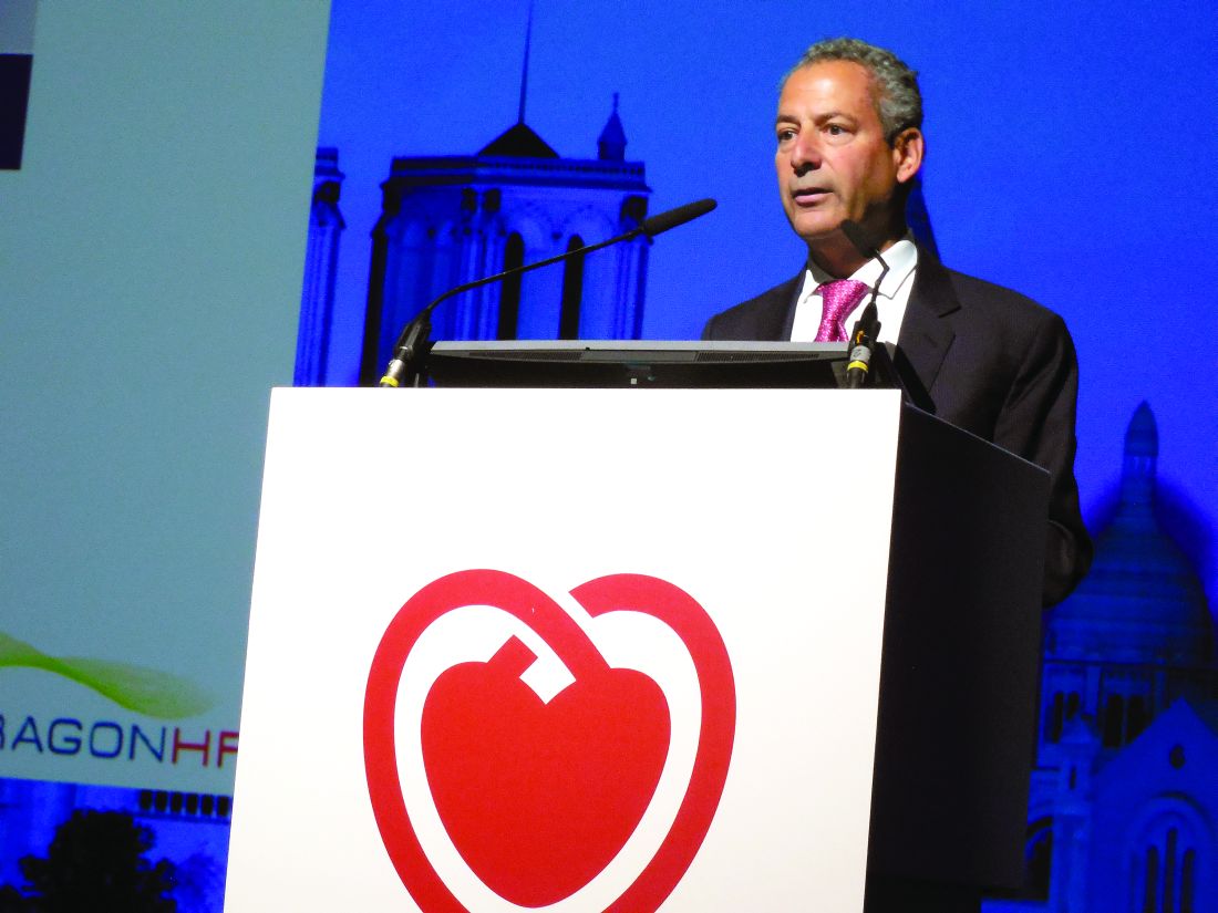
PARIS – Arguably one of the most important and far-reaching studies presented at the annual congress of the European Society of Cardiology didn’t take place in the massive main ballroom with dazzling lights and sound and thousands of cardiologists in attendance, but in a tiny, makeshift, open-sided venue slapped together of cardboard and fiberboard and plunked down in the noisy poster hall.

It was there that Terence Dwyer, MBBS, MD, began by observing, “We know quite a bit about the relationship of cardiovascular risk factors in adults to cardiovascular disease; we know virtually nothing about the relationship of those risk factors in childhood because – until now – there has been no direct evidence relating to this. What I’m going to present to you is some direct evidence.”
The data come from the International Childhood Cardiovascular Cohort (i3C) Consortium, which includes investigators from seven pioneering prospective child cohort studies, which collectively measured major cardiovascular risk factors in more than 42,000 children beginning back in the 1970s.
Some of these studies will be familiar names to many American physicians and epidemiologists. They include the Bogolusa Heart Study, the Muscatine Study, the Princeton Lipid Research Clinic Study, and the Minneapolis Childhood Cohort Studies. Similar studies were launched decades ago in Australia and Finland. The oldest of these cohorts are now in their 50s, and they are developing cardiovascular disease. The new i3C findings based on pooled data from these studies provides the first direct evidence that high serum cholesterol, blood pressure, body mass index, and smoking in childhood are linked to increased risk of hospitalization for acute MI, stroke, and peripheral artery disease in early middle age, said Dr. Dwyer, emeritus professor of epidemiology at the University of Oxford (England).
The analysis showed that each 10% increase above average in serum cholesterol in childhood was associated with a 16% increased risk of hospitalization for a cardiovascular event at a mean age of 49 years. A 2-point rise in BMI was associated with a 20% higher risk. A 10% increase above average in systolic blood pressure in childhood was linked to a 40% increase in risk of a cardiovascular event in later life. And smoking in childhood or adolescence was associated with a 77% higher risk of a cardiovascular event.
The i3C analysis also demonstrated that For example, individuals who both as adults and children had two or more of the four major cardiovascular risk factors studied had a sixfold greater risk of a major cardiovascular event in early middle age than if they had two or more risk factors as adults but none as children. If they had two or more risk factors as adults and one risk factor in childhood, their risk of a cardiovascular event was roughly twice as great as if they had no risk factors as a child. And if they had two or more risk factors present in childhood but none in adulthood, their risk of an event was threefold higher than if none of the four major cardiovascular risk factors were present during both periods of life, he continued.
The investigators consider their findings preliminary because most participants in the cohort studies are just reaching age 50 years.
“As we follow them for another 5 years, because of their age, the number of cardiovascular events will increase dramatically,” Dr. Dwyer explained. “One of the reasons we’re presenting this data now in preliminary form is these cohort studies will be the only data of this kind for about another 20 years. We want it out there when it can be most useful. It’s not like the situation with RCTs [randomized, controlled trials] where you’re able to wait 2 years for the next RCT.”
Clinical and policy implications
Asked about the clinical implications of the i3C findings, he replied, “At the very least, at this stage, consideration should be given to lowering risk factors in childhood as a greater priority in the cardiovascular disease prevention field.”
From my experience on national committees that look at what we do about cardiovascular prevention in childhood, they generally say we’re unprepared to take a strong stance on this because we have no direct evidence that these risk factors and what underpins them are a genuine problem,” according to Dr. Dwyer.
That’s no longer the case. By the end of the year, the i3C investigators expect to publish their results. As word reaches the public, he expects to finally see a growing momentum for cardiovascular prevention in pediatrics.
“Just imagine saying to a parent, ‘It looks highly likely that if you don’t do anything about the weight your children have put on, or other risk factors, they will be left at the end of childhood with a residual risk for cardiovascular disease that it doesn’t appear can be completely eradicated. It can be reduced by interventions in adulthood, but something’s happened there in childhood that was important.’ I think parents will demand action at that time,” he said.
In an interview, Donald Lloyd-Jones, MD, called the i3C data “incredibly important.”
“The risk factor values that they’re looking at in kids are not abnormal, they’re at the higher range of what we consider very normal, and yet those slightly elevated exposures within the normal range are causing damage. These kids are accruing risk for atherosclerosis down the road, even within what’s considered to be normal ranges,” commented Dr. Lloyd-Jones, senior associate dean for clinical and translational research and chair of the department of preventive medicine at Northwestern University, Chicago.
“I think it’s very telling that, early in life, we can delineate trajectories already emerging about how these kids are going to play out the rest of their lives in terms of their atherosclerosis and cardiovascular risk. That’s a very important thing to recognize, and we haven’t always thought that way. We always thought you arrive at your 21st birthday and then things start to matter, and by the time you got to 50, now it really matters. But the truth is the horse is already well out of the barn at age 50 and it’s coming out of the barn at age 21. That’s what the i3C data are starting to tell us: that it’s incredibly important that we move further upstream,” the cardiologist added.
What’s the best way forward?
“We have to create an environment where we tilt the playing field towards healthy choices. Sometimes that means taxation policy: It worked for alcohol and tobacco. Sometimes that means frank prohibition: indoor smoking laws have had a huge beneficial effect on public health. Sometimes it’s more controversial, like taxes on sugar-sweetened beverages, but I think that’s an experiment we have to play out to see if it works,” according to Dr. Lloyd-Jones. “I think our best solutions are going to come through policy, environmental change, and lifestyle in the early years because it’s just not practical to think about introducing foreign substances to mass amounts of kids.”
He noted that the National Heart, Lung and Blood Institute has held two workshops within the past year focused on these very issues.
Dr. Lloyd-Jones, past-honored as the American Heart Association Physician of the Year in recognition of his decades of work with that organization in advancing cardiovascular prevention, said “there’s a very good chance” the AHA will take on a major role in what he anticipates will be a much greater emphasis on cardiovascular prevention starting in early life in order to favorably alter life trajectories.
“Stay tuned in the next few months. We’re coming to a decade change, so as we enter 2020, the AHA will be promulgating its strategic goals for the next decade. The AHA is a much bigger, better-funded organization than it was even 10 years ago, and they’re looking to partner with groups like the Robert Woods Johnson Foundation, the Centers for Disease Control, [and] the NIH, to actually make major policy initiatives on cardiovascular prevention,” he said.
The i3C study was funded by the National Heart, Lung, and Blood Institute. Dr. Dwyer reported having no financial conflicts of interest.
PARIS – Arguably one of the most important and far-reaching studies presented at the annual congress of the European Society of Cardiology didn’t take place in the massive main ballroom with dazzling lights and sound and thousands of cardiologists in attendance, but in a tiny, makeshift, open-sided venue slapped together of cardboard and fiberboard and plunked down in the noisy poster hall.
It was there that Terence Dwyer, MBBS, MD, began by observing, “We know quite a bit about the relationship of cardiovascular risk factors in adults to cardiovascular disease; we know virtually nothing about the relationship of those risk factors in childhood because – until now – there has been no direct evidence relating to this. What I’m going to present to you is some direct evidence.”
The data come from the International Childhood Cardiovascular Cohort (i3C) Consortium, which includes investigators from seven pioneering prospective child cohort studies, which collectively measured major cardiovascular risk factors in more than 42,000 children beginning back in the 1970s.
Some of these studies will be familiar names to many American physicians and epidemiologists. They include the Bogolusa Heart Study, the Muscatine Study, the Princeton Lipid Research Clinic Study, and the Minneapolis Childhood Cohort Studies. Similar studies were launched decades ago in Australia and Finland. The oldest of these cohorts are now in their 50s, and they are developing cardiovascular disease. The new i3C findings based on pooled data from these studies provides the first direct evidence that high serum cholesterol, blood pressure, body mass index, and smoking in childhood are linked to increased risk of hospitalization for acute MI, stroke, and peripheral artery disease in early middle age, said Dr. Dwyer, emeritus professor of epidemiology at the University of Oxford (England).
The analysis showed that each 10% increase above average in serum cholesterol in childhood was associated with a 16% increased risk of hospitalization for a cardiovascular event at a mean age of 49 years. A 2-point rise in BMI was associated with a 20% higher risk. A 10% increase above average in systolic blood pressure in childhood was linked to a 40% increase in risk of a cardiovascular event in later life. And smoking in childhood or adolescence was associated with a 77% higher risk of a cardiovascular event.
The i3C analysis also demonstrated that For example, individuals who both as adults and children had two or more of the four major cardiovascular risk factors studied had a sixfold greater risk of a major cardiovascular event in early middle age than if they had two or more risk factors as adults but none as children. If they had two or more risk factors as adults and one risk factor in childhood, their risk of a cardiovascular event was roughly twice as great as if they had no risk factors as a child. And if they had two or more risk factors present in childhood but none in adulthood, their risk of an event was threefold higher than if none of the four major cardiovascular risk factors were present during both periods of life, he continued.
The investigators consider their findings preliminary because most participants in the cohort studies are just reaching age 50 years.
“As we follow them for another 5 years, because of their age, the number of cardiovascular events will increase dramatically,” Dr. Dwyer explained. “One of the reasons we’re presenting this data now in preliminary form is these cohort studies will be the only data of this kind for about another 20 years. We want it out there when it can be most useful. It’s not like the situation with RCTs [randomized, controlled trials] where you’re able to wait 2 years for the next RCT.”
Clinical and policy implications
Asked about the clinical implications of the i3C findings, he replied, “At the very least, at this stage, consideration should be given to lowering risk factors in childhood as a greater priority in the cardiovascular disease prevention field.”
From my experience on national committees that look at what we do about cardiovascular prevention in childhood, they generally say we’re unprepared to take a strong stance on this because we have no direct evidence that these risk factors and what underpins them are a genuine problem,” according to Dr. Dwyer.
That’s no longer the case. By the end of the year, the i3C investigators expect to publish their results. As word reaches the public, he expects to finally see a growing momentum for cardiovascular prevention in pediatrics.
“Just imagine saying to a parent, ‘It looks highly likely that if you don’t do anything about the weight your children have put on, or other risk factors, they will be left at the end of childhood with a residual risk for cardiovascular disease that it doesn’t appear can be completely eradicated. It can be reduced by interventions in adulthood, but something’s happened there in childhood that was important.’ I think parents will demand action at that time,” he said.
In an interview, Donald Lloyd-Jones, MD, called the i3C data “incredibly important.”
“The risk factor values that they’re looking at in kids are not abnormal, they’re at the higher range of what we consider very normal, and yet those slightly elevated exposures within the normal range are causing damage. These kids are accruing risk for atherosclerosis down the road, even within what’s considered to be normal ranges,” commented Dr. Lloyd-Jones, senior associate dean for clinical and translational research and chair of the department of preventive medicine at Northwestern University, Chicago.
“I think it’s very telling that, early in life, we can delineate trajectories already emerging about how these kids are going to play out the rest of their lives in terms of their atherosclerosis and cardiovascular risk. That’s a very important thing to recognize, and we haven’t always thought that way. We always thought you arrive at your 21st birthday and then things start to matter, and by the time you got to 50, now it really matters. But the truth is the horse is already well out of the barn at age 50 and it’s coming out of the barn at age 21. That’s what the i3C data are starting to tell us: that it’s incredibly important that we move further upstream,” the cardiologist added.
What’s the best way forward?
“We have to create an environment where we tilt the playing field towards healthy choices. Sometimes that means taxation policy: It worked for alcohol and tobacco. Sometimes that means frank prohibition: indoor smoking laws have had a huge beneficial effect on public health. Sometimes it’s more controversial, like taxes on sugar-sweetened beverages, but I think that’s an experiment we have to play out to see if it works,” according to Dr. Lloyd-Jones. “I think our best solutions are going to come through policy, environmental change, and lifestyle in the early years because it’s just not practical to think about introducing foreign substances to mass amounts of kids.”
He noted that the National Heart, Lung and Blood Institute has held two workshops within the past year focused on these very issues.
Dr. Lloyd-Jones, past-honored as the American Heart Association Physician of the Year in recognition of his decades of work with that organization in advancing cardiovascular prevention, said “there’s a very good chance” the AHA will take on a major role in what he anticipates will be a much greater emphasis on cardiovascular prevention starting in early life in order to favorably alter life trajectories.
“Stay tuned in the next few months. We’re coming to a decade change, so as we enter 2020, the AHA will be promulgating its strategic goals for the next decade. The AHA is a much bigger, better-funded organization than it was even 10 years ago, and they’re looking to partner with groups like the Robert Woods Johnson Foundation, the Centers for Disease Control, [and] the NIH, to actually make major policy initiatives on cardiovascular prevention,” he said.
The i3C study was funded by the National Heart, Lung, and Blood Institute. Dr. Dwyer reported having no financial conflicts of interest.
REPORTING FROM THE ESC CONGRESS 2019
Sacubitril/valsartan suggests HFpEF benefit in neutral PARAGON-HF
PARIS – but that didn’t stop some experts from seeing a practice-changing message in its findings.
The results of PARAGON-HF, a major trial of sacubitril/valsartan – a compound already approved for treating heart failure with reduced left ventricular ejection fraction – in patients with heart failure with preserved ejection fraction (HFpEF), showed a statistically neutral result for the study’s primary endpoint, but with an excruciatingly close near miss for statistical significance and clear benefit in a subgroup of HFpEF patients with a modestly reduced left ventricular ejection fraction. These findings seemed to convince some experts to soon try using sacubitril/valsartan (Entresto) to treat selected patients with HFpEF, driven in large part by the lack of any other agent clearly proven to benefit the large number of patients with this form of heart failure.
HFpEF is “a huge unmet need,” and data from the PARAGON-HF trial “suggest that sacubitril/valsartan may be beneficial in some patients with HFpEF, particularly those with a left ventricular ejection fraction that is not frankly reduced, but less than normal,” specifically patients with a left ventricular ejection fraction of 45%-57%, Scott D. Solomon, MD, said at the annual congress of the European Society of Cardiology as he reported the primary PARAGON-HF results.
“I’m not speaking for regulators or for guidelines, but I suspect that in this group of patients [with HFpEF and a left ventricular ejection fraction of 45%-57%] there is at least some rationale to use this treatment,” said Dr. Solomon, professor of medicine at Harvard Medical School and director of noninvasive cardiology at Brigham and Women’s Hospital, both in Boston.
His suggestion, which cut against the standard rules that govern the interpretation of trial results, met a substantial level of receptivity at the congress.
Trial results “are not black and white, where a P value of .049 means the trial was totally positive, and a P of .051 means it’s totally neutral. That’s misleading, and it’s why the field is moving to different types of [statistical] analysis that give us more leeway in interpreting data,” commented Philippe Gabriel Steg, MD, professor of cardiology at the University of Paris.
“Everything in this trial points to substantial potential benefit. I’m not impressed by the P value that just missed significance. I think this is a very important advance,” said Dr. Steg, who had no involvement in the study, during a press conference at the congress.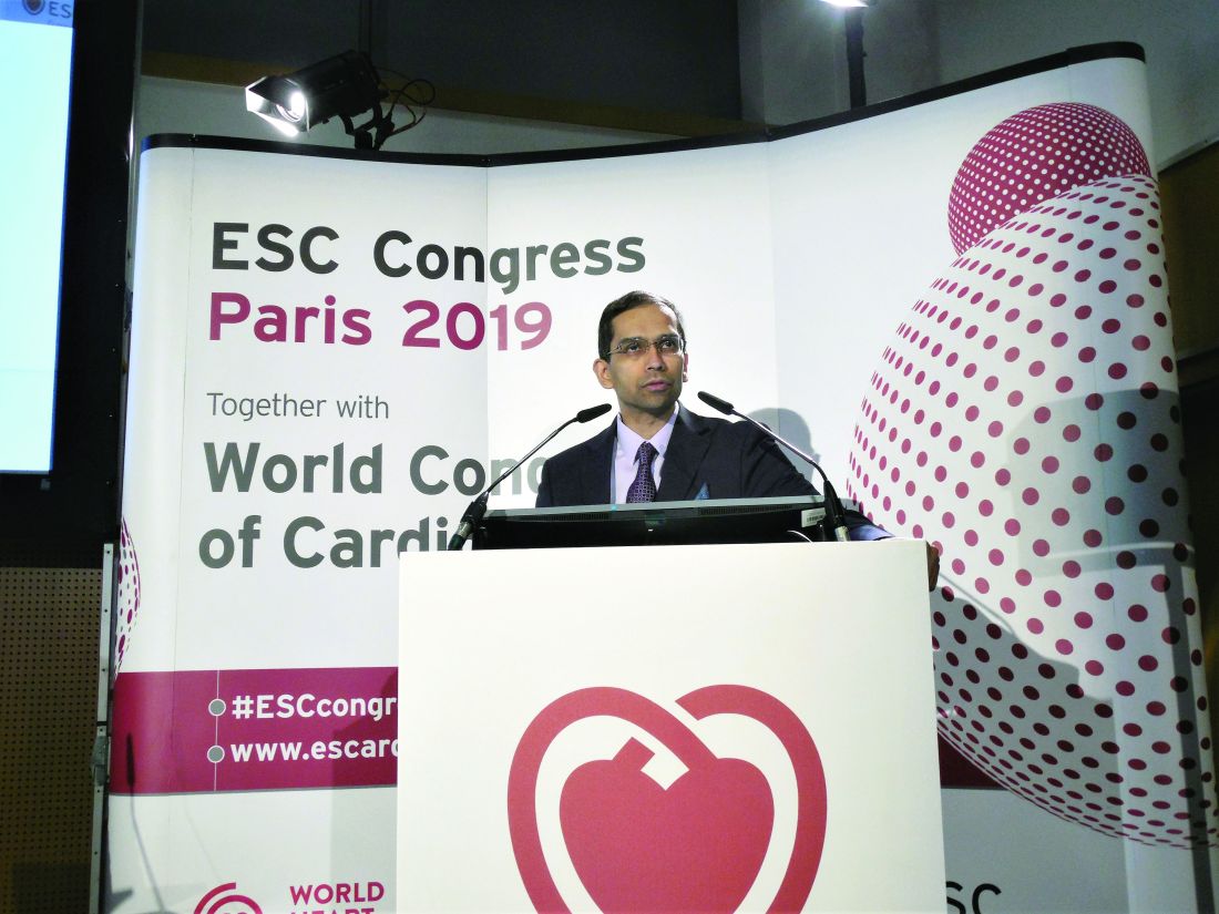
“I agree. I look at the totality of evidence, and to me the PARAGON-HF results were positive in patients with an ejection fraction of 50%, which is not a normal level. The way I interpret the results is, the treatment works in patients with an ejection fraction that is ‘lowish,’ but not at the conventional level of reduced ejection fraction,” commented Deepak L. Bhatt, MD, professor of medicine at Harvard Medical School, who also had no involvement with PARAGON-HF.
Stuart J. Connolly, MD, designated discussant for the report at the congress and professor of medicine at McMaster University, Hamilton, Ont., struck similar notes during his discussion of the report, and called the subgroup analysis by baseline ejection fraction “compelling,” and supported by several secondary findings of the study, the biological plausibility of a link between ejection fraction and treatment response, and by suggestions of a similar effect caused by related drugs in prior studies.
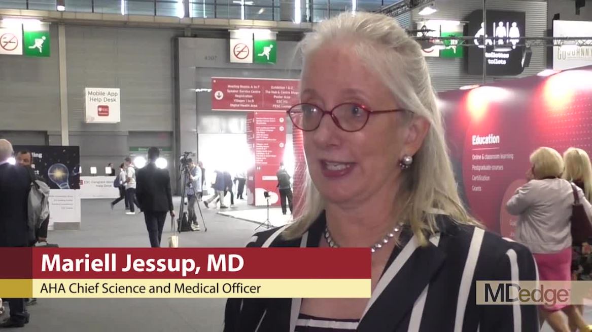
The argument in favor of sacubitril/valsartan’s efficacy in a subgroup of PARAGON-HF patients was also taken up by Mariell Jessup, MD, a heart failure specialist and chief science and medical officer of the American Heart Association in Dallas. “I think it’s legitimate to say that there are HFpEF subgroups that might benefit” from sacubitril/valsartan, such as women. “I think it’s appropriate in this disease to look at subgroups because we have to find something that works for these patients,” she added in a video interview.
But Dr. Jessup also urged caution in interpreting the link between modestly reduced ejection fraction and response to sacubitril/valsartan in HFpEF patients because ejection fraction measurements by echocardiography, as done in the trial, are notoriously unreliable. “We need more precise markers of who responds to this drug and who does not,” she said.
PARAGON-HF randomized 4,796 patients at 848 sites in 43 countries who were aged at least 50 years, had signs and symptoms of heart failure with a left ventricular ejection fraction of at least 45%, had evidence on echocardiography of either left atrial enlargement or left ventricular hypertrophy, and had an elevated blood level of natriuretic peptides.
The study’s primary endpoint was the composite rate of total (both first and recurrent) hospitalizations for heart failure and cardiovascular death. That outcome occurred at a rate of 12.8 events/100 patient-years in patients treated with sacubitril/valsartan and a rate of 14.6 events/100 patient-years in control patients treated with the angiotensin receptor blocking drug valsartan alone. Those results yielded a relative risk reduction by sacubitril/valsartan of 13% with a P value of .059, just missing statistical significance. Concurrently with Dr. Solomon’s report the results appeared in an article online and then subsequently in print (N Engl J Med. 2019 Oct 24;381[17]:1609-20). The primary endpoint was driven primarily by a 15% relative risk reduction in hospitalizations for heart failure; the two treatment arms showed nearly identical rates of cardiovascular disease death.
Notable secondary findings that reached statistical significance included a 16% relative decrease in total heart failure hospitalizations, cardiovascular deaths, and urgent heart failure visits with sacubitril/valsartan treatment, as well as a 16% reduction in all investigator-reported events. Other significant benefits linked with sacubitril/valsartan treatment were a 45% relative improvement in functional class, a 30% relative improvement in patients achieving a meaningful increase in a quality of life measure, and a halving of the incidence of worsening renal function with sacubitril/valsartan.
The safety profile of sacubitril/valsartan in the study matched previous reports on the drug in patients with heart failure with reduced ejection fraction, an approved indication since 2015.
The key subgroup analysis detailed by Dr. Solomon was the incidence of the primary endpoint by baseline ejection fraction. Among the 2,495 patients (52% of the study population) with a left ventricular ejection fraction of 57% or less when they entered the study, treatment with sacubitril/valsartan cut the primary endpoint incidence by 22%, compared with valsartan alone, a statistically significant difference. Among patients with a baseline ejection fraction of 58% or greater, treatment with sacubitril/valsartan had no effect on the primary endpoint, compared with control patients. Dr. Solomon also reported a statistically significant 22% relative improvement in the primary endpoint among the 2,479 women in the study (52% of the total study cohort) while the drug had no discernible impact among men, but he did not highlight any immediate implication of this finding.
Despite how suggestive the finding related to ejection fraction may be for practice, a major impediment to prescribing sacubitril/valsartan to HFpEF patients may come from pharmacy managers, suggested Douglas L. Mann, MD, a heart failure specialist and professor of medicine at Washington University, St. Louis.
“The study did not hit its primary endpoint, so pharmacy managers will face no moral issue by withholding the drug” from HFpEF patients, Dr. Mann said in an interview. Because sacubitril/valsartan is substantially costlier than other renin-angiotensin system inhibitor drugs, which are mostly generic, patients may often find it difficult to pay for sacubitril/valsartan themselves if it receives no insurance coverage.
“It’s heartbreaking that the endpoint missed for a disease with no proven treatment. The study may have narrowly missed, but it still missed, and a lot of us had hoped it would be positive. It’s a slippery slope” when investigators try to qualify a trial result that failed to meet the study’s prespecified definition of a statistically significant effect. “The primary endpoint is the primary endpoint, and we should not overinterpret the data,” Dr. Mann warned.
PARAGON-HF was sponsored by Novartis, which markets sacubitril/valsartan (Entresto). Dr. Solomon has been a consultant to and has received research funding from Novartis and from several other companies. Dr. Steg has received personal fees from Novartis and has received personal fees and research funding from several other companies. Dr. Bhatt has been a consultant to and received research funding from several companies but has had no recent relationship with Novartis. Dr. Connolly and Dr. Jessup had no disclosures. Dr. Mann has been a consultant to Novartis, as well as Bristol-Myers Squibb, LivaNova, and Tenaya Therapeutics.
PARAGON-HF was a well-designed and well-conducted trial that unfortunately showed a modest treatment effect, with sacubitril/valsartan treatment reducing the overall primary endpoint by 13%, compared with control patients, a difference that was not statistically significant. One factor to consider when interpreting this outcome was that the study used an active control arm in which patients received valsartan even though no treatment is specifically approved for or is considered to have proven efficacy in patients with heart failure with preserved ejection fraction. The investigators felt compelled to use this active control because many patients with this form of heart failure receive a drug that inhibits the renin-angiotensin system. It’s possible that if sacubitril/valsartan had been compared with placebo the treatment effect would have been greater.

Although caution is required when interpreting subgroup outcomes in a study that lacks a positive primary endpoint, the data indicate a positive signal in the subgroup analysis that Dr. Solomon presented that took into account left ventricular ejection fraction at entry into the study. Patients with a baseline ejection fraction of 57% or less, roughly half the entire study group, showed a statistically significant benefit in a prespecified analysis, and a finding with some level of biological plausibility. This was a compelling analysis, and it suggested that with this treatment it may be possible to reduce a key outcome – the incidence of heart failure hospitalizations – in patients with modestly reduced ejection fractions.
Stuart J. Connolly, MD , is a professor of medicine at McMaster University, Hamilton, Ont. He had no disclosures. He made these comments as the designated discussant for PARAGON-HF.
PARAGON-HF was a well-designed and well-conducted trial that unfortunately showed a modest treatment effect, with sacubitril/valsartan treatment reducing the overall primary endpoint by 13%, compared with control patients, a difference that was not statistically significant. One factor to consider when interpreting this outcome was that the study used an active control arm in which patients received valsartan even though no treatment is specifically approved for or is considered to have proven efficacy in patients with heart failure with preserved ejection fraction. The investigators felt compelled to use this active control because many patients with this form of heart failure receive a drug that inhibits the renin-angiotensin system. It’s possible that if sacubitril/valsartan had been compared with placebo the treatment effect would have been greater.
Although caution is required when interpreting subgroup outcomes in a study that lacks a positive primary endpoint, the data indicate a positive signal in the subgroup analysis that Dr. Solomon presented that took into account left ventricular ejection fraction at entry into the study. Patients with a baseline ejection fraction of 57% or less, roughly half the entire study group, showed a statistically significant benefit in a prespecified analysis, and a finding with some level of biological plausibility. This was a compelling analysis, and it suggested that with this treatment it may be possible to reduce a key outcome – the incidence of heart failure hospitalizations – in patients with modestly reduced ejection fractions.
Stuart J. Connolly, MD , is a professor of medicine at McMaster University, Hamilton, Ont. He had no disclosures. He made these comments as the designated discussant for PARAGON-HF.
PARIS – but that didn’t stop some experts from seeing a practice-changing message in its findings.
The results of PARAGON-HF, a major trial of sacubitril/valsartan – a compound already approved for treating heart failure with reduced left ventricular ejection fraction – in patients with heart failure with preserved ejection fraction (HFpEF), showed a statistically neutral result for the study’s primary endpoint, but with an excruciatingly close near miss for statistical significance and clear benefit in a subgroup of HFpEF patients with a modestly reduced left ventricular ejection fraction. These findings seemed to convince some experts to soon try using sacubitril/valsartan (Entresto) to treat selected patients with HFpEF, driven in large part by the lack of any other agent clearly proven to benefit the large number of patients with this form of heart failure.
HFpEF is “a huge unmet need,” and data from the PARAGON-HF trial “suggest that sacubitril/valsartan may be beneficial in some patients with HFpEF, particularly those with a left ventricular ejection fraction that is not frankly reduced, but less than normal,” specifically patients with a left ventricular ejection fraction of 45%-57%, Scott D. Solomon, MD, said at the annual congress of the European Society of Cardiology as he reported the primary PARAGON-HF results.
“I’m not speaking for regulators or for guidelines, but I suspect that in this group of patients [with HFpEF and a left ventricular ejection fraction of 45%-57%] there is at least some rationale to use this treatment,” said Dr. Solomon, professor of medicine at Harvard Medical School and director of noninvasive cardiology at Brigham and Women’s Hospital, both in Boston.
His suggestion, which cut against the standard rules that govern the interpretation of trial results, met a substantial level of receptivity at the congress.
Trial results “are not black and white, where a P value of .049 means the trial was totally positive, and a P of .051 means it’s totally neutral. That’s misleading, and it’s why the field is moving to different types of [statistical] analysis that give us more leeway in interpreting data,” commented Philippe Gabriel Steg, MD, professor of cardiology at the University of Paris.
“Everything in this trial points to substantial potential benefit. I’m not impressed by the P value that just missed significance. I think this is a very important advance,” said Dr. Steg, who had no involvement in the study, during a press conference at the congress.
“I agree. I look at the totality of evidence, and to me the PARAGON-HF results were positive in patients with an ejection fraction of 50%, which is not a normal level. The way I interpret the results is, the treatment works in patients with an ejection fraction that is ‘lowish,’ but not at the conventional level of reduced ejection fraction,” commented Deepak L. Bhatt, MD, professor of medicine at Harvard Medical School, who also had no involvement with PARAGON-HF.
Stuart J. Connolly, MD, designated discussant for the report at the congress and professor of medicine at McMaster University, Hamilton, Ont., struck similar notes during his discussion of the report, and called the subgroup analysis by baseline ejection fraction “compelling,” and supported by several secondary findings of the study, the biological plausibility of a link between ejection fraction and treatment response, and by suggestions of a similar effect caused by related drugs in prior studies.
The argument in favor of sacubitril/valsartan’s efficacy in a subgroup of PARAGON-HF patients was also taken up by Mariell Jessup, MD, a heart failure specialist and chief science and medical officer of the American Heart Association in Dallas. “I think it’s legitimate to say that there are HFpEF subgroups that might benefit” from sacubitril/valsartan, such as women. “I think it’s appropriate in this disease to look at subgroups because we have to find something that works for these patients,” she added in a video interview.
But Dr. Jessup also urged caution in interpreting the link between modestly reduced ejection fraction and response to sacubitril/valsartan in HFpEF patients because ejection fraction measurements by echocardiography, as done in the trial, are notoriously unreliable. “We need more precise markers of who responds to this drug and who does not,” she said.
PARAGON-HF randomized 4,796 patients at 848 sites in 43 countries who were aged at least 50 years, had signs and symptoms of heart failure with a left ventricular ejection fraction of at least 45%, had evidence on echocardiography of either left atrial enlargement or left ventricular hypertrophy, and had an elevated blood level of natriuretic peptides.
The study’s primary endpoint was the composite rate of total (both first and recurrent) hospitalizations for heart failure and cardiovascular death. That outcome occurred at a rate of 12.8 events/100 patient-years in patients treated with sacubitril/valsartan and a rate of 14.6 events/100 patient-years in control patients treated with the angiotensin receptor blocking drug valsartan alone. Those results yielded a relative risk reduction by sacubitril/valsartan of 13% with a P value of .059, just missing statistical significance. Concurrently with Dr. Solomon’s report the results appeared in an article online and then subsequently in print (N Engl J Med. 2019 Oct 24;381[17]:1609-20). The primary endpoint was driven primarily by a 15% relative risk reduction in hospitalizations for heart failure; the two treatment arms showed nearly identical rates of cardiovascular disease death.
Notable secondary findings that reached statistical significance included a 16% relative decrease in total heart failure hospitalizations, cardiovascular deaths, and urgent heart failure visits with sacubitril/valsartan treatment, as well as a 16% reduction in all investigator-reported events. Other significant benefits linked with sacubitril/valsartan treatment were a 45% relative improvement in functional class, a 30% relative improvement in patients achieving a meaningful increase in a quality of life measure, and a halving of the incidence of worsening renal function with sacubitril/valsartan.
The safety profile of sacubitril/valsartan in the study matched previous reports on the drug in patients with heart failure with reduced ejection fraction, an approved indication since 2015.
The key subgroup analysis detailed by Dr. Solomon was the incidence of the primary endpoint by baseline ejection fraction. Among the 2,495 patients (52% of the study population) with a left ventricular ejection fraction of 57% or less when they entered the study, treatment with sacubitril/valsartan cut the primary endpoint incidence by 22%, compared with valsartan alone, a statistically significant difference. Among patients with a baseline ejection fraction of 58% or greater, treatment with sacubitril/valsartan had no effect on the primary endpoint, compared with control patients. Dr. Solomon also reported a statistically significant 22% relative improvement in the primary endpoint among the 2,479 women in the study (52% of the total study cohort) while the drug had no discernible impact among men, but he did not highlight any immediate implication of this finding.
Despite how suggestive the finding related to ejection fraction may be for practice, a major impediment to prescribing sacubitril/valsartan to HFpEF patients may come from pharmacy managers, suggested Douglas L. Mann, MD, a heart failure specialist and professor of medicine at Washington University, St. Louis.
“The study did not hit its primary endpoint, so pharmacy managers will face no moral issue by withholding the drug” from HFpEF patients, Dr. Mann said in an interview. Because sacubitril/valsartan is substantially costlier than other renin-angiotensin system inhibitor drugs, which are mostly generic, patients may often find it difficult to pay for sacubitril/valsartan themselves if it receives no insurance coverage.
“It’s heartbreaking that the endpoint missed for a disease with no proven treatment. The study may have narrowly missed, but it still missed, and a lot of us had hoped it would be positive. It’s a slippery slope” when investigators try to qualify a trial result that failed to meet the study’s prespecified definition of a statistically significant effect. “The primary endpoint is the primary endpoint, and we should not overinterpret the data,” Dr. Mann warned.
PARAGON-HF was sponsored by Novartis, which markets sacubitril/valsartan (Entresto). Dr. Solomon has been a consultant to and has received research funding from Novartis and from several other companies. Dr. Steg has received personal fees from Novartis and has received personal fees and research funding from several other companies. Dr. Bhatt has been a consultant to and received research funding from several companies but has had no recent relationship with Novartis. Dr. Connolly and Dr. Jessup had no disclosures. Dr. Mann has been a consultant to Novartis, as well as Bristol-Myers Squibb, LivaNova, and Tenaya Therapeutics.
REPORTING FROM THE ESC 2019 CONGRESS
How P-wave indices can improve AFib-related ischemic stroke prediction
Background: Current AFib management guidelines recommend ischemic stroke risk stratification with CHA2DS2-VASc score; however, emerging studies have highlighted limitations of this score.
Study design: Retrospective review of previously obtained prospective cohort study data.
Setting: Fourteen U.S. communities.
Synopsis: For the 2,929 individuals with new incident AFib without anticoagulant use in the prior year, study authors computed P-wave indices (including P-wave axis, P-wave duration, advanced interatrial block, and P-wave terminal force in lead V1) from the most recent sinus rhythm EKG prior to the diagnosis of AFib. Cox proportional hazard models estimated the hazard ratio between PWIs and ischemic stroke. Of the PWIs tested above, abnormal P-wave axis (hazard ratio, 1.88; 95% confidence interval, 1.36-2.61) and advanced interatrial block (HR, 2.93; 95% CI 1.78-4.81) were associated with increased risk of stroke after adjustment for individual CHA2DS2-VASc variables. A P2-CHA2DS2-VASc score that incorporated abnormal P-wave axis measurements demonstrated superior discrimination, compared with the CHA2DS2-VASc score alone, and resulted in improvement in ischemic stroke risk classification.
Bottom line: Abnormal P-wave axis and advanced interatrial block measured during periods of sinus rhythm may be associated with increased risk of ischemic stroke in patients with atrial fibrillation; the P2-CHA2DS2-VASc score incorporating abnormal P-wave axis may be superior to CHA2DS2-VASc in ischemic stroke risk classification.
Citation: Maheshwari A et al. Refining prediction of atrial fibrillation–related stroke using the P2-CHA2DS2-VASc score. Circulation. 2019 Jan 8;139:180-91.
Dr. Cooke is a hospitalist at Beth Israel Deaconess Medical Center.
A link between A-fib and sleep apnea is no surprise, but why?

Is the relationship between A-fib and sleep apnea more than a coincidence stemming from the number of shared associated comorbidities? Significantly, the treatment of obstructive sleep apnea with continuous positive airway pressure (CPAP) has been shown to decrease the recurrence of A-fib after pharmacologic or electrical conversion and after interventional pulmonary vein interruption.1 This suggests that at least in some cases, sleep apnea plays an active role in initiating and possibly also maintaining A-fib. The immediate culprit mediators that come to mind are hypoxia and hypercapnea; both are at least partially ameliorated by the successful use of CPAP, and both are reasonable physiologic candidates for induction of A-fib. Hypoxia is supported by clinical observation, and hypercapnea by experimental modeling.2
It is easy for clinicians to conceptualize the organ effects of hypoxia and hypercapnea. We are accustomed to seeing clinical ramifications of these in the emergency department and intensive care unit, particularly those affecting the brain and heart, organs critically dependent on transmembrane ion flow. We may recall from biochemistry classes the effects of hypoxia on intracellular metabolism and the implications on energy stores, mitochondrial function, and ion translocation. Recent work on the cellular effects of hypoxia, including research that resulted in a Nobel prize, has drawn major attention to patterned cellular responses to intermittent and persistent hypoxia. This includes recognition of epigenetic changes resulting in localized cardiac remodeling and fibrosis,3 factors that clearly affect the expression of arrhythmias, including A-fib.
But the interrelationship between A-fib and sleep apnea may be even more convoluted and intriguing. It now seems that most things cardiac are associated with inflammation in some guise, and the A-fib connection with sleep apnea may not be an exception. Almost 20 years ago, it was recognized that A-fib is associated with an elevation in circulating C-reactive protein (CRP),4 a biomarker of “inflammation,” although not necessarily an active participant. Recent reviews of this connection have been published,5 and successful anti-inflammatory approaches to preventing A-fib using colchicine have been described.6 So how does this tie in with sleep apnea?
A number of papers have now demonstrated that sleep apnea is also associated with an elevation in CRP,7 perhaps due to increases in tumor necrosis factor (TNF)-alpha in response to the intermittent hypoxia of sleep apnea. TNF can drive the inflammatory response through increased expression of genes regulated by nuclear factor kappa-B.8 While it certainly warrants consideration that the elevated biomarkers of inflammation in patients with sleep apnea actually reflect the presence of the frequent comorbidities, including visceral obesity, treating sleep apnea with CPAP (comparable to what I noted above in patients with A-fib) has been shown to reduce circulating CRP levels.9
As our understanding of the biologic underpinnings of A-fib and sleep apnea continue to grow, the practical clinical implications of the relationship between them, as described by Ayache et al, may achieve greater clarity. The two conditions commonly coexist, and treating the sleep apnea results in better rhythm-directed outcomes in the A-fib.
Stay tuned, there is certainly more to learn about this.
- Shukla A, Aizer A, Holmes D, et al. Effect of sleep apnea treatment on atrial fibrillation recurrence: a meta-analysis. JACC Clin Electropysiol 2015; 1(1–2):41–51. doi:10.1016/j.jacep.2015.02.014
- Stevenson IH, Roberts-Thomson KC, Kistler PM, et al. Atrial electrophysiology is altered by acute hypercapnea but not hypoxemia: implications for promotion of atrial fibrillation in pulmonary disease and sleep apnea. Heart Rhythm 2010; 7(9):1263–1270. doi:10.1016/j.hrthm.2010.03.020
- Zhang W, Song M, Qu J, Liu G. Epigenetic modifications in cardiovascular aging and diseases. Circ Res 2018; 123(7):773–786. doi:10.1161/CIRCRESAHA.118.312497
- Chung MK, Martin DO, Sprecher D, et al. C-reactive protein elevation in patients with atrial arrhythmias: inflammatory mechanisms and persistence of atrial fibrillation. Circulation 2001; 104(24):2886–2891. doi:10.1161/hc4901.101760
- Guo Y, Lip GY, Apostolakis S. Inflammation in atrial fibrillation. J Am Coll Cardiol 2012; 60(22):2263–2270. doi:10.1016/j.jacc.2012.04.063
- Lee JZ, Singh N, Howe CL, et al. Colchicine for prevention of post-operative atrial fibrillation: a meta-analysis. JACC Clin Electrophysiol 2016; 2(1):78–85. doi:10.1016/j.jacep.2015.09.016
- Van der Touw T, Andronicos NM, Smart N. Is C-reactive protein elevated in obstructive sleep apnea? A systematic review and meta-analysis. Biomarkers 2019; 24(5):429–435. doi:10.1080/1354750X.2019.1600025
- Ryan S, Taylor CT, McNicholas WT. Systemic inflammation: a key factor in the pathogenesis of cardiovascular complications in obstructive sleep apnea syndrome? Thorax 2009; 64(7):631–636. doi:10.1136/thx.2008.105577
- Ishida K, Kato M, Kato Y, et al. Appropriate use of nasal continuous positive airway pressure decreases elevated C-reactive protein in patients with obstructive sleep apnea. Chest 2009; 136(1):125–129. doi:10.1378/chest.08-1431
Is the relationship between A-fib and sleep apnea more than a coincidence stemming from the number of shared associated comorbidities? Significantly, the treatment of obstructive sleep apnea with continuous positive airway pressure (CPAP) has been shown to decrease the recurrence of A-fib after pharmacologic or electrical conversion and after interventional pulmonary vein interruption.1 This suggests that at least in some cases, sleep apnea plays an active role in initiating and possibly also maintaining A-fib. The immediate culprit mediators that come to mind are hypoxia and hypercapnea; both are at least partially ameliorated by the successful use of CPAP, and both are reasonable physiologic candidates for induction of A-fib. Hypoxia is supported by clinical observation, and hypercapnea by experimental modeling.2
It is easy for clinicians to conceptualize the organ effects of hypoxia and hypercapnea. We are accustomed to seeing clinical ramifications of these in the emergency department and intensive care unit, particularly those affecting the brain and heart, organs critically dependent on transmembrane ion flow. We may recall from biochemistry classes the effects of hypoxia on intracellular metabolism and the implications on energy stores, mitochondrial function, and ion translocation. Recent work on the cellular effects of hypoxia, including research that resulted in a Nobel prize, has drawn major attention to patterned cellular responses to intermittent and persistent hypoxia. This includes recognition of epigenetic changes resulting in localized cardiac remodeling and fibrosis,3 factors that clearly affect the expression of arrhythmias, including A-fib.
But the interrelationship between A-fib and sleep apnea may be even more convoluted and intriguing. It now seems that most things cardiac are associated with inflammation in some guise, and the A-fib connection with sleep apnea may not be an exception. Almost 20 years ago, it was recognized that A-fib is associated with an elevation in circulating C-reactive protein (CRP),4 a biomarker of “inflammation,” although not necessarily an active participant. Recent reviews of this connection have been published,5 and successful anti-inflammatory approaches to preventing A-fib using colchicine have been described.6 So how does this tie in with sleep apnea?
A number of papers have now demonstrated that sleep apnea is also associated with an elevation in CRP,7 perhaps due to increases in tumor necrosis factor (TNF)-alpha in response to the intermittent hypoxia of sleep apnea. TNF can drive the inflammatory response through increased expression of genes regulated by nuclear factor kappa-B.8 While it certainly warrants consideration that the elevated biomarkers of inflammation in patients with sleep apnea actually reflect the presence of the frequent comorbidities, including visceral obesity, treating sleep apnea with CPAP (comparable to what I noted above in patients with A-fib) has been shown to reduce circulating CRP levels.9
As our understanding of the biologic underpinnings of A-fib and sleep apnea continue to grow, the practical clinical implications of the relationship between them, as described by Ayache et al, may achieve greater clarity. The two conditions commonly coexist, and treating the sleep apnea results in better rhythm-directed outcomes in the A-fib.
Stay tuned, there is certainly more to learn about this.
- Shukla A, Aizer A, Holmes D, et al. Effect of sleep apnea treatment on atrial fibrillation recurrence: a meta-analysis. JACC Clin Electropysiol 2015; 1(1–2):41–51. doi:10.1016/j.jacep.2015.02.014
- Stevenson IH, Roberts-Thomson KC, Kistler PM, et al. Atrial electrophysiology is altered by acute hypercapnea but not hypoxemia: implications for promotion of atrial fibrillation in pulmonary disease and sleep apnea. Heart Rhythm 2010; 7(9):1263–1270. doi:10.1016/j.hrthm.2010.03.020
- Zhang W, Song M, Qu J, Liu G. Epigenetic modifications in cardiovascular aging and diseases. Circ Res 2018; 123(7):773–786. doi:10.1161/CIRCRESAHA.118.312497
- Chung MK, Martin DO, Sprecher D, et al. C-reactive protein elevation in patients with atrial arrhythmias: inflammatory mechanisms and persistence of atrial fibrillation. Circulation 2001; 104(24):2886–2891. doi:10.1161/hc4901.101760
- Guo Y, Lip GY, Apostolakis S. Inflammation in atrial fibrillation. J Am Coll Cardiol 2012; 60(22):2263–2270. doi:10.1016/j.jacc.2012.04.063
- Lee JZ, Singh N, Howe CL, et al. Colchicine for prevention of post-operative atrial fibrillation: a meta-analysis. JACC Clin Electrophysiol 2016; 2(1):78–85. doi:10.1016/j.jacep.2015.09.016
- Van der Touw T, Andronicos NM, Smart N. Is C-reactive protein elevated in obstructive sleep apnea? A systematic review and meta-analysis. Biomarkers 2019; 24(5):429–435. doi:10.1080/1354750X.2019.1600025
- Ryan S, Taylor CT, McNicholas WT. Systemic inflammation: a key factor in the pathogenesis of cardiovascular complications in obstructive sleep apnea syndrome? Thorax 2009; 64(7):631–636. doi:10.1136/thx.2008.105577
- Ishida K, Kato M, Kato Y, et al. Appropriate use of nasal continuous positive airway pressure decreases elevated C-reactive protein in patients with obstructive sleep apnea. Chest 2009; 136(1):125–129. doi:10.1378/chest.08-1431

Should I evaluate my patient with atrial fibrillation for sleep apnea?
Yes. The prevalence of sleep apnea is exceedingly high in patients with atrial fibrillation—50% to 80% compared with 30% to 60% in respective control groups.1–3 Conversely, atrial fibrillation is more prevalent in those with sleep-disordered breathing than in those without (4.8% vs 0.9%).4
Sleep-disordered breathing comprises obstructive sleep apnea and central sleep apnea. Obstructive sleep apnea, characterized by repetitive upper-airway obstruction during sleep, is accompanied by intermittent hypoxia, rises in carbon dioxide, autonomic nervous system fluctuations, and intrathoracic pressure alterations.5 Central sleep apnea may be neurally mediated and, in the setting of cardiac disease, is characterized by alterations in chemosensitivity and chemoresponsiveness, leading to a state of high loop gain—ie, a hypersensitive ventilatory control system leading to ventilatory drive oscillations.6
Both obstructive and central sleep apnea have been associated with atrial fibrillation. Experimental data implicate obstructive sleep apnea as a trigger of atrial arrhythmogenesis,7,8 and epidemiologic studies support an association between central sleep apnea, Cheyne-Stokes respiration, and incident atrial fibrillation.9
HOW SLEEP APNEA COULD LEAD TO ATRIAL FIBRILLATION
In experiments in animals, intermittent upper-airway obstruction led to forced inspiration, substantial negative intrathoracic pressure, subsequent left atrial distention, and increased susceptibility to atrial fibrillation.10 The autonomic nervous system may be a mediator of apnea-induced atrial fibrillation, as apnea-induced atrial fibrillation is suppressed with autonomic blockade.10
Emerging data also support the hypothesis that intermittent hypoxia7 and resolution of hypercapnia,8 as observed in obstructive sleep apnea, exert atrial electrophysiologic changes that increase vulnerability to atrial arrhythmogenesis.
In a case-crossover study,11 the odds of paroxysmal atrial fibrillation occurring after a respiratory disturbance were 17.9 times higher than after normal breathing (95% confidence interval [CI] 2.2–144.2), though the absolute rate of overall arrhythmia events (including both atrial fibrillation and nonsustained ventricular tachycardia) associated with respiratory disturbances was low (1 excess arrhythmia event per 40,000 respiratory disturbances).
EFFECT OF SLEEP APNEA ON ATRIAL FIBRILLATION MANAGEMENT
Sleep apnea also seems to affect the efficacy of a rhythm-control strategy for atrial fibrillation. For example, patients with obstructive sleep apnea have a higher risk of recurrent atrial fibrillation after cardioversion (82% vs 42% in controls)12 and up to a 25% greater risk of recurrence after catheter ablation compared with those without obstructive sleep apnea (risk ratio 1.25, 95% CI 1.08–1.45).13
Several observational studies showed a higher rate of atrial fibrillation after pulmonary vein isolation in obstructive sleep apnea patients who do not use continuous positive airway pressure (CPAP) than in those who do.14–17 CPAP therapy appears to exert beneficial effects on cardiac structural remodeling; cardiac magnetic resonance imaging shows that patients with sleep apnea who received less than 4 hours of CPAP per night had larger left atrial dimensions and increased left ventricular mass compared with those who received more than 4 hours of CPAP at night.17 However, a need remains for high-quality, large randomized controlled trials to eliminate potential unmeasured biases due to differences that may exist between CPAP users and non-users, such as general adherence to medical therapy and healthcare interventions.
An additional consideration is that the overall utility and value of obtaining a diagnosis of obstructive sleep apnea strictly as it pertains to atrial fibrillation management is affected by whether a rhythm- or rate-control strategy is pursued. In other words, if a patient is deemed to be in permanent atrial fibrillation and a rhythm-control strategy is therefore not pursued, the potential effect of untreated obstructive sleep apnea on atrial fibrillation recurrence could be less important. In this case, however, the other beneficial cardiovascular and systemic effects of diagnosing and treating underlying obstructive sleep apnea would remain.
POPULATION STUDIES
Epidemiologic and clinic-based studies have supported an association between sleep apnea (mostly central, but also obstructive) and atrial fibrillation.4,18
Community-based studies such as the Sleep Heart Health Study4 and the Outcomes of Sleep Disorders in Older Men Study (MrOS Sleep),18 involving thousands of participants, have found the strongest cross-sectional associations of both obstructive and central sleep apnea with nocturnal atrial fibrillation. The findings included a 2 to 5 times higher odds of nocturnal atrial fibrillation, particularly in those with a moderate to severe degree of sleep-disordered breathing—even after adjusting for confounding influences (eg, obesity) and self-reported cardiac disease such as heart failure.
In MrOS Sleep, in an older male cohort, both obstructive and central sleep apnea were associated with nocturnal atrial fibrillation, though central sleep apnea and Cheyne-Stokes respirations had a stronger magnitude of association.18
Further insights can be drawn specifically from patients with heart failure. Sin et al,19 in a 1999 study, found that in 450 patients with systolic heart failure (85% men), the prevalence of sleep-disordered breathing was 25% to 33% (depending on the apnea-hypopnea index cutoff used) for central sleep apnea, and similarly 27% to 38% for obstructive sleep apnea. The prevalence of atrial fibrillation in this group was 10% in women and 15% in men. Atrial fibrillation was reported as a significant risk factor for central sleep apnea, but not for obstructive sleep apnea (for which only male sex and increasing body mass index were significant risk factors). Directionality was not clearly reported in this retrospective study in terms of timing of sleep studies and other assessments: ie, the report did not clearly state which came first, the atrial fibrillation or the sleep apnea. Therefore, the possibility that central sleep apnea is a predictor of atrial fibrillation cannot be excluded.
Yumino et al,20 in a study published in 2009, evaluated 218 patients with heart failure (with a left ventricular ejection fraction of ≤ 45%) and reported a prevalence of moderate to severe sleep apnea of 21% for central sleep apnea and 26% for obstructive sleep apnea. In multivariate analysis, atrial fibrillation was independently associated with central sleep apnea but not obstructive sleep apnea.
In recent cohort studies, central sleep apnea was associated with 2 to 3 times higher odds of developing atrial fibrillation, while obstructive sleep apnea was not a predictor of incident atrial fibrillation.9,21
Although most available studies associate sleep apnea with atrial fibrillation, findings of a case-control study22 did not support a difference in the prevalence of sleep apnea syndrome (defined as apnea index ≥ 5 and apnea-hypopnea index ≥ 15, and the presence of sleep symptoms) in patients with lone atrial fibrillation (no evident cardiovascular disease) compared with controls matched for age, sex, and cardiovascular morbidity.
But observational studies are limited by the potential for residual unmeasured confounding factors and lack of objective cardiac structural data, such as left ventricular ejection fraction and atrial enlargement. Moreover, there can be significant differences in sleep apnea definitions among studies, thus limiting the ability to reach a definitive conclusion about the relationship between sleep apnea and atrial fibrillation.
SCREENING AND DIAGNOSIS
The 2014 joint guidelines of the American Heart Association, American College of Cardiology, and Heart Rhythm Society for the management of atrial fibrillation state that a sleep study may be useful if sleep apnea is suspected.23 The 2019 focused update of the 2014 guidelines24 state that for overweight and obese patients with atrial fibrillation, weight loss combined with risk-factor modification is recommended (class I recommendation, level of evidence B-R, ie, data derived from 1 or more randomized trials or meta-analysis of such studies). Risk-factor modification in this case includes assessment and treatment of underlying sleep apnea, hypertension, hyperlipidemia, glucose intolerance, and alcohol and tobacco use.

Laboratory polysomnography has long been considered the gold standard for sleep apnea diagnosis. In one study,13 obstructive sleep apnea was a greater predictor of atrial fibrillation when diagnosed by polysomnography (risk ratio 1.40, 95% CI 1.16–1.68) compared with identification by screening using the Berlin questionnaire (risk ratio 1.07, 95% CI 0.91–1.27). However, a laboratory sleep study is associated with increased patient burden and limited availability.
Home sleep apnea testing is being increasingly used in the diagnostic evaluation of obstructive sleep apnea and may be a less costly, more available alternative. However, since a home sleep apnea test is less sensitive than polysomnography in detecting obstructive sleep apnea, the American Academy of Sleep Medicine guidelines28 state that if a single home sleep apnea test is negative or inconclusive, polysomnography should be done if there is clinical suspicion of sleep apnea. Moreover, current guidelines from this group recommend that patients with significant cardiorespiratory disease should be tested with polysomnography rather than home sleep apnea testing.22
Further study is needed to determine the optimal screening method for sleep apnea in patients with atrial fibrillation and to clarify the role of home sleep apnea testing. While keeping in mind the limitations of a screening questionnaire in this population, as a general approach it is reasonable to use a screening questionnaire for sleep apnea. And if the screen is positive, further evaluation with a sleep study is merited, whether by laboratory polysomnography, a home sleep apnea test, or referral to a sleep specialist.
MULTIDISCIPLINARY CARE MAY BE IDEAL
Overall, given the high prevalence of sleep apnea in patients with atrial fibrillation, the deleterious effects of sleep apnea in general, the influence of sleep apnea on atrial fibrillation, and the cardiovascular and other beneficial effects of adequate treatment of sleep apnea, patients with atrial fibrillation should be assessed for sleep apnea.
While the optimal strategy in evaluating for sleep apnea in these patients needs to be further defined, a multidisciplinary approach to care involving a primary care provider, cardiologist, and sleep specialist may be ideal.
- Braga B, Poyares D, Cintra F, et al. Sleep-disordered breathing and chronic atrial fibrillation. Sleep Med 2009; 10(2):212–216. doi:10.1016/j.sleep.2007.12.007
- Gami AS, Pressman G, Caples SM, et al. Association of atrial fibrillation and obstructive sleep apnea. Circulation 2004; 110(4):364–367. doi:10.1161/01.CIR.0000136587.68725.8E
- Stevenson IH, Teichtahl H, Cunnington D, Ciavarella S, Gordon I, Kalman JM. Prevalence of sleep disordered breathing in paroxysmal and persistent atrial fibrillation patients with normal left ventricular function. Eur Heart J 2008; 29(13):1662–1669. doi:10.1093/eurheartj/ehn214
- Mehra R, Benjamin EJ, Shahar E, et al. Association of nocturnal arrhythmias with sleep-disordered breathing: The Sleep Heart Health Study. Am J Respir Crit Care Med 2006; 173(8):910–916. doi:10.1164/rccm.200509-1442OC
- Cooper VL, Bowker CM, Pearson SB, Elliott MW, Hainsworth R. Effects of simulated obstructive sleep apnoea on the human carotid baroreceptor-vascular resistance reflex. J Physiol 2004; 557(pt 3):1055–1065. doi:10.1113/jphysiol.2004.062513
- Eckert DJ, Jordan AS, Merchia P, Malhotra A. Central sleep apnea: pathophysiology and treatment. Chest 2007; 131(2):595–607. doi:10.1378/chest.06.2287
- Lévy P, Pépin JL, Arnaud C, et al. Intermittent hypoxia and sleep-disordered breathing: current concepts and perspectives. Eur Respir J 2008; 32(4):1082–1095. doi:10.1183/09031936.00013308
- Stevenson IH, Roberts-Thomson KC, Kistler PM, et al. Atrial electrophysiology is altered by acute hypercapnia but not hypoxemia: implications for promotion of atrial fibrillation in pulmonary disease and sleep apnea. Heart Rhythm 2010; 7(9):1263–1270. doi:10.1016/j.hrthm.2010.03.020
- Tung P, Levitzky YS, Wang R, et al. Obstructive and central sleep apnea and the risk of incident atrial fibrillation in a community cohort of men and women. J Am Heart Assoc 2017; 6(7). doi:10.1161/JAHA.116.004500
- Iwasaki YK, Shi Y, Benito B, et al. Determinants of atrial fibrillation in an animal model of obesity and acute obstructive sleep apnea. Heart Rhythm 2012; 9(9):1409–1416.e1. doi:10.1016/j.hrthm.2012.03.024
- Monahan K, Storfer-Isser A, Mehra R, et al. Triggering of nocturnal arrhythmias by sleep-disordered breathing events. J Am Coll Cardiol 2009; 54(19):1797–1804. doi:10.1016/j.jacc.2009.06.038
- Kanagala R, Murali NS, Friedman PA, et al. Obstructive sleep apnea and the recurrence of atrial fibrillation. Circulation 2003; 107(20):2589–2594. doi:10.1161/01.CIR.0000068337.25994.21
- Ng CY, Liu T, Shehata M, Stevens S, Chugh SS, Wang X. Meta-analysis of obstructive sleep apnea as predictor of atrial fibrillation recurrence after catheter ablation. Am J Cardiol 2011; 108(1):47–51. doi:10.1016/j.amjcard.2011.02.343
- Naruse Y, Tada H, Satoh M, et al. Concomitant obstructive sleep apnea increases the recurrence of atrial fibrillation following radiofrequency catheter ablation of atrial fibrillation: clinical impact of continuous positive airway pressure therapy. Heart Rhythm 2013; 10(3):331–337. doi:10.1016/j.hrthm.2012.11.015
- Fein AS, Shvilkin A, Shah D, et al. Treatment of obstructive sleep apnea reduces the risk of atrial fibrillation recurrence after catheter ablation. J Am Coll Cardiol 2013; 62(4):300–305. doi:10.1016/j.jacc.2013.03.052
- Patel D, Mohanty P, Di Biase L, et al. Safety and efficacy of pulmonary vein antral isolation in patients with obstructive sleep apnea: the impact of continuous positive airway pressure. Circ Arrhythm Electrophysiol 2010; 3(5):445–451. doi:10.1161/CIRCEP.109.858381
- Neilan TG, Farhad H, Dodson JA, et al. Effect of sleep apnea and continuous positive airway pressure on cardiac structure and recurrence of atrial fibrillation. J Am Heart Assoc 2013; 2(6):e000421. doi:10.1161/JAHA.113.000421
- Mehra R, Stone KL, Varosy PD, et al. Nocturnal arrhythmias across a spectrum of obstructive and central sleep-disordered breathing in older men: outcomes of sleep disorders in older men (MrOS sleep) study. Arch Intern Med 2009; 169(12):1147–1155. doi:10.1001/archinternmed.2009.138
- Sin DD, Fitzgerald F, Parker JD, Newton G, Floras JS, Bradley TD. Risk factors for central and obstructive sleep apnea in 450 men and women with congestive heart failure. Am J Respir Crit Care Med 1999; 160(4):1101–1106. doi:10.1164/ajrccm.160.4.9903020
- Yumino D, Wang H, Floras JS, et al. Prevalence and physiological predictors of sleep apnea in patients with heart failure and systolic dysfunction. J Card Fail 2009; 15(4):279–285. doi:10.1016/j.cardfail.2008.11.015
- May AM, Blackwell T, Stone PH, et al; MrOS Sleep (Outcomes of Sleep Disorders in Older Men) Study Group. Central sleep-disordered breathing predicts incident atrial fibrillation in older men. Am J Respir Crit Care Med 2016; 193(7):783–791. doi:10.1164/rccm.201508-1523OC
- Porthan KM, Melin JH, Kupila JT, Venho KK, Partinen MM. Prevalence of sleep apnea syndrome in lone atrial fibrillation: a case-control study. Chest 2004; 125(3):879–885. doi:10.1378/chest.125.3.879
- January CT, Wann LS, Alpert JS, et al; ACC/AHA Task Force Members. 2014 AHA/ACC/HRS guideline for the management of patients with atrial fibrillation: a report of the American College of Cardiology/American Heart Association Task Force on practice guidelines and the Heart Rhythm Society. Circulation 2014; 130(23):e199–e267. doi:10.1161/CIR.0000000000000041
- Writing Group Members; January CT, Wann LS, Calkins H, et al. 2019 AHA/ACC/HRS focused update of the 2014 AHA/ACC/HRS guideline for the management of patients with atrial fibrillation: a report of the American College of Cardiology/American Heart Association Task Force on Clinical Practice Guidelines and the Heart Rhythm Society. Heart Rhythm. 2019; 16(8):e66–e93. doi:10.1016/j.hrthm.2019.01.024
- Netzer NC, Stoohs RA, Netzer CM, Clark K, Strohl KP. Using the Berlin Questionnaire to identify patients at risk for the sleep apnea syndrome. Ann Intern Med 1999; 131(7):485–491. doi:10.7326/0003-4819-131-7-199910050-00002
- Chung F, Abdullah HR, Liao P. STOP-bang questionnaire a practical approach to screen for obstructive sleep apnea. Chest 2016; 149(3):631–638. doi:10.1378/chest.15-0903
- Marti-Soler H, Hirotsu C, Marques-Vidal P, et al. The NoSAS score for screening of sleep-disordered breathing: a derivation and validation study. Lancet Respir Med 2016; 4(9):742–748. doi:10.1016/S2213-2600(16)30075-3
- Kapur VK, Auckley DH, Chowdhuri S, et al. Clinical practice guideline for diagnostic testing for adult obstructive sleep apnea: an American Academy of Sleep Medicine clinical practice guideline. J Clin Sleep Med 2017; 13(3):479–504. doi:10.5664/jcsm.6506
Yes. The prevalence of sleep apnea is exceedingly high in patients with atrial fibrillation—50% to 80% compared with 30% to 60% in respective control groups.1–3 Conversely, atrial fibrillation is more prevalent in those with sleep-disordered breathing than in those without (4.8% vs 0.9%).4
Sleep-disordered breathing comprises obstructive sleep apnea and central sleep apnea. Obstructive sleep apnea, characterized by repetitive upper-airway obstruction during sleep, is accompanied by intermittent hypoxia, rises in carbon dioxide, autonomic nervous system fluctuations, and intrathoracic pressure alterations.5 Central sleep apnea may be neurally mediated and, in the setting of cardiac disease, is characterized by alterations in chemosensitivity and chemoresponsiveness, leading to a state of high loop gain—ie, a hypersensitive ventilatory control system leading to ventilatory drive oscillations.6
Both obstructive and central sleep apnea have been associated with atrial fibrillation. Experimental data implicate obstructive sleep apnea as a trigger of atrial arrhythmogenesis,7,8 and epidemiologic studies support an association between central sleep apnea, Cheyne-Stokes respiration, and incident atrial fibrillation.9
HOW SLEEP APNEA COULD LEAD TO ATRIAL FIBRILLATION
In experiments in animals, intermittent upper-airway obstruction led to forced inspiration, substantial negative intrathoracic pressure, subsequent left atrial distention, and increased susceptibility to atrial fibrillation.10 The autonomic nervous system may be a mediator of apnea-induced atrial fibrillation, as apnea-induced atrial fibrillation is suppressed with autonomic blockade.10
Emerging data also support the hypothesis that intermittent hypoxia7 and resolution of hypercapnia,8 as observed in obstructive sleep apnea, exert atrial electrophysiologic changes that increase vulnerability to atrial arrhythmogenesis.
In a case-crossover study,11 the odds of paroxysmal atrial fibrillation occurring after a respiratory disturbance were 17.9 times higher than after normal breathing (95% confidence interval [CI] 2.2–144.2), though the absolute rate of overall arrhythmia events (including both atrial fibrillation and nonsustained ventricular tachycardia) associated with respiratory disturbances was low (1 excess arrhythmia event per 40,000 respiratory disturbances).
EFFECT OF SLEEP APNEA ON ATRIAL FIBRILLATION MANAGEMENT
Sleep apnea also seems to affect the efficacy of a rhythm-control strategy for atrial fibrillation. For example, patients with obstructive sleep apnea have a higher risk of recurrent atrial fibrillation after cardioversion (82% vs 42% in controls)12 and up to a 25% greater risk of recurrence after catheter ablation compared with those without obstructive sleep apnea (risk ratio 1.25, 95% CI 1.08–1.45).13
Several observational studies showed a higher rate of atrial fibrillation after pulmonary vein isolation in obstructive sleep apnea patients who do not use continuous positive airway pressure (CPAP) than in those who do.14–17 CPAP therapy appears to exert beneficial effects on cardiac structural remodeling; cardiac magnetic resonance imaging shows that patients with sleep apnea who received less than 4 hours of CPAP per night had larger left atrial dimensions and increased left ventricular mass compared with those who received more than 4 hours of CPAP at night.17 However, a need remains for high-quality, large randomized controlled trials to eliminate potential unmeasured biases due to differences that may exist between CPAP users and non-users, such as general adherence to medical therapy and healthcare interventions.
An additional consideration is that the overall utility and value of obtaining a diagnosis of obstructive sleep apnea strictly as it pertains to atrial fibrillation management is affected by whether a rhythm- or rate-control strategy is pursued. In other words, if a patient is deemed to be in permanent atrial fibrillation and a rhythm-control strategy is therefore not pursued, the potential effect of untreated obstructive sleep apnea on atrial fibrillation recurrence could be less important. In this case, however, the other beneficial cardiovascular and systemic effects of diagnosing and treating underlying obstructive sleep apnea would remain.
POPULATION STUDIES
Epidemiologic and clinic-based studies have supported an association between sleep apnea (mostly central, but also obstructive) and atrial fibrillation.4,18
Community-based studies such as the Sleep Heart Health Study4 and the Outcomes of Sleep Disorders in Older Men Study (MrOS Sleep),18 involving thousands of participants, have found the strongest cross-sectional associations of both obstructive and central sleep apnea with nocturnal atrial fibrillation. The findings included a 2 to 5 times higher odds of nocturnal atrial fibrillation, particularly in those with a moderate to severe degree of sleep-disordered breathing—even after adjusting for confounding influences (eg, obesity) and self-reported cardiac disease such as heart failure.
In MrOS Sleep, in an older male cohort, both obstructive and central sleep apnea were associated with nocturnal atrial fibrillation, though central sleep apnea and Cheyne-Stokes respirations had a stronger magnitude of association.18
Further insights can be drawn specifically from patients with heart failure. Sin et al,19 in a 1999 study, found that in 450 patients with systolic heart failure (85% men), the prevalence of sleep-disordered breathing was 25% to 33% (depending on the apnea-hypopnea index cutoff used) for central sleep apnea, and similarly 27% to 38% for obstructive sleep apnea. The prevalence of atrial fibrillation in this group was 10% in women and 15% in men. Atrial fibrillation was reported as a significant risk factor for central sleep apnea, but not for obstructive sleep apnea (for which only male sex and increasing body mass index were significant risk factors). Directionality was not clearly reported in this retrospective study in terms of timing of sleep studies and other assessments: ie, the report did not clearly state which came first, the atrial fibrillation or the sleep apnea. Therefore, the possibility that central sleep apnea is a predictor of atrial fibrillation cannot be excluded.
Yumino et al,20 in a study published in 2009, evaluated 218 patients with heart failure (with a left ventricular ejection fraction of ≤ 45%) and reported a prevalence of moderate to severe sleep apnea of 21% for central sleep apnea and 26% for obstructive sleep apnea. In multivariate analysis, atrial fibrillation was independently associated with central sleep apnea but not obstructive sleep apnea.
In recent cohort studies, central sleep apnea was associated with 2 to 3 times higher odds of developing atrial fibrillation, while obstructive sleep apnea was not a predictor of incident atrial fibrillation.9,21
Although most available studies associate sleep apnea with atrial fibrillation, findings of a case-control study22 did not support a difference in the prevalence of sleep apnea syndrome (defined as apnea index ≥ 5 and apnea-hypopnea index ≥ 15, and the presence of sleep symptoms) in patients with lone atrial fibrillation (no evident cardiovascular disease) compared with controls matched for age, sex, and cardiovascular morbidity.
But observational studies are limited by the potential for residual unmeasured confounding factors and lack of objective cardiac structural data, such as left ventricular ejection fraction and atrial enlargement. Moreover, there can be significant differences in sleep apnea definitions among studies, thus limiting the ability to reach a definitive conclusion about the relationship between sleep apnea and atrial fibrillation.
SCREENING AND DIAGNOSIS
The 2014 joint guidelines of the American Heart Association, American College of Cardiology, and Heart Rhythm Society for the management of atrial fibrillation state that a sleep study may be useful if sleep apnea is suspected.23 The 2019 focused update of the 2014 guidelines24 state that for overweight and obese patients with atrial fibrillation, weight loss combined with risk-factor modification is recommended (class I recommendation, level of evidence B-R, ie, data derived from 1 or more randomized trials or meta-analysis of such studies). Risk-factor modification in this case includes assessment and treatment of underlying sleep apnea, hypertension, hyperlipidemia, glucose intolerance, and alcohol and tobacco use.
Laboratory polysomnography has long been considered the gold standard for sleep apnea diagnosis. In one study,13 obstructive sleep apnea was a greater predictor of atrial fibrillation when diagnosed by polysomnography (risk ratio 1.40, 95% CI 1.16–1.68) compared with identification by screening using the Berlin questionnaire (risk ratio 1.07, 95% CI 0.91–1.27). However, a laboratory sleep study is associated with increased patient burden and limited availability.
Home sleep apnea testing is being increasingly used in the diagnostic evaluation of obstructive sleep apnea and may be a less costly, more available alternative. However, since a home sleep apnea test is less sensitive than polysomnography in detecting obstructive sleep apnea, the American Academy of Sleep Medicine guidelines28 state that if a single home sleep apnea test is negative or inconclusive, polysomnography should be done if there is clinical suspicion of sleep apnea. Moreover, current guidelines from this group recommend that patients with significant cardiorespiratory disease should be tested with polysomnography rather than home sleep apnea testing.22
Further study is needed to determine the optimal screening method for sleep apnea in patients with atrial fibrillation and to clarify the role of home sleep apnea testing. While keeping in mind the limitations of a screening questionnaire in this population, as a general approach it is reasonable to use a screening questionnaire for sleep apnea. And if the screen is positive, further evaluation with a sleep study is merited, whether by laboratory polysomnography, a home sleep apnea test, or referral to a sleep specialist.
MULTIDISCIPLINARY CARE MAY BE IDEAL
Overall, given the high prevalence of sleep apnea in patients with atrial fibrillation, the deleterious effects of sleep apnea in general, the influence of sleep apnea on atrial fibrillation, and the cardiovascular and other beneficial effects of adequate treatment of sleep apnea, patients with atrial fibrillation should be assessed for sleep apnea.
While the optimal strategy in evaluating for sleep apnea in these patients needs to be further defined, a multidisciplinary approach to care involving a primary care provider, cardiologist, and sleep specialist may be ideal.
- Braga B, Poyares D, Cintra F, et al. Sleep-disordered breathing and chronic atrial fibrillation. Sleep Med 2009; 10(2):212–216. doi:10.1016/j.sleep.2007.12.007
- Gami AS, Pressman G, Caples SM, et al. Association of atrial fibrillation and obstructive sleep apnea. Circulation 2004; 110(4):364–367. doi:10.1161/01.CIR.0000136587.68725.8E
- Stevenson IH, Teichtahl H, Cunnington D, Ciavarella S, Gordon I, Kalman JM. Prevalence of sleep disordered breathing in paroxysmal and persistent atrial fibrillation patients with normal left ventricular function. Eur Heart J 2008; 29(13):1662–1669. doi:10.1093/eurheartj/ehn214
- Mehra R, Benjamin EJ, Shahar E, et al. Association of nocturnal arrhythmias with sleep-disordered breathing: The Sleep Heart Health Study. Am J Respir Crit Care Med 2006; 173(8):910–916. doi:10.1164/rccm.200509-1442OC
- Cooper VL, Bowker CM, Pearson SB, Elliott MW, Hainsworth R. Effects of simulated obstructive sleep apnoea on the human carotid baroreceptor-vascular resistance reflex. J Physiol 2004; 557(pt 3):1055–1065. doi:10.1113/jphysiol.2004.062513
- Eckert DJ, Jordan AS, Merchia P, Malhotra A. Central sleep apnea: pathophysiology and treatment. Chest 2007; 131(2):595–607. doi:10.1378/chest.06.2287
- Lévy P, Pépin JL, Arnaud C, et al. Intermittent hypoxia and sleep-disordered breathing: current concepts and perspectives. Eur Respir J 2008; 32(4):1082–1095. doi:10.1183/09031936.00013308
- Stevenson IH, Roberts-Thomson KC, Kistler PM, et al. Atrial electrophysiology is altered by acute hypercapnia but not hypoxemia: implications for promotion of atrial fibrillation in pulmonary disease and sleep apnea. Heart Rhythm 2010; 7(9):1263–1270. doi:10.1016/j.hrthm.2010.03.020
- Tung P, Levitzky YS, Wang R, et al. Obstructive and central sleep apnea and the risk of incident atrial fibrillation in a community cohort of men and women. J Am Heart Assoc 2017; 6(7). doi:10.1161/JAHA.116.004500
- Iwasaki YK, Shi Y, Benito B, et al. Determinants of atrial fibrillation in an animal model of obesity and acute obstructive sleep apnea. Heart Rhythm 2012; 9(9):1409–1416.e1. doi:10.1016/j.hrthm.2012.03.024
- Monahan K, Storfer-Isser A, Mehra R, et al. Triggering of nocturnal arrhythmias by sleep-disordered breathing events. J Am Coll Cardiol 2009; 54(19):1797–1804. doi:10.1016/j.jacc.2009.06.038
- Kanagala R, Murali NS, Friedman PA, et al. Obstructive sleep apnea and the recurrence of atrial fibrillation. Circulation 2003; 107(20):2589–2594. doi:10.1161/01.CIR.0000068337.25994.21
- Ng CY, Liu T, Shehata M, Stevens S, Chugh SS, Wang X. Meta-analysis of obstructive sleep apnea as predictor of atrial fibrillation recurrence after catheter ablation. Am J Cardiol 2011; 108(1):47–51. doi:10.1016/j.amjcard.2011.02.343
- Naruse Y, Tada H, Satoh M, et al. Concomitant obstructive sleep apnea increases the recurrence of atrial fibrillation following radiofrequency catheter ablation of atrial fibrillation: clinical impact of continuous positive airway pressure therapy. Heart Rhythm 2013; 10(3):331–337. doi:10.1016/j.hrthm.2012.11.015
- Fein AS, Shvilkin A, Shah D, et al. Treatment of obstructive sleep apnea reduces the risk of atrial fibrillation recurrence after catheter ablation. J Am Coll Cardiol 2013; 62(4):300–305. doi:10.1016/j.jacc.2013.03.052
- Patel D, Mohanty P, Di Biase L, et al. Safety and efficacy of pulmonary vein antral isolation in patients with obstructive sleep apnea: the impact of continuous positive airway pressure. Circ Arrhythm Electrophysiol 2010; 3(5):445–451. doi:10.1161/CIRCEP.109.858381
- Neilan TG, Farhad H, Dodson JA, et al. Effect of sleep apnea and continuous positive airway pressure on cardiac structure and recurrence of atrial fibrillation. J Am Heart Assoc 2013; 2(6):e000421. doi:10.1161/JAHA.113.000421
- Mehra R, Stone KL, Varosy PD, et al. Nocturnal arrhythmias across a spectrum of obstructive and central sleep-disordered breathing in older men: outcomes of sleep disorders in older men (MrOS sleep) study. Arch Intern Med 2009; 169(12):1147–1155. doi:10.1001/archinternmed.2009.138
- Sin DD, Fitzgerald F, Parker JD, Newton G, Floras JS, Bradley TD. Risk factors for central and obstructive sleep apnea in 450 men and women with congestive heart failure. Am J Respir Crit Care Med 1999; 160(4):1101–1106. doi:10.1164/ajrccm.160.4.9903020
- Yumino D, Wang H, Floras JS, et al. Prevalence and physiological predictors of sleep apnea in patients with heart failure and systolic dysfunction. J Card Fail 2009; 15(4):279–285. doi:10.1016/j.cardfail.2008.11.015
- May AM, Blackwell T, Stone PH, et al; MrOS Sleep (Outcomes of Sleep Disorders in Older Men) Study Group. Central sleep-disordered breathing predicts incident atrial fibrillation in older men. Am J Respir Crit Care Med 2016; 193(7):783–791. doi:10.1164/rccm.201508-1523OC
- Porthan KM, Melin JH, Kupila JT, Venho KK, Partinen MM. Prevalence of sleep apnea syndrome in lone atrial fibrillation: a case-control study. Chest 2004; 125(3):879–885. doi:10.1378/chest.125.3.879
- January CT, Wann LS, Alpert JS, et al; ACC/AHA Task Force Members. 2014 AHA/ACC/HRS guideline for the management of patients with atrial fibrillation: a report of the American College of Cardiology/American Heart Association Task Force on practice guidelines and the Heart Rhythm Society. Circulation 2014; 130(23):e199–e267. doi:10.1161/CIR.0000000000000041
- Writing Group Members; January CT, Wann LS, Calkins H, et al. 2019 AHA/ACC/HRS focused update of the 2014 AHA/ACC/HRS guideline for the management of patients with atrial fibrillation: a report of the American College of Cardiology/American Heart Association Task Force on Clinical Practice Guidelines and the Heart Rhythm Society. Heart Rhythm. 2019; 16(8):e66–e93. doi:10.1016/j.hrthm.2019.01.024
- Netzer NC, Stoohs RA, Netzer CM, Clark K, Strohl KP. Using the Berlin Questionnaire to identify patients at risk for the sleep apnea syndrome. Ann Intern Med 1999; 131(7):485–491. doi:10.7326/0003-4819-131-7-199910050-00002
- Chung F, Abdullah HR, Liao P. STOP-bang questionnaire a practical approach to screen for obstructive sleep apnea. Chest 2016; 149(3):631–638. doi:10.1378/chest.15-0903
- Marti-Soler H, Hirotsu C, Marques-Vidal P, et al. The NoSAS score for screening of sleep-disordered breathing: a derivation and validation study. Lancet Respir Med 2016; 4(9):742–748. doi:10.1016/S2213-2600(16)30075-3
- Kapur VK, Auckley DH, Chowdhuri S, et al. Clinical practice guideline for diagnostic testing for adult obstructive sleep apnea: an American Academy of Sleep Medicine clinical practice guideline. J Clin Sleep Med 2017; 13(3):479–504. doi:10.5664/jcsm.6506

Dose-reduced NOACs may be safer than warfarin in some AFib patients
Background: Prior studies have suggested that NOACs have a favorable risk-benefit profile when compared with warfarin, but it is unclear if this advantage also is present for those high-risk patients for whom NOAC dose reduction is recommended.

Study design: A meta-analysis.
Setting: Three phase 3 randomized, control trials.
Synopsis: From the three randomized, control trials, the authors identified 7,351 of the 46,426 patients as being eligible for dose-reduced NOACs. Of these patients, 3,702 were randomized to take a NOAC and 3,649 were randomized to take warfarin. For the primary outcomes of stroke or systemic embolism, there was no significant difference between patients randomized to receive dose-reduced NOAC versus warfarin. For outcomes of major bleeding, hemorrhagic stroke, intracranial hemorrhage, and fatal bleeding, dose-reduced NOACs had a significantly lower risk, compared with warfarin.
Bottom line: In patients eligible for dose-reduced NOACs, the use of dose-reduced NOACs may have a better safety profile without significant difference in the rate of ischemic stroke or systemic embolism.
Citation: Wang KL et al. Efficacy and safety of reduced-dose non–vitamin K antagonist oral anticoagulants in patients with atrial fibrillation: A meta-analysis of randomized controlled trials. Eur Heart J. 2018 Dec 22. doi: 10.1093/eurheartj/ehy802.
Dr. Biddick is a hospitalist at Beth Israel Deaconess Medical Center and instructor in medicine Harvard Medical School.
Background: Prior studies have suggested that NOACs have a favorable risk-benefit profile when compared with warfarin, but it is unclear if this advantage also is present for those high-risk patients for whom NOAC dose reduction is recommended.
Study design: A meta-analysis.
Setting: Three phase 3 randomized, control trials.
Synopsis: From the three randomized, control trials, the authors identified 7,351 of the 46,426 patients as being eligible for dose-reduced NOACs. Of these patients, 3,702 were randomized to take a NOAC and 3,649 were randomized to take warfarin. For the primary outcomes of stroke or systemic embolism, there was no significant difference between patients randomized to receive dose-reduced NOAC versus warfarin. For outcomes of major bleeding, hemorrhagic stroke, intracranial hemorrhage, and fatal bleeding, dose-reduced NOACs had a significantly lower risk, compared with warfarin.
Bottom line: In patients eligible for dose-reduced NOACs, the use of dose-reduced NOACs may have a better safety profile without significant difference in the rate of ischemic stroke or systemic embolism.
Citation: Wang KL et al. Efficacy and safety of reduced-dose non–vitamin K antagonist oral anticoagulants in patients with atrial fibrillation: A meta-analysis of randomized controlled trials. Eur Heart J. 2018 Dec 22. doi: 10.1093/eurheartj/ehy802.
Dr. Biddick is a hospitalist at Beth Israel Deaconess Medical Center and instructor in medicine Harvard Medical School.
Children with single ventricle CHD at risk for behavioral, emotional problems
NEW ORLEANS – Single ventricle congenital heart disease (CHD) and worse social determinants of health are associated with more behavior problems and less total competency in children, and this relationship is mediated by disease-related chronic stress, self-perception, and family environment.

Those are key findings from a large analysis of existing cross-sectional data presented at the annual meeting of the American Academy of Pediatrics. The study set out to assess what factors mediate the relationship between CHD severity, social determinants of health, and behavioral and emotional outcomes.
“We know that worse CHD severity is associated with worse parent-reported and self-reported behavioral and emotional functioning in children and adolescents survivors,” lead author Asad Qadir, MD, said in an interview. “ by taking measures that would decrease their and their caregivers’ disease-related chronic stress, improve family functioning, and improve the self-perception of the child. While social determinants of health are not modifiable, they are important for predicting which children may be at risk for behavior problems.”
Dr. Qadir, a cardiology fellow in the department of pediatrics at Northwestern University, Chicago, and colleagues performed a corollary analysis of the Pediatric Cardiac Quality of Life Inventory Testing study, an international, multicenter, cross-sectional study in which parents and patients with CHD completed questionnaires measuring behavioral and emotional functioning, self-perception, family environment, family coping, posttraumatic stress, and illness-related parenting stress (see Qual Life Res. 2008;17:613-26, Pediatrics. 2010;126[3]:498-508, and Cardiol Young. 2014;[2]:220-8). They assessed the relationships between CHD severity and social determinants of health (predictors), disease-related stress and psychosocial adaptation (mediators), and behavioral and emotional outcomes. They used structural equation modeling to determine the effects of predictors and mediators on outcomes, and created multivariable models for each patient- and parent-reported outcome.
The analysis included 981 patient-parent dyads. Of these, 210 patients had mild biventricular CHD, 620 had moderate biventricular CHD, and 151 had single ventricle CHD. The mean age of patients was 13 years and 55% were male. The researchers found that single ventricle CHD and worse social determinants of health were significant predictors of greater disease-related chronic stress for patients and caregivers and worse psychosocial adaptation in CHD survivors, including self-perception and family functioning constructs of cohesion and expressiveness (P less than .001 for all associations). In addition, single ventricle CHD and worse social determinants of health were associated with worse behavioral and emotional outcomes as reported by patients and parents, including internalizing behaviors, externalizing behaviors, and total competency (P less than .001 for all associations).
In multivariable models for all parent-reported outcomes, significant associations were observed between single ventricle CHD, social determinants of health, disease-related stress, child receiving mental health services, and cohesion/conflict in the family environment (P less than .001). In multivariable models for all patient-reported outcomes, significant associations were seen between single ventricle CHD, self-perception, and cohesion/conflict in the family environment (P less than 0.001).
Patient disease-related stress had the strongest association with externalizing problems, and worse social determinants of health significantly lowered patient-reported total competency.
“Many of the relationships found in the study make intuitive sense,” Dr. Qadir said. “For example, less favorable social determinants of health were associated with more parent disease-related chronic stress, which in turn was associated with parent-reported behavior problems in children. What was surprising was that worse behavioral outcomes were specifically associated with single ventricle disease only. Complex biventricular congenital heart disease patients (CHD that required a surgical- or catheter-based intervention) often have worse behavioral and emotional outcomes, similar to single ventricle patients. However, our model would argue that biventricular congenital heart disease complexity patients have more behavioral and emotional issues not because of their disease complexity, but due to their social determinants of health and the amount of disease-related chronic stress in the child and the parent and the amount of psychosocial adaptation found in the child and parent.”
Parent and patient disease-related chronic stress was not only an important mediator of the effect of CHD severity and social determinants of health on behavioral and emotional outcomes, he added, but it also had indirect effects that were mediated by family cohesion/conflict and patient self-perception.
“These data suggest that for those children with worse social determinants of health and single ventricle congenital heart disease, interventions that mitigate disease-related chronic stress, promote family functioning, and promote self-perception in the child may improve or optimize behavioral and emotional functioning during childhood and adolescence in CHD surgical survivors,” Dr. Qadir concluded.
He acknowledged certain limitations of the analysis, including the fact that it was a corollary cross-sectional analysis of an existing data set. “The results do not reflect possible changes over time,” he added. “There was also selection bias as non-English speakers were excluded, and the study population had a greater percentage of Caucasian and highly educated parents with higher income than the general population, which may affect the generalizability of our results.”
The researchers reported having no relevant financial disclosures.
NEW ORLEANS – Single ventricle congenital heart disease (CHD) and worse social determinants of health are associated with more behavior problems and less total competency in children, and this relationship is mediated by disease-related chronic stress, self-perception, and family environment.
Those are key findings from a large analysis of existing cross-sectional data presented at the annual meeting of the American Academy of Pediatrics. The study set out to assess what factors mediate the relationship between CHD severity, social determinants of health, and behavioral and emotional outcomes.
“We know that worse CHD severity is associated with worse parent-reported and self-reported behavioral and emotional functioning in children and adolescents survivors,” lead author Asad Qadir, MD, said in an interview. “ by taking measures that would decrease their and their caregivers’ disease-related chronic stress, improve family functioning, and improve the self-perception of the child. While social determinants of health are not modifiable, they are important for predicting which children may be at risk for behavior problems.”
Dr. Qadir, a cardiology fellow in the department of pediatrics at Northwestern University, Chicago, and colleagues performed a corollary analysis of the Pediatric Cardiac Quality of Life Inventory Testing study, an international, multicenter, cross-sectional study in which parents and patients with CHD completed questionnaires measuring behavioral and emotional functioning, self-perception, family environment, family coping, posttraumatic stress, and illness-related parenting stress (see Qual Life Res. 2008;17:613-26, Pediatrics. 2010;126[3]:498-508, and Cardiol Young. 2014;[2]:220-8). They assessed the relationships between CHD severity and social determinants of health (predictors), disease-related stress and psychosocial adaptation (mediators), and behavioral and emotional outcomes. They used structural equation modeling to determine the effects of predictors and mediators on outcomes, and created multivariable models for each patient- and parent-reported outcome.
The analysis included 981 patient-parent dyads. Of these, 210 patients had mild biventricular CHD, 620 had moderate biventricular CHD, and 151 had single ventricle CHD. The mean age of patients was 13 years and 55% were male. The researchers found that single ventricle CHD and worse social determinants of health were significant predictors of greater disease-related chronic stress for patients and caregivers and worse psychosocial adaptation in CHD survivors, including self-perception and family functioning constructs of cohesion and expressiveness (P less than .001 for all associations). In addition, single ventricle CHD and worse social determinants of health were associated with worse behavioral and emotional outcomes as reported by patients and parents, including internalizing behaviors, externalizing behaviors, and total competency (P less than .001 for all associations).
In multivariable models for all parent-reported outcomes, significant associations were observed between single ventricle CHD, social determinants of health, disease-related stress, child receiving mental health services, and cohesion/conflict in the family environment (P less than .001). In multivariable models for all patient-reported outcomes, significant associations were seen between single ventricle CHD, self-perception, and cohesion/conflict in the family environment (P less than 0.001).
Patient disease-related stress had the strongest association with externalizing problems, and worse social determinants of health significantly lowered patient-reported total competency.
“Many of the relationships found in the study make intuitive sense,” Dr. Qadir said. “For example, less favorable social determinants of health were associated with more parent disease-related chronic stress, which in turn was associated with parent-reported behavior problems in children. What was surprising was that worse behavioral outcomes were specifically associated with single ventricle disease only. Complex biventricular congenital heart disease patients (CHD that required a surgical- or catheter-based intervention) often have worse behavioral and emotional outcomes, similar to single ventricle patients. However, our model would argue that biventricular congenital heart disease complexity patients have more behavioral and emotional issues not because of their disease complexity, but due to their social determinants of health and the amount of disease-related chronic stress in the child and the parent and the amount of psychosocial adaptation found in the child and parent.”
Parent and patient disease-related chronic stress was not only an important mediator of the effect of CHD severity and social determinants of health on behavioral and emotional outcomes, he added, but it also had indirect effects that were mediated by family cohesion/conflict and patient self-perception.
“These data suggest that for those children with worse social determinants of health and single ventricle congenital heart disease, interventions that mitigate disease-related chronic stress, promote family functioning, and promote self-perception in the child may improve or optimize behavioral and emotional functioning during childhood and adolescence in CHD surgical survivors,” Dr. Qadir concluded.
He acknowledged certain limitations of the analysis, including the fact that it was a corollary cross-sectional analysis of an existing data set. “The results do not reflect possible changes over time,” he added. “There was also selection bias as non-English speakers were excluded, and the study population had a greater percentage of Caucasian and highly educated parents with higher income than the general population, which may affect the generalizability of our results.”
The researchers reported having no relevant financial disclosures.
AT AAP 2019
Geriatric Nutritional Risk Index predicts long-term outcomes in PAD
PARIS – The Geriatric Nutritional Risk Index proved to be an independent predictor of 5-year overall survival as well as the composite of major adverse cardiovascular and limb events in a prospective cohort study of 1,219 patients with peripheral artery disease, Yae Matsuo, MD, reported at the annual congress of the European Society of Cardiology.

The Geriatric Nutritional Risk Index (GNRI) is a score calculated with a formula based upon a patient’s height, serum albumin, and the ratio between ideal and actual body weight (Am J Clin Nutr. 2005 Oct;82(4):777-83). The GNRI tool has been shown to be an accurate prognosticator for clinical outcomes in patients on hemodialysis and those with heart failure. However, it’s predictive accuracy hasn’t been evaluated in patients with PAD, according to Dr. Matsuo, a cardiologist at Kitakanto Cardiovascular Hospital in Shibukawa, Japan.
“The Geriatric Nutritional Risk Index is simple to calculate – so easy – and I think it’s a better predictor than BMI,” she said.
Fifty-six percent of the PAD patients had a GNRI score greater than 98, indicative of no increased risk of malnutrition and nutritional deficiencies. Their 5-year overall survival rate was 81%, compared with 62% in patients with a score of 92-98, 40% in those with a score of 82-91, and 23% with a score of less than 82. Other independent predictors of overall survival in multivariate analysis were age, estimated glomerular filtration rate, ankle brachial index, and C-reactive protein level.
A GNRI score above 98 was also predictive of significantly lower 5-year risk of both major adverse cardiovascular events and the composite of major adverse cardiovascular and limb events than in patients with a score of 98 or less.
The key remaining unanswered question is whether providing timely nutritional support to PAD patients with a low GNRI score will result in improved overall and limb survival and other outcomes.
Dr. Matsuo reported having no financial conflicts.
SOURCE: Matsuo Y. ESC CONGRESS 2019. Abstract P1956.
PARIS – The Geriatric Nutritional Risk Index proved to be an independent predictor of 5-year overall survival as well as the composite of major adverse cardiovascular and limb events in a prospective cohort study of 1,219 patients with peripheral artery disease, Yae Matsuo, MD, reported at the annual congress of the European Society of Cardiology.
The Geriatric Nutritional Risk Index (GNRI) is a score calculated with a formula based upon a patient’s height, serum albumin, and the ratio between ideal and actual body weight (Am J Clin Nutr. 2005 Oct;82(4):777-83). The GNRI tool has been shown to be an accurate prognosticator for clinical outcomes in patients on hemodialysis and those with heart failure. However, it’s predictive accuracy hasn’t been evaluated in patients with PAD, according to Dr. Matsuo, a cardiologist at Kitakanto Cardiovascular Hospital in Shibukawa, Japan.
“The Geriatric Nutritional Risk Index is simple to calculate – so easy – and I think it’s a better predictor than BMI,” she said.
Fifty-six percent of the PAD patients had a GNRI score greater than 98, indicative of no increased risk of malnutrition and nutritional deficiencies. Their 5-year overall survival rate was 81%, compared with 62% in patients with a score of 92-98, 40% in those with a score of 82-91, and 23% with a score of less than 82. Other independent predictors of overall survival in multivariate analysis were age, estimated glomerular filtration rate, ankle brachial index, and C-reactive protein level.
A GNRI score above 98 was also predictive of significantly lower 5-year risk of both major adverse cardiovascular events and the composite of major adverse cardiovascular and limb events than in patients with a score of 98 or less.
The key remaining unanswered question is whether providing timely nutritional support to PAD patients with a low GNRI score will result in improved overall and limb survival and other outcomes.
Dr. Matsuo reported having no financial conflicts.
SOURCE: Matsuo Y. ESC CONGRESS 2019. Abstract P1956.
REPORTING FROM THE ESC CONGRESS 2019
Macitentan produces similar results in PAH-SSc and IPAH/HPAH
NEW ORLEANS – Real-world data support the use of macitentan to treat pulmonary arterial hypertension (PAH) associated with connective tissue disease, according to a speaker at the annual meeting of the American College of Chest Physicians.
Outcomes of macitentan (Opsumit) treatment were similar in patients who had PAH associated with systemic sclerosis (PAH-SSc) and patients who had idiopathic PAH (IPAH) or heritable PAH (HPAH), Vallerie McLaughlin, MD, of the University of Michigan, Ann Arbor, said at the meeting.
“Within the limits of a real-world registry, these data add to the growing body of evidence supporting the use of macitentan for treatment in patients with CTD [connective tissue disease],” Dr. McLaughlin said.
She and her colleagues evaluated data from the prospective OPUS registry (NCT02126943) and the retrospective OrPHeUS study (NCT03197688), both of which included patients who were newly started on macitentan.
Dr. McLaughlin presented data on 2,311 patients with IPAH/HPAH and 668 patients with PAH-SSc. She also presented data on patients with PAH-systemic lupus erythematosus and PAH-mixed CTD, but numbers in these groups were small, and outcomes were similar to those in the PAH-SSc group.
Demographic and disease characteristics at the start of macitentan were similar between the IPAH/HPAH and PAH-SSc groups. The median age was 64 years in both groups. The median time from PAH diagnosis was 7.6 months in the IPAH/HPAH group and 8.5 months in the PAH-SSc group.
The median duration of macitentan exposure was 13.4 months in the IPAH/HPAH group and 14.4 months in the PAH-SSc group. The proportion of patients receiving macitentan in combination with other therapies (double or triple combinations) increased from baseline to 6 months in both groups.
Hepatic adverse events occurred in 7.4% of IPAH/HPAH patients and 7.9% of PAH-SSc patients. The most common adverse events among the IPAH/HPAH and PAH-SSc groups in the OPUS registry alone were dyspnea (19% and 26.1%, respectively), peripheral edema (9.8% and 12.4%), fatigue (6.8% and 11.7%), anemia (6.7% and 11.7%), headache (10.2% and 11%), and dizziness (6.7% and 10.7%).
About 39% of patients in both groups discontinued macitentan. Similar proportions in each group discontinued because of adverse events (17% in the IPAH/HPAH group and 18.3% in the PAH-SSc group) and hepatic adverse events (0.2% and 0.7%, respectively).
The proportion of patients with at least one hospitalization was 36.2% in the IPAH/HPAH group and 40.1% in the PAH-SSc group.
The 12-month Kaplan-Meier survival estimate was 92.9% in the IPAH/HPAH group and 91.3% in the PAH-SSc group. The 24-month estimated survival rate was 85.6% and 82.1%, respectively.
The OPUS registry and OrPHeUS study are sponsored by Actelion. Dr. McLaughlin disclosed relationships with Actelion, Acceleron, Bayer, Caremark, CiVi Biopharma, Reata, Sonovie, and United Therapeutics.
SOURCE: McLaughlin V et al. CHEST 2019. Abstract, doi: 10.1016/j.chest.2019.08.827.
REPORTING FROM CHEST 2019
One third of CVD disease in black adults linked to hypertension
A significant portion of the increased cardiovascular disease risk seen in black adults may stem from hypertension, according to a prospective cohort study published by a team led by Donald Clark III, MD, of the University of Mississippi in Jackson.

The analysis showed that about one-third of cardiovascular disease can be traced to hypertension in black adults, and the influence was much stronger in individuals under 60, suggesting that early interventions to maintain normal blood pressure have the potential to reduce risk in this population.
Hypertension is already known to be the leading contributor to cardiovascular disease (CVD) in the United States, and non-Hispanic black adults experience it at a rate of 55%, higher than any other group.
The researchers used data from the Jackson Heart Study (JHS) and the Reasons for Geographic and Racial Differences in Stroke (REGARDS) study to determine the association between CVD and hypertension, and NHANES 2011-2014 to examine the rate of hypertension among non-Hispanic black adults in the United States.
At baseline, among 12,497 participants In the JHS and REGARDS studies, 33% had normal blood pressure, 41% had elevated BP, and 36% had hypertension. In the NHANES cohort, 35% had normal BP, 12% had elevated BP, and 53% had hypertension.
In the combined JHS and REGARDS cohorts, subjects with elevated BP and hypertension had greater odds of taking cholesterol-lowering medication compared to those with normal BP: 8.5% of normotensive patients and 9.9% of those with elevated BP were on medication, compared with 26.0% of hypertensive patient, emphasizing the importance of effective hypertension management, the investigators noted.
Similarly, 9.9% of patients with normal BP and 14.7% of those with elevated BP had diabetes, compared with 26.0% of hypertensive patients. Hypertensive patients were also less likely to have graduated from high school (81%) than were those with elevated BP (84.5%) and normal BP (89.9%), and they had a higher mean body mass index (31.4 kg/m2) than their counterparts with elevated (29.6) and normal (28.8) BP.
After a maximum of 14.3 years of follow-up, 9.9% of participants experienced a CVD event. The researchers calculated the population attributable risk (PAR) using the prevalence of hypertension from the NHANES dataset and the multivariable-adjusted association between elevated versus normal BP and hypertension versus normal BP in the JHS and REGARDS data.
“Hypertension was independently associated with incident [coronary heart disease], heart failure, and stroke,” the investigators wrote. The PARs associated with hypertension were 32.5% (95% CI, 20.5-43.6%) for CVD, 42.7% (95% CI, 24.0-58.4%) for coronary heart disease, 21.6% (95% CI, 0.6-40.8%) for heart failure, and 38.9% (95% CI, 19.4-55.6%) for stroke.
Men and women had similar PAR values for CVD (33.9% vs. 31.1%). Participants younger than 60 had a higher value of PAR associated with hypertension than older participants (54.6% [95% CI, 37.2-68.7%] vs. 32.0% [95% CI, 11.9-48.1%]). Dr. Clark and his coinvestigators noted that the “most substantial finding” of the study was PAR of 69% for stroke associated with hypertension found in patients younger than 60 years. “These data suggest that interventions to maintain normal BP across the life course may reduce the incidence of CVD in this population,” they concluded.
The REGARDS study was funded by NIH and the American Heart Association. The JHS study was funded by Jackson State University, Tougaloo College, the Mississippi State Department of Health, and the University of Mississippi Medical Center. The authors have extensive financial ties to pharmaceutical companies.
SOURCE: JAMA Card. 2019. October 23, 2019. doi:10.1001/jamacardio.2019.3773.
A significant portion of the increased cardiovascular disease risk seen in black adults may stem from hypertension, according to a prospective cohort study published by a team led by Donald Clark III, MD, of the University of Mississippi in Jackson.
The analysis showed that about one-third of cardiovascular disease can be traced to hypertension in black adults, and the influence was much stronger in individuals under 60, suggesting that early interventions to maintain normal blood pressure have the potential to reduce risk in this population.
Hypertension is already known to be the leading contributor to cardiovascular disease (CVD) in the United States, and non-Hispanic black adults experience it at a rate of 55%, higher than any other group.
The researchers used data from the Jackson Heart Study (JHS) and the Reasons for Geographic and Racial Differences in Stroke (REGARDS) study to determine the association between CVD and hypertension, and NHANES 2011-2014 to examine the rate of hypertension among non-Hispanic black adults in the United States.
At baseline, among 12,497 participants In the JHS and REGARDS studies, 33% had normal blood pressure, 41% had elevated BP, and 36% had hypertension. In the NHANES cohort, 35% had normal BP, 12% had elevated BP, and 53% had hypertension.
In the combined JHS and REGARDS cohorts, subjects with elevated BP and hypertension had greater odds of taking cholesterol-lowering medication compared to those with normal BP: 8.5% of normotensive patients and 9.9% of those with elevated BP were on medication, compared with 26.0% of hypertensive patient, emphasizing the importance of effective hypertension management, the investigators noted.
Similarly, 9.9% of patients with normal BP and 14.7% of those with elevated BP had diabetes, compared with 26.0% of hypertensive patients. Hypertensive patients were also less likely to have graduated from high school (81%) than were those with elevated BP (84.5%) and normal BP (89.9%), and they had a higher mean body mass index (31.4 kg/m2) than their counterparts with elevated (29.6) and normal (28.8) BP.
After a maximum of 14.3 years of follow-up, 9.9% of participants experienced a CVD event. The researchers calculated the population attributable risk (PAR) using the prevalence of hypertension from the NHANES dataset and the multivariable-adjusted association between elevated versus normal BP and hypertension versus normal BP in the JHS and REGARDS data.
“Hypertension was independently associated with incident [coronary heart disease], heart failure, and stroke,” the investigators wrote. The PARs associated with hypertension were 32.5% (95% CI, 20.5-43.6%) for CVD, 42.7% (95% CI, 24.0-58.4%) for coronary heart disease, 21.6% (95% CI, 0.6-40.8%) for heart failure, and 38.9% (95% CI, 19.4-55.6%) for stroke.
Men and women had similar PAR values for CVD (33.9% vs. 31.1%). Participants younger than 60 had a higher value of PAR associated with hypertension than older participants (54.6% [95% CI, 37.2-68.7%] vs. 32.0% [95% CI, 11.9-48.1%]). Dr. Clark and his coinvestigators noted that the “most substantial finding” of the study was PAR of 69% for stroke associated with hypertension found in patients younger than 60 years. “These data suggest that interventions to maintain normal BP across the life course may reduce the incidence of CVD in this population,” they concluded.
The REGARDS study was funded by NIH and the American Heart Association. The JHS study was funded by Jackson State University, Tougaloo College, the Mississippi State Department of Health, and the University of Mississippi Medical Center. The authors have extensive financial ties to pharmaceutical companies.
SOURCE: JAMA Card. 2019. October 23, 2019. doi:10.1001/jamacardio.2019.3773.
FROM JAMA CARDIOLOGY