User login
FFR by wire may soon be obsolete
SAN DIEGO – Angiograms can be deceiving, so it’s best to measure fractional flow reserve (FFR) across coronary obstructions to gauge patients’ true need for intervention. That’s hardly news to cardiologists, but FFR is not often done. The problem is that traditional measurement requires threading wires down coronary arteries; the technique is a bit risky and takes time and training. It also has to be repeated for each lesion.
But now, several companies are developing noninvasive ways to measure FFR.
Findings from one of them, CathWorks, were presented at the Transcatheter Cardiovascular Therapeutics annual meeting sponsored by the Cardiovascular Research Foundation. Its FFRangio system uses high-quality angiograms and an algorithm to estimate resistance and flow across stenoses. After a few cases, the process takes less than 5 minutes (Circulation. 2018 Sep 24. doi: 10.1161/CIRCULATIONAHA.118.037350).
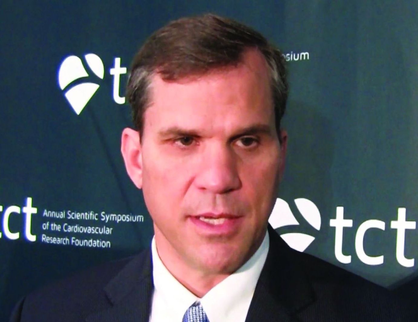
“I think this is a big breakthrough. ... Ultimately, it should lead to better patient outcomes,” said lead investigator William Fearon, MD, professor of cardiology at Stanford University, Calif.
In an interview at TCT 2018, Dr. Fearon explained the importance of FFR, the data for FFRangio, and what’s coming down the pike from other companies. He disclosed institutional research grants from the company.
SAN DIEGO – Angiograms can be deceiving, so it’s best to measure fractional flow reserve (FFR) across coronary obstructions to gauge patients’ true need for intervention. That’s hardly news to cardiologists, but FFR is not often done. The problem is that traditional measurement requires threading wires down coronary arteries; the technique is a bit risky and takes time and training. It also has to be repeated for each lesion.
But now, several companies are developing noninvasive ways to measure FFR.
Findings from one of them, CathWorks, were presented at the Transcatheter Cardiovascular Therapeutics annual meeting sponsored by the Cardiovascular Research Foundation. Its FFRangio system uses high-quality angiograms and an algorithm to estimate resistance and flow across stenoses. After a few cases, the process takes less than 5 minutes (Circulation. 2018 Sep 24. doi: 10.1161/CIRCULATIONAHA.118.037350).
“I think this is a big breakthrough. ... Ultimately, it should lead to better patient outcomes,” said lead investigator William Fearon, MD, professor of cardiology at Stanford University, Calif.
In an interview at TCT 2018, Dr. Fearon explained the importance of FFR, the data for FFRangio, and what’s coming down the pike from other companies. He disclosed institutional research grants from the company.
REPORTING FROM TCT 2018
Time reveals benefit of CABG over PCI for left main disease
SAN DIEGO – In the long run, patients with left main coronary artery disease fare better if they undergo coronary artery bypass grafting (CABG) instead of percutaneous coronary intervention (PCI) with drug-eluting stents, suggest 10-year results of the MAIN-COMPARE trial. Findings were reported at the Transcatheter Cardiovascular Therapeutics annual meeting.
Although CABG is the standard choice for revascularization in this patient population, PCI has been making inroads thanks to advances in stents, antithrombotic drugs, periprocedural management, and operator expertise, noted senior author Seung-Jung Park, MD, PhD, chairman of the Heart Institute at Asan Medical Center in Seoul and professor of medicine at University of Ulsan, South Korea. “Indeed, many studies showed that PCI using drug-eluting stents might be a good alternative for selected patients with left main coronary artery disease.”
Two large, randomized, controlled trials, EXCEL and NOBLE, have compared these treatment strategies and helped clarify outcomes at intermediate follow-up periods of 3-5 years. But long-term data, increasingly important as survival improves, are lacking.
Dr. Park reported the 10-year update of a prospective, observational cohort study that analyzed data from more than 2,000 patients with unprotected left main coronary artery disease in the MAIN-COMPARE registry, which captures revascularization procedures performed at 12 Korean cardiac centers.
In the entire cohort, about a fifth of the patients died, and roughly a fourth experienced a composite adverse outcome of death and cardiovascular events regardless of whether they received PCI or CABG, but the former yielded a rate of target vessel revascularization that was more than three times higher, according to results reported at the meeting and simultaneously published (J Am Coll Cardiol. 2018 Sep 14. doi: 10.1016/j.jacc.2018.09.012). Among the subset of patients treated in the more recent drug-eluting stent era, those who underwent PCI were more likely to die and to experience the composite outcome starting at the 5-year mark.
“Drug-eluting stents were associated with higher risks of death and serious composite outcomes compared to CABG after 5 years. The treatment benefit of CABG has diverged over time during continued follow-up,” Dr. Park noted. “The rate of target-vessel failure was consistently higher in the PCI group.”
“We used mainly first-generation drug-eluting stents,” he acknowledged. “However, many studies have demonstrated there is not too much difference between the first- and second-generation stents.”
Data worth the wait
In the same session, investigators reported the 10-year update of the European and U.S. randomized SYNTAX Extended Survival trial, called SYNTAXES. SYNTAX enrolled patients with three-vessel or left main coronary disease. That trial found no significant difference in survival between PCI with drug-eluting stents and CABG overall. In stratified analysis, mortality was higher with PCI among patients with three-vessel disease, but not among patients with left main disease.
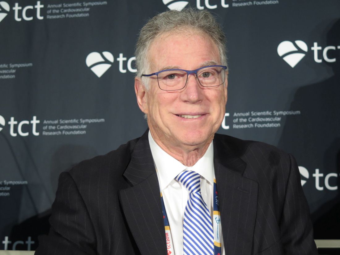
Taken together, these trials help clarify the long-term comparative efficacy of PCI and begin to inform patient selection, according to press conference panelist Morton J. Kern, MD, a professor at the University of California, Irvine Medical Center.
“The fine subgroup analysis of who the best candidates are is still in question,” he elaborated. “The SYNTAXES study told us that surgery for left mains is still pretty good, and even though you can get good results with PCI, the event rates are higher in that three-vessel, high-SYNTAX score group, so we should be careful. Interventionalists need to know their limitations. I think that’s what both studies tell us, actually.”
Study details
The MAIN-COMPARE analyses were based on 2,240 patients with unprotected left main coronary artery disease (stenosis of more than 50% and no coronary artery bypass grafts to the left anterior descending or the left circumflex artery) treated during 2000-2006.
A total of 1,102 patients underwent PCI with stenting: 318 in the era of bare-metal stents and 784 in the era of drug-eluting stents, predominantly sirolimus-eluting stents. A total of 1,138 patients underwent CABG. The minimum follow-up was 10 years in all patients, with a median of 12 years.
In the entire study cohort, PCI and CABG yielded similar rates of death (21.1% vs. 23.2%) and the composite of death, Q-wave myocardial infarction, or stroke (23.8% vs. 26.3%), but the PCI patients had a significantly higher rate of target vessel revascularization (21.1% vs. 5.8%), according to data reported at the meeting, which was sponsored by the New York–based Cardiovascular Research Foundation.
In analyses using a propensity score weighting technique, results for the entire cohort were much the same. But on stratification, PCI with drug-eluting stents versus CABG yielded higher risks of death (hazard ratio, 1.35; P = .05) and the composite adverse outcome (HR, 1.46; P = .009) from 5 years onward, as well as a sharply higher risk of target vessel revascularization for the full duration of follow-up (HR, 5.82; P less than .001).
Dr. Park disclosed that he had no conflicts of interest. The study was supported by the Korean Society of Interventional Cardiology and the CardioVascular Research Foundation of South Korea.
SOURCE: Park SJ et al. J Am Coll Cardiol. 2018 Sep 14. doi: 10.1016/j.jacc.2018.09.012).
SAN DIEGO – In the long run, patients with left main coronary artery disease fare better if they undergo coronary artery bypass grafting (CABG) instead of percutaneous coronary intervention (PCI) with drug-eluting stents, suggest 10-year results of the MAIN-COMPARE trial. Findings were reported at the Transcatheter Cardiovascular Therapeutics annual meeting.
Although CABG is the standard choice for revascularization in this patient population, PCI has been making inroads thanks to advances in stents, antithrombotic drugs, periprocedural management, and operator expertise, noted senior author Seung-Jung Park, MD, PhD, chairman of the Heart Institute at Asan Medical Center in Seoul and professor of medicine at University of Ulsan, South Korea. “Indeed, many studies showed that PCI using drug-eluting stents might be a good alternative for selected patients with left main coronary artery disease.”
Two large, randomized, controlled trials, EXCEL and NOBLE, have compared these treatment strategies and helped clarify outcomes at intermediate follow-up periods of 3-5 years. But long-term data, increasingly important as survival improves, are lacking.
Dr. Park reported the 10-year update of a prospective, observational cohort study that analyzed data from more than 2,000 patients with unprotected left main coronary artery disease in the MAIN-COMPARE registry, which captures revascularization procedures performed at 12 Korean cardiac centers.
In the entire cohort, about a fifth of the patients died, and roughly a fourth experienced a composite adverse outcome of death and cardiovascular events regardless of whether they received PCI or CABG, but the former yielded a rate of target vessel revascularization that was more than three times higher, according to results reported at the meeting and simultaneously published (J Am Coll Cardiol. 2018 Sep 14. doi: 10.1016/j.jacc.2018.09.012). Among the subset of patients treated in the more recent drug-eluting stent era, those who underwent PCI were more likely to die and to experience the composite outcome starting at the 5-year mark.
“Drug-eluting stents were associated with higher risks of death and serious composite outcomes compared to CABG after 5 years. The treatment benefit of CABG has diverged over time during continued follow-up,” Dr. Park noted. “The rate of target-vessel failure was consistently higher in the PCI group.”
“We used mainly first-generation drug-eluting stents,” he acknowledged. “However, many studies have demonstrated there is not too much difference between the first- and second-generation stents.”
Data worth the wait
In the same session, investigators reported the 10-year update of the European and U.S. randomized SYNTAX Extended Survival trial, called SYNTAXES. SYNTAX enrolled patients with three-vessel or left main coronary disease. That trial found no significant difference in survival between PCI with drug-eluting stents and CABG overall. In stratified analysis, mortality was higher with PCI among patients with three-vessel disease, but not among patients with left main disease.
Taken together, these trials help clarify the long-term comparative efficacy of PCI and begin to inform patient selection, according to press conference panelist Morton J. Kern, MD, a professor at the University of California, Irvine Medical Center.
“The fine subgroup analysis of who the best candidates are is still in question,” he elaborated. “The SYNTAXES study told us that surgery for left mains is still pretty good, and even though you can get good results with PCI, the event rates are higher in that three-vessel, high-SYNTAX score group, so we should be careful. Interventionalists need to know their limitations. I think that’s what both studies tell us, actually.”
Study details
The MAIN-COMPARE analyses were based on 2,240 patients with unprotected left main coronary artery disease (stenosis of more than 50% and no coronary artery bypass grafts to the left anterior descending or the left circumflex artery) treated during 2000-2006.
A total of 1,102 patients underwent PCI with stenting: 318 in the era of bare-metal stents and 784 in the era of drug-eluting stents, predominantly sirolimus-eluting stents. A total of 1,138 patients underwent CABG. The minimum follow-up was 10 years in all patients, with a median of 12 years.
In the entire study cohort, PCI and CABG yielded similar rates of death (21.1% vs. 23.2%) and the composite of death, Q-wave myocardial infarction, or stroke (23.8% vs. 26.3%), but the PCI patients had a significantly higher rate of target vessel revascularization (21.1% vs. 5.8%), according to data reported at the meeting, which was sponsored by the New York–based Cardiovascular Research Foundation.
In analyses using a propensity score weighting technique, results for the entire cohort were much the same. But on stratification, PCI with drug-eluting stents versus CABG yielded higher risks of death (hazard ratio, 1.35; P = .05) and the composite adverse outcome (HR, 1.46; P = .009) from 5 years onward, as well as a sharply higher risk of target vessel revascularization for the full duration of follow-up (HR, 5.82; P less than .001).
Dr. Park disclosed that he had no conflicts of interest. The study was supported by the Korean Society of Interventional Cardiology and the CardioVascular Research Foundation of South Korea.
SOURCE: Park SJ et al. J Am Coll Cardiol. 2018 Sep 14. doi: 10.1016/j.jacc.2018.09.012).
REPORTING FROM TCT 2018
Key clinical point: CABG had an edge over PCI with drug-eluting stents in patients with left main disease that became evident with longer follow-up.
Major finding: Compared with CABG, PCI with drug-eluting stents carried higher risks of death (hazard ratio, 1.35; P = .05) and a composite adverse outcome (HR, 1.46; P = .009) from 5 years onward.
Study details: Ten-year follow-up of a multicenter prospective cohort study of 2,240 patients with unprotected left main coronary artery disease who underwent either PCI with stenting or CABG (MAIN-COMPARE study).
Disclosures: Dr. Park disclosed that he had no conflicts of interest. The study was supported by the Korean Society of Interventional Cardiology and the CardioVascular Research Foundation of South Korea.
Source: Park S-J et al. J Am Coll Cardiol. 2018 Sep 14. doi: 10.1016/j.jacc.2018.09.012.
Simplified SYNTAX holds promise, but still has doubters
SAN DIEGO – A simplified version of the SYNTAX score for coronary artery disease complexity strongly correlated with the unmodified SYNTAX score and could make it easier for cardiologists to employ it in everyday practice. The modified version can be easily memorized and has simplified values that a clinician can calculate and use to determine an appropriate treatment without breaking their work flow to consult a computerized system.
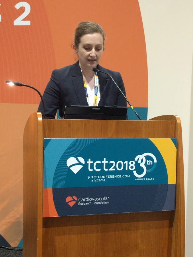
“It’s primarily simplifying the values for the location of the lesions that has allowed it to be more memorizable. That does make it a slightly blunter tool, but only slightly,” said Sonya Burgess, MBCHB, during a presentation at the Transcatheter Cardiovascular Therapeutics annual meeting. Dr. Burgess is an interventional cardiologist at the University of New South Wales, Sydney.
In common practice, SYNTAX scores often go uncalculated because of their complexity, despite guidelines that recommend it. “My guess might be that 1 in every 10 [cardiologists] do it, and that’s not right for our patients,” said Dr. Burgess.
Others at the session agreed that SYNTAX is underutilized, but not all agreed with Dr. Burgess’ solution. Notable among the attendees was Patrick Serruys, MD, of Erasmus University, Rotterdam, the Netherlands, who originally published the SYNTAX system. He doesn’t like the idea of simplifying SYNTAX, based in part on previous experience. “About 10 years ago, Boston Scientific found the score too difficult and they wanted to simplify it, but the score completely lost its prognostic value. I was very emotional about that and I said, ‘no, we have to keep all of the components until we understand [the risk factors] much better,’ ” said Dr. Serruys.
But Dr. Burgess argued that the simplified system, which she and her coinvestigators created, makes concessions to the realities of an interventional cardiology suite. To obtain a standard SYNTAX score, “they have to stop doing something or rebook something, or there’s a lesion there that they just want to treat. [With the simplified score], at least before they go on to treat that lesion, they don’t have to take their gowns and gloves off. You can even have a card hanging on the bar [to refer to].” Dr. Burgess did not provide details of the system.
Dr. Serruys agreed with the source of the problem. “Dr. Burgess is right, it is somewhat demanding,” he allowed, but he also called on clinicians to consider the needs of the patient, even going so far as picturing the patient as a family member. “People complain that doing the SYNTAX is on average 7 minutes, with a standard deviation of 7, so you could work 15 minutes just looking at film. But when you think that the future of your father or brother is depending on the surgeon and the cardiologist looking carefully, I find the argument absolutely obnoxious,” said Dr. Serruys at the meeting sponsored by the Cardiovascular Research Foundation.
Rather than simplification, he stresses teamwork. With at least three people looking at the angiogram, the results are consistent and useful. “You look at the film with the trainees, and that’s how they learn. Never do a SYNTAX score alone,” said Dr. Serruys.
To assess the simplified SYNTAX score, the researchers conducted a retrospective assessment of both the SYNTAX score and the simplified SYNTAX score in 617 patients who had multivessel disease. They performed both assessments in subgroups of patients with 169 patients with ST-segment elevation MI, 78 patients with chronic total occlusion, and 113 patients with left main coronary artery (LMCA) stenosis. They used a 100-patient derivation cohort to determine cutoffs for patients who would not be suitable for percutaneous coronary intervention. They also looked at the accuracy of the simplified version compared with standard SYNTAX in 517 patients from five tertiary centers.
The Spearman’s rho value was 0.93 overall and at least 0.91 for all subgroups (P less than .001 for all). In patients with LMCA stenosis, the simplified version had a sensitivity of 100%, specificity of 85%, negative predictive of 100%, and an area under the curve of 1.0 (P less than .001). For patients with multivessel disease and no LMCA stenosis, the values were 98%, 82%, 99%, and 0.971, respectively (P less than .001).
Dr. Burgess and Dr. Serruys reported no financial conflicts of interest.
SAN DIEGO – A simplified version of the SYNTAX score for coronary artery disease complexity strongly correlated with the unmodified SYNTAX score and could make it easier for cardiologists to employ it in everyday practice. The modified version can be easily memorized and has simplified values that a clinician can calculate and use to determine an appropriate treatment without breaking their work flow to consult a computerized system.
“It’s primarily simplifying the values for the location of the lesions that has allowed it to be more memorizable. That does make it a slightly blunter tool, but only slightly,” said Sonya Burgess, MBCHB, during a presentation at the Transcatheter Cardiovascular Therapeutics annual meeting. Dr. Burgess is an interventional cardiologist at the University of New South Wales, Sydney.
In common practice, SYNTAX scores often go uncalculated because of their complexity, despite guidelines that recommend it. “My guess might be that 1 in every 10 [cardiologists] do it, and that’s not right for our patients,” said Dr. Burgess.
Others at the session agreed that SYNTAX is underutilized, but not all agreed with Dr. Burgess’ solution. Notable among the attendees was Patrick Serruys, MD, of Erasmus University, Rotterdam, the Netherlands, who originally published the SYNTAX system. He doesn’t like the idea of simplifying SYNTAX, based in part on previous experience. “About 10 years ago, Boston Scientific found the score too difficult and they wanted to simplify it, but the score completely lost its prognostic value. I was very emotional about that and I said, ‘no, we have to keep all of the components until we understand [the risk factors] much better,’ ” said Dr. Serruys.
But Dr. Burgess argued that the simplified system, which she and her coinvestigators created, makes concessions to the realities of an interventional cardiology suite. To obtain a standard SYNTAX score, “they have to stop doing something or rebook something, or there’s a lesion there that they just want to treat. [With the simplified score], at least before they go on to treat that lesion, they don’t have to take their gowns and gloves off. You can even have a card hanging on the bar [to refer to].” Dr. Burgess did not provide details of the system.
Dr. Serruys agreed with the source of the problem. “Dr. Burgess is right, it is somewhat demanding,” he allowed, but he also called on clinicians to consider the needs of the patient, even going so far as picturing the patient as a family member. “People complain that doing the SYNTAX is on average 7 minutes, with a standard deviation of 7, so you could work 15 minutes just looking at film. But when you think that the future of your father or brother is depending on the surgeon and the cardiologist looking carefully, I find the argument absolutely obnoxious,” said Dr. Serruys at the meeting sponsored by the Cardiovascular Research Foundation.
Rather than simplification, he stresses teamwork. With at least three people looking at the angiogram, the results are consistent and useful. “You look at the film with the trainees, and that’s how they learn. Never do a SYNTAX score alone,” said Dr. Serruys.
To assess the simplified SYNTAX score, the researchers conducted a retrospective assessment of both the SYNTAX score and the simplified SYNTAX score in 617 patients who had multivessel disease. They performed both assessments in subgroups of patients with 169 patients with ST-segment elevation MI, 78 patients with chronic total occlusion, and 113 patients with left main coronary artery (LMCA) stenosis. They used a 100-patient derivation cohort to determine cutoffs for patients who would not be suitable for percutaneous coronary intervention. They also looked at the accuracy of the simplified version compared with standard SYNTAX in 517 patients from five tertiary centers.
The Spearman’s rho value was 0.93 overall and at least 0.91 for all subgroups (P less than .001 for all). In patients with LMCA stenosis, the simplified version had a sensitivity of 100%, specificity of 85%, negative predictive of 100%, and an area under the curve of 1.0 (P less than .001). For patients with multivessel disease and no LMCA stenosis, the values were 98%, 82%, 99%, and 0.971, respectively (P less than .001).
Dr. Burgess and Dr. Serruys reported no financial conflicts of interest.
REPORTING FROM TCT 2018
Key clinical point: A simplified SYNTAX score could lead to broader implementation of evidence-based decision making for complex coronary artery disease.
Major finding: The simplified score had a Spearman’s rho value of 0.93 overall.
Study details: A retrospective analysis of 617 patients with multivessel disease.
Disclosures: Dr. Burgess and Dr. Serruys reported no financial conflicts of interest.
Drug-coated balloons shown noninferior to DES in thin coronaries
MUNICH – for preventing the clinical consequences of restenosis during 12 months following coronary intervention, according to results from a prospective, randomized, multicenter trial.
The video associated with this article is no longer available on this site. Please view all of our videos on the MDedge YouTube channel
Drug-coated balloons are already used to treat in-stent coronary restenosis. The findings of the current study establish the tested DCB as noninferior to a DES for treating coronary stenoses in narrow arteries less than 3 mm in diameter, Raban V. Jeger, MD, said at the annual congress of the European Society of Cardiology. The DCB approach avoids placing a metal stent in a narrow coronary and thus has no long-term risk for in-stent thrombosis, said Dr. Jeger, a professor of cardiology at Basel (Switzerland) University Hospital. Dr. Jeger acknowledged that the tested DCB is more expensive than the second-generation DES used as the comparator in most of the control patients, “but I think the benefit to patients is worth” the added cost, he said when discussing his report.
The BASKET-SMALL 2 (NCT01574534) study enrolled 758 patients at 14 centers in Switzerland, Germany, and Austria. The trial limited enrollment to patients who were scheduled to undergo percutaneous coronary intervention for stenosis in a coronary artery that was at least 2.0 mm and less than 3.0 mm in diameter and had first undergone successful predilatation without any flow-limiting dissections or residual stenosis, a step in the DCB procedure that adds to the procedure’s cost.
The video associated with this article is no longer available on this site. Please view all of our videos on the MDedge YouTube channel
The study randomized patients to treatment with either a balloon coated with paclitaxel/iopromide (SeQuent Please) or a DES. The first quarter of patients randomized into the DES arm received a first-generation, paclitaxel-eluting DES (Taxus Element); the remaining patients in the comparator arm received a second-generation everolimus-eluting DES (Xience). The DCB tested is not approved for U.S. marketing.
The primary endpoint was the combined rate of cardiac death, nonfatal MI, or target vessel revascularization during 12 months of follow-up. In the intention-to-treat analysis, this occurred in 7.33% of the DCB patients and in 7.45% of the DES patients, a difference that was not statistically significant and that met the prespecified criterion for noninferiority of the DCB. Concurrently with Dr. Jeger’s report at the congress, the results also appeared in an article published in The Lancet (Lancet. 2018 Sep 8;392[10190]:849-56).
One limitation of the study was that the first 25% of patients enrolled into the DES arm received a first-generation DES, while the remaining 75% received a second-generation device. Analysis of the primary endpoint by DES type showed that events occurred more than twice as often in the patients who received a first-generation DES, and their inclusion may have affected the comparator group’s results.
Coronary arteries that need percutaneous intervention and are less than 3 mm in diameter constitute about a third of all target vessels, and they are especially common among women and in patients with diabetes, Dr. Jeger said. Despite this, women made up about a quarter of the study enrollment, and about a third had diabetes. He also noted that a key aspect of adopting the DCB approach into routine practice is that operators would need to have the “courage” to accept some amount of recoil and “minor” dissections after DCB treatment and not feel compelled to correct these with a stent.
Other features of the BASKET-SMALL 2 trial also have raised concerns about the immediate clinical implications of the results, said Roxana Mehran, MD, a professor of medicine at Icahn School of Medicine at Mount Sinai, New York, and the congress’s designated discussant for the report.
The study began in 2012, which means it took more than 5 years to enroll and suggests that the study may have a selection bias. Dr. Mehran also questioned whether it was really a small vessel study, with an enrollment criterion of less than 3 mm in diameter. A future study should be done in “truly” small vessels, those thinner than 2.5 mm, she said.
Dr. Mehran agreed it’s attractive to speculate that, by using a DCB and avoiding stent placement, fewer patients will eventually have very-late adverse events, but this must be proven with longer follow-up and in larger numbers of patients, she said.
Treating thin coronary arteries is a problem because they have a higher risk for in-stent restenosis, although usually we will put a stent in arteries that are at least 2.5 mm wide and sometimes in coronaries as narrow as 2.25 mm. That’s using the narrowest stent we have available. Sometimes in vessels this size, if the result from initial balloon angioplasty looks good on angiography, we accept that outcome and do not place a stent.

Steen Dalby Kristensen, MD , is a professor of cardiology at Aarhus University in Skejby, Denmark. He had no relevant disclosures. He made these comments in a video interview.
Treating thin coronary arteries is a problem because they have a higher risk for in-stent restenosis, although usually we will put a stent in arteries that are at least 2.5 mm wide and sometimes in coronaries as narrow as 2.25 mm. That’s using the narrowest stent we have available. Sometimes in vessels this size, if the result from initial balloon angioplasty looks good on angiography, we accept that outcome and do not place a stent.
Steen Dalby Kristensen, MD , is a professor of cardiology at Aarhus University in Skejby, Denmark. He had no relevant disclosures. He made these comments in a video interview.
MUNICH – for preventing the clinical consequences of restenosis during 12 months following coronary intervention, according to results from a prospective, randomized, multicenter trial.
The video associated with this article is no longer available on this site. Please view all of our videos on the MDedge YouTube channel
Drug-coated balloons are already used to treat in-stent coronary restenosis. The findings of the current study establish the tested DCB as noninferior to a DES for treating coronary stenoses in narrow arteries less than 3 mm in diameter, Raban V. Jeger, MD, said at the annual congress of the European Society of Cardiology. The DCB approach avoids placing a metal stent in a narrow coronary and thus has no long-term risk for in-stent thrombosis, said Dr. Jeger, a professor of cardiology at Basel (Switzerland) University Hospital. Dr. Jeger acknowledged that the tested DCB is more expensive than the second-generation DES used as the comparator in most of the control patients, “but I think the benefit to patients is worth” the added cost, he said when discussing his report.
The BASKET-SMALL 2 (NCT01574534) study enrolled 758 patients at 14 centers in Switzerland, Germany, and Austria. The trial limited enrollment to patients who were scheduled to undergo percutaneous coronary intervention for stenosis in a coronary artery that was at least 2.0 mm and less than 3.0 mm in diameter and had first undergone successful predilatation without any flow-limiting dissections or residual stenosis, a step in the DCB procedure that adds to the procedure’s cost.
The video associated with this article is no longer available on this site. Please view all of our videos on the MDedge YouTube channel
The study randomized patients to treatment with either a balloon coated with paclitaxel/iopromide (SeQuent Please) or a DES. The first quarter of patients randomized into the DES arm received a first-generation, paclitaxel-eluting DES (Taxus Element); the remaining patients in the comparator arm received a second-generation everolimus-eluting DES (Xience). The DCB tested is not approved for U.S. marketing.
The primary endpoint was the combined rate of cardiac death, nonfatal MI, or target vessel revascularization during 12 months of follow-up. In the intention-to-treat analysis, this occurred in 7.33% of the DCB patients and in 7.45% of the DES patients, a difference that was not statistically significant and that met the prespecified criterion for noninferiority of the DCB. Concurrently with Dr. Jeger’s report at the congress, the results also appeared in an article published in The Lancet (Lancet. 2018 Sep 8;392[10190]:849-56).
One limitation of the study was that the first 25% of patients enrolled into the DES arm received a first-generation DES, while the remaining 75% received a second-generation device. Analysis of the primary endpoint by DES type showed that events occurred more than twice as often in the patients who received a first-generation DES, and their inclusion may have affected the comparator group’s results.
Coronary arteries that need percutaneous intervention and are less than 3 mm in diameter constitute about a third of all target vessels, and they are especially common among women and in patients with diabetes, Dr. Jeger said. Despite this, women made up about a quarter of the study enrollment, and about a third had diabetes. He also noted that a key aspect of adopting the DCB approach into routine practice is that operators would need to have the “courage” to accept some amount of recoil and “minor” dissections after DCB treatment and not feel compelled to correct these with a stent.
Other features of the BASKET-SMALL 2 trial also have raised concerns about the immediate clinical implications of the results, said Roxana Mehran, MD, a professor of medicine at Icahn School of Medicine at Mount Sinai, New York, and the congress’s designated discussant for the report.
The study began in 2012, which means it took more than 5 years to enroll and suggests that the study may have a selection bias. Dr. Mehran also questioned whether it was really a small vessel study, with an enrollment criterion of less than 3 mm in diameter. A future study should be done in “truly” small vessels, those thinner than 2.5 mm, she said.
Dr. Mehran agreed it’s attractive to speculate that, by using a DCB and avoiding stent placement, fewer patients will eventually have very-late adverse events, but this must be proven with longer follow-up and in larger numbers of patients, she said.
REPORTING FROM THE ESC CONGRESS 2018
Key clinical point: Drug-coated balloon treatment worked as well as drug-eluting stents in thin coronaries.
Major finding: Twelve-month MACE occurred in 7.33% of balloon-treated patients and in 7.45% of stent-treated patients.
Study details: BASKET-SMALL 2, an international, multicenter randomized trial with 758 patients.
Disclosures: The investigator-initiated study received partial funding from B. Braun, the company that markets the drug-coated balloon (SeQuent Please) tested in the study. Dr. Jeger has received research funding from B. Braun. Dr. Mehran has been a consultant to Abbott, Bayer, BSC, and CSL Behring and has received research funding from Abbott, Astra Zeneca, Bayer, BCC, DSI, and Janssen.

PCI bests medical therapy for FFR grey zone stable angina
PARIS – Patients with stable angina and a fractional flow reserve (FFR) value in the grey zone of 0.75-0.81 experienced a significant reduction in myocardial ischemia and substantially greater quality of life improvement if they were randomized to percutaneous coronary intervention (PCI) plus optimal medical therapy than to optimal medical therapy alone in the Scottish Grey-zone FFR Study.
The Grey-zone FFR Study was a single-center, prospective, unblinded, randomized trial that included 100 patients with stable angina, single-vessel disease, and a fractional flow reserve in the grey zone of 0.75-0.81. While broad consensus exists that an FFR below 0.75 constitutes evidence of a hemodynamically significant coronary lesion warranting revascularization and an FFR greater than 0.80 indicates a lesion isn’t functionally significant and therefore PCI can safely be deferred, there has been uncertainty on what to do about lesions in the grey zone, which are frequently encountered in the cardiac catheterization laboratory.
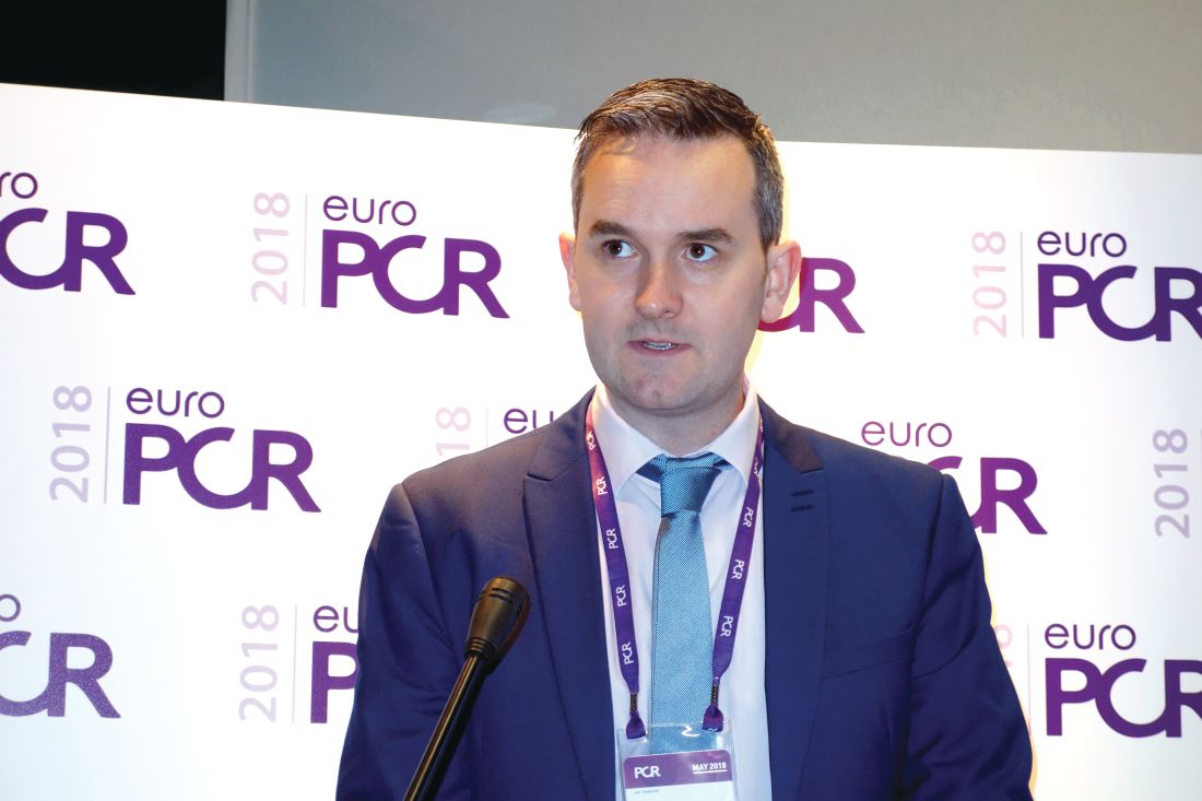
“In my clinical practice, I tend to go ahead with PCI for patients in the grey zone if I felt it was clinically feasible and safe to do so, particularly if I was worried about their lesion morphology,” Barry Hennigan, MD, said in response to questions after presenting the results at the annual meeting of the European Association of Percutaneous Cardiovascular Interventions. “If it’s a proximal LAD [left anterior descending artery] lesion and it’s a grey zone patient, particularly if it’s a lesion morphology that you’re not comfortable with, I think you need to be very careful before you defer a case.”
The twin purposes of the Scottish study were to define the prevalence of major ischemia by stress MRI and invasive flow assessment via a pressure wire in grey zone patients – something which hadn’t been done before – and to determine if PCI deferral in such patients is appropriate in terms of symptom control. The primary outcome was change in angina severity at 3 months follow-up using the Seattle Angina Questionnaire (SAQ).
Scores on two of the five domains of the SAQ – anginal frequency and quality of life – were significantly improved in the PCI group. Anginal frequency scores improved by a mean of 20.58 points in the PCI plus optimal medical therapy (OMT) group, compared with a 9.39-point improvement with OMT alone. Quality of life scores improved by 24.04 points in the PCI group versus 9.39 points in controls, said Dr. Hennigan, an interventional cardiologist at the University of Glasgow and Golden Jubilee National Hospital. Scores in the other three SAQ domains – physical limitations, anginal stability, and treatment satisfaction – didn’t differ significantly between the two study arms, although consistently greater improvements were seen in the PCI group.
Baseline stress perfusion MRI as assessed by two blinded observers demonstrated that 17.4% of patients with stable angina and a grey zone FFR had major ischemia, while any ischemia – major or minor – was present in 24.4%. Follow-up scans at 3 months showed a roughly 50% reduction in the prevalence of ischemia in the PCI group, with 7.3% of treated patients still having major ischemia and 12.2% having any ischemia.
Also, 28% of participants had evidence of ischemia at baseline based upon their coronary flow reserve measurements and 8% had a hyperemic stenosis resistance measurement indicative of ischemia. So the FFR grey zone encompasses a range of cardiovascular risks.
In the PCI plus OMT group, 89% of patients (eight of nine) with baseline ischemia on stress MRI had a greater than 10-point improvement in quality of life scores on the SAQ at follow-up in contrast to 53% of patients without ischemia, which made for a statistically significant difference. An improvement of that magnitude is generally considered clinically meaningful. In contrast, in the OMT-only group, 9 of 14 patients with baseline ischemia (64.2%) had a greater than 10-point quality of life improvement, which wasn’t significantly different from the 45.5% improvement rate in patients with no ischemia.
The lessons? Grey zone patients who benefit most from prompt revascularization are those with demonstrable ischemia. In addition, roughly half of grey zone patients with stable angina will improve their quality of life scores by more than 10 points with OMT alone regardless of the presence of myocardial ischemia or not.
Dr. Hennigan was repeatedly asked how he reconciles the results of the grey zone study with those of the much-discussed ORBITA trial, the first and only randomized trial of real versus sham PCI in patients with stable angina. ORBITA didn’t find a significant quality of life advantage for real PCI over sham PCI.
“It is quite possible that a lot of the effect that we saw in our PCI group was placebo related,” he conceded. “However, we do have objective evidence that we reduced ischemia on MRI. Also, 29% of ORBITA patients had an FFR above 0.8, whereas nearly all our patients were below that threshold. So we perhaps had more prevalent ischemia than the ORBITA cohort.”
Also informative is a comparison of SAQ scores at follow-up in the sham PCI ORBITA control group versus the grey zone Scottish PCI group, Dr. Hennigan continued. The Scottish PCI group had a mean 20.6-point improvement in anginal frequency scores while on an average of 1.3 antianginal medications, compared with a 9.6-point improvement in ORBITA patients on 2.9 drugs. The grey zone group who got PCI plus OMT also had a mean 16.1-point improvement in the SAQ physical limitations domain, versus a 5.0-point improvement in the ORBITA controls.
The Grey-zone FFR Study was supported by the British Heart Foundation. Dr. Hennigan reported having no financial conflicts of interest.
PARIS – Patients with stable angina and a fractional flow reserve (FFR) value in the grey zone of 0.75-0.81 experienced a significant reduction in myocardial ischemia and substantially greater quality of life improvement if they were randomized to percutaneous coronary intervention (PCI) plus optimal medical therapy than to optimal medical therapy alone in the Scottish Grey-zone FFR Study.
The Grey-zone FFR Study was a single-center, prospective, unblinded, randomized trial that included 100 patients with stable angina, single-vessel disease, and a fractional flow reserve in the grey zone of 0.75-0.81. While broad consensus exists that an FFR below 0.75 constitutes evidence of a hemodynamically significant coronary lesion warranting revascularization and an FFR greater than 0.80 indicates a lesion isn’t functionally significant and therefore PCI can safely be deferred, there has been uncertainty on what to do about lesions in the grey zone, which are frequently encountered in the cardiac catheterization laboratory.
“In my clinical practice, I tend to go ahead with PCI for patients in the grey zone if I felt it was clinically feasible and safe to do so, particularly if I was worried about their lesion morphology,” Barry Hennigan, MD, said in response to questions after presenting the results at the annual meeting of the European Association of Percutaneous Cardiovascular Interventions. “If it’s a proximal LAD [left anterior descending artery] lesion and it’s a grey zone patient, particularly if it’s a lesion morphology that you’re not comfortable with, I think you need to be very careful before you defer a case.”
The twin purposes of the Scottish study were to define the prevalence of major ischemia by stress MRI and invasive flow assessment via a pressure wire in grey zone patients – something which hadn’t been done before – and to determine if PCI deferral in such patients is appropriate in terms of symptom control. The primary outcome was change in angina severity at 3 months follow-up using the Seattle Angina Questionnaire (SAQ).
Scores on two of the five domains of the SAQ – anginal frequency and quality of life – were significantly improved in the PCI group. Anginal frequency scores improved by a mean of 20.58 points in the PCI plus optimal medical therapy (OMT) group, compared with a 9.39-point improvement with OMT alone. Quality of life scores improved by 24.04 points in the PCI group versus 9.39 points in controls, said Dr. Hennigan, an interventional cardiologist at the University of Glasgow and Golden Jubilee National Hospital. Scores in the other three SAQ domains – physical limitations, anginal stability, and treatment satisfaction – didn’t differ significantly between the two study arms, although consistently greater improvements were seen in the PCI group.
Baseline stress perfusion MRI as assessed by two blinded observers demonstrated that 17.4% of patients with stable angina and a grey zone FFR had major ischemia, while any ischemia – major or minor – was present in 24.4%. Follow-up scans at 3 months showed a roughly 50% reduction in the prevalence of ischemia in the PCI group, with 7.3% of treated patients still having major ischemia and 12.2% having any ischemia.
Also, 28% of participants had evidence of ischemia at baseline based upon their coronary flow reserve measurements and 8% had a hyperemic stenosis resistance measurement indicative of ischemia. So the FFR grey zone encompasses a range of cardiovascular risks.
In the PCI plus OMT group, 89% of patients (eight of nine) with baseline ischemia on stress MRI had a greater than 10-point improvement in quality of life scores on the SAQ at follow-up in contrast to 53% of patients without ischemia, which made for a statistically significant difference. An improvement of that magnitude is generally considered clinically meaningful. In contrast, in the OMT-only group, 9 of 14 patients with baseline ischemia (64.2%) had a greater than 10-point quality of life improvement, which wasn’t significantly different from the 45.5% improvement rate in patients with no ischemia.
The lessons? Grey zone patients who benefit most from prompt revascularization are those with demonstrable ischemia. In addition, roughly half of grey zone patients with stable angina will improve their quality of life scores by more than 10 points with OMT alone regardless of the presence of myocardial ischemia or not.
Dr. Hennigan was repeatedly asked how he reconciles the results of the grey zone study with those of the much-discussed ORBITA trial, the first and only randomized trial of real versus sham PCI in patients with stable angina. ORBITA didn’t find a significant quality of life advantage for real PCI over sham PCI.
“It is quite possible that a lot of the effect that we saw in our PCI group was placebo related,” he conceded. “However, we do have objective evidence that we reduced ischemia on MRI. Also, 29% of ORBITA patients had an FFR above 0.8, whereas nearly all our patients were below that threshold. So we perhaps had more prevalent ischemia than the ORBITA cohort.”
Also informative is a comparison of SAQ scores at follow-up in the sham PCI ORBITA control group versus the grey zone Scottish PCI group, Dr. Hennigan continued. The Scottish PCI group had a mean 20.6-point improvement in anginal frequency scores while on an average of 1.3 antianginal medications, compared with a 9.6-point improvement in ORBITA patients on 2.9 drugs. The grey zone group who got PCI plus OMT also had a mean 16.1-point improvement in the SAQ physical limitations domain, versus a 5.0-point improvement in the ORBITA controls.
The Grey-zone FFR Study was supported by the British Heart Foundation. Dr. Hennigan reported having no financial conflicts of interest.
REPORTING FROM EUROPCR 2018
Key clinical point:
Major finding: Patients with stable angina and a fractional flow reserve in the grey zone of 0.75-0.81 experienced a 50% reduction in objectively defined myocardial ischemia if they received percutaneous coronary intervention plus medical therapy, compared with medical therapy alone.
Study details: This single-center, prospective, open-label trial randomized 100 stable angina patients with a grey zone fractional flow reserve of 0.75-0.81 to percutaneous coronary intervention plus optimal medical therapy or optimal medical therapy alone.
Disclosures: The study was supported by the British Heart Foundation. The presenter reported having no financial conflicts.
FAST-FFR: Noninvasive FFR nearly as good as wire
SAN DIEGO – A less-invasive way to measure fractional flow reserve using angiography had a sensitivity of 94% and specificity of 91%, compared with standard wire-based techniques, according to a report at the Transcatheter Cardiovascular Therapeutics annual meeting.
The method may provide an easier and potentially faster method for performing physiology-guided assessment of the overall coronary angiogram than with coronary pressure wire–based FFR, William Fearon, MD, said in presenting results of the FAST-FFR trial.
The added bonus of the FFRangio system, from the Israeli company CathWorks, is that it automatically produces a 3-D reconstruction of the entire coronary tree and can calculate FFR values for all occlusions. It requires high-quality angiograms, which are transferred to a proprietary counsel; the system estimates resistance and flow across stenoses using an algorithm. After a few cases, the process takes less than 5 minutes. CathWorks is working on Food and Drug Administration clearance, Dr. Fearon reported.
Several other companies are also developing noninvasive ways to measure FFR, the pressure gradient across lesions. It helps clinicians make the call on revascularization, since angiograms can be deceiving; occlusions that look bad might not really be causing a problem, and vice-versa.
FFR is recommended in assessment guidelines, but it’s not used much. The problem is that traditional measurement requires threading wires down coronary arteries; the technique is a bit risky and takes time and training. It also has to be repeated for each lesion.
The new system “has the potential to eventually replace wire-based FFR measurement and substantially increase physiologic coronary lesion assessment in the catheterization laboratory, thereby leading to improved patient outcomes,” said Dr. Fearon, professor of cardiology at Stanford (Calif.) University.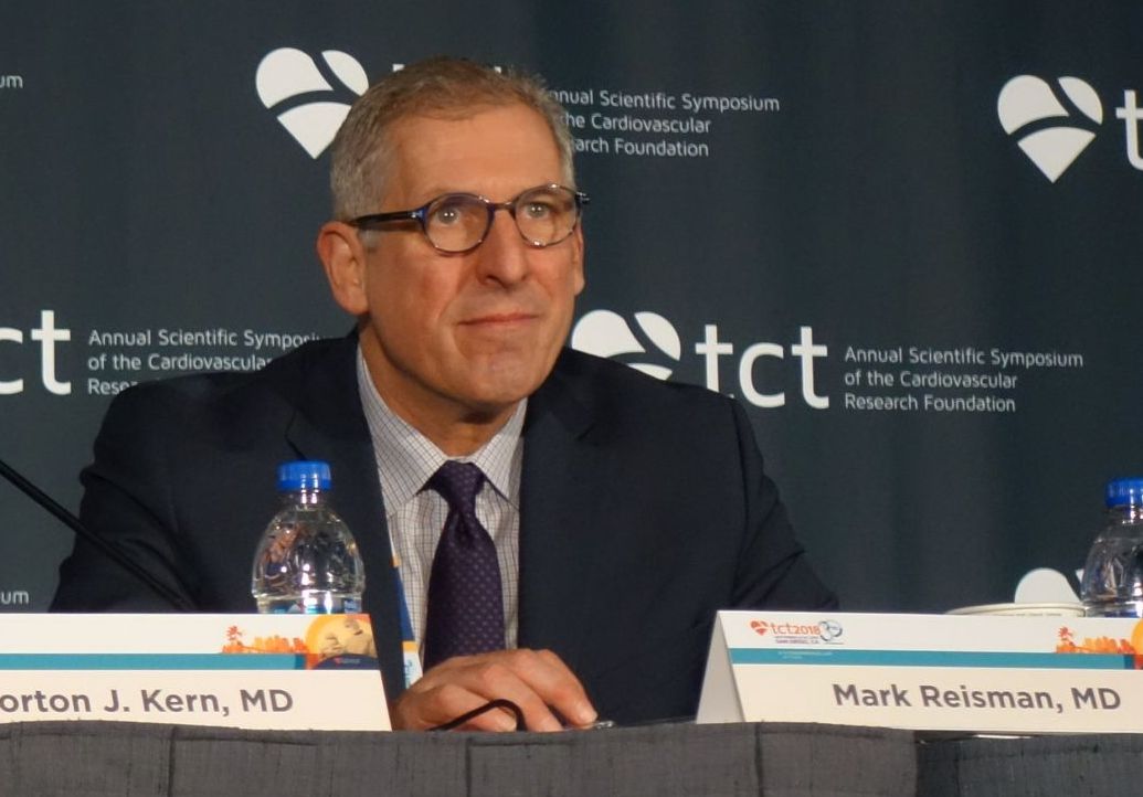
He said that he thought FFRangio could change practice, and several panelists agreed. “This is a real advance in the field. The use of FFR is not as great as it should be. I hope this will improve the ability of the assessor to identify ischemic lesions. I think that’s what’s going to happen. This is going to drive us forward,” said Mark Reisman, MD, head of cardiology and director of the Center for Emerging Cardiovascular Therapies at the University of Washington, Seattle.
The FAST-FFR (FFRangio Accuracy versus Standard FFR) study was conducted at 10 centers in the United States, Europe, and Israel; FFRangio was used to obtain FFRs in 319 vessels among 301 patients; the results were checked against FFR measured by wire. FFRangio operators were blinded to wire results.
The mean FFR was 0.81, and 43% of vessels had an FFR of 0.80 or less, signaling a possible abnormality. The diagnostic accuracy of FFRangio against wire measurement was 92%, and 87% when only gray-zone values between 0.75-0.85 were considered. Correlation with wire measurements was good (r = 0.80; P less than 0.001). Mismatches with the wire were more likely in the right coronary artery. The results were published online at the time of the presentation at TCT, sponsored by the Cardiovascular Research Foundation (Circulation. 2018 Sept 24. doi: 10.1161/circulationaha.118.037350).
“The main issue with this is that we need to do optimal angiographies. We should be doing them on a routine basis, but oftentimes, cardiologists want to be quick; they get lazy. But you do need to fill the entire vessel with contrast,” Dr. Fearon said.
“One of the nice things is you can rotate” the 3-D coronary artery tree reconstruction. “You can get a better idea of the relationship between the lesion and side branches, the length of the lesion, and a lot of additional information you don’t have on the angiogram [alone]. Then, you can pull back the cursor and measure FFR all along the vessel and different branches, all based on one angiography,” he said.
The majority of patients in the study were overweight or obese with complex coronary anatomy, as in daily practice. The investigation was limited to lesions amenable to wire measurement, so it didn’t include left main disease, low ejection fraction, and in-stent restenosis, although assessment may be possible.
Dr. Fearon didn’t know how much FFRangio will cost if it’s cleared or how CathWorks will be made available.
The work was funded by CathWorks. Dr. Fearon disclosed institutional research support from the company. One of the investigators was a cofounder of the company, with shares and intellectual property rights. The TCT meeting is sponsored by the Cardiovascular Research Foundation.
REPORTING FROM TCT 2018
Key clinical point:
Major finding: FFRangio, a less-invasive way to measure fractional flow reserve using angiograms, had a sensitivity of 94% and specificity of 91%, compared with standard, wire-based techniques.
Study details: The prospective, multicenter FAST-FFR compared FFRangio with pressure wire–derived fractional flow reserve in 301 subjects.
Disclosures: The work was funded by FFRangio maker, CathWorks. Dr. Fearon disclosed institutional research support from the company. One of the authors was a cofounder of the company, with shares and intellectual property rights.
IVUS guidance cuts target-vessel failure risk by 47%
SAN DIEGO – An all-comers trial conducted in China confirms that intravascular ultrasound (IVUS)–guided implantation of drug-eluting stents (DES) resulted in better clinical outcomes than did angiography-guided procedures, Jun-Jie Zhang, MD, said in reporting results of the ULTIMATE trial at the Transcatheter Cardiovascular Therapeutics annual meeting.
The ULTIMATE trial is the latest in a line of evidence showing the utility of IVUS guidance, though uptake of the procedure is not high in the United States or Europe. Its broader focus also rounds out the findings of earlier studies, which focused on patients with more complex lesions.
“How can people continue to ignore the importance of imaging-guided stent optimization? Even with second-generation DES, the results are consistent across the studies. This is just another piece of irrefutable evidence,” said discussant Gary Mintz, MD.
That sentiment was generally echoed by the rest of the panel. John McB Hodgson, MD, a professor of medicine at MetroHealth Medical Center in Cleveland, also pointed out the consistency of the body of evidence supporting the use of imaging. The study also represented a variety of cases, and the angiography arm of the study showed that the procedure was performed to a high standard. “It shows that even with a good angiographic approach, IVUS still wins. I’m amazed that there’s still some resistance to image guidance,” said Dr. Hodgson.
In ULTIMATE, 1,448 all-comer patients were randomized to either IVUS-guided or angiography-guided DES implantation. Patients with a life-expectancy shorter than 12 months, who were intolerant of dual-antiplatelet therapy, or who had severe calcification needing rotational atherectomy were excluded.
The procedural time was longer in the IVUS group, at 61 min, compared with 45 min in the angiography group, and the contrast volume was higher, at 178 mL and 162 mL, respectively (P less than .001 for both). At 30 days, the incidence of target vessel failure (TVF) was 0.8% in the IVUS group and 1.9% in the angiographic group, though this difference did not reach statistical significance (P = .08). The trend did reach significance at 1 year, with failure occurring in 2.9% of IVUS patients and 5.4% of angiography patients (P = .019). The hazard ratio for TVF in the IVUS group was 0.53 (95% confidence interval, 0.312-0.901), for a relative risk reduction of 47%, reported Dr. Zhang of Nanjing (China) Medical University.
Patients also underwent a postprocedure IVUS assessment to determine whether the stent was deployed optimally or suboptimally. The criteria for optimal deployment included minimal lumen area in the stented segment at least 5.0 mm2, or 90% of the minimal lumen area at distal reference segment meeting that criteria; a less than 50% plaque burden at the 5 mm of vessel proximal or distal to the stent edge; and no edge dissection involving media greater than 3 mm in length.
More than half (53%) of patients in the IVUS group had optimal placement, and 47% did not. Significantly fewer (1.6%) of patients with optimal IVUS experienced TVF at 12 months, compared with 4.4% of the suboptimal group (HR, 0.35; 95% CI, 0.135-0.898). The results were published online simultaneously with the presentation in the Journal of the American College of Cardiology.
“I’m particularly impressed by the analysis of the optimal versus nonoptimal group. If you don’t use IVUS correctly, you don’t get a benefit. The ones who did not get optimal stenting were very similar to the angiographic group,” said Dr. Mintz, chief medical officer at the Cardiovascular Research Foundation, which sponsored the meeting.
ULTIMATE was funded by the National Science Foundation of China and several other research organizations. Dr. Zhang had no relevant disclosures. Dr. Mintz has received research support and/or consulting fees from Abbott Vascular, Boston Scientific, Volcano, and Infraredx. Dr. Hodgson has received research support and consulted for Volcano.
REPORTING FROM TCT 2018
Key clinical point:
Major finding: IVUS guidance reduced target vessel failure by 47%.
Study details: ULTIMATE, a randomized, controlled trial of 1,448 patients.
Disclosures: ULTIMATE was funded by the National Science Foundation of China and several other research organizations. Dr. Zhang had no relevant disclosures. Dr. Mintz has received research support from Abbott Vascular and Boston Scientific. He has been a consultant for Boston Scientific, Volcano, and Infraredx. Dr. Hodgson has received research support and consulted for Volcano.
Rotablation aids in prepping highly calcified lesions: PREPARE-CALC
SAN DIEGO – When it comes to preparing highly calcified coronary lesions for drug-eluting stents, rotational atherectomy has an early procedural advantage over modified balloons that cut or score the lesion, but yields similar clinical outcomes over time, according to results of the randomized PREPARE-CALC trial.
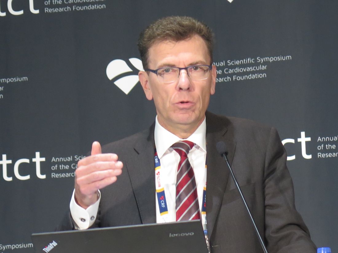
“Because severe calcification is mostly excluded from randomized PCI [percutaneous coronary intervention] trials, there is poor evidence for best PCI practice,” commented presenter Gert Richardt, MD, PhD, chief of cardiology and angiology at the Heart Center Segeberger Kliniken, Bad Segeberg, Germany. “Most interventional cardiologists will agree it is essential to facilitate stent delivery and adequate stent expansion in severely calcified lesions.”
Compared with standard balloon preparation, rotational atherectomy (rotablation) achieves a higher stenting success rate and better acute luminal gain, but its stimulatory effect leads to neointima formation that translates to late lumen loss, he noted. New balloons and drug-eluting stents may alter that calculus, however.
The 200 patients in PREPARE-CALC were randomized evenly to undergo preparation of their severely calcified coronary lesions with rotational atherectomy or a modified cutting/scoring balloon, each followed by placement of drug-eluting stents.
Compared with modified balloons, rotational atherectomy yielded a higher rate of successful stent delivery and expansion, according to data reported at the Transcatheter Cardiovascular Therapeutics annual meeting and simultaneously published online (Circ Cardiovasc Interv. 2018;11:e007415). The two methods did not differ significantly with respect to the 9-month magnitude of restenosis or rate of target vessel failure, at 6% with rotational atherectomy and 8% with modified balloons.
Importantly, about one in six patients in the modified balloon group had to cross over to rotational atherectomy because the balloon could not pass or sufficiently dilate the lesion.
“In patients with severely calcified coronary lesions, elective rotablation is feasible in nearly all patients, and the acute success rate is superior to modified balloons. Both approaches, elective rotational atherectomy and balloon plus bailout rotational atherectomy, are equally safe and effective,” Dr. Richardt summarized at the meeting sponsored by the Cardiovascular Research Foundation. “Use of rotablation is no longer associated with excessive late lumen loss in the era of modern drug-eluting stents.”
An underused tool
“This is a little bit ‘back to the future’ for me,” commented press conference panelist Mark Reisman, MD, director of the Center for Emerging Cardiovascular Therapies and head of UW Cardiology, Seattle. The PREPARE-CALC results are not surprising, but low uptake of rotablation in Europe, at just 1%-2%, is surprising and unexplained.
Importantly, the trial allows a sound comparison of rotational atherectomy with the best available alternatives, he said. “This may significantly impact the behavior and maybe some of the reimbursement in Europe. ... I’m sure that drives a lot of the decisions: cost as well as technique.”
Many of the original rotational atherectomy studies used larger burrs and multiple burrs, whereas the trial investigators applied less aggressive, more refined parameters. It will be interesting to look at their technique as it may have contributed to the favorable findings, according to Dr. Reisman.
“Based on what we just heard, there are very complex lesions that were maybe not being approached historically, which now people will feel a lot more confident in approaching, looking at the durable outcome with the Rotablator [Boston Scientific],” he summarized.
“From the practice of a clinician, it’s very frustrating to hit a calcified lesion and attempt to work on it up front and find you can’t cross it,” commented press conference panelist Morton J. Kern, MD, professor of medicine at the school of medicine and chief of medicine, VA Long Beach Health Care System, University of California, Irvine Medical Center. The high rate of crossover to rotablation in PREPARE-CALC speaks to this problem. Having to resort to this tool after failure of other lesional interventions often translates to a rockier overall procedure and clinical course.
“Lesion preparation is underestimated. I know from my own experience that [in] those calcified long lesions where I didn’t use the Rotablator, I regretted it in a fair number of cases,” Dr. Kern said. “So my view is that we probably don’t use it enough. This trial suggests that you can get away [without it], but I think ultimately, we are going to need this tool, even though it’s not used in a huge number of patients.”
Trial details
Patients enrolled in PREPARE-CALC had severely calcified coronary lesions according to angiographic criteria. For lesion preparation, half underwent high-speed rotational atherectomy (Rotablator), and half underwent treatment with either a cutting balloon (Flextome, Boston Scientific) or a scoring balloon (AngioSculpt, AngioScore, or Scoreflex, OrbusNeich Medical). Thereafter, all received sirolimus-eluting stents (Orsiro, Biotronik).
Results reported at the meeting, which is sponsored by the Cardiovascular Research Foundation, showed that, compared with the modified balloons, rotational atherectomy yielded a higher rate of strategy success, defined as successful stent delivery and expansion with less than 20% in-stent residual stenosis and TIMI 3 flow, without crossover to the other arm or stent failure. The rotational atherectomy patients had a success rate of 98%, compared with 81% for the balloon patients (P = .0001).
“This difference was mainly driven by a high rate of crossovers from balloon to rotablation,” Dr. Richardt, noted, with 16% of patients assigned to the former modality ultimately receiving the latter. “These crossovers were due to noncrossable or nondilated lesions.”
Analyses suggested three subgroups did not benefit from rotational atherectomy when it came to strategy success: women, patients in whom the left anterior descending artery was the target, and patients not having type C lesions.
Quantitative coronary analysis showed that the acute lumen gain, whether in-stent or in-segment, did not differ significantly between the two preparation methods. “This is different from what we know from historical studies where the Rotablator was always achieving more acute gain than standard balloons,” Dr. Richardt commented.
Rotational atherectomy and modified balloons were similar with respect to the magnitude of in-stent late lumen loss (0.22 vs. 0.16 mm, P = .021 for superiority; P = .01 for noninferiority) and the rate of target-vessel failure (6% vs. 8%) at 9 months. Those failure rates were lower than the 10% expected from previous trials, he pointed out. “This might increase over time, but it’s not so far away from what we see in contemporary drug-eluting stent trials.”
Fluoroscopy time was about 4 minutes longer with rotational atherectomy than with modified balloons. “You may say that’s a lot. I would say in these 75-year-old patients, it is acceptable,” Dr. Richardt commented. “But to be honest, we had some cases where we had to place a pacemaker, and this increases the duration of the fluoroscopy time.”
The PREPARE-CALC findings should improve the rate of rotational atherectomy use in Europe, he said. “If you believe my data, then the rate should go up to 10% or 12%.”
Dr. Richardt disclosed that he receives speaker’s honoraria from Boston Scientific and Biotronik. The trial was sponsored by the Herz-Kreislauf-Zentrum Segeberger Kliniken GmbH.
SAN DIEGO – When it comes to preparing highly calcified coronary lesions for drug-eluting stents, rotational atherectomy has an early procedural advantage over modified balloons that cut or score the lesion, but yields similar clinical outcomes over time, according to results of the randomized PREPARE-CALC trial.
“Because severe calcification is mostly excluded from randomized PCI [percutaneous coronary intervention] trials, there is poor evidence for best PCI practice,” commented presenter Gert Richardt, MD, PhD, chief of cardiology and angiology at the Heart Center Segeberger Kliniken, Bad Segeberg, Germany. “Most interventional cardiologists will agree it is essential to facilitate stent delivery and adequate stent expansion in severely calcified lesions.”
Compared with standard balloon preparation, rotational atherectomy (rotablation) achieves a higher stenting success rate and better acute luminal gain, but its stimulatory effect leads to neointima formation that translates to late lumen loss, he noted. New balloons and drug-eluting stents may alter that calculus, however.
The 200 patients in PREPARE-CALC were randomized evenly to undergo preparation of their severely calcified coronary lesions with rotational atherectomy or a modified cutting/scoring balloon, each followed by placement of drug-eluting stents.
Compared with modified balloons, rotational atherectomy yielded a higher rate of successful stent delivery and expansion, according to data reported at the Transcatheter Cardiovascular Therapeutics annual meeting and simultaneously published online (Circ Cardiovasc Interv. 2018;11:e007415). The two methods did not differ significantly with respect to the 9-month magnitude of restenosis or rate of target vessel failure, at 6% with rotational atherectomy and 8% with modified balloons.
Importantly, about one in six patients in the modified balloon group had to cross over to rotational atherectomy because the balloon could not pass or sufficiently dilate the lesion.
“In patients with severely calcified coronary lesions, elective rotablation is feasible in nearly all patients, and the acute success rate is superior to modified balloons. Both approaches, elective rotational atherectomy and balloon plus bailout rotational atherectomy, are equally safe and effective,” Dr. Richardt summarized at the meeting sponsored by the Cardiovascular Research Foundation. “Use of rotablation is no longer associated with excessive late lumen loss in the era of modern drug-eluting stents.”
An underused tool
“This is a little bit ‘back to the future’ for me,” commented press conference panelist Mark Reisman, MD, director of the Center for Emerging Cardiovascular Therapies and head of UW Cardiology, Seattle. The PREPARE-CALC results are not surprising, but low uptake of rotablation in Europe, at just 1%-2%, is surprising and unexplained.
Importantly, the trial allows a sound comparison of rotational atherectomy with the best available alternatives, he said. “This may significantly impact the behavior and maybe some of the reimbursement in Europe. ... I’m sure that drives a lot of the decisions: cost as well as technique.”
Many of the original rotational atherectomy studies used larger burrs and multiple burrs, whereas the trial investigators applied less aggressive, more refined parameters. It will be interesting to look at their technique as it may have contributed to the favorable findings, according to Dr. Reisman.
“Based on what we just heard, there are very complex lesions that were maybe not being approached historically, which now people will feel a lot more confident in approaching, looking at the durable outcome with the Rotablator [Boston Scientific],” he summarized.
“From the practice of a clinician, it’s very frustrating to hit a calcified lesion and attempt to work on it up front and find you can’t cross it,” commented press conference panelist Morton J. Kern, MD, professor of medicine at the school of medicine and chief of medicine, VA Long Beach Health Care System, University of California, Irvine Medical Center. The high rate of crossover to rotablation in PREPARE-CALC speaks to this problem. Having to resort to this tool after failure of other lesional interventions often translates to a rockier overall procedure and clinical course.
“Lesion preparation is underestimated. I know from my own experience that [in] those calcified long lesions where I didn’t use the Rotablator, I regretted it in a fair number of cases,” Dr. Kern said. “So my view is that we probably don’t use it enough. This trial suggests that you can get away [without it], but I think ultimately, we are going to need this tool, even though it’s not used in a huge number of patients.”
Trial details
Patients enrolled in PREPARE-CALC had severely calcified coronary lesions according to angiographic criteria. For lesion preparation, half underwent high-speed rotational atherectomy (Rotablator), and half underwent treatment with either a cutting balloon (Flextome, Boston Scientific) or a scoring balloon (AngioSculpt, AngioScore, or Scoreflex, OrbusNeich Medical). Thereafter, all received sirolimus-eluting stents (Orsiro, Biotronik).
Results reported at the meeting, which is sponsored by the Cardiovascular Research Foundation, showed that, compared with the modified balloons, rotational atherectomy yielded a higher rate of strategy success, defined as successful stent delivery and expansion with less than 20% in-stent residual stenosis and TIMI 3 flow, without crossover to the other arm or stent failure. The rotational atherectomy patients had a success rate of 98%, compared with 81% for the balloon patients (P = .0001).
“This difference was mainly driven by a high rate of crossovers from balloon to rotablation,” Dr. Richardt, noted, with 16% of patients assigned to the former modality ultimately receiving the latter. “These crossovers were due to noncrossable or nondilated lesions.”
Analyses suggested three subgroups did not benefit from rotational atherectomy when it came to strategy success: women, patients in whom the left anterior descending artery was the target, and patients not having type C lesions.
Quantitative coronary analysis showed that the acute lumen gain, whether in-stent or in-segment, did not differ significantly between the two preparation methods. “This is different from what we know from historical studies where the Rotablator was always achieving more acute gain than standard balloons,” Dr. Richardt commented.
Rotational atherectomy and modified balloons were similar with respect to the magnitude of in-stent late lumen loss (0.22 vs. 0.16 mm, P = .021 for superiority; P = .01 for noninferiority) and the rate of target-vessel failure (6% vs. 8%) at 9 months. Those failure rates were lower than the 10% expected from previous trials, he pointed out. “This might increase over time, but it’s not so far away from what we see in contemporary drug-eluting stent trials.”
Fluoroscopy time was about 4 minutes longer with rotational atherectomy than with modified balloons. “You may say that’s a lot. I would say in these 75-year-old patients, it is acceptable,” Dr. Richardt commented. “But to be honest, we had some cases where we had to place a pacemaker, and this increases the duration of the fluoroscopy time.”
The PREPARE-CALC findings should improve the rate of rotational atherectomy use in Europe, he said. “If you believe my data, then the rate should go up to 10% or 12%.”
Dr. Richardt disclosed that he receives speaker’s honoraria from Boston Scientific and Biotronik. The trial was sponsored by the Herz-Kreislauf-Zentrum Segeberger Kliniken GmbH.
REPORTING FROM TCT 2018
Key clinical point:
Major finding: Rotational atherectomy preparation and cutting/scoring balloon preparation did not differ significantly with respect to 9-month in-stent late lumen loss (0.22 vs. 0.16 mm) and target vessel failure (6% vs. 8%).
Study details: PREPARE-CALC, an open-label, randomized, controlled trial in 200 patients with severely calcified coronary lesions undergoing implantation of drug-eluting stents.
Disclosures: Dr. Richardt disclosed that he receives speaker’s honoraria from Boston Scientific and Biotronik. The trial was sponsored by the Herz-Kreislauf-Zentrum Segeberger Kliniken GmbH.
Eluvia drug-eluting vascular stent approved on heels of TCT presentation
Boston Scientific announced the was presented at the Transcatheter Cardiovascular Therapeutics annual meeting in San Diego.

The Eluvia Drug-Eluting Vascular Stent System is designed to restore blood flow in the peripheral arteries above the knee – specifically, the superficial femoral artery and proximal popliteal artery, according to the company.
Lead investigator William A. Gray, MD, a cardiologist at the Lankenau Heart Institute, Wynnewood, Pa., presented the results of IMPERIAL at TCT 2018. The randomized, head-to-head trial compared the performance of Eluvia with the paclitaxel-eluting Zilver PTX, which at the time was the only drug-releasing stent approved in the United States for the indication. Patients had occlusive lesions of the superficial femoral and/or proximal popliteal arteries.
At 12 months, primary patency was 86.8% in the Eluvia group versus 81.5% among those randomized to Zilver PTX, meeting the threshold for noninferiority.
For the endpoint of target lesion revascularization, Eluvia was superior to Zilver PTX, with 4.5% and 9% of patients, respectively, requiring it (Lancet. 2018 Sep 22. doi: 10.1016/S0140-6736[18]32262-1).
According to Boston Scientific’s announcement of the approval, the drug-polymer combination used in the Eluvia stent offers sustained release of paclitaxel over 1 year to prevent tissue regrowth.
Eluvia was approved in Europe in early 2016.
Boston Scientific announced the was presented at the Transcatheter Cardiovascular Therapeutics annual meeting in San Diego.
The Eluvia Drug-Eluting Vascular Stent System is designed to restore blood flow in the peripheral arteries above the knee – specifically, the superficial femoral artery and proximal popliteal artery, according to the company.
Lead investigator William A. Gray, MD, a cardiologist at the Lankenau Heart Institute, Wynnewood, Pa., presented the results of IMPERIAL at TCT 2018. The randomized, head-to-head trial compared the performance of Eluvia with the paclitaxel-eluting Zilver PTX, which at the time was the only drug-releasing stent approved in the United States for the indication. Patients had occlusive lesions of the superficial femoral and/or proximal popliteal arteries.
At 12 months, primary patency was 86.8% in the Eluvia group versus 81.5% among those randomized to Zilver PTX, meeting the threshold for noninferiority.
For the endpoint of target lesion revascularization, Eluvia was superior to Zilver PTX, with 4.5% and 9% of patients, respectively, requiring it (Lancet. 2018 Sep 22. doi: 10.1016/S0140-6736[18]32262-1).
According to Boston Scientific’s announcement of the approval, the drug-polymer combination used in the Eluvia stent offers sustained release of paclitaxel over 1 year to prevent tissue regrowth.
Eluvia was approved in Europe in early 2016.
Trump administration proposes weakening rules governing organ transplant centers
This article was produced in collaboration with the Houston Chronicle.
The Trump administration on Sept. 17 proposed eliminating a decade-old regulation that puts hospitals at risk of losing their Medicare funding if too many of their patients die or suffer organ failure after receiving transplants.
The rule the government is proposing to scrap is the same one that led the Centers for Medicare & Medicaid Services to cut off funding in August for heart transplants at Baylor St. Luke’s Medical Center in Houston after an investigation by ProPublica and the Houston Chronicle revealed an outsized number of patient deaths and complications in recent years.
The proposal was unveiled Sept. 17 as part of the White House’s push to “cut the red tape” and do away with “burdensome regulation” that officials said put paperwork ahead of patients. In a speech announcing the proposed changes, CMS Administrator Seema Verma said the agency’s existing policies have “put lives in danger.”
“We are proposing to remove those inefficiencies to reduce the amount of time patients have to wait, so that they can begin healing,” Ms. Verma said.
The proposal, now subject to public comment and revision before it is finalized, surprised many transplant physicians who have long called for relaxed federal oversight. They’ve argued that the rules requiring that hospitals meet certain survival thresholds for transplants discourage them from taking on risky patients or accepting less-than-perfect organs, lengthening the time patients spend on the waiting list.
The regulation was put in place in 2007 after a series of scandals at transplant programs revealed lax federal oversight. Several transplant programs had compiled abysmal patient survival statistics for years while continuing to receive Medicare funding.
Even though it has the authority to do so, Medicare rarely terminates programs for poor outcomes. It is far more common for the government to force underperforming programs into systems-improvement agreements, in which hospitals agree to make certain changes and be subject to stepped-up oversight.
Medicare bypassed that process and cut off funding for heart transplants at St. Luke’s in August after the hospital’s 1-year patient survival rate fell below national norms from 2014 to mid-2016. A few St. Luke’s cardiologists grew so concerned that they started sending some of their patients to other hospitals for transplants.
St. Luke’s has appealed its Medicare termination, saying, “we do not believe CMS’ recent decisions reflect our ongoing progress and accomplishments to improve the quality of our care.” A spokeswoman said officials are still reviewing the Medicare proposal and declined to comment.
In a statement to reporters Sept. 21, CMS said it would continue to monitoring transplant programs and is strengthening its process for inspecting transplant programs to ensure they provide safe, quality care.
“CMS will continue to collect the data on each transplant program’s performance with regards to patient and graft survival,” the agency said in the statement. “These data, rather than being a stand-alone measure, will be used as a component of the survey process to further inform and direct the survey.”
If the proposed regulatory change had been in place previously, it’s not clear whether St. Luke’s would have faced punitive action from Medicare.
“I am probably in the minority in the transplant community, but I think, based on what is proposed, this is a bad idea,” said Laura J. Aguiar, an Arizona-based transplant consultant who has spent years helping programs improve their outcomes to stave off Medicare penalties. “I have been around long enough to remember that there were very valid reasons why CMS, in the George W. Bush administration, took the steps they took in implementing all of this.”
Since the rules were put in place 11 years ago, the percentage of patients who survive at least 1 year after receiving heart, kidney, lung, and other organ transplants has increased nationally. But some experts say those gains have come at a cost.
Jesse D. Schold, PhD, a researcher in quantitative health sciences at the Cleveland Clinic, has spent years chronicling what he calls the “unintended consequences” of holding transplant programs accountable for poor outcomes. Even though CMS relies on data that has been adjusted to ensure that programs aren’t punished for treating sicker patients or accepting riskier donor organs, Dr. Schold said the rules have created a perception that programs need to turn away some ailing patients and reject less-than-ideal organs in order to meet outcome targets.
As a result, Dr. Schold said, some potentially viable donor organs are discarded while thousands of patients die on waiting lists each year. Dr. Schold said he was surprised to learn a study he coauthored was among the research cited by CMS as justification for the policy change, which he said would be a “significant net positive” for patients.
“I don’t want anything to in any way imply that I’m a fan of the current administration,” Dr. Schold said, referencing the Trump administration’s aggressive and often controversial efforts to slash regulations. “However, in this case, I think it is something that is beneficial.”
Joel Adler, MD, a liver surgeon at the University of Wisconsin–Madison, whose research also was cited by CMS, said a major weakness of the current regulation is that it focuses only on the survival of patients fortunate enough to have received a transplant. Medicare, he said, does not take into account the percentage of patients who die awaiting a transplant. That can discourage programs from accepting organs for patients who might be less likely to survive afterward.
Despite identifying problems with the current rules, Dr. Adler and his coauthor did not propose eliminating Medicare’s standards, but they instead suggested ways to improve outcome measures and create incentives for programs to perform more, not fewer, transplants.
“Should we get rid of (the regulation) entirely?” Dr. Adler asked. “I don’t know. I suspect we’ll swing back to somewhere in the middle, because there has to be some mechanism of capturing things when they are really, really bad. That has to exist.”
Medicare isn’t the only organization that oversees transplant centers. The United Network for Organ Sharing, a federal contractor that operates the national waiting list for organs, can put programs on probation or even revoke their good standing for continued poor results, though it rarely takes such actions. Similarly, another federal contractor, the Scientific Registry of Transplant Recipients, analyzes transplant outcomes and publicly reports which centers have mortality rates that are higher than expected based on the characteristics of their patients.
Even if the proposal is approved, it would not mean CMS would stop regulating transplant programs. Last year, for example, CMS cut off funding to the Medical University Hospital in Charleston, South Carolina, after the program failed to perform the minimum number of heart transplants required by CMS to maintain certification. That provision, also added during the reforms of 2007, would not be affected by the changes proposed Sept. 17.
The Charleston hospital, South Carolina’s only heart transplant program, made necessary changes and regained Medicare certification this year.
Alexander Aussi, a San Antonio–based consultant who specializes in helping transplant programs satisfy regulatory requirements, said he understands the desire to reduce the number of rules and regulations that transplant centers must follow. But he said he fears that the CMS proposal would return the transplant field to an earlier era when “some programs were cowboyish about their outcomes.”
“I cannot tell you in good conscience that if you remove those guidelines and metrics … that you’re going to have better outcomes,” Mr. Aussi said. “On the contrary, I believe you’re going to have a lot of transplant programs reversing course.”
Ms. Aguiar, the Arizona-based consultant, shares those concerns. Even without strict CMS oversight, she said, many transplant programs will continue providing high-quality care; but some won’t.
“There will be others that will take the removal of these requirements as a blessing to go back to the bad old days,” she said, “and it is patients who will end up paying the price for it.”
Mike Hixenbaugh is an investigative reporter at the Houston Chronicle. Email him at mike.hixenbaugh@chron.com and follow him on Twitter at @Mike_Hixenbaugh. Charles Ornstein is a senior editor at ProPublica, overseeing the Local Reporting Network. Follow him on Twitter at @charlesornstein.