User login
Immunotoxin could treat B-cell malignancies, team says
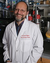
Photo courtesy of the
University of Minnesota
A bispecific ligand-directed diphtheria toxin known as DT2219 shows promise for treating patients with relapsed/refractory B-cell malignancies, according to researchers.
DT2219 produced responses in 2 of 25 patients analyzed in a phase 1 study. The maximum tolerated dose of DT2219 was not reached, although 2 patients experienced dose-limiting toxicities.
“In this phase 1 trial, we found a safe dose of the drug that has biological activity,” said Daniel Vallera, PhD, of the University of Minnesota in Minneapolis.
“We are planning a phase 2 trial with this drug. It will focus on giving more cycles of treatment, which we believe will dramatically enhance the response rates.”
Dr Vallera and his colleagues detailed the phase 1 results in Clinical Cancer Research.
To develop DT2219, the researchers chose 2 antibody fragments that each selectively bind to CD19 and CD22. They used genetic engineering to attach these two antibodies to the bacterial diphtheria toxin.
When the antibody fragments bind to the two targets on the cancer cell, the entire drug enters the cell, and the toxin kills the cell.
To test DT2219, the researchers enrolled 25 patients with chemo-refractory pre-B acute lymphoblastic leukemia (n=10), chronic lymphocytic leukemia (n=5), or non-Hodgkin lymphoma (n=10). All tumors had CD19 and/or CD22 proteins.
Patients had received a median of 3 prior therapies (range, 2 to 5), and 8 patients had undergone an unsuccessful stem cell transplant (5 autologous and 3 allogeneic).
Patients received DT2219 intravenously over 2 hours every other day for 4 total doses. The dose was escalated from 0.5 μg/kg/day to 80 μg/kg/day in 9 dose cohorts until a dose-limiting toxicity occurred.
All but 1 patient received a single course of DT2219. That patient received a second, 4-dose course after attaining a partial response.
Outcomes
The 12 patients who received doses ranging from 0.5 mg/kg/day to 20 mg/kg/day had minimal or no adverse events (AEs). But all 13 patients who received 4 doses of DT2219 at 40 mg/kg or greater every other day experienced treatment-related AEs.
Grade 1-2 treatment-related AEs included capillary leak syndrome (n=7), ALT/AST elevation (n=4), fatigue (n=3), fever (n=3), hypokalemia (n=2), hypoalbuminemia (n=1), hearing loss (n=1), hypocalcemia (n=1), anemia (n=1), and vomiting (n=1).
Grade 3-4 treatment-related AEs were thrombocytopenia (n=2), neutropenia (n=1), neutropenic fever (n=1), capillary leak syndrome (n=1), hypokalemia (n=1), and leg weakness (n=1). The grade 3 leg weakness and grade 3 capillary leak syndrome were dose-limiting toxicities.
The maximum tolerated dose was not reached, but clinical responses occurred between doses of 40 to 80 µg/kg.
All 25 patients were evaluable for response, but only 9 patients in the highest dose cohorts had measurable drug levels.
Two patients had durable, objective responses. One patient had chronic lymphocytic leukemia, and the other had diffuse large B-cell lymphoma. The latter patient’s response was a complete remission that occurred after 2 treatment cycles.
“We were surprised that the drug was effective enough to entirely eliminate the cancer in one of our patients,” Dr Vallera said. “Further, we expected the patients to make antibodies against the bacterial toxin and, thus, reject our drug. Surprisingly, this did not occur in the majority of our patients [70%].”
“We need to study more patients to understand why they did not produce neutralizing antibodies. However, we also have been working to create a less immunogenic form of the toxin for the next-generation drug.”
“Another important fact about our drug is that it was home-grown, meaning there was no commercial partner, which is rare. The drug was funded mostly with private donations, including individuals that have lost loved ones to cancer.” ![]()
Drug incompatible with certain devices, FDA warns
The US Food and Drug Administration (FDA) is warning healthcare professionals not to use Treanda (bendamustine hydrochloride) solution with closed-system transfer devices (CSTD), adapters, and syringes containing polycarbonate or acrylonitrile-butadiene-styrene (ABS).
Most marketed CSTDs contain either polycarbonate or ABS. And these materials dissolve when they come into contact with N, N-dimethylacetamide (DMA), an ingredient in Treanda solution.
This can lead to device failure, possible product contamination, and potential serious adverse health consequences, including skin reactions in healthcare professionals preparing and administering this product and the risk of small blood vessel blockage in patients.
Discovering the incompatibility
Treanda, which is manufactured by Teva, is used to treat patients with chronic lymphocytic leukemia and indolent B-cell non-Hodgkin lymphoma that has progressed during or within 6 months of treatment with rituximab or a rituximab-containing regimen.
Treanda is available as a solution—Treanda Injection (45 mg/0.5 mL or 180 mg/2 mL solution)—and a lyophilized powder—Treanda for Injection (25mg/vial or 100 mg/vial lyophilized powder).
The incompatibility of DMA with polycarbonate and ABS is only an issue with Treanda solution—not the lyophilized powder.
Since December 2014, Teva has received 40 complaints of the incompatibility issue, which was recently brought to the FDA’s attention. The agency also received a notification of device incompatibility with Treanda solution from a pharmacist.
These incompatibility issues included leaking of the CSTD, breaking or operational failure of the CSTD components, and a cloudy appearance or presence of particulate matter in the intravenous bag after dilution. To date, no adverse events have been reported related to the incompatibility.
FDA recommendations
The FDA has required label changes for both the solution and the powder formulations of Treanda to reflect the following safe preparation information.
The agency is recommending that healthcare professionals use Treanda solution only with polypropylene syringes containing a metal needle and a polypropylene hub. Polypropylene syringes are translucent in appearance.
Treanda solution should be inspected visually for particulate matter and discoloration prior to administration whenever the solution and container permit. The solution must be withdrawn and transferred for dilution in a biosafety cabinet or containment isolator.
If they aim to use a CSTD with Treanda solution, healthcare professionals should verify with the CSTD manufacturer or Teva US Medical Information (1-800-896-5855) that the CSTD is compatible with Treanda solution before preparing the drug.
Alternatively, healthcare professionals can use Treanda lyophilized powder with a CSTD. The solution and lyophilized powder formulations of Treanda should not be mixed.
For additional details on safe preparation of Treanda solution and lyophilized powder, see Teva’s Dear Health Care Provider letter.
Adverse events or quality problems associated with the use of Treanda products can be reported to the FDA’s MedWatch Adverse Event Reporting Program. ![]()
Group identifies new subtype of ALL

Photo by Steven Harbour
Researchers say they have discovered a new subtype of acute lymphoblastic leukemia (ALL) that is sensitive to drugs already approved to treat other hematologic malignancies.
The team uncovered cases of ALL that were dependent upon tonic pre-B-cell-receptor (BCR) signaling and therefore sensitive to drugs that inhibit tyrosine kinases downstream of the pre-BCR.
The group also developed a test that can identify patients with this subtype of ALL.
“We hope patients in this newly identified subset can be treated with these targeted drugs, . . . which are powerfully effective in the mouse experiments we have conducted on ALL,” said Markus Müschen, MD, PhD, of the University of California, San Francisco.
Dr Müschen and his colleagues described this work in Cancer Cell.
The researchers studied samples from 830 patients enrolled in 4 ongoing ALL trials and found tonic pre-BCR signaling in 112 patients (13.5%). Virtually all of the bone marrow slices from these patients showed “beautiful staining” of BCL6 expression, Dr Müschen said. (Two of the patients had weak staining.)
On the other hand, no BCL6 staining was observed in patients lacking pre-BCR signaling. These results suggest that BCL6 is a biomarker for pre-BCR signaling. And by testing patients for BCL6, we may be able to identify those who will respond to treatment with pre-BCR signaling inhibitors, the researchers said.
The team tested a range of pre-BCR signaling inhibitors in vitro. And they found a few compounds that were effective against pre-BCR+ ALL—the SYK inhibitor PRT062607, the BTK inhibitor ibrutinib, the SRC inhibitor dasatinib, and the PIK3δ inhibitor idelalisib.
Subsequent experiments revealed that dasatinib had the strongest antileukemic effect, so the researchers tested the drug in mouse models of pre-BCR+ ALL. Dasatinib significantly delayed leukemic expansion and prolonged overall survival in some mice, while completely eradicating the disease in other mice.
Dr Müschen said that dasatinib and other pre-BCR signaling inhibitors may be able to reduce the amount of conventional chemotherapy given to patients with pre-BCR+ ALL, or even replace chemotherapy altogether.
“In our experiments with mice, both combination therapy with low-dose chemotherapy and single-agent targeted therapy each worked very well,” he said. ![]()
New test can better predict cytokine storm, team says

Photo by Juan D. Alfonso
Scientists have developed a test that uses cells from a single donor’s blood to predict whether a new drug will cause a cytokine storm.
The group says this is an improvement over current tests, which use endothelial cells and peripheral blood mononuclear cells (PBMCs) from two separate donors and can therefore produce inaccurate results.
Furthermore, current tests cannot differentiate drugs that induce a mild cytokine storm from those that induce a severe one, but the new test can.
Jane Mitchell, PhD, of the National Heart and Lung Institute at Imperial College London in the UK, and her colleagues described the new test in The FASEB Journal.
Current tests for cytokine storm reactions use endothelial cells taken from the vessels of one donor and PBMCs from a different donor because endothelial cells are normally only grown from tissue removed in surgery or post-mortem, or from umbilical vessels after birth.
When cells from two different donors are used, one may have an immune reaction to the other. And this can result in the test falsely showing a severe immune reaction to a drug that is safe.
Dr Mitchell and her colleagues say they have solved this problem by isolating stem cells from the blood of a volunteer and using them to grow endothelial cells in a dish. The team then added PBMCs to the donor’s own endothelial cells to recreate the unique conditions found in their blood vessels.
When the scientists added the immunomodulatory drug TGN1412, the mixture of cells released a cytokine storm, as would happen inside the human body.
Responses to other drugs were consistent with those observed in humans as well. There was a modest response to alemtuzumab (Campath) and no response to the control antibodies trastuzumab (Herceptin), bevacizumab (Avastin), and ofatumumab (Arzerra).
“As biological therapies become more mainstream, it’s more likely that drugs being tested on humans for the first time will have unexpected and potentially catastrophic effects,” Dr Mitchell said.
“We’ve used adult stem cell technology to develop a laboratory test that could prevent another disaster like the TGN1412 trial [in which 6 healthy young men developed multi-organ failure]. Drug companies have the technical capacity to start using this test now, but we’re working on developing an off-the-shelf kit, which will make it easy to use on a large scale.”
The team has collaborated with the National Institute for Biological Standards and Control to validate the test and are now working with the clinical trials company Quintiles to develop the technology further. ![]()
Similar outcomes from HSCT found with sibling and unrelated donors
Outcomes among 411 pediatric patients with acute lymphoblastic leukemia after hematopoietic stem-cell transplantation were similar from well-matched sibling and nonrelative donors, according to a study published online March 9 in the Journal of Clinical Oncology.
After a median follow up of 4.2 years, investigators found no significant differences in 4-year event-free survival, overall survival, or relapse incidence between 306 patients with transplantations from unrelated donors and 105 with sibling donors. However, nonrelapse mortality rates from matched unrelated donors (MUDs) was 0.10 ± 0.02 vs. 0.03 ± 0.02 from matched sibling donors (MSDs), Dr. Christina Peters, professor of pediatrics at St. Anna Children’s Hospital, Vienna, Austria, and associates reported.
Patients who received MSD-HSCT had significantly faster engraftment than did those who received MUD-HSCT (median time to neutrophil engraftment 17 days vs. 22 days, and 30-day cumulative incidence of 75% vs. 44%, respectively).
“Despite excellent outcomes of MUD-HSCT, our data indicate that MSD BM (bone marrow) transplantation remains superior, which is possibly a result of fewer severe infections. We speculate that this is influenced by the short and limited GVHD prophylaxis in this setting,” the investigators wrote (J. Clin. Oncol. 2015 March 9 [doi: 10.1200/JCO.2014.58.9747]).
Patients in the MUD-HSCT group had superior event-free and overall survival compared to previous studies of children with high-risk ALL, results which may have been influenced by the use of high-resolution HLA typing and the requirement that donors have 9/10 or 10/10 HLA matches. Over 70% of patients who lacked MSDs were matched with MUDs, and no outcome differences were observed in patients who received transplantations from 9/10 or 10/10 matches. Patients older than 2 years and in the absence of contraindications had conditioning by total-body irradiation (TBI) and etoposide.
“Our data demonstrate excellent EFS and OS, and low incidence of relapse in children with high-risk ALL after treatment with TBI and etoposide before allogeneic HSCT from HLA-matched siblings or well-matched unrelated donors. This large, prospective, multicenter trial suggest that MUD-HSCT could be a standard of care for patients with ALL who have a high risk of relapse and who lack MSDs,” the investigators concluded.
FROM JOURNAL OF CLINICAL ONCOLOGY
Key clinical point: Children with high-risk acute lymphoblastic leukemia who received stem cell transplants from matched sibling vs. well-matched unrelated donors had similar outcomes.
Major finding: The 4-year event-free survival rate for patients who received transplants from sibling donors was 0.71 ± 0.05 vs. 0.67 ± 0.03 for unrelated donors (P = .405).
Data source: The prospective study enrolled 411 children with high-risk acute lymphoblastic leukemia from 2003 to 2011; 105 received transplants from siblings and 306 from unrelated donors.
Disclosures: Dr. Peters reported consulting roles or research funding from Medac Pharma, EUSA Pharma, Pfizer, Amgen, Fresenius Biotech, Genzyme, Medac, and RIEMSER Pharma. Coauthors reported relationships and roles with several companies.
Aggressive infant leukemia has few mutations

Photo by Vera Kratochvil
Infants who have acute lymphoblastic leukemia (ALL) with MLL rearrangements have few other mutations, according to new research.
The findings suggest that targeting MLL rearrangements in these patients is likely the key to improving their survival.
“We frequently associate a cancer’s aggressiveness with its mutation rate, but this work indicates that the two don’t always go hand-in-hand,” said Richard K. Wilson, PhD, of the Washington University School of Medicine in St Louis, Missouri.
“Still, our findings provide a new direction for developing more effective treatments for these very young patients.”
Dr Wilson and his colleagues reported their findings in Nature Genetics.
The researchers performed whole-genome, exome, RNA, and targeted DNA sequencing to identify genetic alterations in 65 infants with ALL, including 47 with the MLL rearrangement.
The team was surprised to find that, despite being an aggressive leukemia, the MLL-rearranged subtype had among the lowest mutation rates reported for any cancer. The predominant leukemic clone carried a mean of 1.3 non-silent mutations.
“These results show that, to improve survival for patients with this aggressive leukemia, we need to develop drugs that target the abnormal proteins produced by the MLL fusion gene or that interact with the abnormal MLL fusion protein to shut down the cellular machinery that drives their tumors,” said James R. Downing, MD, of St Jude Research Hospital in Memphis, Tennessee. “That will not be easy, but this study found no obvious cooperating mutations to target.”
Almost half of infants with MLL-rearranged ALL (47%) had activating mutations in the kinase-PI3K-RAS signaling pathway. But the mutations were often present in only some of the leukemic cells.
Furthermore, the researchers analyzed leukemia cells in infants whose cancer returned after treatment and found that, at the time of relapse, the cells lacked these mutations.
“The fact that the mutations were often lost at relapse suggests that patients are unlikely to benefit from therapeutically targeting these mutations at diagnosis,” Dr Downing said.
The researchers also found that older children with MLL-rearranged leukemia had significantly more mutations than infants—a mean of 6.5 mutations per case (P=7.15 × 10⁻⁵).
Furthermore, 45% of the older children had mutations in genes that encode epigenetic regulatory proteins. And, aside from MLL, epigenetic regulators were rarely mutated in infants with MLL-rearranged ALL.
“While MLL belongs to a family of genes that encode epigenetic regulatory proteins, there was a striking difference between infants and older children regarding the frequency of mutations in other epigenetic regulatory genes,” said Anna Andersson, PhD, of Lund University in Sweden.
“This observation raises the possibility of a fundamental difference in the cell targeted for transformation in infants versus older patients,” said Tanja Gruber, MD, PhD, of St Jude.
“Our working hypothesis is that, in infants, the MLL rearrangement occurs in a developing blood cell, a prenatal progenitor cell, which requires fewer additional mutations to fully transform into leukemia. In contrast, in older patients, the MLL rearrangement isn’t enough on its own.” ![]()
FDA approves first biosimilar product
The US Food and Drug Administration (FDA) has approved the leukocyte growth factor Zarxio (filgrastim-sndz), the first biosimilar product to be approved in the US.
A biosimilar product is approved based on data showing that it is highly similar to an already-approved biological product.
Sandoz Inc’s Zarxio is biosimilar to Amgen Inc’s Neupogen (filgrastim), which was originally licensed in 1991. Zarxio is now approved for the same indications as Neupogen.
Zarxio can be prescribed for:
- patients with cancer receiving myelosuppressive chemotherapy
- patients with acute myeloid leukemia receiving induction or consolidation chemotherapy
- patients with cancer undergoing bone marrow transplant
- patients undergoing autologous peripheral blood progenitor cell collection and therapy
- patients with severe chronic neutropenia.
Zarxio is marketed as Zarzio outside the US. The biosimilar is available in more than 60 countries worldwide.
“Biosimilars will provide access to important therapies for patients who need them,” said FDA Commissioner Margaret A. Hamburg, MD.
“Patients and the healthcare community can be confident that biosimilar products approved by the FDA meet the agency’s rigorous safety, efficacy, and quality standards.”
Zarxio data
The FDA’s approval of Zarxio is based on a review of evidence that included structural and functional characterization, in vivo data, human pharmacokinetic and pharmacodynamics data, clinical immunogenicity data, and other clinical safety and effectiveness data that demonstrates Zarxio is biosimilar to Neupogen.
The PIONEER study was the final piece of data the FDA used to approve Zarxio as biosimilar to Neupogen. The data was sufficient to allow extrapolation of the use of Zarxio to all indications of Neupogen.
In the PIONEER study, Zarxio and Neupogen both produced the expected reduction in the duration of severe neutropenia in cancer patients undergoing myelosuppressive chemotherapy—1.17 and 1.20 days, respectively.
The mean time to absolute neutrophil count recovery in cycle 1 was also similar—1.8 ± 0.97 days in the Zarxio arm and 1.7 ± 0.81 days in the Neupogen arm. No immunogenicity or antibodies against rhG-CSF were detected throughout the study.
The most common side effects of Zarxio are aching in the bones or muscles and redness, swelling, or itching at the injection site. Serious side effects may include spleen rupture; serious allergic reactions that may cause rash, shortness of breath, wheezing and/or swelling around the mouth and eyes; fast pulse and sweating; and acute respiratory distress syndrome.
About biosimilar approval
The Biologics Price Competition and Innovation Act of 2009 (BPCI Act) was passed as part of the Affordable Care Act that President Barack Obama signed into law in March 2010. The BPCI Act created an abbreviated licensure pathway for biological products shown to be “biosimilar” to or “interchangeable” with an FDA-licensed biological product, known as the reference product.
This abbreviated licensure pathway under section 351(k) of the Public Health Service Act permits reliance on certain existing scientific knowledge about the safety and effectiveness of the reference product, and it enables a biosimilar biological product to be licensed based on less than a full complement of product-specific preclinical and clinical data.
A biosimilar product can only be approved by the FDA if it has the same mechanism(s) of action, route(s) of administration, dosage form(s) and strength(s) as the reference product, and only for the indication(s) and condition(s) of use that have been approved for the reference product. The facilities where biosimilars are manufactured must also meet the FDA’s standards.
There must be no clinically meaningful differences between the biosimilar and the reference product in terms of safety and effectiveness. Only minor differences in clinically inactive components are allowable.
Zarxio has been approved as a biosimilar, not an interchangeable product. Under the BPCI Act, a biological product that has been approved as “interchangeable” may be substituted for the reference product without the intervention of the healthcare provider who prescribed the reference product.
For Zarxio’s approval, the FDA has designated a placeholder nonproprietary name for this product as “filgrastim-sndz.” The provision of a placeholder nonproprietary name should not be viewed as reflective of the agency’s decision on a comprehensive naming policy for biosimilars and other biological products.
While the FDA has not yet issued draft guidance on how current and future biological products marketed in the US should be named, the agency intends to do so in the near future.
For more details on Zarxio, see the full prescribing information. ![]()
‘Biodegradable’ CAR may aid transplant in AML
Photo courtesy of
BMT Tandem Meetings
SAN DIEGO—Researchers have developed a “biodegradable” chimeric antigen receptor (CAR) T-cell therapy that could potentially serve as a preparative regimen for acute myeloid leukemia (AML) patients undergoing allogeneic transplant.
The team created CAR T cells that target CD33 (CART33) and modified them with RNA so the cells would stop expressing CARs over time.
In mouse models of AML, the RNA-CART33 cells had an antileukemic effect and induced myeloablation.
The cells also stopped expressing CARs by the 2-week mark, which would allow for engraftment after allogeneic transplant, according to the researchers.
Saad S. Kenderian, MD, of the University of Pennsylvania in Philadelphia, presented this research at the 2015 BMT Tandem Meetings as one of the meeting’s “Best Abstracts” (abstract 1). The research was funded by Novartis.
“Allogeneic transplantation is the only potentially curative option in relapsed/refractory AML,” Dr Kenderian noted. “Outcomes are poor if patients are transplanted in residual disease . . . , and these patients are often considered transplant-ineligible. Therefore, novel therapies are desperately needed.”
With this in mind, Dr Kenderian and his colleagues set out to develop a CAR T-cell therapy targeting CD33, which is expressed on AML blasts.
The researchers created a CAR from the anti-CD33 single-chain fragment variable of gemtuzumab ozogamicin, 41BB costimulation, CD3ζ signaling domain, and a lentiviral (LV) vector. They transduced T cells with this construct and expanded them in culture using anti-CD3/CD28 magnetic beads.
The team then tested these CART33 cells in NSGS mice engrafted with primary AML blasts. The mice received CART33 cells, another CAR T-cell therapy known as CART123, or control T cells.
At 4 weeks, mice that had received CART33 or CART123 cells were entirely leukemia-free, but the disease continued to progress in mice that received control T cells.
Likewise, when the experiment ended at 200 days, survival was 100% among mice that received CART33 or CART123, but all of the control mice had died. And at 200 days, CAR T cells were still circulating in the CART33- and CART123-treated mice.
Next, the researchers administered CART33 cells to HIS-NSG mice engrafted with human bone marrow and found the treatment resulted in myeloablation. There was a significant reduction of CD34-positive cells in mice that received CART33 compared to mice that received control T cells or no treatment.
“So based on our preclinical data, when we treat refractory AML with lentivirally transduced CART33, that will result in myeloablation, eradication of AML, and persistence of these CARs,” Dr Kenderian said.
“If allogeneic transplantation is performed at this aplastic stage, it will likely lead to rejection of the graft by persisting CAR therapy, which also means that elimination of CARs is necessary prior to stem cell infusion.”
So the researchers decided to create a transiently expressed, mRNA-modified CAR based on CART33. They electroporated T cells with this construct, and the cells expressed CARs for up to 6 days.
In experiments with the MOLM14 cell line, RNA-modified CART33 cells exhibited transient but comparable killing ability as LV-transduced CART33.
The researchers then tested RNA-CART33 in combination with chemotherapy in vivo. They transplanted NSG mice with MOLM14 and treated them with cyclophosphamide plus RNA-CART33 or cyclophosphamide plus control T cells.
Combination RNA-CART33 and chemotherapy prompted stronger, more durable antileukemic activity than cyclophosphamide and control T cells. Furthermore, there was a significant improvement in survival among RNA-CART33-treated mice (P=0.01).
Finally, Dr Kenderian and his colleagues tested the effect of RNA-CART33 on hematopoiesis. The team treated NSGS mice with busulfan and transplanted them with T-cell-depleted bone marrow. Following engraftment, mice received RNA-CART33 cells, LV-CART33 cells, or control T cells.
The researchers followed the mice for 2 weeks and found that both RNA-CART33 and LV-CART33 induced myeloablation. And at 14 days, LV-CART33-treated mice were still expressing CARs, but RNA-CART33-treated mice were not.
“Based on our preclinical data, if we treat refractory AML with RNA-modified CART33, that results in myeloablation, anti-AML activity, and biodegradable, non-persisting CARs,” Dr Kenderian summarized.
“If allogeneic transplantation follows at this stage, it will likely lead to engraftment. Therefore, we conclude from this study that RNA-CART33 could be incorporated in novel conditioning regimens and will be tested in pilot phase 1 studies.” ![]()
Photo courtesy of
BMT Tandem Meetings
SAN DIEGO—Researchers have developed a “biodegradable” chimeric antigen receptor (CAR) T-cell therapy that could potentially serve as a preparative regimen for acute myeloid leukemia (AML) patients undergoing allogeneic transplant.
The team created CAR T cells that target CD33 (CART33) and modified them with RNA so the cells would stop expressing CARs over time.
In mouse models of AML, the RNA-CART33 cells had an antileukemic effect and induced myeloablation.
The cells also stopped expressing CARs by the 2-week mark, which would allow for engraftment after allogeneic transplant, according to the researchers.
Saad S. Kenderian, MD, of the University of Pennsylvania in Philadelphia, presented this research at the 2015 BMT Tandem Meetings as one of the meeting’s “Best Abstracts” (abstract 1). The research was funded by Novartis.
“Allogeneic transplantation is the only potentially curative option in relapsed/refractory AML,” Dr Kenderian noted. “Outcomes are poor if patients are transplanted in residual disease . . . , and these patients are often considered transplant-ineligible. Therefore, novel therapies are desperately needed.”
With this in mind, Dr Kenderian and his colleagues set out to develop a CAR T-cell therapy targeting CD33, which is expressed on AML blasts.
The researchers created a CAR from the anti-CD33 single-chain fragment variable of gemtuzumab ozogamicin, 41BB costimulation, CD3ζ signaling domain, and a lentiviral (LV) vector. They transduced T cells with this construct and expanded them in culture using anti-CD3/CD28 magnetic beads.
The team then tested these CART33 cells in NSGS mice engrafted with primary AML blasts. The mice received CART33 cells, another CAR T-cell therapy known as CART123, or control T cells.
At 4 weeks, mice that had received CART33 or CART123 cells were entirely leukemia-free, but the disease continued to progress in mice that received control T cells.
Likewise, when the experiment ended at 200 days, survival was 100% among mice that received CART33 or CART123, but all of the control mice had died. And at 200 days, CAR T cells were still circulating in the CART33- and CART123-treated mice.
Next, the researchers administered CART33 cells to HIS-NSG mice engrafted with human bone marrow and found the treatment resulted in myeloablation. There was a significant reduction of CD34-positive cells in mice that received CART33 compared to mice that received control T cells or no treatment.
“So based on our preclinical data, when we treat refractory AML with lentivirally transduced CART33, that will result in myeloablation, eradication of AML, and persistence of these CARs,” Dr Kenderian said.
“If allogeneic transplantation is performed at this aplastic stage, it will likely lead to rejection of the graft by persisting CAR therapy, which also means that elimination of CARs is necessary prior to stem cell infusion.”
So the researchers decided to create a transiently expressed, mRNA-modified CAR based on CART33. They electroporated T cells with this construct, and the cells expressed CARs for up to 6 days.
In experiments with the MOLM14 cell line, RNA-modified CART33 cells exhibited transient but comparable killing ability as LV-transduced CART33.
The researchers then tested RNA-CART33 in combination with chemotherapy in vivo. They transplanted NSG mice with MOLM14 and treated them with cyclophosphamide plus RNA-CART33 or cyclophosphamide plus control T cells.
Combination RNA-CART33 and chemotherapy prompted stronger, more durable antileukemic activity than cyclophosphamide and control T cells. Furthermore, there was a significant improvement in survival among RNA-CART33-treated mice (P=0.01).
Finally, Dr Kenderian and his colleagues tested the effect of RNA-CART33 on hematopoiesis. The team treated NSGS mice with busulfan and transplanted them with T-cell-depleted bone marrow. Following engraftment, mice received RNA-CART33 cells, LV-CART33 cells, or control T cells.
The researchers followed the mice for 2 weeks and found that both RNA-CART33 and LV-CART33 induced myeloablation. And at 14 days, LV-CART33-treated mice were still expressing CARs, but RNA-CART33-treated mice were not.
“Based on our preclinical data, if we treat refractory AML with RNA-modified CART33, that results in myeloablation, anti-AML activity, and biodegradable, non-persisting CARs,” Dr Kenderian summarized.
“If allogeneic transplantation follows at this stage, it will likely lead to engraftment. Therefore, we conclude from this study that RNA-CART33 could be incorporated in novel conditioning regimens and will be tested in pilot phase 1 studies.”
Studies help explain multidrug resistance in cancer

(right) and Sung Chang Lee
Photo courtesy of
The Scripps Research Institute
Scientists have discovered how the primary protein responsible for multidrug chemotherapy resistance changes shape and reacts to drugs.
They believe this information will aid the design of better molecules to inhibit or evade multidrug resistance.
The researchers noted that the proteins at work in multidrug resistance are ABC transporters. An important ABC transporter, P-glycoprotein (P-gp), catches harmful toxins in a “binding pocket” and expels them from cells.
The problem is that, in cancer patients, P-gp sometimes begins recognizing chemotherapy drugs and expelling them too. Over time, more and more cancer cells can develop multidrug resistance, eliminating all possible treatments.
“Virtually all cancer deaths can be attributed to the failure of chemotherapy,” said study author Qinghai Zhang, PhD, of The Scripps Research Institute in La Jolla, California.
He and his colleagues theorized that scientists might be able to design more effective cancer drugs if they had a better understanding of P-gp and how it binds to molecules.
A better look at transporters
For their first study, published in Structure, the researchers looked at P-gp and MsbA, a similar transporter protein found in bacteria, under an electron microscope. This helped them solve a major problem in transporter research.
Until recently, scientists could only compare images of crystal structures made from transporter proteins. These crystallography images showed single snapshots of the transporter but didn’t show how the shape of the transporters could change.
Using electron microscopy, however, a whole range of different conformations of the structures could be visualized, essentially capturing P-gp and MsbA in action.
The research was also aided by the development of new chemical tools. The team used a solution of lipids and peptides to mimic natural conditions in the cell membrane. They used a novel chemical called beta-sheet peptide to stabilize the protein and provide enough stability for a new perspective.
Together with electron microscopy, this technique enabled the researchers to capture a series of images showing how transporter proteins change shape in response to drug and nucleotide binding. They found that transporter proteins have an open binding pocket that constantly switches to face different sides of membranes.
“The transporter goes through many steps,” Dr Zhang said. “It’s like a machine.”
A closer look at binding
In a second study, published in Acta Crystallographica Section D, the scientists investigated the drug binding sites of P-gp using higher-resolution X-ray crystallography. And they discovered how P-gp interacts with ligands.
The researchers studied crystals of the transporter bound to 4 different ligands to see how the transporters reacted. They found that when certain ligands bind to P-gp, they trigger local conformational changes in the transporter.
Binding also increased the rate of ATP hydrolysis, which provides mechanical energy and may be the first step in the process by which the binding pocket closes.
The team also discovered that ligands could bind to different areas of the transporter, leaving nearby slots open for other molecules. This suggests it may be difficult to completely halt the drug expulsion process.
Dr Zhang said the next step for this research is to develop molecules to evade P-gp binding.
Cancer care spending doesn’t correlate to lives saved

Photo by Rhoda Baer
A new analysis suggests that although US spending on cancer treatment has increased greatly in recent years, cancer mortality rates have decreased only modestly.
The study showed that care in the US often failed to prevent cancer-related deaths as well as care in Western Europe.
And when deaths were averted in the US, there was a substantial cost attached, said study author Samir Soneji, PhD, of the Norris Cotton Cancer Center in Lebanon, New Hampshire.
He and JaeWon Yang, a former undergraduate at Dartmouth College in Hanover, New Hampshire, reported these findings in Health Affairs.
The researchers compared cancer deaths and money spent on cancer care in the US and Western Europe between 1982 and 2010. They found that costs were higher in the US than in Europe for all cancers analyzed.
And compared to Western Europe, the US had 64,560 excess leukemia deaths; 164,429 excess non-Hodgkin lymphoma (NHL) deaths; 1,119,599 excess lung cancer deaths; and 39,144 excess melanoma deaths.
On the other hand, the US averted 4859 Hodgkin lymphoma deaths; 66,797 breast cancer deaths; 4354 cervical/uterine cancer deaths; 264,632 colorectal cancer deaths; 59,882 prostate cancer deaths; 621,820 stomach cancer deaths; 3372 testicular cancer deaths; and 18,320 thyroid cancer deaths.
“The greatest number of deaths averted occurred in cancers for which decreasing mortality rates were more likely to be the result of successful prevention and screening rather than advancements in treatment,” Dr Soneji noted.
He and Yang also found that the ratio of incremental cost to quality-adjusted life years (QALYs) saved in the US was $156,045 for Hodgkin lymphoma; $402,369 for breast cancer; $110,009 for colorectal cancer; $1,978,542 for prostate cancer; $4635 for stomach cancer; $222,839 for testicular cancer; and $139,681 for thyroid cancer.
But the US lost QALYs despite additional spending for leukemia, NHL, and a few other cancers. The incremental cost divided by QALYs saved was -$30,790 for leukemia; -$41,362 for NHL; -$855,019 for cervical/uterine cancer; -$18,815 for lung cancer, and -$136,592 for melanoma.
Dr Soneji described these results as, “substantially contrary to previous findings, especially for breast and prostate cancer, despite using the same data” as a previous study published in Health Affairs.
Non-replicability is a serious problem throughout academia, Dr Soneji noted. So to promote open discussion, he makes his data and procedures available to all scholars on an open-access repository called Dataverse.