User login
Tofacitinib shows safety during real-world RA use
MADRID – The Janus kinase inhibitor tofacitinib had a safety profile mostly similar to that of biologic drugs in a review of more than 8,600 U.S. rheumatoid arthritis patients enrolled in a national registry during 2012-2017, the first 5 years when tofacitinib was on the U.S. market.
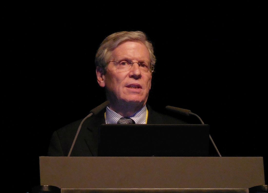
The “reassuring” safety performance of tofacitinib (Xeljanz) compared with biologic agents used to treat rheumatoid arthritis (RA) in these registry data notably showed a similar rate of venous thromboembolism (VTE) with tofacitinib treatment compared with biologic drugs, Joel M. Kremer, MD, reported at the European Congress of Rheumatology. It is an important finding because of recent concerns raised about VTE incidence among patients taking tofacitinib or another Janus kinase (JAK) inhibitor, he noted. The registry analysis Dr. Kremer presented showed that patients treated with tofacitinib had slightly more than double the rate of herpes zoster, compared with patients on biologic agents, a finding consistent with prior reports that tofacitinib treatment linked with an almost threefold increased rate of herpes zoster when compared with placebo-treated patients in a series of clinical trials (Arthritis Rheumatol. 2014 Oct;66[10]:2675-84). None of the herpes zoster activations identified in the registry patients on tofacitinib were rated as “serious,” noted Dr. Kremer , professor of medicine at Albany (N.Y.) Medical College.
The analysis he presented used data collected in the Corrona Rheumatoid Arthritis registry during November 2012, the month when tofacitinib received U.S. marketing approval, through December 2017. Data came from RA patients in the registry who began treatment during November 2012–June 2017 with either tofacitinib (1,544 patients) or a biologic agent (7,083 patients). The safety assessment focused on four outcomes: the combined rate of major adverse cardiovascular events (including MI, strokes, and fatal events); the incidence of serious infection; the incidence of any herpes zoster regardless of severity; and VTE. These outcomes were numerous enough to allow for propensity-score matching of patients from the tofacitinib and biologic subgroups to adjust for baseline differences between patients in these two categories, but as of the end of 2017, the cumulative number of VTEs was not high enough to allow for propensity-score adjustment, Dr. Kremer said, so instead he reported the unadjusted numbers. Further data collection should allow an adjusted analysis of VTE within another couple of years, he added. The currently available VTE data were “underpowered” for more rigorous statistical analysis, Dr. Kremer said.
After adjustment, the patients treated with tofacitinib had a 42% lower rate of major cardiovascular events, compared with patients who received a biologic drug, but the difference was not statistically significant, and the two subgroups had virtually identical rates of all serious infections. The incidence of herpes zoster was 2.26-fold more frequent among tofacitinib-treated patients than among those on other biologic agents, a statistically significant difference. The unadjusted VTE analysis showed a rate of 0.19/100 patient-years with tofacitinib treatment, and 0.33/100 patient-years with other biological agents, a difference that was not statistically significant, Dr. Kremer reported. Comparison of the rates of pulmonary embolism and deep vein thrombosis were each not statistically different between the two treatment subgroups.
Concern about possibly increased rates of VTE with the use of tofacitinib or other JAK inhibitors arose recently primarily because of two reports. In February 2019, the Food and Drug Administration released a safety announcement that data from an ongoing, postmarketing study of tofacitinib showed an elevated rate of RA patients with pulmonary embolism when they received an off-label, 10-mg twice-daily dosage of the drug, twice the labeled maximum dosage for this population. (The labeling for tofacitinib allows for a maximum dosage of 10 mg twice daily for patients treated for ulcerative colitis.) In addition, a recent report on another JAK inhibitor approved for U.S. marketing for RA treatment, baricitinib (Olumiant), documented a possible excess of VTE events among patients treated with this drug, compared with those who received placebo (Arthritis Rheumatol. 2019 Jan 21. doi: 10.1002/art.40841).
Regarding the now well-described excess of herpes zoster with tofacitinib treatment, Dr. Kremer said that perhaps the best way to address this in a patient who seems to be at risk is to prophylactically vaccinate the patient with the recombinant, adjuvanted zoster vaccine (Shingrix). However, this means withdrawing disease-modifying treatment from the RA patient for 3 weeks at the time of each of two vaccinations, with the possibility of flare induced by the adjuvant, he explained in an interview. The risks and benefits of this approach have not been investigated, he noted, and the Corrona data he studied came almost entirely from the period before Shingrix came onto the U.S. market following its approval in late 2017.
Dr. Kremer has been a consultant to and has received research funding from Pfizer, the company that markets tofacitinib. He has been a consultant to AbbVie, Amgen, Bristol-Myers Squibb, Genentech, and Regeneron/Sanofi, and has received research funding from AbbVie, Eli Lilly, Genentech, and Novartis. One of the coauthors on the report is a Pfizer employee.
SOURCE: Kremer JM et al. Ann Rheum Dis. Jun 2019;78(Suppl 2):82-3; Abstract OP0028. doi: 10.1136/annrheumdis-2019-eular.621.
MADRID – The Janus kinase inhibitor tofacitinib had a safety profile mostly similar to that of biologic drugs in a review of more than 8,600 U.S. rheumatoid arthritis patients enrolled in a national registry during 2012-2017, the first 5 years when tofacitinib was on the U.S. market.
The “reassuring” safety performance of tofacitinib (Xeljanz) compared with biologic agents used to treat rheumatoid arthritis (RA) in these registry data notably showed a similar rate of venous thromboembolism (VTE) with tofacitinib treatment compared with biologic drugs, Joel M. Kremer, MD, reported at the European Congress of Rheumatology. It is an important finding because of recent concerns raised about VTE incidence among patients taking tofacitinib or another Janus kinase (JAK) inhibitor, he noted. The registry analysis Dr. Kremer presented showed that patients treated with tofacitinib had slightly more than double the rate of herpes zoster, compared with patients on biologic agents, a finding consistent with prior reports that tofacitinib treatment linked with an almost threefold increased rate of herpes zoster when compared with placebo-treated patients in a series of clinical trials (Arthritis Rheumatol. 2014 Oct;66[10]:2675-84). None of the herpes zoster activations identified in the registry patients on tofacitinib were rated as “serious,” noted Dr. Kremer , professor of medicine at Albany (N.Y.) Medical College.
The analysis he presented used data collected in the Corrona Rheumatoid Arthritis registry during November 2012, the month when tofacitinib received U.S. marketing approval, through December 2017. Data came from RA patients in the registry who began treatment during November 2012–June 2017 with either tofacitinib (1,544 patients) or a biologic agent (7,083 patients). The safety assessment focused on four outcomes: the combined rate of major adverse cardiovascular events (including MI, strokes, and fatal events); the incidence of serious infection; the incidence of any herpes zoster regardless of severity; and VTE. These outcomes were numerous enough to allow for propensity-score matching of patients from the tofacitinib and biologic subgroups to adjust for baseline differences between patients in these two categories, but as of the end of 2017, the cumulative number of VTEs was not high enough to allow for propensity-score adjustment, Dr. Kremer said, so instead he reported the unadjusted numbers. Further data collection should allow an adjusted analysis of VTE within another couple of years, he added. The currently available VTE data were “underpowered” for more rigorous statistical analysis, Dr. Kremer said.
After adjustment, the patients treated with tofacitinib had a 42% lower rate of major cardiovascular events, compared with patients who received a biologic drug, but the difference was not statistically significant, and the two subgroups had virtually identical rates of all serious infections. The incidence of herpes zoster was 2.26-fold more frequent among tofacitinib-treated patients than among those on other biologic agents, a statistically significant difference. The unadjusted VTE analysis showed a rate of 0.19/100 patient-years with tofacitinib treatment, and 0.33/100 patient-years with other biological agents, a difference that was not statistically significant, Dr. Kremer reported. Comparison of the rates of pulmonary embolism and deep vein thrombosis were each not statistically different between the two treatment subgroups.
Concern about possibly increased rates of VTE with the use of tofacitinib or other JAK inhibitors arose recently primarily because of two reports. In February 2019, the Food and Drug Administration released a safety announcement that data from an ongoing, postmarketing study of tofacitinib showed an elevated rate of RA patients with pulmonary embolism when they received an off-label, 10-mg twice-daily dosage of the drug, twice the labeled maximum dosage for this population. (The labeling for tofacitinib allows for a maximum dosage of 10 mg twice daily for patients treated for ulcerative colitis.) In addition, a recent report on another JAK inhibitor approved for U.S. marketing for RA treatment, baricitinib (Olumiant), documented a possible excess of VTE events among patients treated with this drug, compared with those who received placebo (Arthritis Rheumatol. 2019 Jan 21. doi: 10.1002/art.40841).
Regarding the now well-described excess of herpes zoster with tofacitinib treatment, Dr. Kremer said that perhaps the best way to address this in a patient who seems to be at risk is to prophylactically vaccinate the patient with the recombinant, adjuvanted zoster vaccine (Shingrix). However, this means withdrawing disease-modifying treatment from the RA patient for 3 weeks at the time of each of two vaccinations, with the possibility of flare induced by the adjuvant, he explained in an interview. The risks and benefits of this approach have not been investigated, he noted, and the Corrona data he studied came almost entirely from the period before Shingrix came onto the U.S. market following its approval in late 2017.
Dr. Kremer has been a consultant to and has received research funding from Pfizer, the company that markets tofacitinib. He has been a consultant to AbbVie, Amgen, Bristol-Myers Squibb, Genentech, and Regeneron/Sanofi, and has received research funding from AbbVie, Eli Lilly, Genentech, and Novartis. One of the coauthors on the report is a Pfizer employee.
SOURCE: Kremer JM et al. Ann Rheum Dis. Jun 2019;78(Suppl 2):82-3; Abstract OP0028. doi: 10.1136/annrheumdis-2019-eular.621.
REPORTING FROM THE EULAR 2019 CONGRESS
Upadacitinib monotherapy appears promising in RA patients with inadequate methotrexate response
The investigational oral Janus kinase (JAK) inhibitor upadacitinib has shown promise as a monotherapy treatment option for patients with active rheumatoid arthritis despite treatment with methotrexate, based on results from the phase 3 SELECT-MONOTHERAPY trial.

“The SELECT-MONOTHERAPY trial showed that patients who were having an inadequate response to methotrexate could be switched to oral upadacitinib monotherapy (15 mg or 30 mg) once a day, with improvements in clinical signs and symptoms, physical function, and quality of life measures, compared with patients who continued on their previous methotrexate dose,” Josef S. Smolen, MD, of the Medical University of Vienna, and his colleagues wrote in the Lancet.
The phase 3, double-blind, placebo-controlled trial randomized 648 RA patients with active disease despite methotrexate therapy from 138 sites in 24 countries.
Higher proportions of patients receiving upadacitinib 15 mg and 30 mg versus continued methotrexate achieved the primary endpoints of the proportion of patients achieving a 20% improvement in American College of Rheumatology (ACR 20) criteria at 14 weeks and the proportion achieving low disease activity defined as a 28-joint Disease Activity Score using C-reactive protein (DAS28-CRP) of 3.2 or lower.
An ACR 20 response was achieved by 89 (41%) of 216 patients (95% confidence interval, 35%-48%) in the continued methotrexate group, 147 (68%) of 217 patients (95% CI, 62%-74%) receiving upadacitinib 15 mg, and 153 (71%) of 215 patients (95% CI, 65%-77%) receiving upadacitinib 30 mg (P less than .0001 for both doses vs. continued methotrexate).
A DAS28-CRP score of 3.2 or lower was met by 42 (19%) of 216 (95% CI, 14%-25%) receiving methotrexate, 97 (45%) of 217 (95% CI, 38%-51%) receiving upadacitinib 15 mg, and 114 (53%) of 215 (95% CI, 46%-60%) receiving upadacitinib 30 mg (P less than .0001 for both doses vs. continued methotrexate).
The investigators noted that responses were observed with both 15-mg and 30-mg doses of upadacitinib, although numerically higher responses were seen with the 30-mg dose.
“Whether the 15-mg or the 30-mg dose is the more appropriate one for patients who switch from methotrexate to upadacitinib will have to be established in conjunction with data from the other phase 3 upadacitinib trials,” they wrote.
Treatment-emergent adverse events were reported at similar frequencies across the arms (n = 102 with methotrexate; n = 103 with upadacitinib 15 mg; n = 105 with upadacitinib 30 mg).
Serious adverse events were reported in 11 (5%) of 217 patients in the upadacitinib 15 mg arm, 6 (3%) of 216 patients in the continued methotrexate arm, and 6 (3%) of 215 patients in the upadacitinib 30 mg arm.
“This favorable benefit-risk profile of upadacitinib monotherapy has the potential to provide a treatment option for patients who are intolerant to methotrexate or who prefer a treatment without the need for concomitant [conventional synthetic] DMARDs,” the authors wrote.
The study was funded by AbbVie. Dr. Smolen and most authors reported financial ties to AbbVie and other companies marketing rheumatoid arthritis treatments. Five authors are employees of AbbVie.
SOURCE: Smolen JS et al. Lancet. 2019;393[10188]:2303-11. doi: 10.1016/S0140-6736(19)30419-2
Treatment recommendations for the management of RA do not currently include the use of novel disease-modifying antirheumatic drugs (DMARDs) as monotherapy. However, there is growing interest in this concept, most likely because of poor tolerability and contraindications to conventional synthetic DMARDs such as methotrexate.

The observed treatment effects of SELECT-NEXT and the current trial by Prof. Smolen and associates are similar for both control groups (responders: 36% vs. 41% on methotrexate plus placebo) and for the upadacitinib treatment groups, regardless of concomitant methotrexate status (64% on methotrexate plus upadacitinib 15 mg or 66% on methotrexate plus upadacitinib 30 mg vs. 68% on upadacitinib 15 mg or 71% on upadacitinib 30 mg).
Prof. Smolen and associates chose a trial design that assessed the continuation of methotrexate plus placebo versus switching from methotrexate to upadacitinib monotherapy, but does this research question adequately reflect the question often raised in clinical practice: Should a novel DMARD be added to methotrexate or should patients switch from methotrexate to a novel DMARD?
But we now need a trial that compares a switch versus an add-on strategy to adequately answer the question on the treatment of resistant RA.
These comments are adapted from an accompanying editorial by Anna Moltó, MD, and Maxime Dougados, MD, both with the rheumatology department at Cochin Hospital, Paris (Lancet. 2019;393[10188]:2277-8. doi: 10.1016/S0140-6736(19)30768-8). The rheumatology department at Cochin Hospital has received grants to do clinical studies and trials from AbbVie and other manufacturers of drugs for RA. Both Dr. Dougados and Dr. Moltó reported receiving personal fees for advisory boards and symposia from AbbVie and other manufacturers of drugs for RA outside the area of work commented on here.
Treatment recommendations for the management of RA do not currently include the use of novel disease-modifying antirheumatic drugs (DMARDs) as monotherapy. However, there is growing interest in this concept, most likely because of poor tolerability and contraindications to conventional synthetic DMARDs such as methotrexate.
The observed treatment effects of SELECT-NEXT and the current trial by Prof. Smolen and associates are similar for both control groups (responders: 36% vs. 41% on methotrexate plus placebo) and for the upadacitinib treatment groups, regardless of concomitant methotrexate status (64% on methotrexate plus upadacitinib 15 mg or 66% on methotrexate plus upadacitinib 30 mg vs. 68% on upadacitinib 15 mg or 71% on upadacitinib 30 mg).
Prof. Smolen and associates chose a trial design that assessed the continuation of methotrexate plus placebo versus switching from methotrexate to upadacitinib monotherapy, but does this research question adequately reflect the question often raised in clinical practice: Should a novel DMARD be added to methotrexate or should patients switch from methotrexate to a novel DMARD?
But we now need a trial that compares a switch versus an add-on strategy to adequately answer the question on the treatment of resistant RA.
These comments are adapted from an accompanying editorial by Anna Moltó, MD, and Maxime Dougados, MD, both with the rheumatology department at Cochin Hospital, Paris (Lancet. 2019;393[10188]:2277-8. doi: 10.1016/S0140-6736(19)30768-8). The rheumatology department at Cochin Hospital has received grants to do clinical studies and trials from AbbVie and other manufacturers of drugs for RA. Both Dr. Dougados and Dr. Moltó reported receiving personal fees for advisory boards and symposia from AbbVie and other manufacturers of drugs for RA outside the area of work commented on here.
The investigational oral Janus kinase (JAK) inhibitor upadacitinib has shown promise as a monotherapy treatment option for patients with active rheumatoid arthritis despite treatment with methotrexate, based on results from the phase 3 SELECT-MONOTHERAPY trial.
“The SELECT-MONOTHERAPY trial showed that patients who were having an inadequate response to methotrexate could be switched to oral upadacitinib monotherapy (15 mg or 30 mg) once a day, with improvements in clinical signs and symptoms, physical function, and quality of life measures, compared with patients who continued on their previous methotrexate dose,” Josef S. Smolen, MD, of the Medical University of Vienna, and his colleagues wrote in the Lancet.
The phase 3, double-blind, placebo-controlled trial randomized 648 RA patients with active disease despite methotrexate therapy from 138 sites in 24 countries.
Higher proportions of patients receiving upadacitinib 15 mg and 30 mg versus continued methotrexate achieved the primary endpoints of the proportion of patients achieving a 20% improvement in American College of Rheumatology (ACR 20) criteria at 14 weeks and the proportion achieving low disease activity defined as a 28-joint Disease Activity Score using C-reactive protein (DAS28-CRP) of 3.2 or lower.
An ACR 20 response was achieved by 89 (41%) of 216 patients (95% confidence interval, 35%-48%) in the continued methotrexate group, 147 (68%) of 217 patients (95% CI, 62%-74%) receiving upadacitinib 15 mg, and 153 (71%) of 215 patients (95% CI, 65%-77%) receiving upadacitinib 30 mg (P less than .0001 for both doses vs. continued methotrexate).
A DAS28-CRP score of 3.2 or lower was met by 42 (19%) of 216 (95% CI, 14%-25%) receiving methotrexate, 97 (45%) of 217 (95% CI, 38%-51%) receiving upadacitinib 15 mg, and 114 (53%) of 215 (95% CI, 46%-60%) receiving upadacitinib 30 mg (P less than .0001 for both doses vs. continued methotrexate).
The investigators noted that responses were observed with both 15-mg and 30-mg doses of upadacitinib, although numerically higher responses were seen with the 30-mg dose.
“Whether the 15-mg or the 30-mg dose is the more appropriate one for patients who switch from methotrexate to upadacitinib will have to be established in conjunction with data from the other phase 3 upadacitinib trials,” they wrote.
Treatment-emergent adverse events were reported at similar frequencies across the arms (n = 102 with methotrexate; n = 103 with upadacitinib 15 mg; n = 105 with upadacitinib 30 mg).
Serious adverse events were reported in 11 (5%) of 217 patients in the upadacitinib 15 mg arm, 6 (3%) of 216 patients in the continued methotrexate arm, and 6 (3%) of 215 patients in the upadacitinib 30 mg arm.
“This favorable benefit-risk profile of upadacitinib monotherapy has the potential to provide a treatment option for patients who are intolerant to methotrexate or who prefer a treatment without the need for concomitant [conventional synthetic] DMARDs,” the authors wrote.
The study was funded by AbbVie. Dr. Smolen and most authors reported financial ties to AbbVie and other companies marketing rheumatoid arthritis treatments. Five authors are employees of AbbVie.
SOURCE: Smolen JS et al. Lancet. 2019;393[10188]:2303-11. doi: 10.1016/S0140-6736(19)30419-2
FROM THE LANCET
Rituximab serious infection risk predicted by immunoglobulin levels
Monitoring immunoglobulin (Ig) levels at baseline and before each cycle of rituximab could reduce the risk of serious infection events (SIEs) in patients needing repeated treatment, according to research published in Arthritis & Rheumatology.

In a large, single-center, longitudinal study conducted at a tertiary referral center, having low IgG (less than 6 g/L) in particular was associated with a higher rate of SIEs, compared with having normal IgG levels (6-16 g/L). Considering 103 of 700 patients who had low levels of IgG before starting treatment with rituximab for various rheumatic and musculoskeletal diseases (RMDs), there were 16.4 SIEs per 100 patient-years. In those who developed low IgG during subsequent cycles of rituximab therapy, the SIE rate was even higher, at 21.3 per 100 patient-years. By comparison, the SIE rate for those with normal IgG levels was 9.7 per 100 patient-years.
“We really have to monitor immunoglobulins at baseline and also before we re-treat the patients, because higher IgG level is protective of serious infections,” study first author Md Yuzaiful Md Yusof, MBChB, PhD, said in an interview.
Low IgG has been linked to a higher risk of SIEs in the first 12 months of rituximab therapy but, until now, there have been limited data on infection predictors during repeated cycles of treatment. While IgG is a consistent marker of SIEs associated with repeated rituximab treatment, IgM and IgA should also be monitored to give a full picture of any hyperglobulinemia that may be present.
“There is no formal guidance on how to safely monitor patients on rituximab,” observed Dr. Md Yusof, who will present these data at the 2019 European Congress of Rheumatology in Madrid. The study’s findings could help to change that, however, as they offer a practical way to help predict and thus prevent SIEs. The study’s findings not only validate previous work, he noted, but also add new insights into why some patients treated with repeat rituximab cycles but not others may experience a higher rate of such infections.
Altogether, the investigators examined data on 700 patients with RMDs treated with rituximab who were consecutively seen during 2012-2017 at Dr. Md Yusof’s institution – the Leeds (England) Institute of Rheumatic and Musculoskeletal Medicine, which is part of the University of Leeds. Their immunoglobulin levels had been measured before starting rituximab therapy and every 4-6 months after each cycle of rituximab treatment.
Patients with any RMD being treated with at least one cycle of rituximab were eligible for inclusion in the retrospective study, with the majority (72%) taking it for rheumatoid arthritis and some for systemic lupus erythematosus (13%) or antineutrophil cytoplasmic antibody (ANCA)–associated vasculitis (7%).
One of the main aims of the study was to look for predictors of SIEs during the first 12 months and during repeated cycles of rituximab. Dr. Md Yusof and his associates also looked at how secondary hypogammaglobulinemia might affect SIE rates and the humoral response to vaccination challenge and its persistence following treatment discontinuation. Their ultimate aim was to see if these findings could then be used to develop a treatment algorithm for rituximab administration in RMDs.
Over a follow-up period encompassing 2,880 patient-years of treatment, 281 SIEs were recorded in 176 patients, giving a rate of 9.8 infections per 100 patient-years. Most (61%) of these were due to lower respiratory tract infections.
The proportion of patients experiencing their first SIE increased with time: 16% within 6 weeks of starting rituximab therapy, 35% at 12 weeks, 72% at 26 weeks, 83% at 38 weeks, and 100% by 1 year of repeated treatment.
Multivariable analysis showed that the presence of several comorbidities at baseline – notably chronic obstructive pulmonary disease, diabetes, heart failure, and prior cancer – raised the risk for SIEs with repeated rituximab therapy. The biggest factor, however, was a history of SIEs – with a sixfold increased risk of further serious infection.
Higher corticosteroid dose and factors specific to rituximab – low IgG, neutropenia, high IgM, and a longer time to retreatment – were also predictive of SIEs.
“Low IgG also results in poor humoral response to vaccination,” Dr. Md Yusof said, noting that the IgG level remains below the lower limit of normal for several years after rituximab is discontinued in most patients.
In the study, 5 of 8 (64%) patients had impaired humoral response to pneumococcal and haemophilus following vaccination challenge and 4 of 11 patients had IgG normalized after switching to another biologic disease-modifying antirheumatic drug (bDMARD).
Cyclophosphamide is commonly used as a first-line agent to induce remission in patients with severe and refractory systemic lupus erythematosus and ANCA-associated vasculitis, with patients switched to rituximab at relapse. The effect of this prior treatment was examined in 20 patients in the study, with a marked decline in almost all immunoglobulin classes seen up to 18 months. Prior treatment with immunosuppressants such as intravenous cyclophosphamide could be behind progressive reductions in Ig levels seen with repeated rituximab treatment rather than entirely because of rituximab, Dr. Md Yusof said.
Dr. Md Yusof, who is a National Institute for Health Research (NIHR) Academic Clinical Lecturer at the University of Leeds, said the value of the study, compared with others, is that hospital data for all patients treated with rituximab with at least 3 months follow-up were included, making it an almost complete data set.
“By carefully reviewing records of every patient to capture all infection episodes in the largest single-center cohort study to date, our findings provide insights on predictors of SIEs as well as a foundation for safety monitoring of rituximab,” he and his coauthors wrote.
They acknowledge reporting a higher rate of SIEs than seen in registry and clinical studies with rituximab, which may reflect a “channeling bias” as the patients comprised those with multiple comorbidities including those that represent a relative contraindication for bDMARD use. That said, the findings clearly show that Ig levels should be monitored before and after each rituximab cycle, especially in those with comorbid diseases and those with low IgG levels to start with.
They conclude that an “individualized benefit-risk assessment” is needed to determine whether rituximab should be repeated in those with low IgG as this is a “consistent predictor” of SIE and may “increase infection profiles when [rituximab] is switched to different bDMARDs.”
The research was supported by Octapharma, the National Institute for Health Research (NIHR), and NIHR Leeds Biomedical Research Centre based at Leeds Teaching Hospitals NHS Trust in England. Dr. Md Yusof had no conflicts of interest. Several coauthors disclosed financial ties to multiple pharmaceutical companies, including Roche.
SOURCE: Md Yusof MY et al. Arthritis Rheumatol. 2019 May 27. doi: 10.1002/art.40937.
Monitoring immunoglobulin (Ig) levels at baseline and before each cycle of rituximab could reduce the risk of serious infection events (SIEs) in patients needing repeated treatment, according to research published in Arthritis & Rheumatology.
In a large, single-center, longitudinal study conducted at a tertiary referral center, having low IgG (less than 6 g/L) in particular was associated with a higher rate of SIEs, compared with having normal IgG levels (6-16 g/L). Considering 103 of 700 patients who had low levels of IgG before starting treatment with rituximab for various rheumatic and musculoskeletal diseases (RMDs), there were 16.4 SIEs per 100 patient-years. In those who developed low IgG during subsequent cycles of rituximab therapy, the SIE rate was even higher, at 21.3 per 100 patient-years. By comparison, the SIE rate for those with normal IgG levels was 9.7 per 100 patient-years.
“We really have to monitor immunoglobulins at baseline and also before we re-treat the patients, because higher IgG level is protective of serious infections,” study first author Md Yuzaiful Md Yusof, MBChB, PhD, said in an interview.
Low IgG has been linked to a higher risk of SIEs in the first 12 months of rituximab therapy but, until now, there have been limited data on infection predictors during repeated cycles of treatment. While IgG is a consistent marker of SIEs associated with repeated rituximab treatment, IgM and IgA should also be monitored to give a full picture of any hyperglobulinemia that may be present.
“There is no formal guidance on how to safely monitor patients on rituximab,” observed Dr. Md Yusof, who will present these data at the 2019 European Congress of Rheumatology in Madrid. The study’s findings could help to change that, however, as they offer a practical way to help predict and thus prevent SIEs. The study’s findings not only validate previous work, he noted, but also add new insights into why some patients treated with repeat rituximab cycles but not others may experience a higher rate of such infections.
Altogether, the investigators examined data on 700 patients with RMDs treated with rituximab who were consecutively seen during 2012-2017 at Dr. Md Yusof’s institution – the Leeds (England) Institute of Rheumatic and Musculoskeletal Medicine, which is part of the University of Leeds. Their immunoglobulin levels had been measured before starting rituximab therapy and every 4-6 months after each cycle of rituximab treatment.
Patients with any RMD being treated with at least one cycle of rituximab were eligible for inclusion in the retrospective study, with the majority (72%) taking it for rheumatoid arthritis and some for systemic lupus erythematosus (13%) or antineutrophil cytoplasmic antibody (ANCA)–associated vasculitis (7%).
One of the main aims of the study was to look for predictors of SIEs during the first 12 months and during repeated cycles of rituximab. Dr. Md Yusof and his associates also looked at how secondary hypogammaglobulinemia might affect SIE rates and the humoral response to vaccination challenge and its persistence following treatment discontinuation. Their ultimate aim was to see if these findings could then be used to develop a treatment algorithm for rituximab administration in RMDs.
Over a follow-up period encompassing 2,880 patient-years of treatment, 281 SIEs were recorded in 176 patients, giving a rate of 9.8 infections per 100 patient-years. Most (61%) of these were due to lower respiratory tract infections.
The proportion of patients experiencing their first SIE increased with time: 16% within 6 weeks of starting rituximab therapy, 35% at 12 weeks, 72% at 26 weeks, 83% at 38 weeks, and 100% by 1 year of repeated treatment.
Multivariable analysis showed that the presence of several comorbidities at baseline – notably chronic obstructive pulmonary disease, diabetes, heart failure, and prior cancer – raised the risk for SIEs with repeated rituximab therapy. The biggest factor, however, was a history of SIEs – with a sixfold increased risk of further serious infection.
Higher corticosteroid dose and factors specific to rituximab – low IgG, neutropenia, high IgM, and a longer time to retreatment – were also predictive of SIEs.
“Low IgG also results in poor humoral response to vaccination,” Dr. Md Yusof said, noting that the IgG level remains below the lower limit of normal for several years after rituximab is discontinued in most patients.
In the study, 5 of 8 (64%) patients had impaired humoral response to pneumococcal and haemophilus following vaccination challenge and 4 of 11 patients had IgG normalized after switching to another biologic disease-modifying antirheumatic drug (bDMARD).
Cyclophosphamide is commonly used as a first-line agent to induce remission in patients with severe and refractory systemic lupus erythematosus and ANCA-associated vasculitis, with patients switched to rituximab at relapse. The effect of this prior treatment was examined in 20 patients in the study, with a marked decline in almost all immunoglobulin classes seen up to 18 months. Prior treatment with immunosuppressants such as intravenous cyclophosphamide could be behind progressive reductions in Ig levels seen with repeated rituximab treatment rather than entirely because of rituximab, Dr. Md Yusof said.
Dr. Md Yusof, who is a National Institute for Health Research (NIHR) Academic Clinical Lecturer at the University of Leeds, said the value of the study, compared with others, is that hospital data for all patients treated with rituximab with at least 3 months follow-up were included, making it an almost complete data set.
“By carefully reviewing records of every patient to capture all infection episodes in the largest single-center cohort study to date, our findings provide insights on predictors of SIEs as well as a foundation for safety monitoring of rituximab,” he and his coauthors wrote.
They acknowledge reporting a higher rate of SIEs than seen in registry and clinical studies with rituximab, which may reflect a “channeling bias” as the patients comprised those with multiple comorbidities including those that represent a relative contraindication for bDMARD use. That said, the findings clearly show that Ig levels should be monitored before and after each rituximab cycle, especially in those with comorbid diseases and those with low IgG levels to start with.
They conclude that an “individualized benefit-risk assessment” is needed to determine whether rituximab should be repeated in those with low IgG as this is a “consistent predictor” of SIE and may “increase infection profiles when [rituximab] is switched to different bDMARDs.”
The research was supported by Octapharma, the National Institute for Health Research (NIHR), and NIHR Leeds Biomedical Research Centre based at Leeds Teaching Hospitals NHS Trust in England. Dr. Md Yusof had no conflicts of interest. Several coauthors disclosed financial ties to multiple pharmaceutical companies, including Roche.
SOURCE: Md Yusof MY et al. Arthritis Rheumatol. 2019 May 27. doi: 10.1002/art.40937.
FROM ARTHRITIS & RHEUMATOLOGY
Key clinical point: Immunoglobulin should be monitored at baseline and before each rituximab cycle to identify patients at risk of serious infection events (SIEs).
Major finding: SIE rates per 100 patient-years were 16.4 and 21.3 in patients with low (less than 6 g/L) IgG at baseline and during rituximab cycles versus 9.7 for patients with normal (6–16 g/L) IgG levels.
Study details: A retrospective, single-center, longitudinal study involving 700 rituximab-treated patients with rheumatoid arthritis and other rheumatic and musculoskeletal diseases.
Disclosures: The research was supported by Octapharma, the National Institute for Health Research (NIHR), and NIHR Leeds Biomedical Research Centre based at Leeds Teaching Hospitals NHS Trust in the United Kingdom. Dr. Md Yusof had no conflicts of interest. Several coauthors disclosed financial ties to multiple pharmaceutical companies, including Roche.
Source: Md Yusof MY et al. Arthritis Rheumatol. 2019 May 27. doi: 10.1002/art.40937.

Could biosimilar switchbacks due to ‘inefficacy’ be subjective?
BIRMINGHAM, ENGLAND – Patients with rheumatoid arthritis (RA) who switch back to an original tumor necrosis factor inhibitor (TNFi) after using a biosimilar product often do so because of ineffectiveness, but this could be largely subjective, research suggests.
Data from the British Society for Rheumatology Biologics Register–Rheumatoid Arthritis (BSRBR-RA), presented at the annual conference of the British Society for Rheumatology, have shown that the majority (81%) of 760 patients who switched from an etanercept originator (ETN-O) product (Enbrel) to an etanercept biosimilar (ETN-B) product (Benepali or Erelzi) remained on the latter at an average of 2 years’ follow-up.
However, of those who switched back to an ETN-O (8%, n = 58), ineffectiveness was the primary reason for doing so in more than half of patients (53%). Of patients who stopped ETN treatment altogether after the biosimilar switch (11%, n = 84), 29% stopped because of inefficacy. Other important reasons for switching back or stopping treatment were adverse events in 28% and 54% of cases, respectively. The switch back to an ETN-O happened within a median time of 4 months (range, 2–6 months).
Patients switching back to an ETN-O tended to be slightly younger than all patients who had switched to an ETN-B (median age 60 vs. 64 years). They were more likely to be female (79% vs. 75%). Median disease activity score in 28 joints (DAS28) at baseline were highest in those who discontinued ETN (3.9 vs. 3.2 for those who switched back and 3.0 for all who had switched to a biosimilar).
It is not known what is driving the purported ineffectiveness, said research assistant Rebecca Davies of the Arthritis Research UK Epidemiology Unit at the University of Manchester (England).
“It could be due to patient factors and a nocebo effect,” she suggested. This is where “negative patient expectations cause treatment to have a more negative effect.” For example, she added, “if a patient is settled on the originator drug and had to mandatorily switch to a biosimilar, it might be that they are more heightened to symptoms.”
Another explanation could be that consultants may be “overcautious” to continuing the biosimilar in patients who had responded well to the originator product, she hypothesized.
“Our next steps are to look at disease activity between 4 and 9 months post switch to determine” the cause of the described ineffectiveness.
Educate patients to reduce switchbacks?
Meanwhile data presented by a team of researchers from Imperial College Healthcare NHS Trust in London, headed by Maresa Carulli, MD, suggested that patient education may be an important factor in tackling why patients want to switch back to biosimilar treatment.
Over a 20-month period among 202 patients who were switched to an ETN-B after they had been established on an ETN-O, 27 (13.4%) switched back.
“The majority of patients who switched back to Enbrel reported subjective worsening of disease symptoms with Benepali,” Dr. Carulli and coauthors said in their poster. Indeed, 78% of patients who switched back after an average of just under 6 months reported that their symptoms had become worse on the biosimilar product.
Analysis of the RA patients (n = 16) who switched back demonstrated that DAS28 scores had increased by more than 1.2 points during the period of the switch, but this was “mainly due to increases in visual analog scores [VAS],” they said.
The average change in DAS28 was an increase of 1.32 points during the switch period, the researchers noted. The average changes in DAS28 components were: +5 and +0.44 points for the tender and swollen joint counts, +9.89 points for the erythrocyte sedimentation rate, and +25.56 points for VAS.
Although further data are need, the results “may suggest that a subjective component contributes toward intolerance” of the biosimilar product, the researchers said.
“Consideration of patient education during initiation of biosimilar treatment could be a significant factor in improving compliance with it.”
The BSR receives restricted income from multiple U.K. pharmaceutical companies, which is used to fund the BSRBR-RA. The pharmaceutical company funders had no role in the design and conduct of the study; collection, management, analysis, and interpretation of the data; preparation, review, or approval of the manuscript; or any decision to submit manuscripts for publication.
Ms. Davies and Dr. Carulli and team declared having no competing interests.
SOURCES: Davies R et al. Rheumatology. 2019;58(suppl 3), Abstract 022; and Dahanayake C et al. Rheumatology. 2019;58(suppl 3), Abstract 102
REPORTING FROM BSR 2019
Proinflammatory diets up rheumatoid arthritis risk
BIRMINGHAM, ENGLAND – Proinflammatory diets are associated with increased C-reactive protein (CRP) and subsequent rheumatoid arthritis (RA), according to combined data from the European Prospective Investigation of Cancer and Nutrition (EPIC) and Norfolk Arthritis Register (NOAR).
“There has always been a debate around this topic,” Max Yates, MBBS, PhD, said at the annual conference of the British Society for Rheumatology. “A quick online search will reveal a plethora of texts claiming to give definitive or the best advice for arthritis” and diet, he said, often from “questionable experts.”
“I think we’re all interested in diet,” observed Dr. Yates, of the University of East Anglia in Norwich (England), and “although the association between diet and arthritis is open to debate, previous studies have shown an association with those who have a lower intake of vitamin C and fiber.” The problem is one of credibility, he noted, so this was something that the NOAR investigators decided to look into with data from the Dietary Inflammatory Index (DII) collected from the EPIC cohort.
The DII is a literature-based, population-derived tool that has been used to determine the inflammatory potential of diet, Dr. Yates explained. Data show that inflammatory diets are is associated with increased levels of inflammatory markers including C-reactive protein (CRP) and interleukin (IL)-6. These diets include items such as trans and saturated fats, and fats from animal protein versus more anti-inflammatory items such as black tea, thyme, turmeric, and saffron.
“We are fortunate that NOAR is in the same geographic location as EPIC,” Dr. Yates said, and the two cohorts have been running alongside each other since the early 1990s. While NOAR has been collecting data on incident inflammatory polyarthritis since 1989, EPIC has been “intensively” collecting information on dietary and lifestyle factors and blood samples from its participants since 1993.
EPIC investigators have been “trailblazers” in recording of dietary data, Dr. Yates said. First using paper-based questionnaires and now smartphone apps that allow people to upload photos of what they are eating. Data are linked to both primary care practice and hospital records.
For the present study, data on 159 patients who participated in both NOAR and EPIC were used. Participants had RA according to the 2010 American College of Rheumatology criteria and had an average disease onset of 7 years after enrollment into NOAR and EPIC.
“Quite pleasingly, the dietary inflammatory index scores were associated with high-sensitivity CRP taken at baseline enrollment in 1993 to 1997, further validating the index again within another population,” Dr. Yates said.
Results showed that there was a significant association between the baseline DII score and subsequent development of RA, with an odds ratio of 1.90 comparing individuals with the highest and lowest DII scores (P less than .01).
When cases were matched by age, sex, and body mass index, however, there was only a trend for an association between inflammatory diets and RA onset. “We hope to identify more patients within EPIC to strengthen this association,” Dr. Yates said.
The results are consistent with data from the Nurses’ Health Study, Dr. Yates noted, adding that future research is needed to address whether dietary modification can demonstrate causality.
“Diet is one of the modifiable risk factors that we can use to tackle RA, and I think it’s about time we as a community take over this area and gave definitive advice.”
Dr. Yates presented the work on behalf of PhD student Ellie Sayer. Neither Dr. Yates nor Ms. Sayer had any conflicts of interests to disclose.
SOURCE: Sayer E et al. Rheumatology. 2019;58(suppl 3), Abstract 014.
REPORTING FROM BSR 2019
Antimalarials in pregnancy and lactation
According to the World Health Organization, there were about 219 million cases of malaria and an estimated 660,000 deaths in 2010. Although huge, this was a 26% decrease from the rates in 2000. Six countries in Africa account for 47% of malaria cases: Cote d’Ivoire, Democratic Republic of the Congo, Mozambique, Nigeria, Uganda, and the United Republic of Tanzania. The second-most affected region in the world is Southeast Asia, which includes Myanmar, India, and Indonesia. In comparison, about 1,500 malaria cases and 5 deaths are reported annually in the United States, mostly from returned travelers.
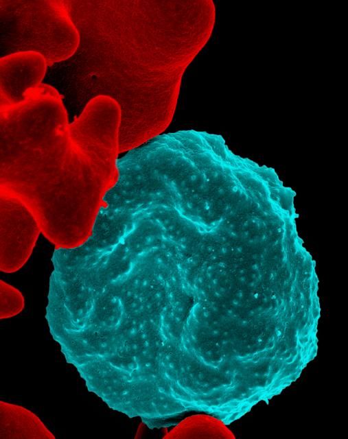
if they will be traveling in any of the above regions. Malaria during pregnancy increases the risk for adverse pregnancy outcomes, including maternal anemia, prematurity, spontaneous abortion, and stillbirth.
As stated by the Centers for Disease Control and Prevention, no antimalarial agent is 100% protective. Therefore, whatever agent is used must be combined with personal protective measures such as wearing insect repellent, long sleeves, and long pants; sleeping in a mosquito-free setting; or using an insecticide-treated bed net.
There are nine antimalarial drugs available in the United States.
Atovaquone/Proguanil Hcl (Malarone and as generic)
This agent is good for last-minute travelers because the drug is started 1-2 days before traveling to areas where malaria transmission occurs. The combination can be classified as compatible in pregnancy. No reports in breastfeeding with atovaquone or the combination have been found. Proguanil is not available in the United States as a single agent.
Chloroquine (generic)
This is the drug of choice to prevent and treat sensitive malaria species during pregnancy. The drug crosses the placenta producing fetal concentrations that are similar to those in the mother. The drug appears to be low risk for embryo-fetal harm.
It is compatible in breastfeeding.
Dapsone (generic)
This agent does not appear to represent a major risk of harm to the fetus. Although it has been used in combination with pyrimethamine (an antiparasitic) or trimethoprim (an antibiotic) to prevent malaria, the efficacy of the combination has not been confirmed.
In breastfeeding, there is one case of mild hemolytic anemia in the mother and her breastfeeding infant that may have been caused by the drug.
Hydroxychloroquine (generic)
This agent is used for the treatment of malaria, systemic erythematosus, and rheumatoid arthritis. For antimalarial prophylaxis, 400 mg/week appears to be low risk for embryo-fetal harm. Doses used to treat malaria have been 200-400 mg/day.
Because very low concentrations of the drug have been found in breast milk, breastfeeding is probably compatible.
Mefloquine (generic)
This agent is a quinoline-methanol agent that does not appear to cause embryo-fetal harm based on a large number of pregnancy exposures.
There are no reports of its use while breastfeeding.
Primaquine (generic)
This agent is best avoided in pregnancy. There is no human pregnancy data, but it may cause hemolytic anemia in patients with glucose-6-phosphate dehydrogenase deficiency (G6PD). Because the fetus is relatively G6PD deficient, it is best avoided in pregnancy regardless of the mother’s status.
There are no reports describing the use of the drug during lactation. Both the mother and baby should be tested for G6PD deficiency before the drug is used during breastfeeding.
Pyrimethamine (generic)
This agent has been used for the treatment or prophylaxis of malaria. Most studies have found this agent to be relatively safe and effective.

It is excreted into breast milk and has been effective in eliminating malaria parasites from breastfeeding infants.
Quinidine (generic)
Reports linking the use of this agent with congenital defects have not been found. Although the drug has data on its use as an antiarrhythmic, its published use to treat malaria is limited.
The drug is excreted into breast milk, but there are no reports of its during breastfeeding.
Quinine (generic)
This agent has a large amount of human pregnancy data (more than 1,000 exposures) that found no increased risk of birth defects. The drug has been replaced by newer agents but still may be used for chloroquine-resistant malaria.
The drug appears to be compatible during breastfeeding.
Mr. Briggs is clinical professor of pharmacy at the University of California, San Francisco, and adjunct professor of pharmacy at the University of Southern California, Los Angeles, as well as at Washington State University, Spokane. Mr. Briggs said he had no relevant financial disclosures. Email him at obnews@mdedge.com.
According to the World Health Organization, there were about 219 million cases of malaria and an estimated 660,000 deaths in 2010. Although huge, this was a 26% decrease from the rates in 2000. Six countries in Africa account for 47% of malaria cases: Cote d’Ivoire, Democratic Republic of the Congo, Mozambique, Nigeria, Uganda, and the United Republic of Tanzania. The second-most affected region in the world is Southeast Asia, which includes Myanmar, India, and Indonesia. In comparison, about 1,500 malaria cases and 5 deaths are reported annually in the United States, mostly from returned travelers.
if they will be traveling in any of the above regions. Malaria during pregnancy increases the risk for adverse pregnancy outcomes, including maternal anemia, prematurity, spontaneous abortion, and stillbirth.
As stated by the Centers for Disease Control and Prevention, no antimalarial agent is 100% protective. Therefore, whatever agent is used must be combined with personal protective measures such as wearing insect repellent, long sleeves, and long pants; sleeping in a mosquito-free setting; or using an insecticide-treated bed net.
There are nine antimalarial drugs available in the United States.
Atovaquone/Proguanil Hcl (Malarone and as generic)
This agent is good for last-minute travelers because the drug is started 1-2 days before traveling to areas where malaria transmission occurs. The combination can be classified as compatible in pregnancy. No reports in breastfeeding with atovaquone or the combination have been found. Proguanil is not available in the United States as a single agent.
Chloroquine (generic)
This is the drug of choice to prevent and treat sensitive malaria species during pregnancy. The drug crosses the placenta producing fetal concentrations that are similar to those in the mother. The drug appears to be low risk for embryo-fetal harm.
It is compatible in breastfeeding.
Dapsone (generic)
This agent does not appear to represent a major risk of harm to the fetus. Although it has been used in combination with pyrimethamine (an antiparasitic) or trimethoprim (an antibiotic) to prevent malaria, the efficacy of the combination has not been confirmed.
In breastfeeding, there is one case of mild hemolytic anemia in the mother and her breastfeeding infant that may have been caused by the drug.
Hydroxychloroquine (generic)
This agent is used for the treatment of malaria, systemic erythematosus, and rheumatoid arthritis. For antimalarial prophylaxis, 400 mg/week appears to be low risk for embryo-fetal harm. Doses used to treat malaria have been 200-400 mg/day.
Because very low concentrations of the drug have been found in breast milk, breastfeeding is probably compatible.
Mefloquine (generic)
This agent is a quinoline-methanol agent that does not appear to cause embryo-fetal harm based on a large number of pregnancy exposures.
There are no reports of its use while breastfeeding.
Primaquine (generic)
This agent is best avoided in pregnancy. There is no human pregnancy data, but it may cause hemolytic anemia in patients with glucose-6-phosphate dehydrogenase deficiency (G6PD). Because the fetus is relatively G6PD deficient, it is best avoided in pregnancy regardless of the mother’s status.
There are no reports describing the use of the drug during lactation. Both the mother and baby should be tested for G6PD deficiency before the drug is used during breastfeeding.
Pyrimethamine (generic)
This agent has been used for the treatment or prophylaxis of malaria. Most studies have found this agent to be relatively safe and effective.
It is excreted into breast milk and has been effective in eliminating malaria parasites from breastfeeding infants.
Quinidine (generic)
Reports linking the use of this agent with congenital defects have not been found. Although the drug has data on its use as an antiarrhythmic, its published use to treat malaria is limited.
The drug is excreted into breast milk, but there are no reports of its during breastfeeding.
Quinine (generic)
This agent has a large amount of human pregnancy data (more than 1,000 exposures) that found no increased risk of birth defects. The drug has been replaced by newer agents but still may be used for chloroquine-resistant malaria.
The drug appears to be compatible during breastfeeding.
Mr. Briggs is clinical professor of pharmacy at the University of California, San Francisco, and adjunct professor of pharmacy at the University of Southern California, Los Angeles, as well as at Washington State University, Spokane. Mr. Briggs said he had no relevant financial disclosures. Email him at obnews@mdedge.com.
Patients rate burden of OA equal to RA
TORONTO – The disease burden of osteoarthritis at the time of the first visit to a rheumatologist is similar to that of a new rheumatoid arthritis patient; the big difference between the two diseases is that a year later the disease burden of RA is significantly diminished, while it remains unchanged over time in OA patients, Theodore Pincus, MD, reported at the OARSI 2019 World Congress.
These divergent trajectories of disease burden, as measured using a validated patient self-assessment instrument, reflect the far superior and more numerous therapies available for treatment of RA. It’s an unfortunate disparity, especially given that OA is more than 20 times as prevalent as RA.
But the side-by-side, patient self-reported disease burden data presented by Dr. Pincus also underscored another point: “The severity of disease burden in OA to the patient appears to be underrated by the medical community, general public, and even patients,” he said at the meeting sponsored by the Osteoarthritis Research Society International.
Dr. Pincus, a pioneer in outcomes assessment in rheumatology, is credited with codeveloping the RAPID 3 (Routine Assessment of Patient Index Data 3) score, widely utilized by rheumatologists as part of routine care in clinical practice (Bull NYU Hosp Jt Dis. 2009;67[2]:211-25).
At OARSI 2019, he presented results of a longitudinal study of disease burden over the course of 2 years in 101 patients with OA and 175 with RA who completed the Multidimensional Health Assessment Questionnaire (MDHAQ) and RAPID 3 in the waiting room before their first and all subsequent office visits with a rheumatologist. The MDHAQ, another self-assessment tool codeveloped by Dr. Pincus, is a two-page questionnaire that includes scores for pain, physical function in 10 activities, fatigue, and a self-reported painful joint count.
At the first visit with a rheumatologist, the mean MDHAQ/RAPID 3 score on a 0-30 scale was 11.9 in the OA group and 13.7 in the RA patients, a difference small enough that Dr. Pincus dismissed it as not clinically significant. After 1 year, the mean score was 11.5 in the OA group, and 2 years later it was 11.9, identical to the OA patients’ score back at their first visit. Meanwhile, the RA patients improved from 13.7 at baseline to 10.9 at 1 year and 9.0 at 2 years, according to Dr. Pincus, professor of rheumatology at Rush Medical College, Chicago.
The between-group differences over time in pain and physical function were particularly telling. Pain on a 0-10 scale stuttered from 5.0 at first visit in the OA group to 4.9 at 1 year and 4.7 at 2 years, while the RA group registered much greater improvement, going from a mean of 5.5 at first visit to 4.3 at 1 year and 3.6 at 2 years. Meanwhile, physical function worsened over time in the OA group while improving in the RA group from 0.78 at first visit to 0.66 at 1 year and 0.53 at 2 years, he noted.
Dr. Pincus reported having no financial conflicts regarding this study.
REPORTING FROM OARSI 2019
Prepare for deluge of JAK inhibitors for RA
MAUI, HAWAII – As it grows increasingly likely that oral Janus kinase inhibitors will constitute a major development in the treatment of rheumatoid arthritis, with a bevy of these agents becoming available for that indication, rheumatologists are asking questions about the coming revolution. Like, when should these agents be used? What are the major safety and efficacy differences, if any, within the class? How clinically relevant is JAK selectivity? And which JAK inhibitor is the best choice?
on these and other related questions at the 2019 Rheumatology Winter Clinical Symposium.
These issues take on growing relevance for clinicians and their RA patients because two oral small molecule JAK inhibitors – tofacitinib (Xeljanz) and baricitinib (Olumiant) – are already approved for RA, and three more – upadacitinib, filgotinib, and peficitinib – are on the horizon. Indeed, AbbVie has already filed for marketing approval of once-daily upadacitinib for RA on the basis of an impressive development program featuring six phase 3 trials, with a priority review decision from the Food and Drug Administration anticipated this fall. Filgotinib is the focus of three phase 3 studies, one of which is viewed as a home run, with the other two yet to report results. Peficitinib is backed by two positive phase 3 trials, although its manufacturer will at least initially seek marketing approval only in Japan and South Korea. And numerous other JAK inhibitors are in development for a variety of indications.
When should a JAK inhibitor be used?
That’s easy, according to Roy M. Fleischmann, MD: If the cost proves comparable, it makes sense to turn to a JAK inhibitor ahead of a tumor necrosis factor inhibitor or other biologic.
He noted that in the double-blind, phase 3 SELECT-COMPARE head-to-head comparison of upadacitinib at 15 mg/day, adalimumab (Humira) at 40 mg every other week, versus placebo, all on top of background methotrexate, upadacitinib proved superior to the market-leading tumor necrosis factor inhibitor in terms of both the American College of Rheumatology–defined 20% level of response (ACR 20) and 28-joint Disease Activity Score based on C-reactive protein (DAS28-CRP).
“The results were very dramatic,” noted Dr. Fleischmann, who presented the SELECT-COMPARE findings at the 2018 annual meeting of the American College of Rheumatology.
Moreover, other major trials have shown that baricitinib at 4 mg/day was superior in efficacy to adalimumab, and tofacitinib and peficitinib were “at least equal” to anti-TNF therapy, he added.
“These numbers are clinically meaningful – not so much for the difference in ACR 20, but in the depth of response: the ACR 50 and 70, the CDAI. I think these drugs are better than adalimumab,” declared Dr. Fleischmann, codirector of the division of rheumatology at Texas Health Presbyterian Medical Center, Dallas.
Mark Genovese, MD, concurred.
“I think that for most patients who don’t have a lot of other comorbidities, they would certainly prefer to take a pill over a shot. And if you have a drug that’s more effective than the standard of care and it comes at a reasonable price point – and ‘reasonable’ is in the eye of the beholder – but if I can get access to it on the formulary, I’d have no qualms about putting them on a JAK inhibitor before I’d move to a TNF inhibitor,” said Dr. Genovese, professor of medicine and director of the rheumatology clinic at Stanford (Calif.) University.
Upadacitinib elicited a better response at 30 mg than at 15 mg once daily in the phase 3 program; however, both speakers indicated they’d be happy with access to the 15-mg dose, should the FDA go that route, since it has a better safety profile.
Dr. Genovese was principal investigator in the previously reported multicenter FINCH2 trial of filgotinib at 100 or 200 mg/day in RA patients with a prior inadequate response to one or more biologic disease-modifying antirheumatic drugs.
“Impressive results in a refractory population,” he said. “I don’t see a big difference in safety between 100 and 200 mg, so I’d opt for the 200 because it worked really well in patients who had refractory disease.”
Other advantages of JAK inhibitors
Speed of onset is another advantage in addition to oral administration and efficacy greater than or equivalent to anti-TNF therapy, according to Dr. Genovese.
“As a class, JAK inhibitors have a faster onset than methotrexate in terms of improvement in disease activity and pain. So in a few weeks you can have a sense of whether folks are going to be responders,” the rheumatologist said.
Does JAK isoform selectivity really make a difference in terms of efficacy and safety?
It’s doubtful, the rheumatologists agreed. All of these oral small molecules target JAK1, and that’s what’s key.
Tofacitinib is relatively selective for JAK1 and JAK3, baricitinib for JAK1 and JAK2, upadacitinib and fibotinib for JAK1, and peficitinib is a pan-JAK inhibitor.
What are the safety concerns with this class of medications?
The risk of herpes zoster is higher than with TNF inhibitors, reinforcing the importance of varicella vaccination in JAK inhibitor candidates. Anemia occurs in a small percentage of patients. As for the risk of venous thromboembolism as a potential side effect of JAK inhibitors, a topic of great concern to the FDA, Dr. Fleischmann dismissed it as vastly overblown.
“I think VTEs are an RA effect. You see it with all the drugs, including methotrexate,” he said.
Idiosyncratic self-limited increases in creatine kinase have been seen in 2%-4% of patients on JAK inhibitors in pretty much all of the clinical trials. “I’m not aware of any cases of myositis, though,” Dr. Fleischmann noted.
As for the teratogenicity potential of JAK inhibitors, Dr. Genovese said that, as is true for most medications, it hasn’t been well studied.
“We don’t know. But I would not choose to use a JAK inhibitor in a woman who is going to conceive, has conceived, or is breastfeeding. I just don’t think that would be a good decision,” according to the rheumatologist.
Which JAK inhibitor is the best choice for treatment of RA?
It’s impossible to say because of the hazards in trying to draw meaningful conclusions from cross-study comparisons, the experts agreed.
“It’s a challenge. I think at the end of the day there will probably be one agent that looks like it might be best in class predicated on having the most number of indications, and that will probably become a preferred agent. The question is, does that happen before tofacitinib goes generic? And I don’t know the answer to that,” Dr. Genovese said.
Notably, upadacitinib is the subject of a plethora of ongoing phase 3 trials in atopic dermatitis, psoriatic arthritis, Crohn’s disease, and ulcerative colitis. It is also in earlier-phase investigation for the treatment of ankylosing spondylitis.
Do we really need all these JAK inhibitors?
“How many TNF inhibitors do you need?” Dr. Genovese retorted. “I think the reality is there’s probably a finite number and additional members add to the class, but there probably will always be one or two that are going to be best in class.”
Both rheumatologists indicated they serve as consultants to more than a dozen pharmaceutical companies and receive research grants from numerous firms.
REPORTING FROM RWCS 2019
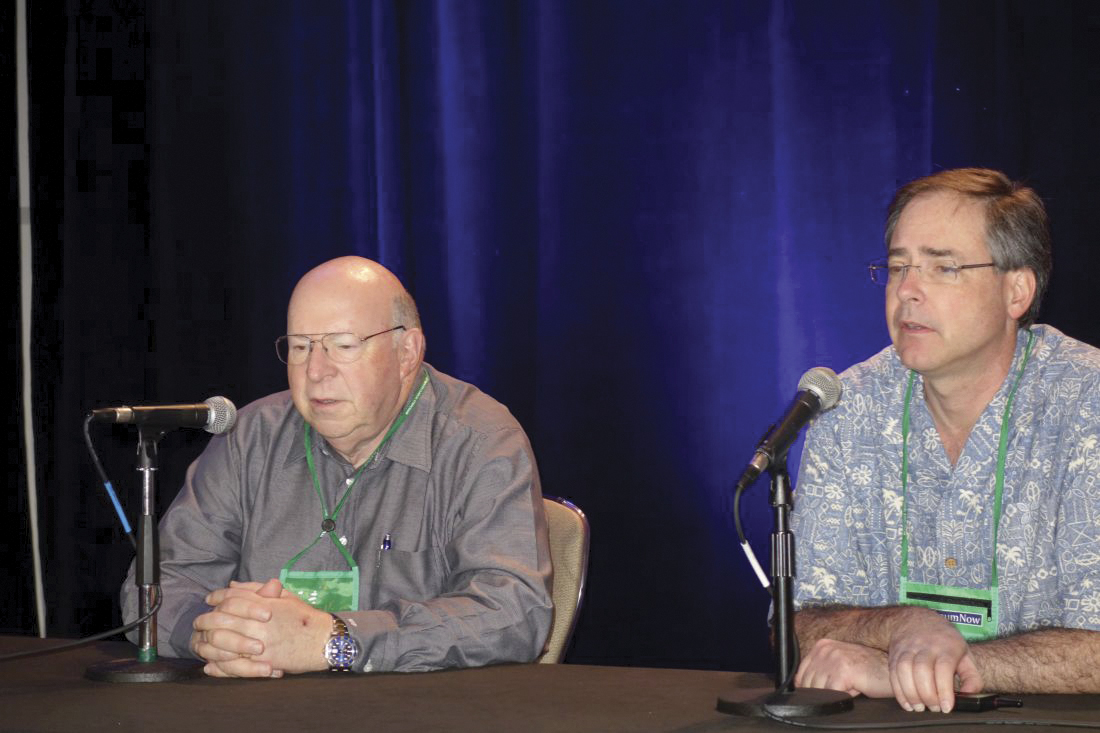
Rheumatoid arthritis treatment less aggressive, not less favorable in older adults
BIRMINGHAM, ENGLAND – Being diagnosed with rheumatoid arthritis at age 75 years or older made it less likely that patients would receive intensive therapy than their younger counterparts, but that did not mean that they were treated any less favorably overall, according to findings derived from the Early RA Network cohort.
“The claim that the elderly are treated less aggressively isn’t completely true throughout the whole treat-to-target strategy,” said Simone Howard of King’s College London at the British Society for Rheumatology annual conference. While older patients were less likely to receive intensive treatment up to 2 years after their diagnosis, there was a shorter delay between the onset of symptoms and the first outpatient visit to a rheumatology clinic.
When compared against patients who were younger than 65 years, those aged 65-74 years and 75 years and older were 11% (P = .02) and 15% (P = .02) more likely to have their first outpatient visit within 10 months.
Furthermore, no significant differences were seen between any age groups in the time to first initiation of a conventional synthetic disease-modifying antirheumatic drug (csDMARD), which averaged nearly 3 months after symptoms appeared.
Ms. Howard, who has previously worked at the European Medicines Agency, noted that, during her time at the EMA, “there was a real push to incorporate the elderly into the regulatory framework more. In parallel, there were also reports of the elderly being treated less aggressively. So the question was, where was that coming from?”
Similar therapeutic approaches are advocated for older and younger RA patients, and to look for any disparities, Ms. Howard and associates turned to the Early RA Network (ERAN) to “investigate potential treatment bias against the elderly.”
ERAN is a hospital-based inception cohort of 1,236 patients with early RA who were recruited across 23 centers in the United Kingdom and Ireland between 2002 and 2014.
Of 1,131 patients used in the analyses, 9.7% (n = 110) were 75 years or older, 21.5% (n = 243) were aged 65-74 years, and 68.8% (n = 778) were 65 years or younger. The majority (67.7%) of patients were female.
Patients aged 75 years and older were more likely to present with comorbidities than the youngest group, and they had higher health assessment questionnaire scores at baseline. However, they were no more likely to have high disease activity at the first visit, which was defined as a disease activity score in 28 joints of more than 5, and the older patients were 27% less likely to be seropositive (P = .004).
“It’s when we come to pharmacological aspects of care that we are seeing treatment biases,” Ms. Howard noted. Patients over 75 years were significantly more likely than the youngest age group to be treated with glucocorticoids or csDMARD monotherapy at 1 year, and 23% more likely to be on less aggressive therapy (P equal to or less than .0001). Aggressive therapy was defined as the use of a combination of csDMARDs or the use of biologic drugs.
At 2 years, the oldest patients were 46% more likely than those under 65 years to be on less-intensive therapies (P equal to or less than .0001), with those aged 65-74 years 19% more likely to be on glucocorticoid or csDMARD therapy (P = .005).
Factors such as patient choice and tolerance were not considered in the analyses and could be important, Ms. Howard conceded in response to a question after her presentation.
Another point raised was that perhaps the prescribing of aggressive therapy would rationally be different in someone diagnosed with RA at age 85 versus 65 because the duration of time that would be likely to be lived with accumulating joint damage would be shorter at the older age and that would be balanced against the other effects of the therapy. So, there may be important reasons in shared decision making that influenced the treatment choices other than the age of patients.
Ms. Howard agreed, noting that this demonstrated the need to be careful around the language used for defining what constituted aggressive or intensive therapy.
She and her coauthors reported no conflicts of interest.
SOURCE: Howard S et al. Rheumatology. 2019;58(suppl 3), Abstract 011.
REPORTING FROM BSR 2019
Methotrexate does not cause rheumatoid interstitial lung disease
BIRMINGHAM, ENGLAND – Data from two early RA inception cohorts provide reassurance that methotrexate does not cause interstitial lung disease and suggest that treatment with methotrexate might even be protective.
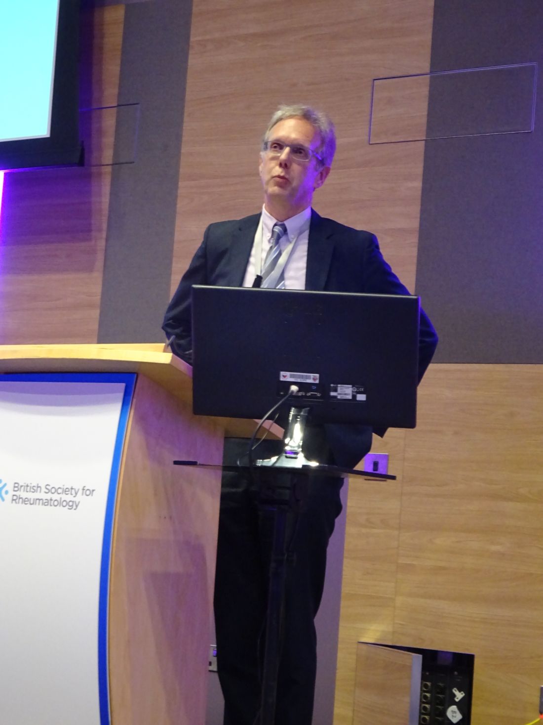
In the Early RA Study (ERAS) and Early RA Network (ERAN), which together include 2,701 patients with RA, 101 (3.7%) had interstitial lung disease (ILD). There were 92 patients with RA-ILD who had information available on exposure to any conventional synthetic disease-modifying antirheumatic drug (csDMARD); of these, 39 (2.5%) had been exposed to methotrexate (n = 1,578) and 53 (4.8%) to other csDMARDs (n = 1,114).
Multivariate analysis showed that methotrexate exposure was associated with a reduced risk of developing ILD, with an odds ratio of 0.48 (P = .004). In a separate analysis that excluded 25 patients who had ILD before they received any csDMARD therapy (n = 67), there was no association between methotrexate use and ILD (OR, 0.85; P = .578). In fact, there was a nonsignificant trend for a delayed onset of ILD in patients who had been treated with methotrexate (OR, 0.54; P = .072).
Methotrexate use is associated with an acute hypersensitivity pneumonitis in patients with RA, explained Patrick Kiely, MBBS, PhD, of St. George’s University Hospitals NHS Foundation Trust in London at the British Society for Rheumatology annual conference. “This is well recognized, it’s very rare [0.43%-1.00%], it’s easy to spot, and usually goes away if you stop methotrexate,” said Dr. Kiely, adding that “it’s not benign, and severe cases can be life threatening.”
Because of the association between methotrexate and pneumonitis, there has been concern that methotrexate may exacerbate or even cause ILD in RA but there are sparse data available to confirm this. The bottom line is that you should not start someone on methotrexate if you think their existing lung capacity is not up to treatment with methotrexate, Dr. Kiely said.
ILD is not always symptomatic in RA, but when it is, it is associated with very poor survival. The lung disease can be present before joint symptoms, Dr. Kiely said. Although less than 10% of cases may be symptomatic, this “is a big deal, because it has a high mortality, with death within 5 years. It’s the second-commonest cause of excess mortality in RA after cardiovascular disease.”
To look at the association between incident RA-ILD and the use of methotrexate, Dr. Kiely and associates analyzed data from ERAS (1986-2001) and ERAN (2002-2013), that together have more than 25 years of follow-up data on patients who were recruited at the first sign of RA symptoms. Patients within these cohorts have been treated according to best practice, and a range of outcomes – including RA-ILD – have been assessed at annual intervals.
In the patients who developed ILD after any csDMARD exposure, older age at RA onset (OR, 1.04; P less than .001) and having ever smoked (OR, 1.91; P = .016) were associated with the development of the lung disease. Incident ILD was also associated with being positive for rheumatoid factor (OR, 2.02; P = .029) at baseline. Being male was also associated with a higher risk for developing ILD, Dr. Kiely reported, as was a longer duration of time between the onset of first RA symptoms and the first secondary care visit. Conversely, the presence of nonrespiratory, major comorbidities at baseline appeared to be protective (OR, 0.62; P = .027).
“We found no association between methotrexate treatment and incident RA-ILD and a possibility that it may be protective,” Dr. Kiely concluded, noting that these data were now published in BMJ Open (2019;9:e028466. doi: 10.1136/bmjopen-2018-028466).
Following Dr. Kiely’s presentation, an audience member asked if the protective effect seen with methotrexate could have been caused by better disease control overall.
Dr. Kiely answered that, up until 2001, the time when ERAS was ongoing, standard practice in the United Kingdom was to use sulfasalazine, but then methotrexate started to be used in higher and higher doses, as seen in ERAN.
The interesting thing is that in ERAN more methotrexate was used in higher doses, but less RA-ILD was seen, Dr. Kiely observed. The overall prevalence of RA-ILD in the later early RA cohort was 3.2% and the median dose of methotrexate used was 20 mg. In ERAS, the prevalence was 4.2% and the median dose of methotrexate used was 10 mg.
There was a suggestion that disease control was slightly better in ERAN than ERAS, but that wasn’t statistically significant, Dr. Kiely said.
So, should a patient with RA and ILD be given methotrexate? There’s no reason not to, Dr. Kiely suggested, based on the evidence shown. Part of the challenge will now be convincing chest physician colleagues that methotrexate is not problematic in terms of causing ILD.
These findings are completely on board with the ILD group’s findings that methotrexate doesn’t cause pulmonary fibrosis in patients with RA, commented Julie Dawson, MD, of St. Helens and Knowsley Teaching Hospitals NHS Trust, St. Helens, England. Her own research, which includes a 10-year follow-up of patients with inflammatory arthritis, has shown that methotrexate does not appear to increase the risk of pulmonary fibrosis.
The study had no specific outside funding. Dr. Kiely reported having no conflicts of interest.
SOURCE: Kiely P et al. Rheumatology. 2019;58(suppl 3), Abstract 009.
BIRMINGHAM, ENGLAND – Data from two early RA inception cohorts provide reassurance that methotrexate does not cause interstitial lung disease and suggest that treatment with methotrexate might even be protective.
In the Early RA Study (ERAS) and Early RA Network (ERAN), which together include 2,701 patients with RA, 101 (3.7%) had interstitial lung disease (ILD). There were 92 patients with RA-ILD who had information available on exposure to any conventional synthetic disease-modifying antirheumatic drug (csDMARD); of these, 39 (2.5%) had been exposed to methotrexate (n = 1,578) and 53 (4.8%) to other csDMARDs (n = 1,114).
Multivariate analysis showed that methotrexate exposure was associated with a reduced risk of developing ILD, with an odds ratio of 0.48 (P = .004). In a separate analysis that excluded 25 patients who had ILD before they received any csDMARD therapy (n = 67), there was no association between methotrexate use and ILD (OR, 0.85; P = .578). In fact, there was a nonsignificant trend for a delayed onset of ILD in patients who had been treated with methotrexate (OR, 0.54; P = .072).
Methotrexate use is associated with an acute hypersensitivity pneumonitis in patients with RA, explained Patrick Kiely, MBBS, PhD, of St. George’s University Hospitals NHS Foundation Trust in London at the British Society for Rheumatology annual conference. “This is well recognized, it’s very rare [0.43%-1.00%], it’s easy to spot, and usually goes away if you stop methotrexate,” said Dr. Kiely, adding that “it’s not benign, and severe cases can be life threatening.”
Because of the association between methotrexate and pneumonitis, there has been concern that methotrexate may exacerbate or even cause ILD in RA but there are sparse data available to confirm this. The bottom line is that you should not start someone on methotrexate if you think their existing lung capacity is not up to treatment with methotrexate, Dr. Kiely said.
ILD is not always symptomatic in RA, but when it is, it is associated with very poor survival. The lung disease can be present before joint symptoms, Dr. Kiely said. Although less than 10% of cases may be symptomatic, this “is a big deal, because it has a high mortality, with death within 5 years. It’s the second-commonest cause of excess mortality in RA after cardiovascular disease.”
To look at the association between incident RA-ILD and the use of methotrexate, Dr. Kiely and associates analyzed data from ERAS (1986-2001) and ERAN (2002-2013), that together have more than 25 years of follow-up data on patients who were recruited at the first sign of RA symptoms. Patients within these cohorts have been treated according to best practice, and a range of outcomes – including RA-ILD – have been assessed at annual intervals.
In the patients who developed ILD after any csDMARD exposure, older age at RA onset (OR, 1.04; P less than .001) and having ever smoked (OR, 1.91; P = .016) were associated with the development of the lung disease. Incident ILD was also associated with being positive for rheumatoid factor (OR, 2.02; P = .029) at baseline. Being male was also associated with a higher risk for developing ILD, Dr. Kiely reported, as was a longer duration of time between the onset of first RA symptoms and the first secondary care visit. Conversely, the presence of nonrespiratory, major comorbidities at baseline appeared to be protective (OR, 0.62; P = .027).
“We found no association between methotrexate treatment and incident RA-ILD and a possibility that it may be protective,” Dr. Kiely concluded, noting that these data were now published in BMJ Open (2019;9:e028466. doi: 10.1136/bmjopen-2018-028466).
Following Dr. Kiely’s presentation, an audience member asked if the protective effect seen with methotrexate could have been caused by better disease control overall.
Dr. Kiely answered that, up until 2001, the time when ERAS was ongoing, standard practice in the United Kingdom was to use sulfasalazine, but then methotrexate started to be used in higher and higher doses, as seen in ERAN.
The interesting thing is that in ERAN more methotrexate was used in higher doses, but less RA-ILD was seen, Dr. Kiely observed. The overall prevalence of RA-ILD in the later early RA cohort was 3.2% and the median dose of methotrexate used was 20 mg. In ERAS, the prevalence was 4.2% and the median dose of methotrexate used was 10 mg.
There was a suggestion that disease control was slightly better in ERAN than ERAS, but that wasn’t statistically significant, Dr. Kiely said.
So, should a patient with RA and ILD be given methotrexate? There’s no reason not to, Dr. Kiely suggested, based on the evidence shown. Part of the challenge will now be convincing chest physician colleagues that methotrexate is not problematic in terms of causing ILD.
These findings are completely on board with the ILD group’s findings that methotrexate doesn’t cause pulmonary fibrosis in patients with RA, commented Julie Dawson, MD, of St. Helens and Knowsley Teaching Hospitals NHS Trust, St. Helens, England. Her own research, which includes a 10-year follow-up of patients with inflammatory arthritis, has shown that methotrexate does not appear to increase the risk of pulmonary fibrosis.
The study had no specific outside funding. Dr. Kiely reported having no conflicts of interest.
SOURCE: Kiely P et al. Rheumatology. 2019;58(suppl 3), Abstract 009.
REPORTING FROM BSR 2019