User login
VIDEO: When early-stage vulvar cancer recurs, the prognosis is poor
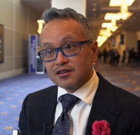
NATIONAL HARBOR, MD – Cancer of the vulva is becoming more common, and as incidence is rising, so are death rates from the relatively rare malignancy. A new examination of women with early-stage vulvar cancer showed that recurrence was common, and carried a poor prognosis.
Cancer in “nearly 25% of women with stage IB squamous cell carcinoma of the vulva will recur within 3 years of initial diagnosis,” said the study’s lead author, Rebecca Stone, MD, at the annual meeting of the Society of Gynecologic Oncology. Although most (85%) of the cancers that recur do so locally, salvage rates for patients with recurrences are poor, she said in a video interview.
Dr. Stone of Johns Hopkins University, Baltimore, said that the study looked at the subset of 59 patients with recurrence of their stage IB vulvar cancer, drawing from a total of 244 stage IB patients for whom complete data were available.
Current National Comprehensive Cancer Network (NCCN) guidelines for the treatment of early-stage vulvar squamous cell carcinoma (meaning T1 or 4 cm or smaller T2 lesions) call for adjuvant radiotherapy for patients with positive margins or unresectable lesions. For patients with negative margins after the primary procedure or re-excision, the guidelines say that radiotherapy decisions should be guided by risk factors including margin width, tumor size, the presence of lymphovascular invasion, and the pattern and depth of invasion.
However, Dr. Stone said, “There’s a knowledge gap in our current management of early-stage disease.” Some unknowns include what the threshold should be for tumor recurrence in terms of depth of invasion and size of tumor, whether perineural invasion is a risk factor, and whether adverse clinical factors are important. Also, she said, it’s not known how risk factors combine to increase recurrence risk.
Tumor size, invasion depth over 6 mm, and patient age were all risk factors identified by the study; perineural and lymphovascular invasion were also potential risk factors for recurrence.
The retrospective study, said Dr. Stone, is the largest multi-institutional cohort study examining stage IB vulvar cancer that has been completed, and it accomplished a detailed examination of which patients have recurrences, factors associated with recurrence, and postrecurrence salvage rates.
Dr. Stone reported no disclosures.
The video associated with this article is no longer available on this site. Please view all of our videos on the MDedge YouTube channel
koakes@frontlinemedcom.com
On Twitter @karioakes
AT THE ANNUAL MEETING ON WOMEN’S CANCER
VIDEO: How to start an oncology sexual health clinic
NATIONAL HARBOR, MD – Start small, but anticipate growth. Engage your administration from the start. Be smart about resources, and consider using advanced practice providers to keep costs down. Above all, keep lines of communication open with physicians and other members of the care team.
In a video interview, Joanne Rash, PA-C, a certified physician assistant at the University of Wisconsin–Madison offers these and other tips. She explains her collaborative work with David Kushner, MD, director of the gynecologic oncology program and professor at the University of Wisconsin School of Medicine and Public Health, and a colleague to develop the Women’s Integrative Sexual Health (WISH) program.
WISH is modeled on the University of Chicago’s Program in Integrative Sex and Medicine for Women and Girls with Cancer (PRISM) and participates in the PRISM registry, which studies ways to prevent and treat sexual problems for women and girls with cancer.
“I think what makes the WISH program unique is that we carve time out,” said Ms. Rash. “Of course, we address some of these issues in my gynecologic oncology practice, but, when we do it in WISH, the format is different,” and there’s just more time for discussion.
Communication is key to the model’s success in safe integration of sexual health into cancer care, she said. “We certainly don’t want to do something that compromises cancer care, and so, it’s important that we have those conversations with that woman’s team. And now we get to be a part of that team, which is a real privilege.”
Ms. Rash reported no conflicts of interest.
The video associated with this article is no longer available on this site. Please view all of our videos on the MDedge YouTube channel
koakes@frontlinemedcom.com
On Twitter @karioakes
AT THE ANNUAL MEETING ON WOMEN’S CANCER
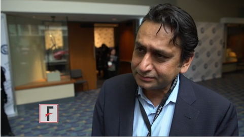
VIDEO: Sex, intimacy part of history in gynecologic oncology care
NATIONAL HARBOR, MD. – Cancer care can feel like a sequence of crises, large and small; a comprehensive cancer care approach can keep the patient at the center of this flurry of events. But understanding a patient’s sexual history and assessing how a gynecologic malignancy has affected that patient’s sexual health can, too often, get lost in the shuffle, said Don Dizon, MD, of Massachusetts General Hospital, Boston.
“I think there are multiple reasons for that, but it has to be the job of the clinician – not necessarily the oncologist-physician – but somebody has to be able to say, ‘What is your sexual history? Can I ask about your sexual history?’ ” he said in a video interview at the annual meeting of the Society of Gynecologic Oncology.
Dr. Dizon, director of the Oncology Sexual Health Clinic at Massachusetts General Hospital*, emphasized the importance of providing sensitive sexual health care while staying true to one’s own framework of care. “I teach clinicians not to run away from their schema,” he said. “Having said that, the job of a clinician is to normalize the history, and to normalize that sex, intimacy, relationships are a part of someone’s history.”
Dr. Dizon sits on the board of the Patty Brisben Foundation and the Young Survival Coalition.
koakes@frontlinemedcom.com
On Twitter @karioakes
*A previous version of this article misstated Dr. Dizon's affiliation.
AT THE ANNUAL MEETING ON WOMEN'S CANCER
Video: Try ‘PLISSIT’ to address postcancer sexual health
NATIONAL HARBOR, MD. – Comprehensive care of patients with gynecologic malignancies should include a sensitive and thorough assessment of sexual health.
In a video interview at the annual meeting of the Society of Gynecologic Oncology, Don Dizon, MD, of Massachusetts General Hospital, Boston, gives a series of practical tips to help physicians take a thorough sexual health history and provide information and guidance for patients and their partners.
Dr. Dizon, professor of gynecologic oncology and director of the oncology sexual health clinic at Brigham and Women’s Hospital, also in Boston, said that he likes to begin with the PLISSIT model, where patients are given permission (P) to talk about sexual problems. Then, the clinician gives the patient limited (LI) scientific or clinical information about the situation, followed by specific suggestions (SS) that might help. Finally, patients may be referred to mental health providers or sex counselors for intensive therapy (IT) if needed.
“It’s also important not to confuse terminology,” said Dr. Dizon. “Intimacy is experienced very differently between men and women. Women experience intimacy through arousal, desire, and, when desire is satisfied, that’s intimacy. Intercourse is not a part of that equation.” For men, he said, intimacy is more often experienced through intercourse. “So the disconnect is greater after cancer is diagnosed,” making it especially important to acknowledge problems sensitively, and to helps patients and partners find a way forward.
“The word I like to use is ‘play,’ ” said Dr. Dizon. When a renegotiation of an intimate relationship is framed in terms of play, the pressure is off, and “men can wrap their hands around that idea,” he said.
Dr. Dizon sits on the board of the Patty Brisben Foundation and the Young Survival Coalition.
The video associated with this article is no longer available on this site. Please view all of our videos on the MDedge YouTube channel
koakes@frontlinemedcom.com
On Twitter @karioakes
AT THE ANNUAL MEETING ON WOMEN’S CANCER
VIDEO: Picowave laser uses are expanding beyond tattoo removal
ORLANDO – The applications for picowavelength lasers are expanding, with emerging data on their uses for cosmetic indications other than tattoo removal, according to Anne Chapas, MD, who is in private practice in New York.
First introduced and cleared by the Food and Drug Administration for tattoo removal, “picowave devices ... are now being studied in multiple different cosmetic conditions, including their use in acne scars, fine lines and wrinkles, and melasma,” Dr. Chapas said in a video interview at the annual meeting of the American Academy of Dermatology.
The No. 1 thing dermatologists need to know is that these types of lasers are delivering energy extremely quickly,” at 1,000 times faster than nanosecond lasers, she said. Another difference between the two is that “the laser tissue interaction between the two types of devices is completely different.”
In the interview, she highlighted other important points about picowave lasers, including less downtime after treatment.
At the meeting, Dr. Chapas spoke during a session entitled “the Science Behind New Devices in Dermatology.”
Her disclosures include serving as a consultant and investigator for Syneron and Candela.
The video associated with this article is no longer available on this site. Please view all of our videos on the MDedge YouTube channel
AT AAD 17

VIDEO: Internet-based intervention shows antihypertensive efficacy
WASHINGTON – Patients who regularly accessed 30 minute, Internet-based behavioral counseling videos cut their systolic blood pressure, compared with baseline over 1 year by an average 4 mm Hg more than control patients in a randomized, phase II study with 264 patients.
Electronic counseling (e-counseling) “enhanced the efficacy of usual care for hypertension,” Robert P. Nolan, Ph.D., said at the annual meeting of the American College of Cardiology.
“We hope to optimize the efficacy of medical treatments with a behavioral intervention,” said Dr. Nolan, who added that the magnitude of the added benefit from the e-counseling program was “like adding an additional antihypertensive medication.”
“We know antihypertensive treatments work, but compliance is a huge challenge” for health care providers, commented E. Magnus Ohman, MBBS, a professor and cardiologist at Duke University in Durham, N.C. “Having a new way to enhance compliance would be fantastic,” he said.
The Internet-based counseling program devised by Dr. Nolan and his associates consisted of a year-long series of 28 videos, each about 30 minutes long, that participants in the active arm accessed over the Internet. During the study, participants received a series of emailed messages that sent links to the videos on a set schedule over 12 months: During the first 4 months they received an emailed link weekly, during the next 4 months they received an emailed link to a new video every other week, and during the final 4 months of the intervention participants received emailed links once a month. Patients could access each video more than once if they wished, and they were free to share the links with any family members or friends who helped the patients with lifestyle management of their hypertension.
Patients in the e-counseling intervention arm received links to videos that focused on motivational messages and teaching cognitive behavioral skills. Patients in the control arm received emails with generic messages on blood pressure management and without links to videos.
The REACH (E-Counseling Promotes Blood Pressure Reduction and Therapeutic Lifestyle Change in Hypertension) study ran at four Canadian sites. It sent invitations to participate to 609 patients with stage 1 or 2 hypertension, with a blood pressure prior to treatment of 140/90-180/110 mm Hg. Among the invited patients, 264 elected to participate; 100 patients in the e-counseling arm and 97 control patients completed the 1-year program. Participants averaged 58 years of age, their average body mass index was 31 kg/m2, and 9% smoked. Their blood pressure at entry averaged 141/87 mm Hg, their average pulse pressure was about 54 mm Hg, and their average 10-year risk for a cardiovascular disease event, measured by the Framingham Risk Score, was about 16%. At entry, patients in the study received an average of 1.5 antihypertensive drugs each.
At the end of the 1-year program, systolic blood pressure fell by an average of 6 mm Hg from baseline among patients who completed the control program, and by an average of 10.1 mm Hg among the patients who completed the e-counseling arm, a statistically significant difference for one of the study’s primary endpoints. Change in pulse pressure from baseline showed an average 1.5–mm Hg drop in the control patients and an average 4.3–mm Hg decline in the e-counseling patients, another statistically significant difference for a second primary endpoint, reported Dr. Nolan, a clinical psychologist and director of the cardiac eHealth program at the University of Toronto.
A third primary endpoint was change in the Framingham Risk Score, which fell by an average of 1.9% after 12 months in the e-counseling patients and rose by an average of 0.2% among the controls.
The final primary endpoint was the change in diastolic blood pressure from baseline, which showed a better than 4–mm Hg incremental decline in the men who received e-counseling, compared with controls, but among women in the study, the drop in diastolic blood pressure from baseline was nearly the same – about 6 mm Hg – in both the controls and e-counseling patients.
“This tells us that we need to better tailor the [e-counseling] intervention to men and to women,” Dr. Nolan said in a video interview. He also envisions better tailoring of the e-counseling videos to various socioeconomic and ethnic groups. He plans to continue testing of a revised version of the e-counseling intervention in a larger, phase III study, but he also hopes that the intervention videos can soon be available at no charge for use in routine practice.
REACH received no commercial funding. Dr. Nolan had no disclosures.
The video associated with this article is no longer available on this site. Please view all of our videos on the MDedge YouTube channel
mzoler@frontlinemedcom.com
On Twitter @mitchelzoler
I love the REACH study. Hypertension is incredibly prevalent among the patients I see, and they often need three different antihypertensive drugs to control their blood pressure. Lifestyle interventions can be very effective at helping to lower blood pressure, but during the 10-minute visits I have with most of my patients, it’s hard for me to have much impact on their behavior.
What I especially like about the Internet-based counseling used in this study was its application of evidence-based approaches to change patient behavior. This was a well-designed and exciting trial.
Sandra J. Lewis, MD, chief of cardiology at Legacy Good Samaritan Hospital in Portland, Ore., made these comments during a press conference. She had no disclosures.
WASHINGTON – Patients who regularly accessed 30 minute, Internet-based behavioral counseling videos cut their systolic blood pressure, compared with baseline over 1 year by an average 4 mm Hg more than control patients in a randomized, phase II study with 264 patients.
Electronic counseling (e-counseling) “enhanced the efficacy of usual care for hypertension,” Robert P. Nolan, Ph.D., said at the annual meeting of the American College of Cardiology.
“We hope to optimize the efficacy of medical treatments with a behavioral intervention,” said Dr. Nolan, who added that the magnitude of the added benefit from the e-counseling program was “like adding an additional antihypertensive medication.”
“We know antihypertensive treatments work, but compliance is a huge challenge” for health care providers, commented E. Magnus Ohman, MBBS, a professor and cardiologist at Duke University in Durham, N.C. “Having a new way to enhance compliance would be fantastic,” he said.
The Internet-based counseling program devised by Dr. Nolan and his associates consisted of a year-long series of 28 videos, each about 30 minutes long, that participants in the active arm accessed over the Internet. During the study, participants received a series of emailed messages that sent links to the videos on a set schedule over 12 months: During the first 4 months they received an emailed link weekly, during the next 4 months they received an emailed link to a new video every other week, and during the final 4 months of the intervention participants received emailed links once a month. Patients could access each video more than once if they wished, and they were free to share the links with any family members or friends who helped the patients with lifestyle management of their hypertension.
Patients in the e-counseling intervention arm received links to videos that focused on motivational messages and teaching cognitive behavioral skills. Patients in the control arm received emails with generic messages on blood pressure management and without links to videos.
The REACH (E-Counseling Promotes Blood Pressure Reduction and Therapeutic Lifestyle Change in Hypertension) study ran at four Canadian sites. It sent invitations to participate to 609 patients with stage 1 or 2 hypertension, with a blood pressure prior to treatment of 140/90-180/110 mm Hg. Among the invited patients, 264 elected to participate; 100 patients in the e-counseling arm and 97 control patients completed the 1-year program. Participants averaged 58 years of age, their average body mass index was 31 kg/m2, and 9% smoked. Their blood pressure at entry averaged 141/87 mm Hg, their average pulse pressure was about 54 mm Hg, and their average 10-year risk for a cardiovascular disease event, measured by the Framingham Risk Score, was about 16%. At entry, patients in the study received an average of 1.5 antihypertensive drugs each.
At the end of the 1-year program, systolic blood pressure fell by an average of 6 mm Hg from baseline among patients who completed the control program, and by an average of 10.1 mm Hg among the patients who completed the e-counseling arm, a statistically significant difference for one of the study’s primary endpoints. Change in pulse pressure from baseline showed an average 1.5–mm Hg drop in the control patients and an average 4.3–mm Hg decline in the e-counseling patients, another statistically significant difference for a second primary endpoint, reported Dr. Nolan, a clinical psychologist and director of the cardiac eHealth program at the University of Toronto.
A third primary endpoint was change in the Framingham Risk Score, which fell by an average of 1.9% after 12 months in the e-counseling patients and rose by an average of 0.2% among the controls.
The final primary endpoint was the change in diastolic blood pressure from baseline, which showed a better than 4–mm Hg incremental decline in the men who received e-counseling, compared with controls, but among women in the study, the drop in diastolic blood pressure from baseline was nearly the same – about 6 mm Hg – in both the controls and e-counseling patients.
“This tells us that we need to better tailor the [e-counseling] intervention to men and to women,” Dr. Nolan said in a video interview. He also envisions better tailoring of the e-counseling videos to various socioeconomic and ethnic groups. He plans to continue testing of a revised version of the e-counseling intervention in a larger, phase III study, but he also hopes that the intervention videos can soon be available at no charge for use in routine practice.
REACH received no commercial funding. Dr. Nolan had no disclosures.
The video associated with this article is no longer available on this site. Please view all of our videos on the MDedge YouTube channel
mzoler@frontlinemedcom.com
On Twitter @mitchelzoler
AT ACC 17
Key clinical point:
Major finding: The Internet-based program reduced systolic blood pressure from baseline by an average additional 4.1 mm Hg, compared with controls.
Data source: REACH, a multicenter, randomized trial with 264 hypertensive patients.
Disclosures: REACH received no commercial funding. Dr. Nolan had no disclosures.

VIDEO: Looking at keloids from a different perspective
ORLANDO – It may be time to start considering new options for treating keloids, according to Amy McMichael, MD, professor of dermatology at Wake Forest University, Winston-Salem, N.C.
At the annual meeting of the American Academy of Dermatology, Dr. McMichael discussed one of the highlights of the annual symposium of the Skin of Color Society, held right before the annual meeting, a presentation by Michael Tirgan, MD, who has treated patients with keloids for about 10 years.
Dr. Tirgan, an oncologist based in New York, has a large database and registry of patients and shared some interesting data at the symposium. “What he’s found is that those who come with the supermassive and massive keloids are those who have had the most surgery on their keloid,” Dr. McMichael said in a video interview at the meeting.
His findings shed light on what she described as “a new perspective in the way that we think about keloids” and a new approach to treatment – considering keloids as tumors – not just a scar.
Dr. McMichael had no relevant disclosures.
The video associated with this article is no longer available on this site. Please view all of our videos on the MDedge YouTube channel
AT AAD 17

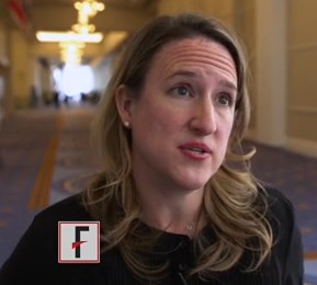
VIDEO: Pain and impaired QOL persist after open endometrial cancer surgery
NATIONAL HARBOR, MD. – Patient-reported outcomes from a prospective cohort study of minimally invasive versus open surgery for women with endometrial cancer showed that the disability from open surgery persisted for longer than had previously been recognized. Further, for a subset of patients, impairment in sexual functioning was significant, and persistent, regardless of the type of surgery.
At 3 weeks after surgery, patients who had open surgery had greater pain as measured by the Brief Pain Inventory (minimally important difference greater than 1, P = .0004). By 3 months post surgery, responses on the Functional Assessment of Cancer Therapy–General were still significantly lower for the open-surgery group, compared with the minimally invasive group (P = .0011).
Although patients’ pain and overall state of health were better at 3 weeks post surgery, regardless of whether women had open, laparoscopic, or robotic surgery, the reduced overall quality of life experienced by patients who had open surgery persisted.
“What was a bit different from other studies … is that we found that this is maintained even at 3 months, and it was clinically and statistically different,” Sarah Ferguson, MD, said in a video interview at the annual meeting of the Society of Gynecologic Oncology. “So that was really, I think, an interesting finding, that this doesn’t just impact the very short term. Three months is a fairly long time after a primary surgery, and [it’s] important for women to know this.”
The video associated with this article is no longer available on this site. Please view all of our videos on the MDedge YouTube channel
Patients in the eight-center study had histologically confirmed clinical stage I or II endometrial cancer. The open-surgery arm of the study involved 106 patients, and 414 had minimally invasive surgery (168 laparascopic, 246 robotic).
The robotic and laparoscopic arms showed no statistically significant differences for any patient-reported outcome, even after adjusting for potentially confounding variables, said Dr. Ferguson of Princess Margaret Cancer Centre at the University of Toronto. Accordingly, investigators compared both minimally invasive arms grouped together against open surgery.
Overall, about 80% of patients completed the quality-of-life questionnaires. The response rate for the sexual-functioning questionnaires, however, was much lower, ranging from about a quarter to a half of the participants.
When Dr. Ferguson and her colleagues examined the characteristics of the patients who did complete the sexual-functioning questionnaires, they found that these women were more likely to be younger, partnered, premenopausal and sexually active at the time of diagnosis. Both of the surgical groups “met the clinical cutoff for sexual dysfunction” on the Female Sexual Function Index questionnaire, she said.
For the sexual function questionnaires, differences between the open and minimally invasive groups were not significant at any time point throughout the 26 weeks that patients were studied. “Though it’s a small population, I think these results are important,” said Dr. Ferguson. “These variables may be helpful for us to target patients in our practice, or in future studies, who require intervention.”
Though the study was not randomized, Dr. Ferguson said that the baseline characteristics were similar between groups, and the investigators’ intention-to-treat analysis used a statistical model that adjusted for many potential confounding variables.
Dr. Ferguson reported having no conflicts of interest.
koakes@frontlinemedcom.com
On Twitter @karioakes
AT THE ANNUAL MEETING ON WOMEN'S CANCER

VIDEO: Postop troponin T spike flags high mortality risk
WASHINGTON – A rise in the blood level of troponin T immediately after patients underwent noncardiac surgery identified a high risk group with a 30-day mortality rate nearly fivefold higher than that of patients who did not have a postoperative troponin T spike, according to a prospective study of more than 21,000 patients.
For 93% of the patients who have these perioperative spikes in troponin T, a marker of myocardial ischemia, the increased level was the only indicator of a heart problem, P.J. Devereaux, MD, said at the annual meeting of the American College of Cardiology. The painkillers that patients receive following surgery generally mask the chest discomfort they might otherwise feel from their heart damage, he explained. The clinical condition is called myocardial injury after noncardiac surgery.
“We’re seeing more older patients with a high burden of vascular disease undergoing surgery, and surgery is a very significant stress, so a large proportion of these patients will have [myocardial injury after noncardiac surgery] and that affects 30-day survival,” said Dr. Devereaux, professor and director of cardiology at McMaster University in Hamilton, Ont.
Based on the new findings, he recommended performing a baseline assessment of troponin T levels in patients scheduled for noncardiac surgery if they are at least 65 years of age, or if they are age 45-64 years with known vascular disease, followed by repeat testing 1 and 2 days after surgery to check whether a spike in the measure had occurred. The high sensitivity troponin T (hsTnT) test he used in the study is relatively costly (and received Food and Drug Administration approval for U.S. marketing in January 2017), but Dr. Devereaux believed that, used in this way, the cost for testing would be reasonable, given its powerful ability to identify high-risk patients and relative to the cost of other screening tools routinely used in U.S. medical practice.
“It looks very cost-effective,” he said in a video interview.
If the baseline and two follow-up measures of hsTnT showed a postoperative level of at least 20 ng/L that rose above the baseline level by at least 5 ng/L, or if the postoperative level was at least 65 ng/L, Dr. Devereaux recommended starting daily treatment with aspirin and a statin to try to contain any perioperative myocardial damage the patient may have, and follow with comprehensive assessment of the patient by a cardiologist or other internal medicine physician.
“Given the risks associated with a rise in hsTnT in this study, Dr. Devereaux’s recommendations are very reasonable until we collect more data on this,” said Frank W. Sellke, MD, professor of surgery and chief of cardiothoracic surgery at Brown Medical School and the Lifespan Hospitals in Providence, R.I. “What was surprising was how few patients had symptoms” of myocardial ischemia. “You can’t do hsTnT measurements on every patient who goes in for a hernia operation; it’s not practical. But his findings are fairly compelling, and hopefully the cost of this testing will come down,” Dr. Sellke said in an interview.
In the multicenter study of 21,842 patients, 24% had a postoperative hsTnT level of 20 ng/L or greater, including 5% with a level of 65 ng/L or greater. The 30-day mortality rate was 3% among those with a perioperative level of 20-64 ng/L, 9% among patients with perioperative hsTnT levels of 65-999 ng/L, and 30% among the 54 patients (0.2% of the study group) with perioperative levels that reached 1,000 ng/L or greater. Dr. Devereaux reported.
This iteration of the Vascular Events In Noncardiac Surgery Patients Cohort Evaluation Study (VISION) enrolled patients who were at least 45 years of age and underwent noncardiac surgery at 23 centers in 13 countries, including the United States and Canada. All patients underwent hsTnT testing 6-12 hours after surgery and 1, 2, and 3 days after surgery, but only 40% also had a baseline measurement before their surgery began. Full 30-day follow-up occurred for 21,050. The patients’ average age was 63 years. The most common surgery was “low-risk,” in 35%, followed by “major” general surgery in 20%, and “major” orthopedic surgery in 16%. At 30 days, 266 patients (1.2%) had died.
These findings “help define a cutoff for hsTnT that will be clinically useful to change practice,” said Athena Poppas, MD, a cardiologist and director of the Cardiovascular Institute at Rhode Island Hospital in Providence. Dr. Poppas was a designated discussant for Dr. Devereaux’s report at the meeting.
In January 2017, the Canadian Cardiovascular Society issued guidelines for perioperative cardiac risk assessment and management for patients undergoing noncardiac surgery (Can J Cardiol. 2017 Jan;33[1]:17-32). Dr. Devereaux was a member of the writing panel for these guidelines. This is what the guidelines said about using troponin T measurements:
“We recommend obtaining daily troponin measurements for 48-72 hours after noncardiac surgery in patients with a baseline risk greater than 5% for cardiovascular death or nonfatal myocardial infarction at 30 days after surgery (i.e., patients with an elevated NT-proBNP/BNP measurement before surgery or, if there is no NT-proBNP/BNP measurement before surgery, in those who have an RCRI [revised cardiac risk index] score of 1 or greater, age 45-64 years with significant cardiovascular disease, or age 65 years or older).”
The VISION study is sponsored by Roche Diagnostics, which markets the high sensitivity troponin T assay used in the study. Dr. Devereaux has received research funding from Roche Diagnostics and from Abbott Diagnostics and Boehringer Ingelheim. Dr. Sellke and Dr. Poppas had no relevant disclosures.
The video associated with this article is no longer available on this site. Please view all of our videos on the MDedge YouTube channel
mzoler@frontlinemedcom.com
On Twitter @mitchelzoler
AT ACC 17
Key clinical point:
Major finding: A postoperative high sensitivity troponin T rise of 5 ng/L or more linked with a 4.7-fold increase in 30-day mortality.
Data source: VISION, a prospective, multicenter observational study of 21,842 patients undergoing noncardiac surgery.
Disclosures: The VISION study is sponsored by Roche Diagnostics, which markets the high sensitivity troponin T assay used in the study. Dr. Devereaux has received research funding from Roche Diagnostics and from Abbott Diagnostics and Boehringer Ingelheim.

VIDEO: Subclinical leaflet thrombosis frequent after TAVR, SAVR
WASHINGTON – Subclinical leaflet thrombosis, as evidenced by reduced leaflet motion and corresponding hypoattenuating lesions on high-resolution CT, occurs frequently in bioprosthetic aortic valves implanted either by the transcatheter route or surgically, according to a report presented at the annual meeting of the American College of Cardiology and simultaneously published March 19 in the Lancet.
In an observational cohort study involving 890 patients enrolled in two single-center registries, reduced leaflet motion signaling the presence of thrombosis was detected in 106 patients (12%) by four-dimensional, volume-rendered CT. The thrombosis was hemodynamically silent and undetected by transthoracic echocardiograms done in a large subgroup of the study participants. Mean aortic valve gradients were numerically higher in affected patients, but still fell within the normal range in most of them, said Raj R. Makkar, MD, of Cedars-Sinai Heart Institute, Los Angeles.
The video associated with this article is no longer available on this site. Please view all of our videos on the MDedge YouTube channel
Dr. Makkar and his associates studied this issue by analyzing data in the RESOLVE (Assessment of Transcatheter and Surgical Aortic Bioprosthetic Valve Thrombosis and its Treatment with Anticoagulation) registry at Cedars-Sinai and the SAVORY (Subclinical Aortic Valve Biosprosthesis Thrombosis Assessed with Four-Dimensional CT) registry at the Rigshospitalet Heart Center in Copenhagen. Their study focused on enrolled patients who underwent either TAVR (626 patients) or SAVR (264 patients) and then had high-resolution CT scans done at various times after implantation. All the CT scans were analyzed in blinded fashion, and the study participants were followed for a mean of 540 days.
The prevalence of subclinical leaflet thrombosis was significantly higher in TAVR valves (13%) than in SAVR valves (4%). The severity of the thrombosis also was significantly higher in TAVR valves, when measured by the extent of leaflet motion restriction (71.0% vs. 56.9%).
One possible reason for this discrepancy is that traumatic injury to the pericardial leaflets, which theoretically could predispose to thrombus formation, may be more likely to occur during crimping and deployment of balloon-expandable and self-expanding stent valves in TAVR. Another possibility is that resection of the calcified native aortic valves during SAVR might improve flow dynamics after valve replacement, compared with leaving native aortic valve cusps in situ during TAVR. A third possibility is that inadvertent incomplete expansion or overexpansion of transcatheter valves might alter mechanical stress on the leaflets, predisposing them to thrombus formation, compared with the uniform expansion of valves placed surgically, the investigator said.
“Nevertheless, these findings should be interpreted in the context of findings from multiple randomized controlled trials showing similar mortality and stroke rates, better hemodynamics, and equivalent durability of transcatheter aortic valves at 5 years, compared with surgical valves,” he noted.
Patients taking novel oral anticoagulants (NOACs) or warfarin for anticoagulation before valve replacement were less likely to develop subclinical leaflet thrombosis than patients not taking those anticoagulants. And the thrombosis appeared to resolve in all patients treated with NOACs or warfarin for 30 days after it was found on CT, but it persisted or progressed in those not treated with anticoagulation. Thus, “anticoagulation with NOACs or warfarin was effective in prevention and treatment of reduced leaflet motion, but dual-antiplatelet therapy, which is the standard of care, was not,” the investigators said (Lancet 2017 Mar 19. doi: 10.1016/S0140-6736(17)30757-2.
“Our study challenges the American College of Cardiology and American Heart Association and European Society of Cardiology and European Association for Cardio-Thoracic Surgery guidelines, which recommend dual-antiplatelet therapy after TAVR and do not recommend routine anticoagulation after TAVR,” he noted.
However, routine anticoagulation for all TAVR and SAVR patients, or even for all who develop leaflet thrombosis, cannot be recommended on the basis of results from a single observational study. The benefit of anticoagulation may not offset the risk of bleeding in this predominantly elderly population with multiple comorbidities, he said.
There were no significant differences between patients who developed subclinical leaflet thrombosis and those who did not regarding rates of death, MI, or stroke. However, “reduced leaflet wall motion was significantly associated with increased rates of all TIAs [transient ischemic attacks], nonprocedural TIAs, and post-CT TIAs.” Dr. Makkar said.
Based on his new findings, Dr. Makkar suggested that clinicians keep possible leaflet thrombosis on a replaced aortic valve in mind if they see clinical evidence for it, such as an increase in the valve’s pressure gradient or development of a TIA or stroke.* These events should trigger further investigation of the valve with CT angiography, and if thrombosis is then confirmed, the patient should receive treatment with an anticoagulant, although the appropriate duration of anticoagulant treatment remains unclear, he said in a video interview. Dr. Makkar also said that trials are needed to determine whether routine CT angiography assessment or routine prophylactic treatment with an anticoagulant is warranted because the antiplatelet therapy that patients currently receive after aortic valve replacement seems unable to prevent leaflet thrombosis.
Since this was an observational study based on data from nonrandomized registries, the findings cannot prove causality but only an association between leaflet thrombosis and TIA. They must be substantiated in further studies, including “the current Food and Drug Administration–mandated imaging substudies in randomized clinical trials.”
Dr. Makkar has received consultant fees and/or honoraria from Abbott Vascular, Medtronic, Cordis, and Entourage Medical, and research grants from Edwards Lifesciences and St. Jude Medical.
Mitchel L. Zoler contributed to this report.
Correction, 3/20/17: An earlier version of this article misstated the clinical evidence for possible leaflet thrombosis on a replaced aortic valve.
The study by Dr. Makkar and his coinvestigators provides important new information to guide future research, but changing clinical practice or guidelines on the basis of this information would be premature.
In particular, whether to recommend anticoagulation therapy to TAVR and SAVR patients is unknown, given the risks of chronic anticoagulation. We do not yet know whether such a recommendation should be guided by CT or echocardiographic imaging, and, if so, at what intervals? We don’t yet know the optimal duration of anticoagulation treatment, or whether NOAC’s are as good as or better than warfarin in this setting. And we don’t yet know whether the risk of bleeding is offset by a reduction in stroke or any other meaningful clinical sequelae.
Jeroen J. Bax, MD, is in the department of cardiology at Leiden (the Netherlands) University Medical Center. Gregg W. Stone, MD, is at Columbia University Medical Center and the Cardiovascular Research Foundation, both in New York. Dr. Bax’s institution has received unrestricted research grants from Biotronik, Medtronic, Boston Scientific, and Edwards LifeScience; Dr. Stone reported ties to numerous industry sources. Dr. Bax and Dr. Stone made these remarks in an editorial accompanying the Lancet report (Lancet 2017 Mar 19. doi: 10.1016/S0140-6736(17)30757-2).
WASHINGTON – Subclinical leaflet thrombosis, as evidenced by reduced leaflet motion and corresponding hypoattenuating lesions on high-resolution CT, occurs frequently in bioprosthetic aortic valves implanted either by the transcatheter route or surgically, according to a report presented at the annual meeting of the American College of Cardiology and simultaneously published March 19 in the Lancet.
In an observational cohort study involving 890 patients enrolled in two single-center registries, reduced leaflet motion signaling the presence of thrombosis was detected in 106 patients (12%) by four-dimensional, volume-rendered CT. The thrombosis was hemodynamically silent and undetected by transthoracic echocardiograms done in a large subgroup of the study participants. Mean aortic valve gradients were numerically higher in affected patients, but still fell within the normal range in most of them, said Raj R. Makkar, MD, of Cedars-Sinai Heart Institute, Los Angeles.
The video associated with this article is no longer available on this site. Please view all of our videos on the MDedge YouTube channel
Dr. Makkar and his associates studied this issue by analyzing data in the RESOLVE (Assessment of Transcatheter and Surgical Aortic Bioprosthetic Valve Thrombosis and its Treatment with Anticoagulation) registry at Cedars-Sinai and the SAVORY (Subclinical Aortic Valve Biosprosthesis Thrombosis Assessed with Four-Dimensional CT) registry at the Rigshospitalet Heart Center in Copenhagen. Their study focused on enrolled patients who underwent either TAVR (626 patients) or SAVR (264 patients) and then had high-resolution CT scans done at various times after implantation. All the CT scans were analyzed in blinded fashion, and the study participants were followed for a mean of 540 days.
The prevalence of subclinical leaflet thrombosis was significantly higher in TAVR valves (13%) than in SAVR valves (4%). The severity of the thrombosis also was significantly higher in TAVR valves, when measured by the extent of leaflet motion restriction (71.0% vs. 56.9%).
One possible reason for this discrepancy is that traumatic injury to the pericardial leaflets, which theoretically could predispose to thrombus formation, may be more likely to occur during crimping and deployment of balloon-expandable and self-expanding stent valves in TAVR. Another possibility is that resection of the calcified native aortic valves during SAVR might improve flow dynamics after valve replacement, compared with leaving native aortic valve cusps in situ during TAVR. A third possibility is that inadvertent incomplete expansion or overexpansion of transcatheter valves might alter mechanical stress on the leaflets, predisposing them to thrombus formation, compared with the uniform expansion of valves placed surgically, the investigator said.
“Nevertheless, these findings should be interpreted in the context of findings from multiple randomized controlled trials showing similar mortality and stroke rates, better hemodynamics, and equivalent durability of transcatheter aortic valves at 5 years, compared with surgical valves,” he noted.
Patients taking novel oral anticoagulants (NOACs) or warfarin for anticoagulation before valve replacement were less likely to develop subclinical leaflet thrombosis than patients not taking those anticoagulants. And the thrombosis appeared to resolve in all patients treated with NOACs or warfarin for 30 days after it was found on CT, but it persisted or progressed in those not treated with anticoagulation. Thus, “anticoagulation with NOACs or warfarin was effective in prevention and treatment of reduced leaflet motion, but dual-antiplatelet therapy, which is the standard of care, was not,” the investigators said (Lancet 2017 Mar 19. doi: 10.1016/S0140-6736(17)30757-2.
“Our study challenges the American College of Cardiology and American Heart Association and European Society of Cardiology and European Association for Cardio-Thoracic Surgery guidelines, which recommend dual-antiplatelet therapy after TAVR and do not recommend routine anticoagulation after TAVR,” he noted.
However, routine anticoagulation for all TAVR and SAVR patients, or even for all who develop leaflet thrombosis, cannot be recommended on the basis of results from a single observational study. The benefit of anticoagulation may not offset the risk of bleeding in this predominantly elderly population with multiple comorbidities, he said.
There were no significant differences between patients who developed subclinical leaflet thrombosis and those who did not regarding rates of death, MI, or stroke. However, “reduced leaflet wall motion was significantly associated with increased rates of all TIAs [transient ischemic attacks], nonprocedural TIAs, and post-CT TIAs.” Dr. Makkar said.
Based on his new findings, Dr. Makkar suggested that clinicians keep possible leaflet thrombosis on a replaced aortic valve in mind if they see clinical evidence for it, such as an increase in the valve’s pressure gradient or development of a TIA or stroke.* These events should trigger further investigation of the valve with CT angiography, and if thrombosis is then confirmed, the patient should receive treatment with an anticoagulant, although the appropriate duration of anticoagulant treatment remains unclear, he said in a video interview. Dr. Makkar also said that trials are needed to determine whether routine CT angiography assessment or routine prophylactic treatment with an anticoagulant is warranted because the antiplatelet therapy that patients currently receive after aortic valve replacement seems unable to prevent leaflet thrombosis.
Since this was an observational study based on data from nonrandomized registries, the findings cannot prove causality but only an association between leaflet thrombosis and TIA. They must be substantiated in further studies, including “the current Food and Drug Administration–mandated imaging substudies in randomized clinical trials.”
Dr. Makkar has received consultant fees and/or honoraria from Abbott Vascular, Medtronic, Cordis, and Entourage Medical, and research grants from Edwards Lifesciences and St. Jude Medical.
Mitchel L. Zoler contributed to this report.
Correction, 3/20/17: An earlier version of this article misstated the clinical evidence for possible leaflet thrombosis on a replaced aortic valve.
AT ACC 17
Key clinical point: Subclinical leaflet thrombosis occurs frequently in bioprosthetic aortic valves implanted either by the transcatheter route or surgically.
Major finding: Reduced leaflet motion signaling the presence of thrombosis was detected in 106 patients (12%).
Data source: An observational cohort study involving 890 patients enrolled in two registries who underwent high-resolution CT to detect leaflet thrombosis after either TAVR or SAVR.
Disclosures: The RESOLVE study was funded by Cedars-Sinai Heart Institute and the SAVORY study was funded by Rigshospitalet. Dr. Makkar has received consultant fees and/or honoraria from Abbott Vascular, Medtronic, Cordis, and Entourage Medical, and research grants from Edwards Lifesciences and St. Jude Medical.