User login
Aspirin desensitization making headway in U.S.
HOUSTON – About 63% of allergists and fellows in training perform aspirin desensitization for aspirin-exacerbated respiratory disease, according to a national survey.
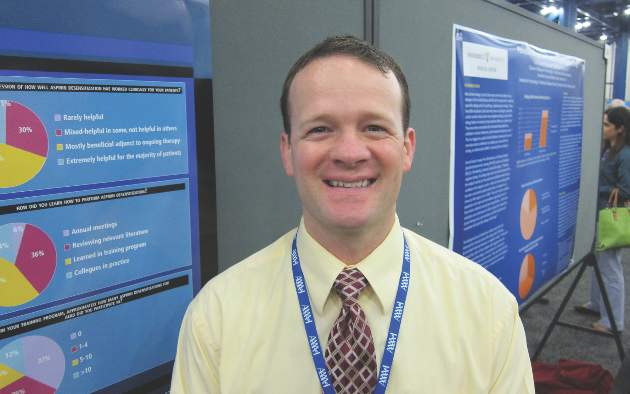
That figure is lower than it should be, given the wealth of published evidence that aspirin desensitization is a safe and effective component of the treatment of aspirin-exacerbated respiratory disease (AERD), Dr. Jeremy D. Waldram asserted in presenting the survey findings at the annual meeting of the American Academy of Allergy, Asthma, and Immunology.
Moreover, the figure likely overcalls the true rate, since participation in the survey was voluntary and fans of aspirin desensitization were probably more inclined to complete the 16-item questionnaire, added Dr. Waldram, a fellow in allergy and immunology at the Scripps Clinic in San Diego.

Was he surprised to find that aspirin desensitization isn’t more widely utilized?
“I think the number that surprised me more was that among the 37.5% of allergists who don’t do aspirin desensitization, almost 30% of them don’t even refer their patients to others who do the procedure. We don’t know why they don’t refer out; it wasn’t a question included in the survey. Perhaps they see patients who are of a less severe phenotype,” he said in an interview.
The 684 survey responses represented a 15% response rate. While 37.5% of respondents indicated they don’t perform aspirin desensitization, 73% of those who reported doing the procedure said they do an average of 1-5 cases annually.
Among allergists who don’t perform aspirin desensitization, safety concerns were the leading reason cited. Indeed, 70% of those who don’t do aspirin desensitization indicated safety risks were the main reason. More than one reason could be given, however, and 30% of allergists cited poor compensation for the procedure as a deterrent, nearly 60% said the logistics of monitoring care were too onerous, and one-third said they didn’t perform aspirin desensitization because they hadn’t been trained to do it.
Of allergists who reported doing aspirin desensitization, 52% perform the procedure in an outpatient setting unattached to a hospital. Another 21% do so in an outpatient clinic that’s physically attached to a hospital.
Within the past 5 years, 9% of respondents said that they’ve had a patient react severely to aspirin desensitization, requiring an unanticipated transfer to a higher level of care. That’s contrary to the experience among allergists at the Scripps Clinic, which is widely credited with pioneering the outpatient approach.
“We essentially do all our aspirin desensitizations for AERD in the outpatient setting. In 1,500 treated patients we’ve never had one that we had to transfer to a higher level of care. We don’t have any special setup. It’s a typical outpatient clinic. We usually don’t start IVs or do anything above and beyond,” Dr. Waldram said.
While 26% of respondents reported they generally recommend aspirin desensitization immediately upon identifying a patient history that supports the diagnosis of AERD, another 54% said they usually recommend the procedure to patients only after they’ve failed to improve on typical medical therapy.
Twenty percent of physicians rated aspirin desensitization as “extremely helpful for the majority of patients,” another 49% said they find it most beneficial as an adjuvant to ongoing medical therapy.
Forty-four percent of allergists who perform aspirin desensitization reported that they learned to do the procedure during fellowship training. Fourteen percent said they learned to the procedure at an annual meeting, and 36% picked it up by reviewing the relevant literature.
Several allergists commented that had Dr. Waldram’s survey been conducted even a couple of years ago the rate of utilization of aspirin desensitization would have been far lower. They interpreted his reported 62.5% rate as a sign of progress. Dr. Waldram said he believes the key to further boosting utilization of aspirin desensitization lies in increasing exposure to the procedure during fellowship training. He noted that internal medicine-trained fellows who responded to the survey had a significantly higher aspirin desensitization utilization rate than those who came to their allergy fellowship with a background in pediatrics.
The hallmarks of AERD are difficult-to-treat nasal polyps, chronic eosinophilic sinusitis, and asthma in a patient with sensitivity to aspirin and other COX-1 inhibitors.
Dr. Waldram reported having no financial conflicts with regard to his study, which was conducted free of commercial support.
HOUSTON – About 63% of allergists and fellows in training perform aspirin desensitization for aspirin-exacerbated respiratory disease, according to a national survey.
That figure is lower than it should be, given the wealth of published evidence that aspirin desensitization is a safe and effective component of the treatment of aspirin-exacerbated respiratory disease (AERD), Dr. Jeremy D. Waldram asserted in presenting the survey findings at the annual meeting of the American Academy of Allergy, Asthma, and Immunology.
Moreover, the figure likely overcalls the true rate, since participation in the survey was voluntary and fans of aspirin desensitization were probably more inclined to complete the 16-item questionnaire, added Dr. Waldram, a fellow in allergy and immunology at the Scripps Clinic in San Diego.
Was he surprised to find that aspirin desensitization isn’t more widely utilized?
“I think the number that surprised me more was that among the 37.5% of allergists who don’t do aspirin desensitization, almost 30% of them don’t even refer their patients to others who do the procedure. We don’t know why they don’t refer out; it wasn’t a question included in the survey. Perhaps they see patients who are of a less severe phenotype,” he said in an interview.
The 684 survey responses represented a 15% response rate. While 37.5% of respondents indicated they don’t perform aspirin desensitization, 73% of those who reported doing the procedure said they do an average of 1-5 cases annually.
Among allergists who don’t perform aspirin desensitization, safety concerns were the leading reason cited. Indeed, 70% of those who don’t do aspirin desensitization indicated safety risks were the main reason. More than one reason could be given, however, and 30% of allergists cited poor compensation for the procedure as a deterrent, nearly 60% said the logistics of monitoring care were too onerous, and one-third said they didn’t perform aspirin desensitization because they hadn’t been trained to do it.
Of allergists who reported doing aspirin desensitization, 52% perform the procedure in an outpatient setting unattached to a hospital. Another 21% do so in an outpatient clinic that’s physically attached to a hospital.
Within the past 5 years, 9% of respondents said that they’ve had a patient react severely to aspirin desensitization, requiring an unanticipated transfer to a higher level of care. That’s contrary to the experience among allergists at the Scripps Clinic, which is widely credited with pioneering the outpatient approach.
“We essentially do all our aspirin desensitizations for AERD in the outpatient setting. In 1,500 treated patients we’ve never had one that we had to transfer to a higher level of care. We don’t have any special setup. It’s a typical outpatient clinic. We usually don’t start IVs or do anything above and beyond,” Dr. Waldram said.
While 26% of respondents reported they generally recommend aspirin desensitization immediately upon identifying a patient history that supports the diagnosis of AERD, another 54% said they usually recommend the procedure to patients only after they’ve failed to improve on typical medical therapy.
Twenty percent of physicians rated aspirin desensitization as “extremely helpful for the majority of patients,” another 49% said they find it most beneficial as an adjuvant to ongoing medical therapy.
Forty-four percent of allergists who perform aspirin desensitization reported that they learned to do the procedure during fellowship training. Fourteen percent said they learned to the procedure at an annual meeting, and 36% picked it up by reviewing the relevant literature.
Several allergists commented that had Dr. Waldram’s survey been conducted even a couple of years ago the rate of utilization of aspirin desensitization would have been far lower. They interpreted his reported 62.5% rate as a sign of progress. Dr. Waldram said he believes the key to further boosting utilization of aspirin desensitization lies in increasing exposure to the procedure during fellowship training. He noted that internal medicine-trained fellows who responded to the survey had a significantly higher aspirin desensitization utilization rate than those who came to their allergy fellowship with a background in pediatrics.
The hallmarks of AERD are difficult-to-treat nasal polyps, chronic eosinophilic sinusitis, and asthma in a patient with sensitivity to aspirin and other COX-1 inhibitors.
Dr. Waldram reported having no financial conflicts with regard to his study, which was conducted free of commercial support.
AT 2015 AAAAI ANNUAL MEETING
Key clinical point: Aspirin desensitization for patients with aspirin-exacerbated respiratory disease is catching on among U.S. allergists.
Major finding: Roughly 63% of allergists and allergy fellows who responded to a national survey indicated they perform aspirin desensitization for aspirin-exacerbated respiratory disease.
Data source: This was a 16-question survey of aspirin desensitization practices among U.S. allergists and allergy fellows. The national survey drew 684 responses.
Disclosures: The presenter reported having no financial conflicts with regard to his study, which was funded without commercial support.
Novel oral anticoagulants best warfarin for AF in heart failure
SAN DIEGO – The novel oral anticoagulants clearly outperformed warfarin for stroke prevention and safety endpoints in patients with atrial fibrillation and comorbid heart failure in a meta-analysis of four recent landmark Phase 3 clinical trials.
Collectively the four novel oral anticoagulants (NOACs) approved for stroke prophylaxis in nonvalvular atrial fibrillation (AF) reduced the risk of stroke and systemic embolism by 14%, compared with patients randomized to warfarin. Moreover, the NOACs decreased the risks of major bleeding and intracranial bleeding by 23% and 45%, respectively, Dr. Gianluigi Savarese reported at the annual meeting of the American College of Cardiology.

“NOACs represent a valuable therapeutic option in patients with nonvalvular atrial fibrillation and heart failure,” concluded Dr. Savarese of Federico II University, Naples.
There has never been a randomized trial comparing a NOAC to warfarin specifically in patients with these dual diagnoses. In the absence of such a definitive study, the next best thing is a meta-analysis of the pivotal Phase 3 trials in which warfarin was compared to dabigatran (Pradaxa, the RE-LY study), apixaban (Eliquis, ARISTOTLE), rivaroxaban (Xarelto, ROCKET AF), and edoxaban (Savaysa, ENGAGE AF-TIMI 48).
The meta-analysis focused on a subset population of 26,384 randomized patients with AF and heart failure. It’s important to know how the NOACs stack up against warfarin in this population because symptomatic heart failure is common: indeed, it’s present in 30% of patients with AF. Patients with AF and comorbid heart failure are generally older, frailer, have more comorbidities, and are at higher risk of both stroke and bleeding, compared with AF patients without heart failure. Since heart failure is a recognized risk factor for reduced time in the therapeutic international normalized ratio (INR) range for patients on warfarin, it’s likely that warfarin-treated dual diagnosis patients would be exposed to further increased risks of stroke and bleeding, according to Dr. Savarese.
In the meta-analysis, in addition to the NOAC-treated patients’ significantly reduced risks of stroke, major bleeding, and intracranial bleeding, they showed a 12% decrease in total bleeding and an 8% reduction in cardiovascular death, compared with warfarin-treated controls, although neither of those latter two favorable trends achieved statistical significance.
The four NOACs didn’t differ significantly on any of the prespecified outcomes in the meta-analysis.
One audience member noted that while the relative risk reductions for stroke and major bleeding seen with the NOACs in the meta-analysis were large and impressive, the absolute risk reductions were actually quite small. For example, warfarin-treated controls in RE-LY, the first of the major trials, had a stroke/systemic embolism rate of 1.69%/year and a major bleeding rate of 3.4%/year (N. Engl. J. Med. 2009;361:1139-51), while controls in ENGAGE AF-TIMI 48 had annualized stroke and major bleeding rates of 1.5% and 3.4%, respectively (N. Engl. J. Med. 2013;369:2093-2104).
Dr. Savarese replied that he and his coinvestigators consider those absolute risk reductions to be clinically meaningful, especially in light of the enormous and rapidly growing number of patients with both AF and heart failure.
He reported having no financial conflicts regarding this meta-analysis, which was carried out free of commercial support.
SAN DIEGO – The novel oral anticoagulants clearly outperformed warfarin for stroke prevention and safety endpoints in patients with atrial fibrillation and comorbid heart failure in a meta-analysis of four recent landmark Phase 3 clinical trials.
Collectively the four novel oral anticoagulants (NOACs) approved for stroke prophylaxis in nonvalvular atrial fibrillation (AF) reduced the risk of stroke and systemic embolism by 14%, compared with patients randomized to warfarin. Moreover, the NOACs decreased the risks of major bleeding and intracranial bleeding by 23% and 45%, respectively, Dr. Gianluigi Savarese reported at the annual meeting of the American College of Cardiology.
“NOACs represent a valuable therapeutic option in patients with nonvalvular atrial fibrillation and heart failure,” concluded Dr. Savarese of Federico II University, Naples.
There has never been a randomized trial comparing a NOAC to warfarin specifically in patients with these dual diagnoses. In the absence of such a definitive study, the next best thing is a meta-analysis of the pivotal Phase 3 trials in which warfarin was compared to dabigatran (Pradaxa, the RE-LY study), apixaban (Eliquis, ARISTOTLE), rivaroxaban (Xarelto, ROCKET AF), and edoxaban (Savaysa, ENGAGE AF-TIMI 48).
The meta-analysis focused on a subset population of 26,384 randomized patients with AF and heart failure. It’s important to know how the NOACs stack up against warfarin in this population because symptomatic heart failure is common: indeed, it’s present in 30% of patients with AF. Patients with AF and comorbid heart failure are generally older, frailer, have more comorbidities, and are at higher risk of both stroke and bleeding, compared with AF patients without heart failure. Since heart failure is a recognized risk factor for reduced time in the therapeutic international normalized ratio (INR) range for patients on warfarin, it’s likely that warfarin-treated dual diagnosis patients would be exposed to further increased risks of stroke and bleeding, according to Dr. Savarese.
In the meta-analysis, in addition to the NOAC-treated patients’ significantly reduced risks of stroke, major bleeding, and intracranial bleeding, they showed a 12% decrease in total bleeding and an 8% reduction in cardiovascular death, compared with warfarin-treated controls, although neither of those latter two favorable trends achieved statistical significance.
The four NOACs didn’t differ significantly on any of the prespecified outcomes in the meta-analysis.
One audience member noted that while the relative risk reductions for stroke and major bleeding seen with the NOACs in the meta-analysis were large and impressive, the absolute risk reductions were actually quite small. For example, warfarin-treated controls in RE-LY, the first of the major trials, had a stroke/systemic embolism rate of 1.69%/year and a major bleeding rate of 3.4%/year (N. Engl. J. Med. 2009;361:1139-51), while controls in ENGAGE AF-TIMI 48 had annualized stroke and major bleeding rates of 1.5% and 3.4%, respectively (N. Engl. J. Med. 2013;369:2093-2104).
Dr. Savarese replied that he and his coinvestigators consider those absolute risk reductions to be clinically meaningful, especially in light of the enormous and rapidly growing number of patients with both AF and heart failure.
He reported having no financial conflicts regarding this meta-analysis, which was carried out free of commercial support.
AT ACC 15
Key clinical point: Patients with nonvalvular atrial fibrillation and heart failure clearly fare better on any of the novel oral anticoagulants than with warfarin for stroke prophylaxis.
Major finding: Dual diagnosis patients randomized to a novel oral anticoagulant had a 14% reduction in stroke/systemic embolism and a 23% decrease in major bleeding compared with those on warfarin.
Data source: This was a meta-analysis of the 26,384 patients with both atrial fibrillation and heart failure who were included in four pivotal Phase 3 clinical trials that led to approval of dabigatran, apixaban, rivaroxaban, and edoxaban.
Disclosures: The presenter reported having no financial conflicts regarding this meta-analysis, which was carried out free of commercial support.
Risk factors identified for gestational eczema
HOUSTON– New-onset eczema during pregnancy is a common phenomenon with several newly identified risk factors.
This disease entity deserves a proper name: gestational eczema, Dr. Wilfried J.J. Karmaus asserted at the annual meeting of the American Academy of Allergy, Asthma, and Immunology.
In contrast, the likelihood of new-onset asthma arising during pregnancy isn’t significantly more common than in an affected woman’s male partner during the same time frame.
“There was no large difference in wheezing between the women and men. Therefore, we cannot propose the term ‘gestational asthma,’” said Dr. Karmaus, professor of epidemiology at the University of Memphis. “Investigations into how to prevent eczema and asthma in pregnancy are really important, because eczema and asthma in pregnancy can increase the risk of these diseases in the offspring. This is a totally undeveloped field.”
He presented new findings from the Isle of Wight study, a prospective study in which a cohort of women has been followed from birth through pregnancy across three generations.
Eczema and asthma are common atopic diseases, and they are particularly common during pregnancy. Indeed, eczema is the most common skin disease seen in pregnancy, accounting for 35%-50% of all dermatoses in previous studies by other investigators. In those studies, only 20%-40% of women with eczema during pregnancy had a prepregnancy history of the disease.
In the Isle of Wight cohort, women were evaluated for asthma and eczema symptoms at ages 1, 2, 4, 10, and 18 years and again during pregnancy at gestational weeks 20 and 28. A total of 26 of 116 women developed eczema during pregnancy, with eight of them (31%) experiencing the skin disease for the first time in their lives. In contrast, only six of their male partners had eczema during the pregnancy time frame, and just one of them had new-onset eczema.
A history of maternal eczema in the preceding generation was associated with a 52% increased relative risk of having eczema by age 18 and a 3.1-fold increased likelihood of eczema during pregnancy. Also, methylation of the filaggrin gene at the cytosine-phosphate-guanine site cg13447818 when assessed at age 18 was associated with a significantly increased likelihood of eczema in a subject’s mother as well as increased risk of gestational eczema 1-7 years later, Dr. Karmaus continued.
Eighteen percent of women in the Isle of Wight cohort had asthma during pregnancy, as did a similar proportion of their male partners. Twenty-seven percent of women with asthma during pregnancy had no previous history of the respiratory disease, a rate which was again comparable in their male partners with asthma.
DNA methylation of the IL1RL1 gene at cg17738684 was significantly associated with asthma heritability across three generations in the Isle of Wight study. The IL1RL1 gene at cg17738684 is a candidate gene for asthma that encodes for interleukin-33. This finding raises the possibility that addressing this DNA methylation could prove fruitful as a transgenerational asthma prevention strategy.
The Isle of Wight birth cohort study is funded by the National Institutes of Health, Asthma UK, and the Isle of Wight Trust. Dr. Karmaus reported having no financial conflicts of interest.
AT 2015 AAAAI ANNUAL MEETING
Key clinical point: Thirty-one percent of cases of eczema and 27% of asthma in a group of pregnant women occurred for the first time in the woman’s life.
Major finding: A history of maternal eczema was associated with a 3.1-fold increased likelihood of eczema during the offspring’s pregnancy.
Data source: The Isle of Wight birth cohort study is a prospective study following three generations from birth through pregnancy.
Disclosures: The study is funded by the National Institutes of Health, Asthma UK, and the Isle of Wight Trust. The presenter reported having no financial conflicts of interest.
Aspirin for AF Fading Away
SNOWMASS, COLO.– The sun may be setting on the use of aspirin for thromboprophylaxis in patients with nonvalvular atrial fibrillation. The 2014 American College of Cardiology/American Heart Association/Heart Rhythm Society guidelines are unique among the major international guidelines in giving a modest IIb, Level of Evidence C endorsement to the option of aspirin or no treatment in patients with atrial fibrillation (AF) and a CHA2DS2-VASc score of 1. In contrast, the 2014 U.K. NICE (National Institute for Health and Care Excellence) guidelines, the 2013 Asian Pacific Heart Rhythm Society guidelines, and the 2012 European Society of Cardiology guidelines all recommend consideration of oral anticoagulation – eschewing aspirin – in patients with a CHA2DS2-VASc score of 1 or more, Dr. Bernard J. Gersh noted at the Annual Cardiovascular Conference at Snowmass.

The NICE guidelines put the matter succinctly, stating, “The main departure from prior guidelines is that aspirin should not be used in AF simply to reduce the risk of stroke, as it is not as effective as NOACs [novel oral anticoagulants] ... and can cause more bleeding side effects.”
Dr. Gersh was coauthor of a recently published think piece that argued that exaggerated misperceptions of aspirin’s efficacy and safety have led to its inappropriate status as “the easy option” for stroke prevention in AF (Eur. Heart J. 2015;36:653-6).
“By giving physicians a soft option of aspirin in CHA2DS2-VASc 1 patients, we’re allowing them an excuse not to use an oral anticoagulant, which is what they should be using,” Dr. Gersh explained at the conference.
He noted that an analysis of more than 41,000 Medicare beneficiaries with AF in 2007-2008 showed that only 66.8% were on warfarin.
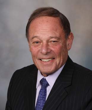
“There’s no doubt that warfarin, and for that matter the NOACs, are underutilized, and it’s possible that the misperception that aspirin is effective may contribute to that underutilization,” asserted Dr. Gersh, professor of medicine at the Mayo Clinic, Rochester, Minn.
The cardiologist added that the widely held belief that aspirin reduces stroke risk by roughly 20%, compared with placebo, in patients with AF is based upon seriously flawed data. True, a meta-analysis of six placebo-controlled randomized trials done in an earlier era concluded that aspirin reduced the relative risk of stroke by 19%, but what’s often overlooked is that aspirin significantly outperformed placebo in only one of those six studies – the SPAF 1 trial – where in one arm aspirin achieved an “almost implausible” 94% relative risk reduction.
“Nothing reduces risk by 94%,” Dr. Gersh observed, adding that the SPAF 1 methodology was, upon careful examination, “completely unacceptable” by contemporary standards.
He was a coinvestigator in a study of 7,347 AF patients on oral anticoagulation therapy in 176 U.S. practices participating in the ORBIT-AF registry (Outcomes Registry for Better Informed Treatment of Atrial Fibrillation). The analysis showed that concomitant use of aspirin and an oral anticoagulant in patients with AF is common in everyday clinical practice, being employed in 35% of the study population. Of note, 39% of these patients had no history of CAD and therefore weren’t on aspirin for secondary cardiovascular prevention, and 17% of them were at elevated bleeding risk because of an ATRIA (Anticoagulation and Risk Factors in Atrial Fibrillation) bleeding risk score of 5 or more.
The key, disturbing finding was this: The adjusted risks of major bleeding and hospitalization for bleeding were 53% and 52% greater, respectively, in patients on an oral anticoagulant plus aspirin than with an oral anticoagulant alone (Circulation 2013;128:721-8).
On the basis of this study and other evidence, Dr. Gersh believes the use of aspirin in patients with AF should be considered only in those with CAD, where a case can be made for its use in secondary prevention alongside an oral anticoagulant to reduce stroke risk. But even then, aspirin’s use is reasonable only in those at low risk of bleeding, and extra vigilance and prophylaxis with a proton pump inhibitor are called for.
“If a patient is at high risk of bleeding, I personally would not give aspirin,” the cardiologist added.
The big remaining question – and an area of current controversy – concerns the safety of halting aspirin in AF patients on an oral anticoagulant who’ve undergone coronary stenting. Ongoing studies are looking at this issue, and answers are expected within a year or 2.
Dr. Gersh reported serving as a consultant to Merck and Ortho-McNeil-Janssen and on data safety monitoring boards for Baxter, Medtronic, and Teva.
SNOWMASS, COLO.– The sun may be setting on the use of aspirin for thromboprophylaxis in patients with nonvalvular atrial fibrillation. The 2014 American College of Cardiology/American Heart Association/Heart Rhythm Society guidelines are unique among the major international guidelines in giving a modest IIb, Level of Evidence C endorsement to the option of aspirin or no treatment in patients with atrial fibrillation (AF) and a CHA2DS2-VASc score of 1. In contrast, the 2014 U.K. NICE (National Institute for Health and Care Excellence) guidelines, the 2013 Asian Pacific Heart Rhythm Society guidelines, and the 2012 European Society of Cardiology guidelines all recommend consideration of oral anticoagulation – eschewing aspirin – in patients with a CHA2DS2-VASc score of 1 or more, Dr. Bernard J. Gersh noted at the Annual Cardiovascular Conference at Snowmass.
The NICE guidelines put the matter succinctly, stating, “The main departure from prior guidelines is that aspirin should not be used in AF simply to reduce the risk of stroke, as it is not as effective as NOACs [novel oral anticoagulants] ... and can cause more bleeding side effects.”
Dr. Gersh was coauthor of a recently published think piece that argued that exaggerated misperceptions of aspirin’s efficacy and safety have led to its inappropriate status as “the easy option” for stroke prevention in AF (Eur. Heart J. 2015;36:653-6).
“By giving physicians a soft option of aspirin in CHA2DS2-VASc 1 patients, we’re allowing them an excuse not to use an oral anticoagulant, which is what they should be using,” Dr. Gersh explained at the conference.
He noted that an analysis of more than 41,000 Medicare beneficiaries with AF in 2007-2008 showed that only 66.8% were on warfarin.
“There’s no doubt that warfarin, and for that matter the NOACs, are underutilized, and it’s possible that the misperception that aspirin is effective may contribute to that underutilization,” asserted Dr. Gersh, professor of medicine at the Mayo Clinic, Rochester, Minn.
The cardiologist added that the widely held belief that aspirin reduces stroke risk by roughly 20%, compared with placebo, in patients with AF is based upon seriously flawed data. True, a meta-analysis of six placebo-controlled randomized trials done in an earlier era concluded that aspirin reduced the relative risk of stroke by 19%, but what’s often overlooked is that aspirin significantly outperformed placebo in only one of those six studies – the SPAF 1 trial – where in one arm aspirin achieved an “almost implausible” 94% relative risk reduction.
“Nothing reduces risk by 94%,” Dr. Gersh observed, adding that the SPAF 1 methodology was, upon careful examination, “completely unacceptable” by contemporary standards.
He was a coinvestigator in a study of 7,347 AF patients on oral anticoagulation therapy in 176 U.S. practices participating in the ORBIT-AF registry (Outcomes Registry for Better Informed Treatment of Atrial Fibrillation). The analysis showed that concomitant use of aspirin and an oral anticoagulant in patients with AF is common in everyday clinical practice, being employed in 35% of the study population. Of note, 39% of these patients had no history of CAD and therefore weren’t on aspirin for secondary cardiovascular prevention, and 17% of them were at elevated bleeding risk because of an ATRIA (Anticoagulation and Risk Factors in Atrial Fibrillation) bleeding risk score of 5 or more.
The key, disturbing finding was this: The adjusted risks of major bleeding and hospitalization for bleeding were 53% and 52% greater, respectively, in patients on an oral anticoagulant plus aspirin than with an oral anticoagulant alone (Circulation 2013;128:721-8).
On the basis of this study and other evidence, Dr. Gersh believes the use of aspirin in patients with AF should be considered only in those with CAD, where a case can be made for its use in secondary prevention alongside an oral anticoagulant to reduce stroke risk. But even then, aspirin’s use is reasonable only in those at low risk of bleeding, and extra vigilance and prophylaxis with a proton pump inhibitor are called for.
“If a patient is at high risk of bleeding, I personally would not give aspirin,” the cardiologist added.
The big remaining question – and an area of current controversy – concerns the safety of halting aspirin in AF patients on an oral anticoagulant who’ve undergone coronary stenting. Ongoing studies are looking at this issue, and answers are expected within a year or 2.
Dr. Gersh reported serving as a consultant to Merck and Ortho-McNeil-Janssen and on data safety monitoring boards for Baxter, Medtronic, and Teva.
EXPERT ANALYSIS FROM THE CARDIOVASCULAR CONFERENCE AT SNOWMASS

Aspirin for AF fading away
SNOWMASS, COLO.– The sun may be setting on the use of aspirin for thromboprophylaxis in patients with nonvalvular atrial fibrillation. The 2014 American College of Cardiology/American Heart Association/Heart Rhythm Society guidelines are unique among the major international guidelines in giving a modest IIb, Level of Evidence C endorsement to the option of aspirin or no treatment in patients with atrial fibrillation (AF) and a CHA2DS2-VASc score of 1. In contrast, the 2014 U.K. NICE (National Institute for Health and Care Excellence) guidelines, the 2013 Asian Pacific Heart Rhythm Society guidelines, and the 2012 European Society of Cardiology guidelines all recommend consideration of oral anticoagulation – eschewing aspirin – in patients with a CHA2DS2-VASc score of 1 or more, Dr. Bernard J. Gersh noted at the Annual Cardiovascular Conference at Snowmass.

The NICE guidelines put the matter succinctly, stating, “The main departure from prior guidelines is that aspirin should not be used in AF simply to reduce the risk of stroke, as it is not as effective as NOACs [novel oral anticoagulants] ... and can cause more bleeding side effects.”
Dr. Gersh was coauthor of a recently published think piece that argued that exaggerated misperceptions of aspirin’s efficacy and safety have led to its inappropriate status as “the easy option” for stroke prevention in AF (Eur. Heart J. 2015;36:653-6).
“By giving physicians a soft option of aspirin in CHA2DS2-VASc 1 patients, we’re allowing them an excuse not to use an oral anticoagulant, which is what they should be using,” Dr. Gersh explained at the conference.
He noted that an analysis of more than 41,000 Medicare beneficiaries with AF in 2007-2008 showed that only 66.8% were on warfarin.
“There’s no doubt that warfarin, and for that matter the NOACs, are underutilized, and it’s possible that the misperception that aspirin is effective may contribute to that underutilization,” asserted Dr. Gersh, professor of medicine at the Mayo Clinic, Rochester, Minn.
The cardiologist added that the widely held belief that aspirin reduces stroke risk by roughly 20%, compared with placebo, in patients with AF is based upon seriously flawed data. True, a meta-analysis of six placebo-controlled randomized trials done in an earlier era concluded that aspirin reduced the relative risk of stroke by 19%, but what’s often overlooked is that aspirin significantly outperformed placebo in only one of those six studies – the SPAF 1 trial – where in one arm aspirin achieved an “almost implausible” 94% relative risk reduction.
“Nothing reduces risk by 94%,” Dr. Gersh observed, adding that the SPAF 1 methodology was, upon careful examination, “completely unacceptable” by contemporary standards.
He was a coinvestigator in a study of 7,347 AF patients on oral anticoagulation therapy in 176 U.S. practices participating in the ORBIT-AF registry (Outcomes Registry for Better Informed Treatment of Atrial Fibrillation). The analysis showed that concomitant use of aspirin and an oral anticoagulant in patients with AF is common in everyday clinical practice, being employed in 35% of the study population. Of note, 39% of these patients had no history of CAD and therefore weren’t on aspirin for secondary cardiovascular prevention, and 17% of them were at elevated bleeding risk because of an ATRIA (Anticoagulation and Risk Factors in Atrial Fibrillation) bleeding risk score of 5 or more.
The key, disturbing finding was this: The adjusted risks of major bleeding and hospitalization for bleeding were 53% and 52% greater, respectively, in patients on an oral anticoagulant plus aspirin than with an oral anticoagulant alone (Circulation 2013;128:721-8).
On the basis of this study and other evidence, Dr. Gersh believes the use of aspirin in patients with AF should be considered only in those with CAD, where a case can be made for its use in secondary prevention alongside an oral anticoagulant to reduce stroke risk. But even then, aspirin’s use is reasonable only in those at low risk of bleeding, and extra vigilance and prophylaxis with a proton pump inhibitor are called for.
“If a patient is at high risk of bleeding, I personally would not give aspirin,” the cardiologist added.
The big remaining question – and an area of current controversy – concerns the safety of halting aspirin in AF patients on an oral anticoagulant who’ve undergone coronary stenting. Ongoing studies are looking at this issue, and answers are expected within a year or 2.
Dr. Gersh reported serving as a consultant to Merck and Ortho-McNeil-Janssen and on data safety monitoring boards for Baxter, Medtronic, and Teva.
SNOWMASS, COLO.– The sun may be setting on the use of aspirin for thromboprophylaxis in patients with nonvalvular atrial fibrillation. The 2014 American College of Cardiology/American Heart Association/Heart Rhythm Society guidelines are unique among the major international guidelines in giving a modest IIb, Level of Evidence C endorsement to the option of aspirin or no treatment in patients with atrial fibrillation (AF) and a CHA2DS2-VASc score of 1. In contrast, the 2014 U.K. NICE (National Institute for Health and Care Excellence) guidelines, the 2013 Asian Pacific Heart Rhythm Society guidelines, and the 2012 European Society of Cardiology guidelines all recommend consideration of oral anticoagulation – eschewing aspirin – in patients with a CHA2DS2-VASc score of 1 or more, Dr. Bernard J. Gersh noted at the Annual Cardiovascular Conference at Snowmass.
The NICE guidelines put the matter succinctly, stating, “The main departure from prior guidelines is that aspirin should not be used in AF simply to reduce the risk of stroke, as it is not as effective as NOACs [novel oral anticoagulants] ... and can cause more bleeding side effects.”
Dr. Gersh was coauthor of a recently published think piece that argued that exaggerated misperceptions of aspirin’s efficacy and safety have led to its inappropriate status as “the easy option” for stroke prevention in AF (Eur. Heart J. 2015;36:653-6).
“By giving physicians a soft option of aspirin in CHA2DS2-VASc 1 patients, we’re allowing them an excuse not to use an oral anticoagulant, which is what they should be using,” Dr. Gersh explained at the conference.
He noted that an analysis of more than 41,000 Medicare beneficiaries with AF in 2007-2008 showed that only 66.8% were on warfarin.
“There’s no doubt that warfarin, and for that matter the NOACs, are underutilized, and it’s possible that the misperception that aspirin is effective may contribute to that underutilization,” asserted Dr. Gersh, professor of medicine at the Mayo Clinic, Rochester, Minn.
The cardiologist added that the widely held belief that aspirin reduces stroke risk by roughly 20%, compared with placebo, in patients with AF is based upon seriously flawed data. True, a meta-analysis of six placebo-controlled randomized trials done in an earlier era concluded that aspirin reduced the relative risk of stroke by 19%, but what’s often overlooked is that aspirin significantly outperformed placebo in only one of those six studies – the SPAF 1 trial – where in one arm aspirin achieved an “almost implausible” 94% relative risk reduction.
“Nothing reduces risk by 94%,” Dr. Gersh observed, adding that the SPAF 1 methodology was, upon careful examination, “completely unacceptable” by contemporary standards.
He was a coinvestigator in a study of 7,347 AF patients on oral anticoagulation therapy in 176 U.S. practices participating in the ORBIT-AF registry (Outcomes Registry for Better Informed Treatment of Atrial Fibrillation). The analysis showed that concomitant use of aspirin and an oral anticoagulant in patients with AF is common in everyday clinical practice, being employed in 35% of the study population. Of note, 39% of these patients had no history of CAD and therefore weren’t on aspirin for secondary cardiovascular prevention, and 17% of them were at elevated bleeding risk because of an ATRIA (Anticoagulation and Risk Factors in Atrial Fibrillation) bleeding risk score of 5 or more.
The key, disturbing finding was this: The adjusted risks of major bleeding and hospitalization for bleeding were 53% and 52% greater, respectively, in patients on an oral anticoagulant plus aspirin than with an oral anticoagulant alone (Circulation 2013;128:721-8).
On the basis of this study and other evidence, Dr. Gersh believes the use of aspirin in patients with AF should be considered only in those with CAD, where a case can be made for its use in secondary prevention alongside an oral anticoagulant to reduce stroke risk. But even then, aspirin’s use is reasonable only in those at low risk of bleeding, and extra vigilance and prophylaxis with a proton pump inhibitor are called for.
“If a patient is at high risk of bleeding, I personally would not give aspirin,” the cardiologist added.
The big remaining question – and an area of current controversy – concerns the safety of halting aspirin in AF patients on an oral anticoagulant who’ve undergone coronary stenting. Ongoing studies are looking at this issue, and answers are expected within a year or 2.
Dr. Gersh reported serving as a consultant to Merck and Ortho-McNeil-Janssen and on data safety monitoring boards for Baxter, Medtronic, and Teva.
EXPERT ANALYSIS FROM THE CARDIOVASCULAR CONFERENCE AT SNOWMASS
Anxiety hits 42% of adults with congenital heart disease
SAN DIEGO– Elevated symptoms of anxiety are markedly more common than depression in adults with congenital heart disease, Lisa Deng reported at the annual meeting of the American College of Cardiology.
This observation isn’t reflected in the ACC/American Heart Association guidelines on the management of adult congenital heart disease (ACHD), which recommend that “a careful assessment of depressive symptoms and their possible overlap with symptoms of medical illness or side effects of medications must be part of the clinical evaluation of ACHD patients” (J. Am. Coll. Cardiol. 2008;52:e143-263).
Anxiety and depression have been shown to be associated with worse clinical outcomes in patients with heart disease. Given the high prevalence, deleterious impact, and treatable nature of anxiety symptoms, it makes sense to incorporate evaluation for both anxiety and depression in the clinical assessment of ACHD patients, according to Ms. Deng of Children’s Hospital of Philadelphia.
She reported on 134 patients (mean age, 35 years) attending an outpatient ACHD clinic, where they were assessed using the validated Hospital Anxiety and Depression Scale (HADS), the Satisfaction with Life Scale (SLS), and the Linear Analog Scale for Quality of Life (LAS). Of those patients, 45% had a history of arrhythmia and 20% had heart failure; 42% of subjects demonstrated elevated levels of anxiety as reflected in a HADS-Anxiety score of 8 or more out of a possible 21. In contrast, 12% had an elevated HADS-Depression score of 8 or more. Thus, anxiety was 3.5-fold more common than depression.
Of note, 15 of the 16 patients with depression had comorbid elevated levels of anxiety, but even though 42% of the ACHD patients had elevated anxiety scores, only 1 in 8 study participants had a note in their chart mentioning anxiety.
Patients with elevated anxiety reported significantly lower ratings than those with a HADS-Anxiety score of less than 8 on one quality-of-life measure – the SLS – but not on the LAS. In contrast, patients with depression scored significantly lower on both quality-of-life measures.
The clinical correlates of anxiety and depression differed in these ACHD patients. Half of the patients with elevated anxiety scores had a history of two or more surgical or interventional procedures, compared with 25% of subjects with a normal-range HADS-Anxiety score. One-fourth of patients with high depression scores were unemployed, a prevalence threefold greater than in nondepressive individuals. Also, 19% of patients with a HADS-Depression score of 8 or more had a history of arrhythmia, compared with 7% of nondepressive patients.
This study was supported by a research grant from Big Hearts in Little Bodies. Ms. Deng reported having no financial conflicts.
AT ACC 15
Key clinical point: Elevated anxiety levels are much more common than depression in patients with adult congenital heart disease.
Major finding: Elevated levels of anxiety were found in 42% of ACHD patients; only 12% had elevated depression.
Data source: An outpatient adult congenital heart disease clinic in which 134 patients attending were assessed for anxiety, depression, and quality of life using validated instruments.
Disclosures: This study was supported by a research grant from Big Hearts in Little Bodies. The presenter reported having no financial conflicts.
Bronchial thermoplasty gains momentum for severe asthma
HOUSTON – Bronchial thermoplasty has emerged as an important treatment option for patients with severe asthma at specialized centers, Dr. Mario Castro observed at the annual meeting of the American Academy of Allergy, Asthma, and Immunology.
The most recent international European Respiratory Society/American Thoracic Society practice guidelines on severe asthma recommend that bronchial thermoplasty for severe persistent asthma be utilized only in the setting of a clinical study or independent registry. The guidelines cited “very low confidence” in the available estimates of the novel treatment’s longer-term benefits and harms, as well as the lack of data regarding the phenotypes of asthma patients most likely to benefit (Eur. Respir. J. 2014 Feb;43:343-73).

Dr. Castro, a member of the task force that developed the ERS/ATS guidelines, said the group’s cautious stance was appropriate given the evidence available at the time of deliberations. However, at the AAAAI meeting, he highlighted more recent study results that address many of the task force’s concerns and that he said might lead to a more enthusiastic recommendation for bronchial thermoplasty in future guidelines.
One key piece of evidence unavailable to the task force comes from results reported in the 5-year prospective follow-up of 162 bronchial thermoplasty-treated patients in the international Asthma Intervention Research 2 (AIR2) trial.
“It’s quite striking that the exacerbation rate did not start to creep back up over time in this severe asthma population. We believe this study shows for the first time that this therapy may actually be a disease modifier, and that you can do this procedure in an identified population and the benefits of this one-time treatment are sustained over at least a 5-year time period,” said Dr. Castro, an AIR2 investigator and professor of pulmonary and critical care medicine and pediatrics at Washington University in St. Louis.
Compared with the baseline established during the year prior to bronchial thermoplasty, at 5 years post procedure, there was a 44% decrease in the percentage of AIR2 participants with severe exacerbations requiring oral corticosteroids, and a 48% reduction in the severe exacerbation event rate. Moreover, there was a 78% reduction in the percentage of patients with an emergency department visit for asthma and an 88% drop in the ED visit event rate (J. Allergy Clin. Immunol. 2013;132:1295-302).
With regard to safety, annual high-resolution CT scans showed no structural abnormalities from baseline to 5 years post-bronchial thermoplasty that could be attributed to the procedure. Prebronchodilator forced expiratory volume in 1 second (FEV1) values remained steady between years 1 and 5 post procedure despite an 18% decrease in the average daily dose of inhaled corticosteroids.
In a separate study, Dr. Castro and coinvestigators at Washington University identified a number of predictors of who will get the best responses to bronchial thermoplasty. This was a small study involving 42 patients with severe persistent asthma as reflected in their baseline mean inhaled corticosteroid dose of 2,185 mcg/day. Eighty percent of patients required bursts of oral corticosteroids during the year prior to the procedure. Their average baseline Asthma Quality of Life Questionnaire (AQLQ) score was 3.42. The baseline FEV1 postbronchodilator averaged 70%, with a range of 44%-121%.
Predictors of a clinically meaningful improvement in quality of life as defined by at least a 0.5-point improvement in AQLQ score 1 year post procedure included a shorter duration of asthma – 19 years, as compared with an average of 45 years in nonresponders – and a greater number of severe exacerbations during the year prior to bronchial thermoplasty.
Using another important yardstick of clinical improvement – at least a 240 mcg/day dose reduction in inhaled corticosteroids or a 2.5 mg/day decrease in oral corticosteroids at 1 year post procedure – significant predictors of benefit included older age (55 vs. 43 years), a lower baseline AQLQ score (2.4 vs. 4.0), and greater need for oral corticosteroids.
In addition, several quantitative metrics obtained through multidetector CT scans of the chest showed promise as predictors of a corticosteroid dose reduction. Responders showed less baseline air trapping, with an average of 6.1% of the lung having a density below –850 Hounsfield units, compared with 12.1% in nonresponders. Responders also had less baseline emphysema-like lung, with 3.2% of the lung having a density below –950 Hounsfield units at total lung capacity, compared with 5.8% in nonresponders, according to Dr. Castro.
This as-yet unpublished study was funded by the National Institutes of Health. AIR2 was sponsored by Boston Scientific. Dr. Castro reported research grants from the NIH, the American Lung Association, Boston Scientific, and other companies.
An estimated 5% of asthma patients are categorized as having severe disease. Bronchial thermoplasty has been approved by the FDA for the treatment of severe asthma since 2010. The outpatient procedure entails delivery of radiofrequency energy to the lungs in three sessions spaced several weeks apart.
HOUSTON – Bronchial thermoplasty has emerged as an important treatment option for patients with severe asthma at specialized centers, Dr. Mario Castro observed at the annual meeting of the American Academy of Allergy, Asthma, and Immunology.
The most recent international European Respiratory Society/American Thoracic Society practice guidelines on severe asthma recommend that bronchial thermoplasty for severe persistent asthma be utilized only in the setting of a clinical study or independent registry. The guidelines cited “very low confidence” in the available estimates of the novel treatment’s longer-term benefits and harms, as well as the lack of data regarding the phenotypes of asthma patients most likely to benefit (Eur. Respir. J. 2014 Feb;43:343-73).
Dr. Castro, a member of the task force that developed the ERS/ATS guidelines, said the group’s cautious stance was appropriate given the evidence available at the time of deliberations. However, at the AAAAI meeting, he highlighted more recent study results that address many of the task force’s concerns and that he said might lead to a more enthusiastic recommendation for bronchial thermoplasty in future guidelines.
One key piece of evidence unavailable to the task force comes from results reported in the 5-year prospective follow-up of 162 bronchial thermoplasty-treated patients in the international Asthma Intervention Research 2 (AIR2) trial.
“It’s quite striking that the exacerbation rate did not start to creep back up over time in this severe asthma population. We believe this study shows for the first time that this therapy may actually be a disease modifier, and that you can do this procedure in an identified population and the benefits of this one-time treatment are sustained over at least a 5-year time period,” said Dr. Castro, an AIR2 investigator and professor of pulmonary and critical care medicine and pediatrics at Washington University in St. Louis.
Compared with the baseline established during the year prior to bronchial thermoplasty, at 5 years post procedure, there was a 44% decrease in the percentage of AIR2 participants with severe exacerbations requiring oral corticosteroids, and a 48% reduction in the severe exacerbation event rate. Moreover, there was a 78% reduction in the percentage of patients with an emergency department visit for asthma and an 88% drop in the ED visit event rate (J. Allergy Clin. Immunol. 2013;132:1295-302).
With regard to safety, annual high-resolution CT scans showed no structural abnormalities from baseline to 5 years post-bronchial thermoplasty that could be attributed to the procedure. Prebronchodilator forced expiratory volume in 1 second (FEV1) values remained steady between years 1 and 5 post procedure despite an 18% decrease in the average daily dose of inhaled corticosteroids.
In a separate study, Dr. Castro and coinvestigators at Washington University identified a number of predictors of who will get the best responses to bronchial thermoplasty. This was a small study involving 42 patients with severe persistent asthma as reflected in their baseline mean inhaled corticosteroid dose of 2,185 mcg/day. Eighty percent of patients required bursts of oral corticosteroids during the year prior to the procedure. Their average baseline Asthma Quality of Life Questionnaire (AQLQ) score was 3.42. The baseline FEV1 postbronchodilator averaged 70%, with a range of 44%-121%.
Predictors of a clinically meaningful improvement in quality of life as defined by at least a 0.5-point improvement in AQLQ score 1 year post procedure included a shorter duration of asthma – 19 years, as compared with an average of 45 years in nonresponders – and a greater number of severe exacerbations during the year prior to bronchial thermoplasty.
Using another important yardstick of clinical improvement – at least a 240 mcg/day dose reduction in inhaled corticosteroids or a 2.5 mg/day decrease in oral corticosteroids at 1 year post procedure – significant predictors of benefit included older age (55 vs. 43 years), a lower baseline AQLQ score (2.4 vs. 4.0), and greater need for oral corticosteroids.
In addition, several quantitative metrics obtained through multidetector CT scans of the chest showed promise as predictors of a corticosteroid dose reduction. Responders showed less baseline air trapping, with an average of 6.1% of the lung having a density below –850 Hounsfield units, compared with 12.1% in nonresponders. Responders also had less baseline emphysema-like lung, with 3.2% of the lung having a density below –950 Hounsfield units at total lung capacity, compared with 5.8% in nonresponders, according to Dr. Castro.
This as-yet unpublished study was funded by the National Institutes of Health. AIR2 was sponsored by Boston Scientific. Dr. Castro reported research grants from the NIH, the American Lung Association, Boston Scientific, and other companies.
An estimated 5% of asthma patients are categorized as having severe disease. Bronchial thermoplasty has been approved by the FDA for the treatment of severe asthma since 2010. The outpatient procedure entails delivery of radiofrequency energy to the lungs in three sessions spaced several weeks apart.
EXPERT ANALYSIS FROM THE 2015 AAAAI ANNUAL MEETING
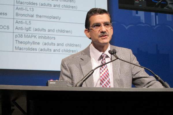
‘Mossy Oak sign’ suggests delayed anaphylaxis to red meat
HOUSTON– In central Virginia, where IgE-mediated delayed allergic reactions to red meat have become the most common cause of anaphylaxis in adults, physicians have taken to looking for what they call the ‘Mossy Oak sign.’
“If a patient shows up in a blaze-orange cap and hunter’s camouflage fatigues, an allergy fellow will tell me, ‘There’s a positive Mossy Oak sign in room 2,’ and I know that probably means the patient has delayed anaphylaxis to alpha-gal,” Dr. Scott P. Commins said at the annual meeting of the American Academy of Allergy, Asthma, and Immunology.

Alpha-gal is short for galactose-alpha-1,3,-galactose, an oligosaccharide present on thyroglobulin and other tissues in nonprimate mammals. It’s not normally present in humans, but when a lone star tick (Amblyomma americanum) that has fed on a nonprimate mammal bites a human, the alpha-gal is transferred, eliciting serum IgE antibodies.
Mossy Oak is a popular brand of hunter’s camouflage clothing. A positive Mossy Oak sign is useful in clinical practice because a patient who presents to a medical clinic dressed in hunting regalia is someone who spends a lot of time outdoors in the woods and fields where ticks lurk. He’s also typically someone who enjoys eating red meat. And whether it’s venison, beef, pork, lamb, goat, or bison, it contains alpha-gal. The result, in a patient who’s been primed via tick bite, can be a life-threatening anaphylactic or urticarial reaction arising 3-6 hours later, explained Dr. Commins, an allergist at the University of Virginia, Charlottesville.
He and his coinvestigators have played a central role in the still-unfolding story of this novel disease involving late-onset anaphylaxis to mammalian meat. Dr. Commins was the lead author of the paper that first described the syndrome (J. Allergy Clin. Immunol. 2009;123:426-33), as well as a subsequent paper that established the lone star tick as the culprit, making this syndrome the first known example of a response to an ectoparasite giving rise to a serious form of food allergy (J. Allergy Clin. Immunol. 2011;127:1286-93). More recently, the investigators have shown that alpha-gal-specific IgE does not cause or worsen asthma (Am. J. Respir. Crit. Care Med. 2012;185:723-30).
In a wide-ranging talk, Dr. Commins addressed the diagnosis and management of delayed anaphylaxis to red meat. He also touched upon some provocative emerging issues, including the possible risks posed by placing a porcine heart valve or bioprosthetic ligament in a patient with serum IgE antibodies to alpha-gal.

The investigators stumbled upon the phenomenon of tick-transmitted delayed anaphylaxis to red meat while they were trying to unravel the explanation for the markedly regional occurrence of IgE-mediated hypersensitivity reactions to the chimeric monoclonal antibody cetuximab (Erbitux) previously reported in the oncology literature (J. Clin. Oncol. 2007;25:3644-8). Dr. Commins and his colleagues realized that the same southeastern and south-central states where reactions to the initial infusion of cetuximab were concentrated were the states where the lone star tick abounds.
Incidentally, scientists at the Centers for Disease Control and Prevention follow the lone star tick closely because it is the primary vector for ehrlichiosis. CDC researchers say the tick’s range is steadily expanding and now includes 28 states, with New York’s Long Island a hot spot.
There is no public health requirement to report serum IgE-mediated delayed reactions to red meat, so the exact number of affected patients is unknown. But it’s clear that many thousands of individuals are affected, and estimates are being revised upward as the novel syndrome becomes more widely known. The disorder is common in Europe and Australia as well.
Classically, IgE-mediated anaphylactic reactions occur within 5-30 minutes after exposure to the offending agent. Thus, the 3- to 6-hour delay in symptom onset in patients with a reaction to the alpha-gal in red meat is remarkable; the explanation for the time lag remains unclear.
Dr. Commins and others have shown that individuals with serum IgE antibodies to alpha-gal also typically have serum IgE antibodies to cat, dog, beef, and pork, but not to egg, peanut, chicken, fish, or house dust mite.
Diagnosis of IgE-mediated urticarial or anaphylactic reactions to mammalian meat is made on the basis of the presence of serum IgE antibodies to alpha-gal. Dr. Commins recommended considering the diagnosis and ordering the blood test in the setting of new-onset anaphylaxis in a patient who enjoys hunting or other outdoor activities in a state where the lone star tick is found, particularly if the symptoms occur at night, hours after a big meat-heavy meal. A history of recent or persistent tick bites is an obvious clue.
“Also, it’s striking how many patients develop palmar erythema and itching during an episode. Not all report it, but when they do it’s usually a pretty good giveaway that they might have IgE to alpha-gal,” according to the allergist.
He added that it’s entirely reasonable to order a screening test for IgE to alpha-gal in patients in lone star tick–abiding states whose anaphylactic reactions seem to occur randomly without an apparent trigger.
Dr. Commins and his coinvestigators have assembled a database of roughly 500 of their patients with IgE to alpha-gal, about half of whom have a history of atopy. The investigators have found that an individual’s atopic status has no bearing on IgE antibody titer or the severity of the delayed reactions. Moreover, neither the alpha-gal IgE antibody level, the ratio of alpha-gal-specific IgE to total IgE, nor IgG antibodies correlate with reaction severity, he continued.
Based upon their study of 45 affected children, Dr. Commins and coworkers concluded that the clinical presentation and serum IgE pattern are the same as in adults (Pediatrics 2013 May [doi:10.1542/peds.2012-2585]). Since that publication, however, the investigators have realized there is a subgroup of affected teenagers who present with GI symptoms, he added.
Turning to disease management, Dr. Commins said he advocates an avoidance diet that eliminates mammalian meats, rich desserts, and super-premium ice cream.
“I also counsel patients to avoid broths, gravies, and anything that might be a mystery sauce,” he said. “You’d be surprised at how many people with alpha-gal order chicken at a Mexican restaurant thinking that they’re doing the right thing and end up reacting. I don’t know exactly why it happens, so I just say ‘avoid mystery sauces.’ Dairy and cheese are actually fairly well tolerated, although soft cheeses, like brie, can cause a reaction.”
Reactions are inconsistent, and symptoms can vary from episode to episode. Cofactors are a concern, with exercise and alcohol tending to make patients more sensitive to an alpha-gal exposure. The degree of risk posed by vaccines containing gelatin constitutes an emerging and unresolved issue.
“We’ve heard of several reactions to the shingles vaccine because of the gelatin, and the MMR vaccine is also on the radar,” Dr. Commins said.
Implantation of porcine bioprosthetic heart valves in patients with serum IgE antibodies to alpha-gal has been associated with reports of early valve failure; all bioprosthetic valves contain alpha-gal unless they’ve been decellularized. In addition, Dr. Commins is familiar with a case at another university in which three separate attempts to place a bioprosthetic ligament during repeated arthroscopic knee surgeries failed in a patient who had a “screamingly high” level of IgE to alpha-gal.
“I think this bioprosthesis issue is yet to be resolved,” he added.
One audience member, a Texas allergist, said she has a lot of trouble convincing her patients who are avid hunters to give up eating red meats. Dr. Commins said he faces the same issue.
“There is a recalcitrant group that just wants to eat a side of beef every day. I tell them if you’re not going to be on an avoidance diet, at least avoid the fattier cuts and don’t eat tremendous amounts. We believe that the antigen is possibly a glycolipid. Those cuts of meat that are high in fat are the ones patients tell us over and over again give them the worst reactions,” he said.
“The inconsistency of the allergic reactions keeps some patients from taking this disease seriously,” the allergist added. “What eventually happens for some patients is they end up having a really bad reaction. And then that convinces them.”
He monitors affected patients’ alpha-gal IgE levels over time. If and when the IgE becomes negative, he recommends a food challenge test. The patient comes in at 8 a.m., eats three pork sausage patties, and spends the day under observation at the clinic, walking the stairs periodically since exercise is a cofactor. If the challenge goes off without a hitch, the patient is free to go home at 4 or 5 p.m. In the past, that was the patient’s ticket to clearance to safely eat a big meat meal with alcohol, but Dr. Commins has pulled back of late from that recommendation.
“We believe that additional tick bites can make the allergy come back. So if someone passes a challenge in October and then the following spring gets more tick bites, you may have set them up to have a reaction because you’ve told them they can eat meat again. So the utility of a negative food challenge is unclear unless you’re pretty confident a patient is not going to have more tick bites,” he explained.
Dr. Commins reported receiving research grants from the National Institutes of Health to conduct his studies on delayed anaphylactic reactions to red meat. He serves on speakers bureaus for Genentech and Teva.
HOUSTON– In central Virginia, where IgE-mediated delayed allergic reactions to red meat have become the most common cause of anaphylaxis in adults, physicians have taken to looking for what they call the ‘Mossy Oak sign.’
“If a patient shows up in a blaze-orange cap and hunter’s camouflage fatigues, an allergy fellow will tell me, ‘There’s a positive Mossy Oak sign in room 2,’ and I know that probably means the patient has delayed anaphylaxis to alpha-gal,” Dr. Scott P. Commins said at the annual meeting of the American Academy of Allergy, Asthma, and Immunology.
Alpha-gal is short for galactose-alpha-1,3,-galactose, an oligosaccharide present on thyroglobulin and other tissues in nonprimate mammals. It’s not normally present in humans, but when a lone star tick (Amblyomma americanum) that has fed on a nonprimate mammal bites a human, the alpha-gal is transferred, eliciting serum IgE antibodies.
Mossy Oak is a popular brand of hunter’s camouflage clothing. A positive Mossy Oak sign is useful in clinical practice because a patient who presents to a medical clinic dressed in hunting regalia is someone who spends a lot of time outdoors in the woods and fields where ticks lurk. He’s also typically someone who enjoys eating red meat. And whether it’s venison, beef, pork, lamb, goat, or bison, it contains alpha-gal. The result, in a patient who’s been primed via tick bite, can be a life-threatening anaphylactic or urticarial reaction arising 3-6 hours later, explained Dr. Commins, an allergist at the University of Virginia, Charlottesville.
He and his coinvestigators have played a central role in the still-unfolding story of this novel disease involving late-onset anaphylaxis to mammalian meat. Dr. Commins was the lead author of the paper that first described the syndrome (J. Allergy Clin. Immunol. 2009;123:426-33), as well as a subsequent paper that established the lone star tick as the culprit, making this syndrome the first known example of a response to an ectoparasite giving rise to a serious form of food allergy (J. Allergy Clin. Immunol. 2011;127:1286-93). More recently, the investigators have shown that alpha-gal-specific IgE does not cause or worsen asthma (Am. J. Respir. Crit. Care Med. 2012;185:723-30).
In a wide-ranging talk, Dr. Commins addressed the diagnosis and management of delayed anaphylaxis to red meat. He also touched upon some provocative emerging issues, including the possible risks posed by placing a porcine heart valve or bioprosthetic ligament in a patient with serum IgE antibodies to alpha-gal.
The investigators stumbled upon the phenomenon of tick-transmitted delayed anaphylaxis to red meat while they were trying to unravel the explanation for the markedly regional occurrence of IgE-mediated hypersensitivity reactions to the chimeric monoclonal antibody cetuximab (Erbitux) previously reported in the oncology literature (J. Clin. Oncol. 2007;25:3644-8). Dr. Commins and his colleagues realized that the same southeastern and south-central states where reactions to the initial infusion of cetuximab were concentrated were the states where the lone star tick abounds.
Incidentally, scientists at the Centers for Disease Control and Prevention follow the lone star tick closely because it is the primary vector for ehrlichiosis. CDC researchers say the tick’s range is steadily expanding and now includes 28 states, with New York’s Long Island a hot spot.
There is no public health requirement to report serum IgE-mediated delayed reactions to red meat, so the exact number of affected patients is unknown. But it’s clear that many thousands of individuals are affected, and estimates are being revised upward as the novel syndrome becomes more widely known. The disorder is common in Europe and Australia as well.
Classically, IgE-mediated anaphylactic reactions occur within 5-30 minutes after exposure to the offending agent. Thus, the 3- to 6-hour delay in symptom onset in patients with a reaction to the alpha-gal in red meat is remarkable; the explanation for the time lag remains unclear.
Dr. Commins and others have shown that individuals with serum IgE antibodies to alpha-gal also typically have serum IgE antibodies to cat, dog, beef, and pork, but not to egg, peanut, chicken, fish, or house dust mite.
Diagnosis of IgE-mediated urticarial or anaphylactic reactions to mammalian meat is made on the basis of the presence of serum IgE antibodies to alpha-gal. Dr. Commins recommended considering the diagnosis and ordering the blood test in the setting of new-onset anaphylaxis in a patient who enjoys hunting or other outdoor activities in a state where the lone star tick is found, particularly if the symptoms occur at night, hours after a big meat-heavy meal. A history of recent or persistent tick bites is an obvious clue.
“Also, it’s striking how many patients develop palmar erythema and itching during an episode. Not all report it, but when they do it’s usually a pretty good giveaway that they might have IgE to alpha-gal,” according to the allergist.
He added that it’s entirely reasonable to order a screening test for IgE to alpha-gal in patients in lone star tick–abiding states whose anaphylactic reactions seem to occur randomly without an apparent trigger.
Dr. Commins and his coinvestigators have assembled a database of roughly 500 of their patients with IgE to alpha-gal, about half of whom have a history of atopy. The investigators have found that an individual’s atopic status has no bearing on IgE antibody titer or the severity of the delayed reactions. Moreover, neither the alpha-gal IgE antibody level, the ratio of alpha-gal-specific IgE to total IgE, nor IgG antibodies correlate with reaction severity, he continued.
Based upon their study of 45 affected children, Dr. Commins and coworkers concluded that the clinical presentation and serum IgE pattern are the same as in adults (Pediatrics 2013 May [doi:10.1542/peds.2012-2585]). Since that publication, however, the investigators have realized there is a subgroup of affected teenagers who present with GI symptoms, he added.
Turning to disease management, Dr. Commins said he advocates an avoidance diet that eliminates mammalian meats, rich desserts, and super-premium ice cream.
“I also counsel patients to avoid broths, gravies, and anything that might be a mystery sauce,” he said. “You’d be surprised at how many people with alpha-gal order chicken at a Mexican restaurant thinking that they’re doing the right thing and end up reacting. I don’t know exactly why it happens, so I just say ‘avoid mystery sauces.’ Dairy and cheese are actually fairly well tolerated, although soft cheeses, like brie, can cause a reaction.”
Reactions are inconsistent, and symptoms can vary from episode to episode. Cofactors are a concern, with exercise and alcohol tending to make patients more sensitive to an alpha-gal exposure. The degree of risk posed by vaccines containing gelatin constitutes an emerging and unresolved issue.
“We’ve heard of several reactions to the shingles vaccine because of the gelatin, and the MMR vaccine is also on the radar,” Dr. Commins said.
Implantation of porcine bioprosthetic heart valves in patients with serum IgE antibodies to alpha-gal has been associated with reports of early valve failure; all bioprosthetic valves contain alpha-gal unless they’ve been decellularized. In addition, Dr. Commins is familiar with a case at another university in which three separate attempts to place a bioprosthetic ligament during repeated arthroscopic knee surgeries failed in a patient who had a “screamingly high” level of IgE to alpha-gal.
“I think this bioprosthesis issue is yet to be resolved,” he added.
One audience member, a Texas allergist, said she has a lot of trouble convincing her patients who are avid hunters to give up eating red meats. Dr. Commins said he faces the same issue.
“There is a recalcitrant group that just wants to eat a side of beef every day. I tell them if you’re not going to be on an avoidance diet, at least avoid the fattier cuts and don’t eat tremendous amounts. We believe that the antigen is possibly a glycolipid. Those cuts of meat that are high in fat are the ones patients tell us over and over again give them the worst reactions,” he said.
“The inconsistency of the allergic reactions keeps some patients from taking this disease seriously,” the allergist added. “What eventually happens for some patients is they end up having a really bad reaction. And then that convinces them.”
He monitors affected patients’ alpha-gal IgE levels over time. If and when the IgE becomes negative, he recommends a food challenge test. The patient comes in at 8 a.m., eats three pork sausage patties, and spends the day under observation at the clinic, walking the stairs periodically since exercise is a cofactor. If the challenge goes off without a hitch, the patient is free to go home at 4 or 5 p.m. In the past, that was the patient’s ticket to clearance to safely eat a big meat meal with alcohol, but Dr. Commins has pulled back of late from that recommendation.
“We believe that additional tick bites can make the allergy come back. So if someone passes a challenge in October and then the following spring gets more tick bites, you may have set them up to have a reaction because you’ve told them they can eat meat again. So the utility of a negative food challenge is unclear unless you’re pretty confident a patient is not going to have more tick bites,” he explained.
Dr. Commins reported receiving research grants from the National Institutes of Health to conduct his studies on delayed anaphylactic reactions to red meat. He serves on speakers bureaus for Genentech and Teva.
EXPERT ANALYSIS FROM THE 2015 AAAAI ANNUAL MEETING

Heart surgeons get serious about RCTs
SAN DIEGO – The days when cardiologists could look down their noses at cardiac surgeons as primitive when it comes to conducting high-quality clinical research have come and gone.
“Cardiologists have been much more sophisticated than we have in doing randomized controlled trials. But cardiac surgeons are finally making strong progress in conducting randomized clinical trials, an area we’re not typically known for,” Dr. Vinod H. Thourani asserted at the annual meeting of the American College of Cardiology.
Much of this progress can be credited to the relatively recent creation of the Cardiothoracic Surgical Trials Network (CSTN), funded by the U.S. National Institutions of Health and the Canadian Institutes of Health Research. This surgical network is carrying out cutting-edge RCTs that will change the practice of cardiology as well as heart surgery, said Dr. Thourani, professor of surgery and codirector of the Structural Heart and Valve Center at Emory University, Atlanta.
To illustrate the breadth of current research in cardiothoracic surgery, he presented thumbnail sketches of RCTs due to report findings as early as this spring and no later than the latter part of next year or early 2017. The trials he selected, some conducted by the CSTN and others with industry sponsorship, address the aortic valve, the mitral valve, postoperative atrial fibrillation, revascularization in patients with unprotected left main coronary artery disease, and advanced heart failure.
• Heart failure: Outcomes of the ENDURANCE destination trial are due to be presented this spring at the annual meeting of the International Society for Heart and Lung Transplantation in Nice, France. This study will compare 12-month outcomes of continuous-flow ventricular assist devices as destination therapy in advanced heart failure. In this HeartWare-sponsored study, 310 patients received the investigational HeartWare ventricular assist system and 155 controls were implanted with the FDA-approved HeartMate II device marketed by Thoratec.
• Atrial fibrillation: The Rate Control Versus Rhythm Control for Postoperative Atrial Fibrillation trial compares the two management strategies in patients with new-onset AF following cardiac surgery. This CSTN-conducted study, to be presented next year, looks at which treatment approach results in fewer days in the hospital, as well as heart rhythm at discharge and through 60 days of follow-up, economic costs, and the incidence of postoperative clinical events.
• Coronary artery disease: The EXCEL trial has randomized 2,600 patients with unprotected left main coronary artery disease to coronary artery bypass surgery or percutaneous intervention with a XIENCE everolimus-eluting stent. The primary outcome is the composite of all-cause mortality, acute MI, or stroke. First results of this Abbott Vascular–sponsored trial are due to be reported next year.“This is a study that will clearly be impactful for you,” Dr. Thourani observed.
• Aortic valve disease: Two major RCTs are looking at the impact of extending transcatheter aortic valve replacement (TAVR) to an intermediate-surgical-risk population of patients with symptomatic severe aortic stenosis. The PARTNER II trial compares transfemoral or transapical/transaortic TAVR with a SAPIEN XT valve to surgical aortic valve replacement in patients with a Society of Thoracic Surgeons mortality risk score of 4%-8%. The primary endpoint is all-cause mortality and disabling stroke at 2 years. Results of this Edwards Lifesciences–sponsored trial will be presented by early 2016.
The Medtronic-sponsored SURTAVI trial compares TAVR using the company’s CoreValve alone or with PCI if revascularization is indicated versus surgical aortic valve replacement alone or with coronary artery bypass grafting if revascularization is indicated. This is a randomized trial involving 2,500 intermediate-risk patients. Of note, the SURTAVI investigators have revised the study protocol to open the trial to patients who are age 75 years or older or have an STS score of 2%-10%, which really redefines the concept of intermediate risk, Dr. Thourani said. Results will be presented by early 2017.
• Mitral valve disease: The Evaluation of Outcomes Following Mitral Valve Repair/Replacement in Severe Chronic Ischemic Mitral Regurgitation trial, carried out by the CSTN, will present 2-year outcome data later this year or in early 2016. The 1-year results caused major consternation in the surgical world. The eye opener was that 33% of patients in the repair group had moderate or severe mitral regurgitation at 12 months, compared with just 2% in the replacement group (N. Engl. J. Med. 2014;370:23-32). Mitral valve repair has traditionally been by far the more popular strategy. If the 2-year results show a growing disparity in terms of rates of severe mitral regurgitation, that may change.
Another CSTN study, the Surgical Intervention in Moderate Ischemic Mitral Regurgitation trial, found no demonstrable clinical benefit in adding a mitral valve repair operation to CABG surgery at 1 year of follow-up. The incidence of moderate or severe mitral regurgitation was lower at 1 year with concomitant valve repair and CABG, but this was offset by more neurologic events, longer ICU and total hospital stays, and no differences in the degree of reverse remodeling, mortality, or quality of life (N. Engl. J. Med. 2014;371:2178-88).
“Two-year follow-up is ongoing. It really becomes very important that later this year or next year we’re going to have the results available for you to determine if the lower incidence of moderate or severe mitral regurgitation at 1 year translates into a net clinical benefit for patients undergoing CABG and mitral repair. This study has big implications for the practice of thoracic surgery and for how cardiologists refer patients,” according to Dr. Thourani.
The COAPT trial is randomizing patients with symptomatic functional mitral regurgitation and very high surgical risk to percutaneous catheter-based treatment with the MitraClip or to a standard-care control group. One-year outcomes will be presented in 2016, and follow-up out to 5 years is planned.
“You can see now that in cardiac surgery there’s a lot going on,” Dr. Thourani concluded. “It’s not only good for us, but it’s good for you. As a cardiovascular community, we need to work together more.”
He reported serving as a consultant to Edwards Lifesciences and St. Jude Medical and receiving research grants from Abbott Medical, Boston Scientific, Medtronic, and Sorin.
EXPERT ANALYSIS FROM ACC 15

Ventricular gel injections improve advanced heart failure
SAN DIEGO – Beefing up a sick left ventricle via a set of injections of an inert alginate hydrogel resulted in significantly improved functional capacity, compared with optimal medical therapy through 6 months of follow-up in patients with advanced heart failure in the randomized AUGMENT-HF trial.
Investigators also noted “an interesting and striking reduction” in hospitalizations for worsening heart failure in the group that received left ventricular (LV) augmentation with the material, known as Algisyl-LVR, Dr. Stefan D. Anker reported at the annual meeting of the American College of Cardiology.

Indeed, among 78 patients with advanced heart failure randomized to hydrogel injections plus optimal medical therapy or to optimal medical therapy alone, there were 14 hospitalizations for worsening heart failure in eight controls, compared with 5 hospitalizations in four patients in the LV augmentation group. The between-group difference is large, but the number of hospitalizations is still small. AUGMENT-HF will continue for 2 years of follow-up.
“This gives us hope for the future,” said Dr. Anker, professor of cardiology and cachexia research at Charité Medical School, Berlin.
In addition, based upon the favorable 6-month study results, planning is underway for a larger, pivotal phase III U.S. trial of Algisyl-LVR, classified as a medical device, to start later this year.
At present, surgeons implant the hydrogel through a minithoracotomy. The procedure involves 10-20 injections totaling 4-5 mL of the inert, permanent material, which is placed as a ring of beads along a circumferential line at the left ventricular midwall.
“We make the wall thicker and the cavity of the ventricle a little smaller, thereby reducing wall stress. We basically try to change the physics of the pump action of the heart to improve patient status and perhaps patient outcome,” Dr. Anker explained.
Surgeons say it’s an easily learned procedure. The surgical morbidity and mortality seen in AUGMENT-HF were deemed acceptable by investigators and the study sponsor, so this new therapy will initially be developed as a surgical procedure. But it’s certainly a treatment that lends itself to delivery by percutaneous catheter in the future, according to the cardiologist.
Study participants had moderate to severe heart failure, with an average LV ejection fraction of 25%. Most were New York Heart Association (NYHA) functional class III.
The primary study endpoint was change in peak oxygen uptake (VO2) at 6 months from a baseline of 12.2 mL/kg/min. The value improved to 13.5 mL/kg/min in the LV augmentation group, compared with 12.4 mL/kg/min in controls, a between-group difference that Dr. Anker characterized as clinically relevant. He noted that one of the study’s strengths was that each peak VO2 result was the average of two tests performed on the same occasion, a method that markedly improves test reproducibility.
Also, 6-minute walk distance improved in the LV augmentation group by a mean of 84.7 meters from a baseline 280 meters, while decreasing by 15.4 meters in controls.
“This is quite a positive result rarely seen with other therapies. For everybody involved, this was a very positive finding,” Dr. Anker said.
Among controls, NYHC class stayed steady over the course of 6 months while showing a 0.9-class improvement in the LV augmentation group.
Heart failure etiology – ischemic versus nonischemic – had no bearing on LV augmentation’s effectiveness. Baseline 6-minute walk distance did, though. Patients with a baseline walk distance of less than 287 meters experienced a much larger treatment effect: a mean 2.42 mL/kg/min greater improvement from baseline to 6 months with LV augmentation than in controls, as compared with a nonsignificant 0.4 mL/kg/min advantage among patients who covered more than 287 meters at baseline.
The mean procedure time was 80 minutes, with 190 minutes of anesthesia time. Patients spent an average of 2 days in the ICU.
Three deaths occurred in the surgical group within the first 30 days. Excluding the index hospitalization, there were 22 major adverse cardiovascular events in the control group and 9 in the LV augmentation group. Among these were three cardiovascular deaths in each study arm, for a total of six deaths through 6 months in the LV augmentation patients. However, with additional study follow-up beyond the 6 months presented at ACC 15, mortality has evened out in the two groups, according to Dr. Anker. Sustained ventricular tachycardia occurred in four controls and one patient who received LV augmentation. Several audience members expressed surprise at the low arrhythmia rate in the LV augmentation group, but Dr. Anker’s coinvestigator Dr. Douglas L. Mann explained that the implantation doesn’t create an isthmus, thus there is no nidus for arrhythmia formation.
“No arrhythmia signal has been seen. There is actually a reduction in both atrial and ventricular arrhythmias,” said Dr. Mann, professor of internal medicine and chief of the division of cardiovascular medicine at Washington University in St. Louis.
The AUGMENT-HF trial was sponsored by LoneStar Heart. Dr. Anker reported having no financial relationship with LoneStar, although he serves as a consultant to half a dozen other health care companies.
SAN DIEGO – Beefing up a sick left ventricle via a set of injections of an inert alginate hydrogel resulted in significantly improved functional capacity, compared with optimal medical therapy through 6 months of follow-up in patients with advanced heart failure in the randomized AUGMENT-HF trial.
Investigators also noted “an interesting and striking reduction” in hospitalizations for worsening heart failure in the group that received left ventricular (LV) augmentation with the material, known as Algisyl-LVR, Dr. Stefan D. Anker reported at the annual meeting of the American College of Cardiology.
Indeed, among 78 patients with advanced heart failure randomized to hydrogel injections plus optimal medical therapy or to optimal medical therapy alone, there were 14 hospitalizations for worsening heart failure in eight controls, compared with 5 hospitalizations in four patients in the LV augmentation group. The between-group difference is large, but the number of hospitalizations is still small. AUGMENT-HF will continue for 2 years of follow-up.
“This gives us hope for the future,” said Dr. Anker, professor of cardiology and cachexia research at Charité Medical School, Berlin.
In addition, based upon the favorable 6-month study results, planning is underway for a larger, pivotal phase III U.S. trial of Algisyl-LVR, classified as a medical device, to start later this year.
At present, surgeons implant the hydrogel through a minithoracotomy. The procedure involves 10-20 injections totaling 4-5 mL of the inert, permanent material, which is placed as a ring of beads along a circumferential line at the left ventricular midwall.
“We make the wall thicker and the cavity of the ventricle a little smaller, thereby reducing wall stress. We basically try to change the physics of the pump action of the heart to improve patient status and perhaps patient outcome,” Dr. Anker explained.
Surgeons say it’s an easily learned procedure. The surgical morbidity and mortality seen in AUGMENT-HF were deemed acceptable by investigators and the study sponsor, so this new therapy will initially be developed as a surgical procedure. But it’s certainly a treatment that lends itself to delivery by percutaneous catheter in the future, according to the cardiologist.
Study participants had moderate to severe heart failure, with an average LV ejection fraction of 25%. Most were New York Heart Association (NYHA) functional class III.
The primary study endpoint was change in peak oxygen uptake (VO2) at 6 months from a baseline of 12.2 mL/kg/min. The value improved to 13.5 mL/kg/min in the LV augmentation group, compared with 12.4 mL/kg/min in controls, a between-group difference that Dr. Anker characterized as clinically relevant. He noted that one of the study’s strengths was that each peak VO2 result was the average of two tests performed on the same occasion, a method that markedly improves test reproducibility.
Also, 6-minute walk distance improved in the LV augmentation group by a mean of 84.7 meters from a baseline 280 meters, while decreasing by 15.4 meters in controls.
“This is quite a positive result rarely seen with other therapies. For everybody involved, this was a very positive finding,” Dr. Anker said.
Among controls, NYHC class stayed steady over the course of 6 months while showing a 0.9-class improvement in the LV augmentation group.
Heart failure etiology – ischemic versus nonischemic – had no bearing on LV augmentation’s effectiveness. Baseline 6-minute walk distance did, though. Patients with a baseline walk distance of less than 287 meters experienced a much larger treatment effect: a mean 2.42 mL/kg/min greater improvement from baseline to 6 months with LV augmentation than in controls, as compared with a nonsignificant 0.4 mL/kg/min advantage among patients who covered more than 287 meters at baseline.
The mean procedure time was 80 minutes, with 190 minutes of anesthesia time. Patients spent an average of 2 days in the ICU.
Three deaths occurred in the surgical group within the first 30 days. Excluding the index hospitalization, there were 22 major adverse cardiovascular events in the control group and 9 in the LV augmentation group. Among these were three cardiovascular deaths in each study arm, for a total of six deaths through 6 months in the LV augmentation patients. However, with additional study follow-up beyond the 6 months presented at ACC 15, mortality has evened out in the two groups, according to Dr. Anker. Sustained ventricular tachycardia occurred in four controls and one patient who received LV augmentation. Several audience members expressed surprise at the low arrhythmia rate in the LV augmentation group, but Dr. Anker’s coinvestigator Dr. Douglas L. Mann explained that the implantation doesn’t create an isthmus, thus there is no nidus for arrhythmia formation.
“No arrhythmia signal has been seen. There is actually a reduction in both atrial and ventricular arrhythmias,” said Dr. Mann, professor of internal medicine and chief of the division of cardiovascular medicine at Washington University in St. Louis.
The AUGMENT-HF trial was sponsored by LoneStar Heart. Dr. Anker reported having no financial relationship with LoneStar, although he serves as a consultant to half a dozen other health care companies.
AT ACC 15
Key clinical point: Left ventricular augmentation achieved through injections of an inert hydrogel shows promise as a novel therapy for advanced heart failure.
Major finding: Mean peak VO2 improved over the course of 6 months from a baseline of 12.2 mL/kg/min to 13.5 mL/kg/min in patients on optimal medical therapy who underwent the left ventricular augmentation procedure, compared with a 6-month value of 12.4 mL/kg/min in controls on optimal medical therapy alone.
Data source: AUGMENT-HF, a randomized, prospective, 78-patient clinical trial conducted in five countries.
Disclosures: AUGMENT-HF was sponsored by LoneStar Heart. The presenter reported having no financial relationship with LoneStar, although he serves as a consultant to half a dozen other health care companies.