User login
Doug Brunk is a San Diego-based award-winning reporter who began covering health care in 1991. Before joining the company, he wrote for the health sciences division of Columbia University and was an associate editor at Contemporary Long Term Care magazine when it won a Jesse H. Neal Award. His work has been syndicated by the Los Angeles Times and he is the author of two books related to the University of Kentucky Wildcats men's basketball program. Doug has a master’s degree in magazine journalism from the S.I. Newhouse School of Public Communications at Syracuse University. Follow him on Twitter @dougbrunk.
Left ventricular endocardial pacing found to be effective, safe
SAN FRANCISCO – Lead pacing from inside the heart’s left ventricle – an alternate site compared with traditional implants – was successful in 89% of implant attempts, results from a multicenter study of 138 patients showed.
"The difficulty with conventional coronary sinus pacing is that we are limited by coronary sinus anatomy," Dr. John M. Morgan said in a press briefing at the annual scientific sessions of the Heart Rhythm Society. "But with the ability to get to inside the left ventricle, we have the landscape to choose the most appropriate pacing site. The reason that this outcome is important is that many patients who currently get cardiac pacing therapy do not benefit from it. About 10% of patients don’t get an implant because there is failure to achieve a pacing site with the conventional coronary sinus approach, and roughly one-third of patients, even when they have their implant, do not respond."

In a trial known as ALSYNC (Alternate Site Cardiac Resynchronization), Dr. Morgan and his associates at 18 centers in Europe and Canada investigated the safety and efficacy of left ventricular endocardial pacing using a Medtronic Model 3830 lead delivered using a novel transseptal system. The system is not currently available for investigational or commercial use in the United States.
The 138 study participants were indicated for cardiac resynchronization therapy (CRT) but were unable to receive a conventional system or did not respond to the therapy at least 6 months post implant. "This so-called nonresponder group has been steadily present over the last decade and a half in all of the evaluations of this otherwise valuable therapy," he said. "We believe that we may be able to address those issues by offering patients who are very sick with heart failure the benefit of cardiac resynchronization by placing the device inside the left ventricle."
The potential downside of this technique, he continued, "is that it’s perhaps slightly more technically challenging, so we need good tools to do that. The other downside is that there is a risk of perhaps developing blood clots in the left side of the heart because we’re putting tools into the left side of the heart, and therefore exposing the circulation to potential development of blood clots that can cause stroke. That has been a major concern for clinicians. The other issue is that, with this technology, we are putting a pacing lead across the mitral valve. There is a possibility that the mitral valve would in some way be impeded or interfered with by this pacing lead."
The ALSYNC system consists of a deflectable catheter, a preshaped inner catheter, and radiofrequency-powered transseptal puncture guidewire/dilator, enabling a subclavian approach and targeted LVE lead delivery. The primary study objective was to show that the complication rates were less than 30% at 6 months in patients with an implant attempt.

The mean age of the patients in the study was 68 years, 78% were male, and 40% had ischemic disease. Dr. Morgan, a cardiologist at the University of Southampton (England), reported that, at 6 months’ follow-up, LVE pacing was successful in 118 out of 133 (89%) implant attempts. The study objective was met with an observed complication rate of 17.7%, which is comparable to the complication rates of conventional CRT implants, he said.
Based on the study results to date, "there is no unexpected adverse complication rate that bothers us about the procedure in clinical terms," Dr. Morgan said.
Concerns about stroke risk "are unfounded as long as our patients are anticoagulated in order to reduce thromboembolic risk. The ALSYNC study offers the potential for us to use this new tool and this new set of technologies to give cardiac resynchronization to patients who either don’t respond or who are not able to have conventional coronary sinus-placed pacing, and thereby should extend the reach and the benefit of cardiac resynchronization therapy to many patients," he said.
The learning curve for the ALSYNC system, he estimated, is "a few cases" for clinicians who have experience with transeptal puncture procedures. "Most interventional electrophysiologists will have that experience," Dr. Morgan said, but "doctors who implant devices may not. We do have some suggestion from the trial data that the higher-volume implanting centers in the trial were getting faster procedure times."
The study was funded by Medtronic. Dr. Morgan disclosed that he is a paid consultant to the company.
SAN FRANCISCO – Lead pacing from inside the heart’s left ventricle – an alternate site compared with traditional implants – was successful in 89% of implant attempts, results from a multicenter study of 138 patients showed.
"The difficulty with conventional coronary sinus pacing is that we are limited by coronary sinus anatomy," Dr. John M. Morgan said in a press briefing at the annual scientific sessions of the Heart Rhythm Society. "But with the ability to get to inside the left ventricle, we have the landscape to choose the most appropriate pacing site. The reason that this outcome is important is that many patients who currently get cardiac pacing therapy do not benefit from it. About 10% of patients don’t get an implant because there is failure to achieve a pacing site with the conventional coronary sinus approach, and roughly one-third of patients, even when they have their implant, do not respond."
In a trial known as ALSYNC (Alternate Site Cardiac Resynchronization), Dr. Morgan and his associates at 18 centers in Europe and Canada investigated the safety and efficacy of left ventricular endocardial pacing using a Medtronic Model 3830 lead delivered using a novel transseptal system. The system is not currently available for investigational or commercial use in the United States.
The 138 study participants were indicated for cardiac resynchronization therapy (CRT) but were unable to receive a conventional system or did not respond to the therapy at least 6 months post implant. "This so-called nonresponder group has been steadily present over the last decade and a half in all of the evaluations of this otherwise valuable therapy," he said. "We believe that we may be able to address those issues by offering patients who are very sick with heart failure the benefit of cardiac resynchronization by placing the device inside the left ventricle."
The potential downside of this technique, he continued, "is that it’s perhaps slightly more technically challenging, so we need good tools to do that. The other downside is that there is a risk of perhaps developing blood clots in the left side of the heart because we’re putting tools into the left side of the heart, and therefore exposing the circulation to potential development of blood clots that can cause stroke. That has been a major concern for clinicians. The other issue is that, with this technology, we are putting a pacing lead across the mitral valve. There is a possibility that the mitral valve would in some way be impeded or interfered with by this pacing lead."
The ALSYNC system consists of a deflectable catheter, a preshaped inner catheter, and radiofrequency-powered transseptal puncture guidewire/dilator, enabling a subclavian approach and targeted LVE lead delivery. The primary study objective was to show that the complication rates were less than 30% at 6 months in patients with an implant attempt.
The mean age of the patients in the study was 68 years, 78% were male, and 40% had ischemic disease. Dr. Morgan, a cardiologist at the University of Southampton (England), reported that, at 6 months’ follow-up, LVE pacing was successful in 118 out of 133 (89%) implant attempts. The study objective was met with an observed complication rate of 17.7%, which is comparable to the complication rates of conventional CRT implants, he said.
Based on the study results to date, "there is no unexpected adverse complication rate that bothers us about the procedure in clinical terms," Dr. Morgan said.
Concerns about stroke risk "are unfounded as long as our patients are anticoagulated in order to reduce thromboembolic risk. The ALSYNC study offers the potential for us to use this new tool and this new set of technologies to give cardiac resynchronization to patients who either don’t respond or who are not able to have conventional coronary sinus-placed pacing, and thereby should extend the reach and the benefit of cardiac resynchronization therapy to many patients," he said.
The learning curve for the ALSYNC system, he estimated, is "a few cases" for clinicians who have experience with transeptal puncture procedures. "Most interventional electrophysiologists will have that experience," Dr. Morgan said, but "doctors who implant devices may not. We do have some suggestion from the trial data that the higher-volume implanting centers in the trial were getting faster procedure times."
The study was funded by Medtronic. Dr. Morgan disclosed that he is a paid consultant to the company.
AT HEART RHYTHM 2014
Key clinical point: A high proportion of patients receiving CRT benefitted from left ventricular endocardial pacing.
Major finding: Successful LVE pacing among patients indicated for CRT was achieved in 89% of attempts.
Data source: A prospective analysis of 138 patients who participated in the Alternate Site Cardiac Resynchronization study at 18 centers in Europe and Canada.
Disclosures: The study was funded by Medtronic. Dr. Morgan disclosed that he is a paid consultant to the company.

Survival benefit from contralateral prophylactic mastectomy small
The absolute 20-year survival benefit from contralateral prophylactic mastectomy stands at less than 1%, regardless of age, estrogen receptor status, and cancer stage, a decision analysis demonstrated.
"Long-term survival in women with unilateral breast cancer treated with or without CPM depends upon several factors, including mortality of the primary breast cancer, risk of CBC [contralateral breast cancer], stage and mortality of the CBC, and the individual patient’s overall life expectancy," wrote Dr. Pamela R. Portschy of the University of Minnesota, Minneapolis.
The report was published July 16 in the Journal of the National Cancer Institute.
"Prospective randomized trials comparing CPM with no CPM are not feasible. Retrospective studies evaluating a potential survival benefit with CPM are limited by short follow-up, potential selection bias, and lack of important clinical information," noted Dr. Portschy and her associates.
They limited their analysis to women with stage I and II breast cancer without BRCA mutations. They developed a Markov model to simulate survival outcomes among those who did and did not have contralateral prophylactic mastectomy (CPM), and they used published studies to estimate probabilities for developing CBC, dying from CBC, dying from primary breast cancer, and age-specific mortality rates. Data were extracted from numerous sources including Surveillance, Epidemiology, and End Results (SEER), the Early Breast Cancer Trialists’ Collaborative Group, and the Oregon State Cancer Registry.
The researchers estimated the 20-year overall survival and life expectancy, but not quality of life or cost, and their analysis considered variation in age, estrogen receptor status, and cancer stage (J. Natl. Cancer Inst. 2014 July 16 [doi:10.1093/jnci/dju160]).
The predicted life expectancy gain from CPM ranged from .13 to .59 years for women with stage I breast cancer, and .08 to .29 years for those with stage II breast cancer. CPM conferred a life expectancy benefit among younger women and among those who had stage I and estrogen receptor–positive disease. "The potential benefit of CPM was consistently lower for patients with stage II breast cancer because of the worse prognosis associated with the primary breast cancer," the researchers wrote. "Similarly, the potential benefits of CPM are more modest for older women because they have relatively fewer years remaining of [life expectancy]."
Dr. Portschy and her associates could not identify any cohort of women that had a greater than 1% absolute survival difference at 20 years. In fact, the predicted 20-year survival differences ranged from .56 to .94% for women with stage I breast cancer and .36 to .61% for those with stage II breast cancer.
The researchers acknowledged limitations of the study, including the fact that the results "do not apply to BRCA gene mutation carriers with unilateral breast cancer who have a cumulative 10-year risk of CBC of approximately 30% to 40%," they wrote. "The outcomes of this analysis were limited to overall and disease-specific survival; we did not evaluate other important outcomes such as surgical complications and quality of life. Also, we assumed the mortality of CBC was the same as the mortality of the index cancer reported by SEER."
They also noted that survival is not the only potential benefit of a cancer risk reduction strategy. "Effects on cancer-related anxiety, cosmesis, and self-image are also important in the decision-making process," they wrote. "For some women, the negative impact of CPM on quality of life may outweigh a potential survival benefit. For others who are very anxious about CBC, CPM may result in a psychological benefit even if survival benefits are minimal."
They concluded that the survival estimates from their Markov model "may be useful for physicians and breast cancer patients to arrive at evidence-based informed decisions regarding CPM. Moreover, the use of accurate and easily understood decision aids may reverse some of the mastectomy trends recently observed in the United States."
The researchers stated that they had no relevant financial conflicts to disclose.
The decision of whether or not to undergo a contralateral prophylactic mastectomy after being treated for breast cancer is a difficult one for many women. The goal of such aggressive therapy is to lower the likelihood of a second primary carcinoma. The downsides are operative risk, impairment of the woman’s self-image, and short-term and long-term morbidities.
This is a well done analysis from an experienced group of investigators and is based on the currently available data. Given the JNCI audience, we shall refrain from niggling points about modeling. Rather, we will stick to the big picture and clinical implications. Although the survival benefit from CPM is small as demonstrated in this model, it is greater than zero, which suggests that for some patients even that small gain may be enough to make it a not unreasonable choice.
From a societal perspective, which was not addressed by Portschy et al., the associated costs of CPM, including the procedure, its complications, reconstruction, and perhaps psychotherapy, may outweigh the modest benefit CPM provides. The small denominator of the cost-effectiveness ratio, were one to be calculated, would imply that the ratio would be very high, making CPM a suboptimal use of health care dollars. Further, we suspect that adding quality of life to the analysis would diminish the benefit and well might turn it into a net harm, in particular for patients with high concern for negative impact of CPM on cosmesis, self image, and morbidity. However, in a fraction of patients who are very troubled by a 0.7% risk of a second, contralateral cancer, CPM might provide an acceptable benefit. The balance between harm and benefit depends on the patient’s preferences and highlights the importance of capturing the patient’s values and expectations before considering CPM.
Of course, these conclusions are based on analysis of women who are at average risk for a contralateral second primary. In women at substantially higher risk (based either on family history or genetics), the benefit of CPM might be far greater, and CPM might be a good choice for the patient or for society.
Dr. Stephen G. Pauker and Dr. Mohamed Alseiari are with the division of clinical decision making in the department of medicine at Tufts Medical Center, Boston. They reported no relevant financial conflicts. This was excerpted from an editorial (J. Natl. Cancer Inst. 2014 July 16 [doi:10.1093/jnci/dju175]).
The absolute 20-year survival benefit from contralateral prophylactic mastectomy stands at less than 1%, regardless of age, estrogen receptor status, and cancer stage, a decision analysis demonstrated.
"Long-term survival in women with unilateral breast cancer treated with or without CPM depends upon several factors, including mortality of the primary breast cancer, risk of CBC [contralateral breast cancer], stage and mortality of the CBC, and the individual patient’s overall life expectancy," wrote Dr. Pamela R. Portschy of the University of Minnesota, Minneapolis.
The report was published July 16 in the Journal of the National Cancer Institute.
"Prospective randomized trials comparing CPM with no CPM are not feasible. Retrospective studies evaluating a potential survival benefit with CPM are limited by short follow-up, potential selection bias, and lack of important clinical information," noted Dr. Portschy and her associates.
They limited their analysis to women with stage I and II breast cancer without BRCA mutations. They developed a Markov model to simulate survival outcomes among those who did and did not have contralateral prophylactic mastectomy (CPM), and they used published studies to estimate probabilities for developing CBC, dying from CBC, dying from primary breast cancer, and age-specific mortality rates. Data were extracted from numerous sources including Surveillance, Epidemiology, and End Results (SEER), the Early Breast Cancer Trialists’ Collaborative Group, and the Oregon State Cancer Registry.
The researchers estimated the 20-year overall survival and life expectancy, but not quality of life or cost, and their analysis considered variation in age, estrogen receptor status, and cancer stage (J. Natl. Cancer Inst. 2014 July 16 [doi:10.1093/jnci/dju160]).
The predicted life expectancy gain from CPM ranged from .13 to .59 years for women with stage I breast cancer, and .08 to .29 years for those with stage II breast cancer. CPM conferred a life expectancy benefit among younger women and among those who had stage I and estrogen receptor–positive disease. "The potential benefit of CPM was consistently lower for patients with stage II breast cancer because of the worse prognosis associated with the primary breast cancer," the researchers wrote. "Similarly, the potential benefits of CPM are more modest for older women because they have relatively fewer years remaining of [life expectancy]."
Dr. Portschy and her associates could not identify any cohort of women that had a greater than 1% absolute survival difference at 20 years. In fact, the predicted 20-year survival differences ranged from .56 to .94% for women with stage I breast cancer and .36 to .61% for those with stage II breast cancer.
The researchers acknowledged limitations of the study, including the fact that the results "do not apply to BRCA gene mutation carriers with unilateral breast cancer who have a cumulative 10-year risk of CBC of approximately 30% to 40%," they wrote. "The outcomes of this analysis were limited to overall and disease-specific survival; we did not evaluate other important outcomes such as surgical complications and quality of life. Also, we assumed the mortality of CBC was the same as the mortality of the index cancer reported by SEER."
They also noted that survival is not the only potential benefit of a cancer risk reduction strategy. "Effects on cancer-related anxiety, cosmesis, and self-image are also important in the decision-making process," they wrote. "For some women, the negative impact of CPM on quality of life may outweigh a potential survival benefit. For others who are very anxious about CBC, CPM may result in a psychological benefit even if survival benefits are minimal."
They concluded that the survival estimates from their Markov model "may be useful for physicians and breast cancer patients to arrive at evidence-based informed decisions regarding CPM. Moreover, the use of accurate and easily understood decision aids may reverse some of the mastectomy trends recently observed in the United States."
The researchers stated that they had no relevant financial conflicts to disclose.
FROM THE JOURNAL OF THE NATIONAL CANCER INSTITUTE
Key clinical point: The long-term survival benefit of contralateral prophylactic mastectomy is small.
Major Finding: The absolute 20-year survival benefit from contralateral prophylactic mastectomy was less than 1% among all age groups, regardless of estrogen receptor status and cancer stage.
Data Source: Results from a Markov model designed to simulate 20-year survival outcomes among those who did and did not have CPM, with considerations for variation in age, estrogen receptor status, and cancer stage.
Disclosures: The researchers disclosed no relevant financial conflicts.
Study tackles yield of epileptiform abnormalities in serial EEG recordings
The cumulative yield of any epileptiform abnormality through the third EEG study is about 70% for both incident epilepsy and a single unprovoked seizure, results from a novel analysis demonstrated.
"The yield of epileptiform abnormalities in serial EEG recordings has not been addressed in a population-based setting for subjects with incident epilepsy or a single unprovoked seizure," researchers led by neurologist Elisa Baldin, a postdoctoral research scientist at Columbia University, New York, wrote July 9 in Epilepsia. "In addition, there are no studies of the predictors of epileptiform abnormalities on clinical EEG studies in an unselected population followed after a single unprovoked seizure or incident epilepsy. Such data are critical for evaluating these issues, because all patients, regardless of their later prognosis, are included, and selection bias related to factors other than the presence of epilepsy is minimized. Thus, this design is crucial for obtaining valid results that form the basis for planning cost-effective diagnostic workups for patients with incident epilepsy or a single unprovoked seizure."
In an effort to assess the yield and predictors of epileptiform abnormalities for the first and subsequent EEG studies, the researchers evaluated comprehensive medical data from the Rochester Epidemiology Project to identify residents of Rochester, Minn., who were born in 1920 or later and received a diagnosis of incident epilepsy or a single unprovoked seizure between 1960 and 1994, and who had at least one EEG study. The investigators defined an unprovoked seizure as one occurring in the absence of an identified proximate precipitating factor, which excluded seizures that were associated only with an acute insult to the central nervous system or a generalized systemic metabolic disturbance. They defined a single unprovoked seizure as one that occurred between 1960 and 1994 and did not recur during the follow-up period, which extended to 2008 (Epilepsia 2014 July 9 [doi:10.1111/epi.12720]).
The study population included 478 patients with a diagnosis of incident epilepsy and 141 with a diagnosis of a single unprovoked seizure. Their mean ages at diagnosis were 19.6 and 17.3 years, respectively, and the entire study population was evenly divided between men and women. Among patients with a diagnosis of incident epilepsy, the cumulative yields of epileptiform abnormalities after the first and third EEG studies were 53% and 72%, respectively. "Subjects aged 20 years and older were less likely than younger subjects to have an epileptiform abnormality," Dr. Baldin and her associates wrote. "By the third EEG study, the cumulative yield was 82.4% among subjects aged 1-19 years and 78.0% among those younger than 1 year, compared to 58.4% for those 20 years or older (P = less than .0001)."
Among patients with a single unprovoked seizure, the cumulative yield of epileptiform abnormalities after the first and third EEG studies were 39% and 68%.
When all EEG studies were considered, the adjusted risk of finding any epileptiform abnormality was higher in subjects aged 1-19 years at diagnosis, compared with those aged 20 years or older (hazard ratio, 1.80). Patients who had epilepsies of unknown origin had a lower risk of any epileptiform abnormality than did those with idiopathic epilepsies (HR, 0.7).
"This is the first population-based study of the risk of finding epileptiform abnormalities over multiple EEG recordings in people with incident epilepsy or a single unprovoked seizure," the researchers wrote. "Clinically it may be worthwhile to consider the low probability of finding an epileptiform abnormality after the third nonepileptiform EEG. This is most evident in incident epilepsy, and specifically, in younger subjects. The utility of ordering multiple EEG recordings over time should be considered carefully, to avoid potentially unnecessary testing."
They acknowledged certain limitations of the study, including the fact that no data on the use of antiepileptic drugs were collected during the follow-up period. "Antiepileptic drugs could affect the detection of an epileptiform abnormality on the EEG, either increasing or decreasing their presence, depending on the specific drugs used, although this has not been shown consistently," they wrote.
The study was supported by a grant from the National Institute of Neurological Disorders and Stroke. Dr. Baldin stated that she had no relevant financial conflicts to disclose. Other authors disclosed financial relationships with manufacturers of antiepileptic drugs as well as receiving research support from various institutions, organizations, and associations.
FROM EPILEPSIA
Key clinical point: The probability of finding an epileptiform abnormality after the third nonepileptiform EEG recording is low.
Major finding: The cumulative yield of any epileptiform abnormality through the third EEG study was 72% among subjects with incident epilepsy and 68% among subjects with a single unprovoked seizure.
Data source: A population-based analysis of 619 participants in the Rochester (Minn.) Epidemiology Project.
Disclosures: The research was supported by a grant from the National Institute of Neurological Disorders and Stroke. Dr. Baldin stated that she had no relevant financial conflicts to disclose. Other authors disclosed financial relationships with manufacturers of antiepileptic drugs as well as receiving research support from various institutions, organizations, and associations.
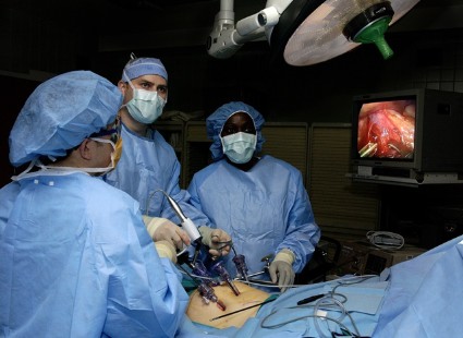
Hospital use of minimally invasive surgery shows disparity in surgical care nationwide
The use of minimally invasive surgery for appendectomy, colectomy, hysterectomy, and lung lobectomy varies widely in the United States, even though the complication rates were lower from each procedure than with open surgery, results from a large retrospective study demonstrated.
"This study has important implications for quality improvement," researchers led by Dr. Martin A. Makary, professor of surgery at Johns Hopkins University, Baltimore, wrote. "Based on our findings, many hospitals have an opportunity to decrease surgical complications by increasing utilization of minimally invasive surgery."

To investigate the levels of variation in the use of minimally invasive surgery across the United States, Dr. Makary and his associates used the National Inpatient Sample database, which is administered by the Agency for Healthcare Research & Quality, to evaluate hospitalizations at hospitals that performed at least 10 of these procedures in 2010. The sample included 1,051 hospitals in 45 states, and was limited to appendectomy, colectomy, hysterectomy, and lung lobectomy. The researchers used a propensity score model to calculate the predicted proportion of minimally invasive operations for each hospital based on patient characteristics. For each procedure, they categorized hospitals as low, medium, or high based on their actual to predicted proportion of minimally invasive surgery use (BMJ 2014;349:g4198).
On average, the use of minimally invasive surgery by the hospitals sampled was 71% for appendectomy, 28% for colectomy, 13% for hysterectomy, and 32% for lung lobectomy. Overall surgical complications for minimally invasive surgery, compared with open surgery, were, respectively, for appendectomy: 3.94% vs. 7.90% (P less than .001); colectomy: 13.8% vs. 35.8% (P less than .001); hysterectomy: 4.69% vs. 6.64% (P less than .001); and lung lobectomy: 17.1% vs. 25.4% (P less than .05). "In our analysis using Agency for Healthcare Research & Quality patient safety indicators for surgical care, we noted fewer wound, infectious, thrombotic, pulmonary, and mortality complications associated with minimally invasive surgery," the researchers wrote. "Based on our findings, increased hospital utilization of minimally invasive surgery at many U.S. hospitals represents a tremendous opportunity to prevent surgical site infection events."
The use of minimally invasive surgery was highly variable among the sampled hospitals. In fact, some never used minimally invasive surgery for some of the four procedures, while others used minimally invasive surgery for more than 75% of these procedures. Factors associated with the use of minimally invasive surgery were urban location, large hospital size, teaching hospital, and, for certain procedures, the hospital being located in the Midwest, South, or West.
"This [regional] disparity may be due to the broad range of surgical services some surgeons in rural areas are required to provide, and a scarcity of surgical specialists in such areas with advanced skills in minimally invasive surgery. Alternatively, the disparity may be a function of a lack of patient awareness about surgical options, decreased competition for patients, or a lack of minimally invasive surgery equipment, staff, or support in rural areas," the researchers wrote.
The findings of underutilization of minimally invasive surgery may also have something to do with a training gap.
"One reason that hospitals may be underperforming minimally invasive surgery is variability in appropriate training in residency and fellowship," Dr. Makary and his associates wrote. "One strategy that hospitals may consider in managing surgeons who cannot or choose not to acquire skills for performing minimally invasive surgery is to create a division of labor where patients who are not candidates for minimally invasive surgery are cared for by these surgeons. Increased standardization of competencies in minimally invasive surgery in surgical residency is needed to tackle wide variations in training."
The researchers acknowledged certain limitations of the study, including the fact that administrative claims data "can have incomplete coding, particularly of preexisting conditions," they wrote. "Another limitation is the lack of information available in the database for physician factors, such as laparoscopic training and experience that may influence the choice of procedure."
The researchers stated that they had no relevant financial conflicts to disclose.
The use of minimally invasive surgery for appendectomy, colectomy, hysterectomy, and lung lobectomy varies widely in the United States, even though the complication rates were lower from each procedure than with open surgery, results from a large retrospective study demonstrated.
"This study has important implications for quality improvement," researchers led by Dr. Martin A. Makary, professor of surgery at Johns Hopkins University, Baltimore, wrote. "Based on our findings, many hospitals have an opportunity to decrease surgical complications by increasing utilization of minimally invasive surgery."
To investigate the levels of variation in the use of minimally invasive surgery across the United States, Dr. Makary and his associates used the National Inpatient Sample database, which is administered by the Agency for Healthcare Research & Quality, to evaluate hospitalizations at hospitals that performed at least 10 of these procedures in 2010. The sample included 1,051 hospitals in 45 states, and was limited to appendectomy, colectomy, hysterectomy, and lung lobectomy. The researchers used a propensity score model to calculate the predicted proportion of minimally invasive operations for each hospital based on patient characteristics. For each procedure, they categorized hospitals as low, medium, or high based on their actual to predicted proportion of minimally invasive surgery use (BMJ 2014;349:g4198).
On average, the use of minimally invasive surgery by the hospitals sampled was 71% for appendectomy, 28% for colectomy, 13% for hysterectomy, and 32% for lung lobectomy. Overall surgical complications for minimally invasive surgery, compared with open surgery, were, respectively, for appendectomy: 3.94% vs. 7.90% (P less than .001); colectomy: 13.8% vs. 35.8% (P less than .001); hysterectomy: 4.69% vs. 6.64% (P less than .001); and lung lobectomy: 17.1% vs. 25.4% (P less than .05). "In our analysis using Agency for Healthcare Research & Quality patient safety indicators for surgical care, we noted fewer wound, infectious, thrombotic, pulmonary, and mortality complications associated with minimally invasive surgery," the researchers wrote. "Based on our findings, increased hospital utilization of minimally invasive surgery at many U.S. hospitals represents a tremendous opportunity to prevent surgical site infection events."
The use of minimally invasive surgery was highly variable among the sampled hospitals. In fact, some never used minimally invasive surgery for some of the four procedures, while others used minimally invasive surgery for more than 75% of these procedures. Factors associated with the use of minimally invasive surgery were urban location, large hospital size, teaching hospital, and, for certain procedures, the hospital being located in the Midwest, South, or West.
"This [regional] disparity may be due to the broad range of surgical services some surgeons in rural areas are required to provide, and a scarcity of surgical specialists in such areas with advanced skills in minimally invasive surgery. Alternatively, the disparity may be a function of a lack of patient awareness about surgical options, decreased competition for patients, or a lack of minimally invasive surgery equipment, staff, or support in rural areas," the researchers wrote.
The findings of underutilization of minimally invasive surgery may also have something to do with a training gap.
"One reason that hospitals may be underperforming minimally invasive surgery is variability in appropriate training in residency and fellowship," Dr. Makary and his associates wrote. "One strategy that hospitals may consider in managing surgeons who cannot or choose not to acquire skills for performing minimally invasive surgery is to create a division of labor where patients who are not candidates for minimally invasive surgery are cared for by these surgeons. Increased standardization of competencies in minimally invasive surgery in surgical residency is needed to tackle wide variations in training."
The researchers acknowledged certain limitations of the study, including the fact that administrative claims data "can have incomplete coding, particularly of preexisting conditions," they wrote. "Another limitation is the lack of information available in the database for physician factors, such as laparoscopic training and experience that may influence the choice of procedure."
The researchers stated that they had no relevant financial conflicts to disclose.
FROM THE BRITISH MEDICAL JOURNAL
Key clinical point: Hospital use of minimally invasive surgical procedures appears to vary widely in the United States.
Major Finding:. The use of minimally invasive surgery by the hospitals sampled was 71% for appendectomy, 28% for colectomy, 13% for hysterectomy, and 32% for lung lobectomy.
Data Source: An analysis of data from the National Inpatient Sample in 2010 that included 1,051 hospitals in 45 states, and was limited to appendectomy, colectomy, hysterectomy, and lung lobectomy.
Disclosures: The authors stated that they had no relevant financial conflicts to disclose.
KIR4.1 perhaps not an effective immune target for MS
Contrary to findings from previous studies, serologic testing for KIR4.1-specific IgG did not help in the diagnosis of multiple sclerosis in a new comparative study.
"No neural autoantigen has been clinically validated as a target of immunoglobulins in serum or CSF [cerebrospinal fluid] in multiple sclerosis," researchers led by Adipong Brickshawana, Ph.D., wrote July 7 in the Lancet Neurology. "The absence of a disease specific biomarker to aid multiple sclerosis diagnosis makes therapeutic trial design and outcome interpretation difficult," they said.
In their own analysis, the researchers determined that the lack of patient-derived IgG reactivity with KIR4.1 "in plasma membrane, cytoplasm, or endoplasmic reticulum of cultured glia argues against antigenic differences in recombinant and native glial KIR4.1." The finding runs counter to 2012 study that used enzyme-linked immunosorbent assay (ELISA). It found that serum antibodies against KIR4.1 were detected in 47% of 397 patients with multiple sclerosis, 1% of 329 patients with other neurologic diseases, and none of the 59 healthy donors (N. Engl. J. Med. 2012;367:115-23).
For the current analysis, Dr. Brickshawana of the departments of immunology and neurology at the Mayo Clinic, Rochester, Minn., and his associates used ELISA with a KIR4.1 peptide to test archival serum from 229 population-based and 57-clinic-based patients with multiple sclerosis, 99 healthy controls, and 109 disease controls, as well as CSF from 25 patients with multiple sclerosis and 22 disease controls (Lancet Neurol. 2014 July 7 [doi:10/1016/S1474-4422(14)70141-3]).
The researchers used cell-based immunofluorescence and immunoprecipitation to test all CSF and serum samples from 50 of the clinic-based patients with multiple sclerosis on cells expressing functional KIR4.1. They also looked for KIR4.1 immunoreactivity in archival brain samples from 15 patients with multiple sclerosis confirmed by histopathology, as well as from three controls with non-neurologic diseases.
Of the serum samples collected, only three from the 286 patients with multiple sclerosis (1.05%) and two from the 208 controls (0.96%) demonstrated KIR4.1 reactivity on ELISA, while none of the CSF samples from patients or controls demonstrated KIR4.1 reactivity. In addition, the researchers did not detect KIR4.1 loss from glia in supratentorial white-matter lesions of archival multiple sclerosis cases.
"Our findings provide neither serological nor neuropathological evidence to lend support to the idea that autoantibodies target glial KIR4.1 K+ channels in patients with multiple sclerosis," the researchers concluded.
Dr. Brickshawana and his colleagues went on to note that different analytical approaches "might, in part, explain these contradictory findings. However, before drawing conclusions, it is essential that KIR4.1 immunoreactivity is examined relative to control white-matter expression as we did in the current study."
The study was funded by the National Institutes of Health, the National Multiple Sclerosis Society, and the Mayo Clinic’s Robert and Arlene Kogod Center on Aging.
KIR4.1 is an inward rectifying potassium channel expressed on oligodendrocytes and the endfeet of astrocytes (including retinal Müller glia and Hensen’s and Claudius’ cells in the cochlea). It contributes to the maintenance of electrochemical gradient by removing potassium from the extracellular space and to the maintenance of potassium and water balance by acting in concert with aquaporin-4 to maintain osmotic homeostasis.
Similar to aquaporin-4, KIR4.1 is not uniquely expressed in CNS cells – it is present also in the retina, kidney, and parietal cells of gastric mucosal epithelium. Antibodies against parietal cells have been detected in the sera of patients with multiple sclerosis, particularly in those with gastrointestinal disturbances, so the occurrence of antibodies against KIR4.1 might merely be an epiphenomenon rather than a sign of causality.
A study by Elodie Nerrant of Gui-de-Chauliac Hospital, Montpellier, France, and her colleagues, using ELISA, reported that only 7.5% of 296 patients with multiple sclerosis had anti-KIR4.1 antibodies, and that this proportion did not significantly differ from those of healthy controls or patients with other neurologic diseases; immunofluorescence analysis did not detect any specific staining (Mult. Scler. 2014 April 22 [doi:10.1177/135245851453086]). Dr. Brickshawana and his associates detected reactivity to KIR4.1 in sera of 3 of 268 people with multiple sclerosis and 2 of 208 controls.
Do these findings, from independent research groups, mean that research into KIR4.1 in multiple sclerosis should end? We hope not.
The reasons for the discrepancies between the investigations might be at least partly technical and call for additional work. Pathologic studies using different analytical approaches also are warranted to deepen the understanding of this potentially revolutionary aspect of multiple sclerosis research. Many unanswered questions related to KIR4.1 function still remain.
The coexpression of KIR4.1 and aquaporin-4 channels in endfeet of astrocytes and their synergistic effect in maintaining osmotic homeostasis is intriguing, especially when considering that most retinal pathologic changes characterized by Müller cell damage are accompanied by changes of the amount or spatial distribution of both channels. Finally, the potential relation between anti-KIR4.1 antibodies and a more general dysfunction of immune-mediated mechanisms in patients with multiple sclerosis deserves further investigation.
Dr. Massimo Filippi and Dr. Maria A. Rocca are with the department of neurology at the Scientific Institute and University Ospedale San Raffaele, Milan, and Dr. Hans Lassmann is with the Center for Brain Research at the Medical University of Vienna. The researchers disclosed several financial conflicts from pharmaceutical companies. This was extracted from their commentary on Dr. Brickshawana’s report (Lancet Neurol. 2014 July 7 [doi:10/1016/S1474-4422(14)70141-3]).
Contrary to findings from previous studies, serologic testing for KIR4.1-specific IgG did not help in the diagnosis of multiple sclerosis in a new comparative study.
"No neural autoantigen has been clinically validated as a target of immunoglobulins in serum or CSF [cerebrospinal fluid] in multiple sclerosis," researchers led by Adipong Brickshawana, Ph.D., wrote July 7 in the Lancet Neurology. "The absence of a disease specific biomarker to aid multiple sclerosis diagnosis makes therapeutic trial design and outcome interpretation difficult," they said.
In their own analysis, the researchers determined that the lack of patient-derived IgG reactivity with KIR4.1 "in plasma membrane, cytoplasm, or endoplasmic reticulum of cultured glia argues against antigenic differences in recombinant and native glial KIR4.1." The finding runs counter to 2012 study that used enzyme-linked immunosorbent assay (ELISA). It found that serum antibodies against KIR4.1 were detected in 47% of 397 patients with multiple sclerosis, 1% of 329 patients with other neurologic diseases, and none of the 59 healthy donors (N. Engl. J. Med. 2012;367:115-23).
For the current analysis, Dr. Brickshawana of the departments of immunology and neurology at the Mayo Clinic, Rochester, Minn., and his associates used ELISA with a KIR4.1 peptide to test archival serum from 229 population-based and 57-clinic-based patients with multiple sclerosis, 99 healthy controls, and 109 disease controls, as well as CSF from 25 patients with multiple sclerosis and 22 disease controls (Lancet Neurol. 2014 July 7 [doi:10/1016/S1474-4422(14)70141-3]).
The researchers used cell-based immunofluorescence and immunoprecipitation to test all CSF and serum samples from 50 of the clinic-based patients with multiple sclerosis on cells expressing functional KIR4.1. They also looked for KIR4.1 immunoreactivity in archival brain samples from 15 patients with multiple sclerosis confirmed by histopathology, as well as from three controls with non-neurologic diseases.
Of the serum samples collected, only three from the 286 patients with multiple sclerosis (1.05%) and two from the 208 controls (0.96%) demonstrated KIR4.1 reactivity on ELISA, while none of the CSF samples from patients or controls demonstrated KIR4.1 reactivity. In addition, the researchers did not detect KIR4.1 loss from glia in supratentorial white-matter lesions of archival multiple sclerosis cases.
"Our findings provide neither serological nor neuropathological evidence to lend support to the idea that autoantibodies target glial KIR4.1 K+ channels in patients with multiple sclerosis," the researchers concluded.
Dr. Brickshawana and his colleagues went on to note that different analytical approaches "might, in part, explain these contradictory findings. However, before drawing conclusions, it is essential that KIR4.1 immunoreactivity is examined relative to control white-matter expression as we did in the current study."
The study was funded by the National Institutes of Health, the National Multiple Sclerosis Society, and the Mayo Clinic’s Robert and Arlene Kogod Center on Aging.
FROM LANCET NEUROLOGY
Key clinical point: KIR4.1-specific IgG did not aid in diagnosis of multiple sclerosis.
Major finding: Using ELISA with a KIR4.1 peptide, KIR4.1-specific IgG in MS patients was observed in only 3 of 286 serum samples and none of 25 CSF samples, and no loss of KIR4.1 was observed from glia in supratentorial white-matter lesions of 15 archival MS cases.
Data source: Tests of archival serum from 229 population-based and 57 clinic-based patients with MS, 99 healthy controls, and 109 disease controls, and tests of cerebrospinal fluid from 25 patients with MS and 22 disease controls.
Disclosures: The study was funded by the National Institutes of Health, the National Multiple Sclerosis Society, and the Mayo Clinic’s Robert and Arlene Kogod Center on Aging.
Levetiracetam found effective in controlling seizures in pregnancy
Levetiracetam monotherapy appears to be an effective strategy for controlling seizures in pregnancy, results from a large Australian study demonstrated.
In fact, levetiracetam provided seizure control comparable with that of older antiepileptic drugs (AEDs) carbamazepine and valproate but superior when compared with other newer AEDs, including lamotrigine and topiramate. The findings suggest that levetiracetam can be considered "among the agents of first choice in women contemplating pregnancy, and to be suitable for use in most circumstances in women who have the potential for pregnancy, unless the type of seizure disorder present, or other consideration, makes a different agent clinically preferable," wrote Dr. Frank J. E. Vajda of the department of medicine at the University of Melbourne and his associates (Epilepsia 2014 July 3 [doi:10.1111/epi.12711]).
Using a national registry, the researchers analyzed data regarding seizure control in 1,111 pregnancies that began between the middle of 1998 and November of 2013, during which the patients took only one AED for at least the first trimester. The seizure occurrence rates for patients on levetiracetam monotherapy were 31.7% for any seizure and 13.4% for convulsive seizures, which were similar to the rates of all older AEDs combined (34.8% and 18.9%, respectively). Compared with levetiracetam, however, the rate of seizure-affected pregnancy was about 50% and 30% higher among patients on lamotrigine and topiramate, respectively.
The authors acknowledged certain limitations of the study, including the fact that the allocation of particular AEDs "was not randomized, and there can be no guarantee that the drugs were always taken as prescribed or necessarily employed optimally, guided by plasma AED concentration monitoring when that was available."
The researchers stated that they had no relevant financial conflicts.
FROM EPILEPSIA
Key clinical point: Compared with older antiepileptic drugs, levetiracetam was equally effective in keeping pregnant women with the major types of epilepsy seizure free.
Major finding: Seizure occurrence rates for patients on levetiracetam monotherapy were 31.7% for any seizure and 13.4% for convulsive seizures, similar to the rates of all older AEDs combined (34.8% and 18.9%, respectively).
Data source: A study of 1,111 pregnancies in Australian women that began between mid-1998 and November of 2013.
Disclosures: The authors stated that they had no relevant financial conflicts.
Lifestyle Change Program Participation Associated With Reduced CVD Incidence
SAN FRANCISCO – Veterans who participated in a lifestyle change program to reduce weight and increase exercise experienced a 17% lower total incidence of cardiovascular disease over 5 years, results from a large analysis demonstrated.
"Two-thirds of U.S. adults are overweight or obese, yet we know little about how to bring help to them," Sandra L. Jackson, Ph.D., said at the annual scientific sessions of the American Diabetes Association.
Lifestyle change is a recommended strategy for prevention of cardiovascular disease, but the evidence of effect of lifestyle change programs on CVD "is mixed, and few studies have had sufficient sample size to study CVD incidence as compared to change in CVD risk factors. In addition, we know little about what lifestyle change programs can achieve in health care settings, where participants are patients and providers recommend a change in lifestyle."
For the current study, she and her associates used data from the U.S. Department of Veterans Affairs’ national weight management program, MOVE! The program has enrolled more than 400,000 veterans since 2005, making it the largest lifestyle change program in the United States. Implementation varies across VA facilities, but most use a standard 10-session core curriculum, with topics that include reading food labels, reducing fat intake, evaluating portion sizes, walking with a pedometer, and setting physical activity goals.
"The program is based on principles of motivational interviewing, so VA patients are encouraged to set their own goals for physical activity and weight loss, rather than follow a set program of prescribed goals," said Dr. Jackson, who conducted the study during her doctoral work in the Nutrition and Health Sciences Program at Emory University, Atlanta. "Nearly three-quarters of patients in the VA health system are overweight or obese and in theory, could benefit from MOVE!"
The researchers evaluated MOVE! participants with data from the National VA Informatics and Computing Infrastructure, which collects data from medical records, including demographics, outpatient visits, inpatient records, diagnoses, procedures, and prescriptions, and also features statistical tools. The study included 1,463,003 eligible patients who did not have CVD at baseline. Their mean age was 52 years, 89% were men, and their mean BMI was 33.6 kg/m2. The average follow-up was about 5 years. Of the 1,463,003 patients, 169,248 were MOVE! participants and 1,293,755 were not.
Dr. Jackson reported that MOVE! participants who engaged in at least 8 sessions over 6 months lost about 2.5% of their body weight, and mostly sustained the loss over 3 years. The less active MOVE! participants lost a little over 0.5% of their body weight and maintained the loss over 3 years, while the eligible nonparticipants gained about 0.5% of body weight over the 3 years.
"Even with the substantial differences between the participant and nonparticipant groups, direct comparisons of the risk factors are hard to interpret," Dr. Jackson noted. For this reason, she and her associates used least-square means to compute averages, and adjusted for differences in baseline CVD risk factors, as well as differences in baseline BMI, age, gender, and race/ethnicity between the populations. They also stratified the analyses by diabetes status.
Systolic BP was a little lower among MOVE! participants than among nonparticipants, particularly at 6 and 12 months, "although the effect disappeared over 24 and 36 months," Dr. Jackson said. HDL cholesterol was a little higher among participants, compared with nonparticipants, while non-HDL cholesterol and plasma glucose levels were a little lower among participants, compared with nonparticipants.
Cox proportional hazards modeling revealed that there was a 17% lower incidence of CVD among participants, compared with nonparticipants (hazard ratio, 0.83). "When we looked separately at coronary artery disease, stroke, peripheral vascular disease, and heart failure, the effects remained significant," she said. These models were also adjusted for age, gender, BMI, smoking status, and a propensity score indicating likelihood of MOVE! participation, as well as other demographic and clinical characteristics.
A subgroup analysis revealed that the effect of MOVE! participation was stronger for men than for women (HR 0.83 vs. HR 0.93, respectively; P = .001).
"The VA population does have substantially fewer women than men, and women in the VA tend to be younger and may be at lower CVD risk than men," Dr. Jackson noted. "We also saw a stronger effect of participation among VA patients without diabetes, compared with those with diabetes, and we saw a stronger effect of participation among current smokers compared with nonsmokers."
Dr. Jackson said that she had no relevant financial conflicts to disclose.
On Twitter @dougbrunk
AT THE ADA ANNUAL SCIENTIFIC SESSIONS

Lifestyle change program participation associated with reduced CVD incidence
SAN FRANCISCO – Veterans who participated in a lifestyle change program to reduce weight and increase exercise experienced a 17% lower total incidence of cardiovascular disease over 5 years, results from a large analysis demonstrated.
"Two-thirds of U.S. adults are overweight or obese, yet we know little about how to bring help to them," Sandra L. Jackson, Ph.D., said at the annual scientific sessions of the American Diabetes Association.

Lifestyle change is a recommended strategy for prevention of cardiovascular disease, but the evidence of effect of lifestyle change programs on CVD "is mixed, and few studies have had sufficient sample size to study CVD incidence as compared to change in CVD risk factors. In addition, we know little about what lifestyle change programs can achieve in health care settings, where participants are patients and providers recommend a change in lifestyle."
For the current study, she and her associates used data from the U.S. Department of Veterans Affairs’ national weight management program, MOVE! The program has enrolled more than 400,000 veterans since 2005, making it the largest lifestyle change program in the United States. Implementation varies across VA facilities, but most use a standard 10-session core curriculum, with topics that include reading food labels, reducing fat intake, evaluating portion sizes, walking with a pedometer, and setting physical activity goals.
"The program is based on principles of motivational interviewing, so VA patients are encouraged to set their own goals for physical activity and weight loss, rather than follow a set program of prescribed goals," said Dr. Jackson, who conducted the study during her doctoral work in the Nutrition and Health Sciences Program at Emory University, Atlanta. "Nearly three-quarters of patients in the VA health system are overweight or obese and in theory, could benefit from MOVE!"
The researchers evaluated MOVE! participants with data from the National VA Informatics and Computing Infrastructure, which collects data from medical records, including demographics, outpatient visits, inpatient records, diagnoses, procedures, and prescriptions, and also features statistical tools. The study included 1,463,003 eligible patients who did not have CVD at baseline. Their mean age was 52 years, 89% were men, and their mean BMI was 33.6 kg/m2. The average follow-up was about 5 years. Of the 1,463,003 patients, 169,248 were MOVE! participants and 1,293,755 were not.
Dr. Jackson reported that MOVE! participants who engaged in at least 8 sessions over 6 months lost about 2.5% of their body weight, and mostly sustained the loss over 3 years. The less active MOVE! participants lost a little over 0.5% of their body weight and maintained the loss over 3 years, while the eligible nonparticipants gained about 0.5% of body weight over the 3 years.
"Even with the substantial differences between the participant and nonparticipant groups, direct comparisons of the risk factors are hard to interpret," Dr. Jackson noted. For this reason, she and her associates used least-square means to compute averages, and adjusted for differences in baseline CVD risk factors, as well as differences in baseline BMI, age, gender, and race/ethnicity between the populations. They also stratified the analyses by diabetes status.
Systolic BP was a little lower among MOVE! participants than among nonparticipants, particularly at 6 and 12 months, "although the effect disappeared over 24 and 36 months," Dr. Jackson said. HDL cholesterol was a little higher among participants, compared with nonparticipants, while non-HDL cholesterol and plasma glucose levels were a little lower among participants, compared with nonparticipants.
Cox proportional hazards modeling revealed that there was a 17% lower incidence of CVD among participants, compared with nonparticipants (hazard ratio, 0.83). "When we looked separately at coronary artery disease, stroke, peripheral vascular disease, and heart failure, the effects remained significant," she said. These models were also adjusted for age, gender, BMI, smoking status, and a propensity score indicating likelihood of MOVE! participation, as well as other demographic and clinical characteristics.
A subgroup analysis revealed that the effect of MOVE! participation was stronger for men than for women (HR 0.83 vs. HR 0.93, respectively; P = .001).
"The VA population does have substantially fewer women than men, and women in the VA tend to be younger and may be at lower CVD risk than men," Dr. Jackson noted. "We also saw a stronger effect of participation among VA patients without diabetes, compared with those with diabetes, and we saw a stronger effect of participation among current smokers compared with nonsmokers."
Dr. Jackson said that she had no relevant financial conflicts to disclose.
On Twitter @dougbrunk
SAN FRANCISCO – Veterans who participated in a lifestyle change program to reduce weight and increase exercise experienced a 17% lower total incidence of cardiovascular disease over 5 years, results from a large analysis demonstrated.
"Two-thirds of U.S. adults are overweight or obese, yet we know little about how to bring help to them," Sandra L. Jackson, Ph.D., said at the annual scientific sessions of the American Diabetes Association.
Lifestyle change is a recommended strategy for prevention of cardiovascular disease, but the evidence of effect of lifestyle change programs on CVD "is mixed, and few studies have had sufficient sample size to study CVD incidence as compared to change in CVD risk factors. In addition, we know little about what lifestyle change programs can achieve in health care settings, where participants are patients and providers recommend a change in lifestyle."
For the current study, she and her associates used data from the U.S. Department of Veterans Affairs’ national weight management program, MOVE! The program has enrolled more than 400,000 veterans since 2005, making it the largest lifestyle change program in the United States. Implementation varies across VA facilities, but most use a standard 10-session core curriculum, with topics that include reading food labels, reducing fat intake, evaluating portion sizes, walking with a pedometer, and setting physical activity goals.
"The program is based on principles of motivational interviewing, so VA patients are encouraged to set their own goals for physical activity and weight loss, rather than follow a set program of prescribed goals," said Dr. Jackson, who conducted the study during her doctoral work in the Nutrition and Health Sciences Program at Emory University, Atlanta. "Nearly three-quarters of patients in the VA health system are overweight or obese and in theory, could benefit from MOVE!"
The researchers evaluated MOVE! participants with data from the National VA Informatics and Computing Infrastructure, which collects data from medical records, including demographics, outpatient visits, inpatient records, diagnoses, procedures, and prescriptions, and also features statistical tools. The study included 1,463,003 eligible patients who did not have CVD at baseline. Their mean age was 52 years, 89% were men, and their mean BMI was 33.6 kg/m2. The average follow-up was about 5 years. Of the 1,463,003 patients, 169,248 were MOVE! participants and 1,293,755 were not.
Dr. Jackson reported that MOVE! participants who engaged in at least 8 sessions over 6 months lost about 2.5% of their body weight, and mostly sustained the loss over 3 years. The less active MOVE! participants lost a little over 0.5% of their body weight and maintained the loss over 3 years, while the eligible nonparticipants gained about 0.5% of body weight over the 3 years.
"Even with the substantial differences between the participant and nonparticipant groups, direct comparisons of the risk factors are hard to interpret," Dr. Jackson noted. For this reason, she and her associates used least-square means to compute averages, and adjusted for differences in baseline CVD risk factors, as well as differences in baseline BMI, age, gender, and race/ethnicity between the populations. They also stratified the analyses by diabetes status.
Systolic BP was a little lower among MOVE! participants than among nonparticipants, particularly at 6 and 12 months, "although the effect disappeared over 24 and 36 months," Dr. Jackson said. HDL cholesterol was a little higher among participants, compared with nonparticipants, while non-HDL cholesterol and plasma glucose levels were a little lower among participants, compared with nonparticipants.
Cox proportional hazards modeling revealed that there was a 17% lower incidence of CVD among participants, compared with nonparticipants (hazard ratio, 0.83). "When we looked separately at coronary artery disease, stroke, peripheral vascular disease, and heart failure, the effects remained significant," she said. These models were also adjusted for age, gender, BMI, smoking status, and a propensity score indicating likelihood of MOVE! participation, as well as other demographic and clinical characteristics.
A subgroup analysis revealed that the effect of MOVE! participation was stronger for men than for women (HR 0.83 vs. HR 0.93, respectively; P = .001).
"The VA population does have substantially fewer women than men, and women in the VA tend to be younger and may be at lower CVD risk than men," Dr. Jackson noted. "We also saw a stronger effect of participation among VA patients without diabetes, compared with those with diabetes, and we saw a stronger effect of participation among current smokers compared with nonsmokers."
Dr. Jackson said that she had no relevant financial conflicts to disclose.
On Twitter @dougbrunk
AT THE ADA ANNUAL SCIENTIFIC SESSIONS
Key clinical point: It works: Adopting a more healthful lifestyle prevented CVD.
Major finding: Veterans who participated in a lifestyle change program to reduce weight and increase exercise experienced a 17% lower incidence of CVD over a period of 5 years, compared with those who did not participate in the program (hazard ratio, 0.83).
Data source: An analysis of 1.5 million patients who were eligible for the VA’s national weight management program, MOVE!, who did not have CVD at baseline.
Disclosures: Dr. Jackson said that she had no relevant financial conflicts to disclose.

Anxiety Heightens Risk of Stroke
Earn 0.25 hours AMA PRA Category 1 credit: Read this article, and click the link at the end to take the posttest.
The more anxious you are, the greater your chances of an incident stroke, results from a prospective analysis demonstrated. The association was independent of other known risk factors including depression, researchers led by Maya J. Lambiase, Ph.D., reported online Dec. 19, 2013, in the journal Stroke.
"Results indicated a dose-response relation between anxiety and stroke," according to Dr. Lambiase, a cardiovascular behavioral medicine researcher in the department of psychiatry at the University of Pittsburgh, and her associates. "Exploratory analyses suggest that behavioral factors, particularly smoking and physical activity, may be important pathways to consider."
Over a period of 22 years, the researchers studied a nationally representative sample of 6,019 people aged 25-74 years from the first National Health and Nutrition Examination Survey (NHANES I), which was conducted from 1971-1975. At baseline, all study participants underwent an in-person structured interview, physical exam, and blood draw, and they completed psychological questionnaires including the General Well-Being Schedule (GWB), a four-item tool that asks respondents to rate the severity of their anxiety-related symptoms during the past month. Strokes were identified through hospital or nursing home reports and death certificates. Numerous covariates were accounted for, including use of blood medications, diagnosis of diabetes by a physician, total serum cholesterol, body mass index, and depressive symptoms as measured by the GWB’s depressed mood subscale and by the Center for Epidemiologic Studies of Depression scale (CESD).
During a mean 16-year follow-up, the researchers identified 419 cases of incident strokes (221 in men and 198 in women). They observed that every one standard deviation increase in anxiety was associated with a 17% increase in stroke risk following adjustment for demographic factors. Men and women in the highest tertile of anxiety symptoms had a 33% higher risk of stroke, compared with those in the lowest tertile following adjustment for other cardiovascular risk factors.
"Poor health behaviors may be one pathway linking anxiety with stroke risk," the researchers wrote. "In the present study, behaviors (particularly smoking and physical activity) had the most sizable attenuating effect on the relationship between anxiety and incident stroke. However, since these behaviors did not account fully for the association between anxiety and incident stroke, direct biologic effects of anxiety should also be considered. Chronic anxiety could lead to excess activation of the hypothalamic-pituitary-adrenal axis and sympathetic nervous system, which may increase the risk for stroke. Anxiety could also contribute to stroke or other cardiovascular disease by lowering the threshold for arrhythmia or by reducing heart rate variability."
The researchers acknowledged certain limitations of the study, including the fact that baseline history of stroke and coronary heart disease were self-reported and that bias "may have occurred due to excluding individuals lost to follow-up or with missing data. We could not formally test mediation due to insufficiencies in temporal ordering of data collection. Stroke cases were identified based on discharge reports/death certificates and were not confirmed by imaging or a neurologist. Additionally, we may not have accurately captured silent strokes (resulting in an underestimate of cases in the population), although such misclassification would likely bias results toward the null."
The study coauthors were Laura D. Kubzansky, Ph.D., and Rebecca Thurston, Ph.D.
The study was funded by the National Heart, Lung, and Blood Institute and by the National Institute of Mental Health. The researchers stated that they had no relevant financial disclosures.
To earn 0.25 hours AMA PRA Category 1 credit after reading this article, take the post-test here.
FROM STROKE

Immunological testing alone missed TB cases in HIV population
SAN DIEGO – Mycobacterium tuberculosis can still be grown in the sputum of people living with HIV who have negative TB immunological testing, and in people who have been treated with TB previously, results from a study of nearly 200 patients demonstrated.
"Previous studies in low TB incidence areas have tested people with positive tuberculin skin tests or interferon release assays for active TB, but we have tested people for TB irrespective of this – and found a case of active tuberculosis and someone with TB in their sputum who would otherwise not have been tested using previous guidelines," lead author Dr. Santino Capocci, a research registrar in respiratory medicine at Royal Free Hospital, London, said in an interview in advance of an international conference of the American Thoracic Society, where the research was presented.

Guidelines from the United Kingdom recommend using blood interferon-gamma release assay with or without tuberculin skin testing for latent TB diagnosis, but it does not cover screening for active TB. "Implementation is patchy with little robust evidence for effectiveness," the researchers wrote in their abstract.
In an effort to determine the impact of comprehensive assessment for TB disease and infection in an ambulatory HIV clinic population with high antiretroviral usage, Dr. Capocci and his associates tested 194 patients, mean age 46 years, who had no clinical suspicion of active tuberculosis. Testing consisted of tuberculin skin testing (TST), interferon-gamma release assay (IGRA), frontal chest radiograph (CXR), and sputum induction for TB culture. Latent TB was defined as a positive IGRA and/or TST of 5 mm or greater in the absence of previously active TB disease.
Nearly one-quarter of patients (24%) were black African, 81% had received a previous Bacillus Calmette-Guerin vaccination, 94% were on antiretroviral medication, and 8% had previous TB disease. Dr. Capocci reported that latent TB infection was detected in 12 (6%) of the 194 patients. Of these, four were diagnosed by positive IGRA and TST of 5 mm or greater; one was diagnosed by positive IGRA and negative TST; five were diagnosed by TST of 10 mm or greater (with negative or borderline IGRA), and two were diagnosed by TST 5-9 mm (with negative IGRA). Two subjects had a positive sputum culture.
"One subject had evidence of tuberculosis in his sputum but no symptoms, normal blood tests and x-rays, and negative immunological testing (tuberculin skin test and IGRA)," Dr. Capocci said. "This has been seen in Africa in people living with HIV, but is not reported as far as we know in low TB incidence areas such as the UK. The case of active TB that we saw was in someone who has had tuberculosis previously, but without persistent symptoms. He may not have been tested using national testing guidelines."
Dr. Capocci acknowledged certain limitations of the study, including the fact that the sample size is "fairly small compared to studies that have used IGRAs or tuberculin skin testing. "It is also important to evaluate the cost effectiveness of this approach [for] future health care policy."
Dr. Capocci said that he had no relevant financial conflicts to disclose.
SAN DIEGO – Mycobacterium tuberculosis can still be grown in the sputum of people living with HIV who have negative TB immunological testing, and in people who have been treated with TB previously, results from a study of nearly 200 patients demonstrated.
"Previous studies in low TB incidence areas have tested people with positive tuberculin skin tests or interferon release assays for active TB, but we have tested people for TB irrespective of this – and found a case of active tuberculosis and someone with TB in their sputum who would otherwise not have been tested using previous guidelines," lead author Dr. Santino Capocci, a research registrar in respiratory medicine at Royal Free Hospital, London, said in an interview in advance of an international conference of the American Thoracic Society, where the research was presented.
Guidelines from the United Kingdom recommend using blood interferon-gamma release assay with or without tuberculin skin testing for latent TB diagnosis, but it does not cover screening for active TB. "Implementation is patchy with little robust evidence for effectiveness," the researchers wrote in their abstract.
In an effort to determine the impact of comprehensive assessment for TB disease and infection in an ambulatory HIV clinic population with high antiretroviral usage, Dr. Capocci and his associates tested 194 patients, mean age 46 years, who had no clinical suspicion of active tuberculosis. Testing consisted of tuberculin skin testing (TST), interferon-gamma release assay (IGRA), frontal chest radiograph (CXR), and sputum induction for TB culture. Latent TB was defined as a positive IGRA and/or TST of 5 mm or greater in the absence of previously active TB disease.
Nearly one-quarter of patients (24%) were black African, 81% had received a previous Bacillus Calmette-Guerin vaccination, 94% were on antiretroviral medication, and 8% had previous TB disease. Dr. Capocci reported that latent TB infection was detected in 12 (6%) of the 194 patients. Of these, four were diagnosed by positive IGRA and TST of 5 mm or greater; one was diagnosed by positive IGRA and negative TST; five were diagnosed by TST of 10 mm or greater (with negative or borderline IGRA), and two were diagnosed by TST 5-9 mm (with negative IGRA). Two subjects had a positive sputum culture.
"One subject had evidence of tuberculosis in his sputum but no symptoms, normal blood tests and x-rays, and negative immunological testing (tuberculin skin test and IGRA)," Dr. Capocci said. "This has been seen in Africa in people living with HIV, but is not reported as far as we know in low TB incidence areas such as the UK. The case of active TB that we saw was in someone who has had tuberculosis previously, but without persistent symptoms. He may not have been tested using national testing guidelines."
Dr. Capocci acknowledged certain limitations of the study, including the fact that the sample size is "fairly small compared to studies that have used IGRAs or tuberculin skin testing. "It is also important to evaluate the cost effectiveness of this approach [for] future health care policy."
Dr. Capocci said that he had no relevant financial conflicts to disclose.
AT ATS 2014
Key clinical point: Use of immunological testing alone to detect TB in people with HIV missed cases of subclinical disease.
Major finding: Despite the convenience of immunological tests to screen for TB in people with HIV, the use of comprehensive assessments detected 12 cases of latent TB in 194 patients with HIV, even in those with good CD4 and no symptoms.
Data source: A study of 194 HIV patients, mean age 46 years, who had no clinical suspicion of active tuberculosis.
Disclosures: Dr. Capocci had financial conflicts.
