User login
Mitchel is a reporter for MDedge based in the Philadelphia area. He started with the company in 1992, when it was International Medical News Group (IMNG), and has since covered a range of medical specialties. Mitchel trained as a virologist at Roswell Park Memorial Institute in Buffalo, and then worked briefly as a researcher at Boston Children's Hospital before pivoting to journalism as a AAAS Mass Media Fellow in 1980. His first reporting job was with Science Digest magazine, and from the mid-1980s to early-1990s he was a reporter with Medical World News. @mitchelzoler
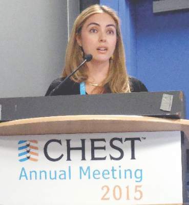
CHEST: Losartan slows emphysema progression in pilot trial
MONTREAL – Daily treatment with the antihypertensive drug losartan for a year appeared to slow progression of emphysema, compared with placebo, in a controlled pilot study involving 46 patients at one U.S. center, Dr. Allison Lambert reported at the annual meeting of the American College of Chest Physicians.
One year of daily, 100-mg oral treatment with the angiotensin receptor–blocking drug losartan in patients with emphysema produced a statistically significant regression of the disease in their right-middle lung lobes, compared with progression of right-middle lung lobe disease among control patients, as assessed by CT lung scans. In this lung segment, emphysema regressed by 0.72% in patients on losartan (Cozaar), compared with an average 3.34% rate of progression in patients on placebo.

The losartan-treated patients also showed a consistent pattern of either substantially slowed or reversed emphysema throughout multiple lung segments, although the between-group differences did not reach statistical significance in any other segment, said Dr. Lambert, a pulmonologist at Johns Hopkins Hospital in Baltimore.
Throughout the entire lung, 12 months of losartan treatment was linked to an average 0.32% reduction in emphysema extent from baseline when measured by CT, compared with a 2.18% rate of emphysema progression in control patients on usual care, which just missed statistical significance (P = .064).
Data from other researchers “suggest the right-middle lobe most commonly progresses in emphysema,” which may explain why that lung segment showed the most dramatic effect from treatment, she suggested. Another finding Dr. Lambert found striking was the consistent trend toward slowed emphysema progression in multiple segments throughout patients’ lungs.
Dr. Lambert called this a phase II, “proof of concept” trial. She and her associates have begun a larger, phase III version that will study the effect of 100-mg daily losartan during 1 year of treatment in 220 patients with emphysema, she said. This trial received funding from the Pulmonary Trials Cooperative of the National Heart Lung and Blood Institute.
“These are some of the most interesting and exciting data I’ve seen,” commented Dr. David P.L. Sachs, a pulmonologist who practices in Stanford, Calif., and cochaired the session in which Dr. Lambert gave her report. “Having an agent that could slow progression of emphysema would be unique,” he said in an interview. One aspect that makes this treatment especially attractive is losartan’s extensive safety record as an antihypertensive drug that is also often used to treat patients with heart failure, Dr. Sachs noted.
To put the 100-mg/day dosage used in the current study in perspective, results from a multicenter randomized trial of more than 3,800 heart failure patients published in 2009 showed that a losartan dosage of 150 mg once daily was safe and effective and produced outcomes superior to those seen with a 50 mg once-daily dosage (Lancet. 2009;374[9704]:1840-8).
Dr. Lambert and her associates designed this pilot study because of previously reported results from other groups showing favorable effects of losartan on animal models of emphysema or other lung disease, as well as suggestions from nonprospective clinical studies of benefits from angiotensin-receptor blockers on lung function and on chronic obstructive pulmonary disease (COPD) in particular.
The current study enrolled patients with mild to severe COPD who were current or former cigarette smokers who had at least a 10 pack-year history and were on stable treatment for their COPD. The researchers excluded patients already taking an angiotensin-receptor blocker or angiotensin-converting enzyme inhibitor.
The study included a total of 106 patients with COPD, including the 46 with emphysema at baseline. Their average age was about 58 years old, the enrolled patients included roughly equal numbers of men and women, and about two-thirds were current smokers. All patients underwent CT lung scans at baseline and after 6 and 12 months, as well as other lung function assessments. The study’s primary endpoint was the amount of additional emphysema in their lungs beyond that seen at baseline using CT imaging.
The entire group of 106 COPD patients showed no significant differences in emphysema progression at either 6 or 12 months between the 54 patients treated with losartan and the 52 controls on placebo, but a second, prespecified analysis that focused only on the 46 enrolled patients who had visible emphysema at baseline showed a significant slowing of progression at the 1-year follow-up, Dr. Lambert said.
The study received partial funding from Merck, which markets losartan (Cozaar). Dr. Lambert had no disclosures. Dr. Sachs had no disclosures.
On Twitter @mitchelzoler
MONTREAL – Daily treatment with the antihypertensive drug losartan for a year appeared to slow progression of emphysema, compared with placebo, in a controlled pilot study involving 46 patients at one U.S. center, Dr. Allison Lambert reported at the annual meeting of the American College of Chest Physicians.
One year of daily, 100-mg oral treatment with the angiotensin receptor–blocking drug losartan in patients with emphysema produced a statistically significant regression of the disease in their right-middle lung lobes, compared with progression of right-middle lung lobe disease among control patients, as assessed by CT lung scans. In this lung segment, emphysema regressed by 0.72% in patients on losartan (Cozaar), compared with an average 3.34% rate of progression in patients on placebo.
The losartan-treated patients also showed a consistent pattern of either substantially slowed or reversed emphysema throughout multiple lung segments, although the between-group differences did not reach statistical significance in any other segment, said Dr. Lambert, a pulmonologist at Johns Hopkins Hospital in Baltimore.
Throughout the entire lung, 12 months of losartan treatment was linked to an average 0.32% reduction in emphysema extent from baseline when measured by CT, compared with a 2.18% rate of emphysema progression in control patients on usual care, which just missed statistical significance (P = .064).
Data from other researchers “suggest the right-middle lobe most commonly progresses in emphysema,” which may explain why that lung segment showed the most dramatic effect from treatment, she suggested. Another finding Dr. Lambert found striking was the consistent trend toward slowed emphysema progression in multiple segments throughout patients’ lungs.
Dr. Lambert called this a phase II, “proof of concept” trial. She and her associates have begun a larger, phase III version that will study the effect of 100-mg daily losartan during 1 year of treatment in 220 patients with emphysema, she said. This trial received funding from the Pulmonary Trials Cooperative of the National Heart Lung and Blood Institute.
“These are some of the most interesting and exciting data I’ve seen,” commented Dr. David P.L. Sachs, a pulmonologist who practices in Stanford, Calif., and cochaired the session in which Dr. Lambert gave her report. “Having an agent that could slow progression of emphysema would be unique,” he said in an interview. One aspect that makes this treatment especially attractive is losartan’s extensive safety record as an antihypertensive drug that is also often used to treat patients with heart failure, Dr. Sachs noted.
To put the 100-mg/day dosage used in the current study in perspective, results from a multicenter randomized trial of more than 3,800 heart failure patients published in 2009 showed that a losartan dosage of 150 mg once daily was safe and effective and produced outcomes superior to those seen with a 50 mg once-daily dosage (Lancet. 2009;374[9704]:1840-8).
Dr. Lambert and her associates designed this pilot study because of previously reported results from other groups showing favorable effects of losartan on animal models of emphysema or other lung disease, as well as suggestions from nonprospective clinical studies of benefits from angiotensin-receptor blockers on lung function and on chronic obstructive pulmonary disease (COPD) in particular.
The current study enrolled patients with mild to severe COPD who were current or former cigarette smokers who had at least a 10 pack-year history and were on stable treatment for their COPD. The researchers excluded patients already taking an angiotensin-receptor blocker or angiotensin-converting enzyme inhibitor.
The study included a total of 106 patients with COPD, including the 46 with emphysema at baseline. Their average age was about 58 years old, the enrolled patients included roughly equal numbers of men and women, and about two-thirds were current smokers. All patients underwent CT lung scans at baseline and after 6 and 12 months, as well as other lung function assessments. The study’s primary endpoint was the amount of additional emphysema in their lungs beyond that seen at baseline using CT imaging.
The entire group of 106 COPD patients showed no significant differences in emphysema progression at either 6 or 12 months between the 54 patients treated with losartan and the 52 controls on placebo, but a second, prespecified analysis that focused only on the 46 enrolled patients who had visible emphysema at baseline showed a significant slowing of progression at the 1-year follow-up, Dr. Lambert said.
The study received partial funding from Merck, which markets losartan (Cozaar). Dr. Lambert had no disclosures. Dr. Sachs had no disclosures.
On Twitter @mitchelzoler
AT CHEST 2015
Key clinical point: A year of daily treatment with losartan produced significant slowing of emphysema progression in a placebo-controlled pilot study with 46 patients.
Major finding: Right middle-lobe emphysema regressed by 0.72% in patients using losartan, compared with 3.34% progression in controls, measured by CT imaging.
Data source: A single-center, prospective, controlled study involving 46 patients with emphysema and 60 additional patients with less severe COPD.
Disclosures: The study received partial funding from Merck, which markets losartan (Cozaar). Dr. Lambert had no disclosures. Dr. Sachs had no disclosures.
VIDEO: ASAP 2 trial will test Watchman in warfarin-contraindicated patients
Now that the Watchman device for left atrial appendage closure is on the U.S. market, target patients are those with atrial fibrillation who can tolerate at least a brief, 6-week course of treatment with warfarin – which is what the device’s labeling demands – but are poor candidates for long-term treatment with oral anticoagulation because they have had a serious bleeding episode while on anticoagulant treatment, Dr. Vivek Y. Reddy said in an interview.
Another type of atrial fibrillation patient who is potentially a prime target for Watchman placement are those with a complete contraindication to warfarin treatment, but as of now this makes then ineligible to receive the device. This category of patient will be the target of the ASAP 2 trial, a large, multicenter trial planned to start by the end of 2015 that will randomize atrial fibrillation patients ineligible to receive any oral anticoagulation to receive Watchman followed by a 6-month period of dual antiplatelet therapy or to current standard therapy for such patients with aspirin alone, said Dr. Reddy, professor of medicine and director of the cardiac arrhythmia service at Mount Sinai Hospital in New York.
The ASAP 2 trial follows the pilot study ASAP (ASA Plavix Feasibility Study With WATCHMAN Left Atrial Appendage Closure Technology) that Dr. Reddy led and ran at four centers in Europe placing Watchman in patients ineligible to receive any oral anticoagulant treatment followed by 6 months of dual antiplatelet therapy. The ASAP results showed that this approach could be safe and effective (J Am Coll Cardiol. 2013 Jun 25;61[25]:2551-6.).
Patients with a total contraindication against treatment with warfarin or another oral anticoagulant “have the greatest need,” said Dr. Reddy. “The problem is we don’t have much safety data” for these patients, and while the results from the ASAP trial showed the device can be safely placed just using dual antiplatelet therapy the numbers were small and the device is not approved for use in this setting, he said.
Dr. Reddy has been an advisor to and received research grants from Atritech/Boston Scientific, the companies that developed and now market Watchman.
The video associated with this article is no longer available on this site. Please view all of our videos on the MDedge YouTube channel
On Twitter @mitchelzoler
HFSA: Emphasizing "acute" in acute decompensated heart failure
NATIONAL HARBOR, MD. – Acute decompensated heart failure is becoming more of an emergency.
Traditionally, it has been seen as a lumbering event that could be treated at a relatively leisurely pace, but heart failure physicians increasingly see the moment when patients arrive in the hospital with an episode of acute decompensated heart failure as a time-sensitive event that requires rapid intervention in a manner much more akin to an acute MI than to chronic heart failure.
While the tide is slowly shifting to put a premium on rapid treatment to try to decongest acute heart failure patients, the treatment options clinicians have available for these patients often remain inadequate.
“Development of drugs for acutely decompensated heart failure has been extremely difficult. We have done a really horrible job treating this disease,” Dr. Milton Packer said at the HFSA annual scientific meeting.
Treatment of acute heart failure patients has generally focused on relieving dyspnea, but part of the new appreciation of this state as an emergency event involves understanding that the pathology patients have when they reach the hospital is much more global and has profoundly morbid consequences.
“We want more from treatment than for patients to feel a little bit better an hour or two sooner” by relieving dyspnea, said Dr. Packer, professor of medicine and a heart failure specialist at the University of Texas Southwestern Medical Center in Dallas. “In the first 6 hours [of acute heart failure hospitalization], many patients are spilling troponin. We don’t know what this means, but the patients who spill troponin have a markedly increased risk for a more complicated hospital course.” About 10%-25% of patients hospitalized with acute heart failure have recurrent worsening heart failure, and many of these are also the patients who have a spike in their troponin level during initial hospitalization, he noted.
The troponin release in many patients and its association with worse outcomes is a clue that these patients are experiencing an ischemic myocardial event similar to an acute MI, possibly caused by myocardial-wall stretch, Dr. Packer said in an interview.
“If we can reduce this early acute cardiac dilatation, maybe we can reduce myocardial injury, reduce troponin release, and have favorable effects on clinically relevant events both short-term and long-term,” he said. “That’s why in trials [of investigational drugs for acute heart failure] we are treating patients earlier. Before we said we could enroll patients [into acute-treatment trials] within 48 hours of hospital admission. Now we enroll within 16 hours, or within 12 hours. We’ve learned that early intervention is important. That makes acute heart failure a lot more similar to an acute MI.”
European Society recommends faster acute heart failure management
European heart failure specialists have also become convinced that acute heart failure is an emergency that needs a rapid response. In June, the Heart Failure Association of the European Society of Cardiology published new recommendations on the in-hospital management of patients with acute heart failure (Eur J Heart Fail 2015 June;17[6];544-58). In the document, the association’s writing panel said, “The potentially greater benefit of early treatment is of conceptual importance in many cardiovascular presentations (e.g., myocardial infarction). Unfortunately, acute heart failure has not been considered with this regard until recently.” Breaking with the past, the association’s new recommendations now say that “all acute heart failure patients should receive appropriate therapy as early as possible,” an approach that involves starting acute management in the prehospital setting.
One member of the writing group for these recommendations put it more succinctly while speaking at the annual congress of the European Society of Cardiology in London in August: “Time is muscle in acute heart failure,” said Dr. Piotr Ponikowski, professor and heart failure specialist at the Medical University in Wroclaw, Poland. “When a patient has acute coronary syndrome everyone rushes, but we have patients with acute heart failure and no one rushes. We give furosemide, maybe something else, and then we wait and see.” He recommended adhering to a schedule that would have a patient assessed and initially treated within the first hour of hospitalization, and even sooner if treatment could start at the prehospital stage.
Like Dr. Packer, Dr. Ponikowski also lamented the inadequate tools now available for treating acute heart failure and the pressing need to identify better approaches to treatment, especially for selected acute heart failure patients.
“It is too simple to think that one drug or one treatment will help the entire spectrum of acute heart failure patients,” he said. “Our hypothesis is that profiling patients at every step of acute heart failure is crucial.”
He itemized five distinct types of acute heart failure patients based on their precipitating triggers of decompensation:
• Rapid arrhythmia or rhythm disturbance.
• Hypertension emergency.
• Pulmonary embolism.
• Pulmonary infection.
• Mechanical cause of acute heart failure.
“We need to clinically profile” patients into these subgroups to better tailor management, he said.
Another important aspect of patient heterogeneity is that fluid congestion may be less important in many patients compared with fluid redistribution from the splanchnic circulation. This distinction is important because fluid redistribution may be better treated with a vasodilator than with a diuretic, he noted. He voiced hope that two phase III trials now in progress with two unique vasodilator drugs, the TRUE-AHF trial of ularitide, and the RELAX-AHF-2 trial of serelaxin, may identify two new vasodilators with “unique effects” that could potentially launch a new era in management of selected patients with acute heart failure. Dr. Packer, the principal investigator for the ularitide trial, offered similar hope.
The responsiveness of acute heart failure patients to in-hospital treatment may vary depending on what end-organ damage they experience, Dr. Ponikowski said.
This end-organ damage is often an acute process occurring during hospitalization caused by the fluid congestion and redistribution that occurs during acute heart failure, said Dr. Alexandre Mebazaa, professor of anesthesiology and critical care medicine at Lariboisière Hospital in Paris.
“Fluid overload leads to organ dysfunction. In the past, we thought that kidney dysfunction [occurring during acute heart failure] was due to low cardiac output, but we know that dysfunction in the kidney and liver is due to congestion, and diuretics do not remove water from the liver and kidney,” Dr. Mebazaa said in an interview. “Diuretics may remove fluid from vessels, but not from organs. We need new approaches to remove fluid from organs – from the kidney, liver, and lungs” – during acute heart failure. This is another reason why heart failure physicians are excited about the possibility of finding new vasodilators, such a ularitide and serelaxin, that might address the issue of venous congestion in peripheral organs.
Faster management endorsed by U.S. clinicians, too

“We used to think that the reason why patients with acute heart failure were not voiding well and became diuretic resistant was because of poor cardiac output. Now we know that there is a lot of venous congestion with an impact on the liver and kidneys,” agreed Dr. Mariell L. Jessup, professor and medical director of the Penn Heart and Vascular Center at the University of Pennsylvania in Philadelphia. “We’ve begun to appreciate how important venous congestion is in causing high pressures on the right side” of the circulatory system, she said in an interview.
Other U.S. physicians echo the call by Dr. Packer and the European cardiologists for faster treatment of acute heart failure. “I collected data at U.S. hospitals and found it took an average of 22 hours for decompensated heart failure patients to receive treatment,” said Dr. Maria Rosa Costanzo, medical director of the heart failure and pulmonary hypertension program at Advocate Heart Institute in Naperville, Ill. “I have tried to convey the message that these patients must be treated early, and this is associated with better outcomes,” she said in an interview during the HFSA meeting.
“Early treatment means at least two doses of intravenous diuretic in the emergency department. We’ve seen that the two immediate doses can make a big difference, producing shorter lengths of stay in the intensive care unit, fewer rehospitalizations, and fewer deaths,” according to data collected in the ADHERE (Acute Decompensated Heart Failure National Registry), she said. “But this has not yet been picked up in a lot of U.S. practice.” Although the hemodynamic abnormalities that lead up to an acute decompensation event can take several weeks of steady worsening before severe symptoms drive a patient to the hospital, once the patient requires hospitalization “it should be treated as an emergency,” she said.
Dr. Costanzo is a major advocate for using ultrafiltration as a second-line treatment for acute decompensated heart failure patients who do not adequately respond to diuretic treatment, but for the time being, ultrafiltration remains a controversial option that at least some other heart failure physicians do not endorse, and it can involve reimbursement issues as many insurers consider it investigational.
“Try to get the patient decongested within the first 6 hours [after arrival at the hospital] or even sooner, within the first 1-2 hours,” recommended Dr. Christopher M. O’Connor, chief executive officer of Inova Heart and Vascular Institute in Falls Church, Va. He suggested treating patients with a combination of diuretics and vasodilators. “Some people are talking about instituting a performance measure for treating acute heart failure within the first 6 hours,” Dr. O’Connor said in an interview.
Currently, vasodilator treatment is limited to standard agents such as intravenous nitroglycerin, but Dr. O’Connor shared the hope that sometime soon a new vasodilator may be shown effective for acutely decompensated patients. He is a coinvestigator on the TRUE-AHF study of ularitide. “We hope that these new vasodilators, ularitide and serelaxin, will be good complements to diuretics, he said. Dr. O’Connor also recommended that clinicians shy away from using ultrafiltration as a back-up therapy, believing that it was shown ineffective and potentially harmful in results from the CARRESS-HF trial (N Engl J Med 2012 Dec 13;367[24]:2296-304).
But not all heart failure specialists see acute heart failure as a new frontier for early treatment and new drug discovery.

“So much energy has already been spent on acute heart failure with very little return,” said Dr. Clyde W. Yancy, professor and chief of cardiology at Northwestern University in Chicago. “I think that our best opportunities in heart failure are in prevention and in better chronic care. The hospitalized patient is so broad and complex; if we’re looking at how to best spend our resources I think it’s best to focus on prevention,” he said in an interview.
“The hospital experience needs to shift toward better use of systems of care and focus less on the biology. The biggest challenge is how to coordinate all the systems to make sure that patients have access to the resources and can obtain [existing] medications. Patients don’t often have the literacy to understand discharge instructions, and our systems are overwhelmed by trying to have 7-day follow-up visits. Focusing on management of the hospitalized patient does not give us a good return on the investment. There is no question that acute heart failure is an unmet need, but the greater unmet need is prevention and improved chronic care. No single intervention will dramatically change the acute heart failure experience. Focusing on the hospitalization does not offer us management opportunities that are as robust as we once thought,” Dr. Yancy said.
Dr. Packer has been a consultant to 22 companies. Dr. Ponikowski has been a consultant to, speaker for, or has received research grants from 11 companies. Dr. Mebazaa has received speaking honoraria and consulting fees from 11 companies. Dr. Jessup, Dr. Costanzo, and Dr. Yancy had no disclosures. Dr. O’Connor has been a consultant to ResMed, Roche Diagnostics, Cardiorentis, Bayer, and Actelion and has received research grants from Otsuka, ResMed, and Roche Diagnostics.
On Twitter@mitchelzoler
NATIONAL HARBOR, MD. – Acute decompensated heart failure is becoming more of an emergency.
Traditionally, it has been seen as a lumbering event that could be treated at a relatively leisurely pace, but heart failure physicians increasingly see the moment when patients arrive in the hospital with an episode of acute decompensated heart failure as a time-sensitive event that requires rapid intervention in a manner much more akin to an acute MI than to chronic heart failure.
While the tide is slowly shifting to put a premium on rapid treatment to try to decongest acute heart failure patients, the treatment options clinicians have available for these patients often remain inadequate.
“Development of drugs for acutely decompensated heart failure has been extremely difficult. We have done a really horrible job treating this disease,” Dr. Milton Packer said at the HFSA annual scientific meeting.
Treatment of acute heart failure patients has generally focused on relieving dyspnea, but part of the new appreciation of this state as an emergency event involves understanding that the pathology patients have when they reach the hospital is much more global and has profoundly morbid consequences.
“We want more from treatment than for patients to feel a little bit better an hour or two sooner” by relieving dyspnea, said Dr. Packer, professor of medicine and a heart failure specialist at the University of Texas Southwestern Medical Center in Dallas. “In the first 6 hours [of acute heart failure hospitalization], many patients are spilling troponin. We don’t know what this means, but the patients who spill troponin have a markedly increased risk for a more complicated hospital course.” About 10%-25% of patients hospitalized with acute heart failure have recurrent worsening heart failure, and many of these are also the patients who have a spike in their troponin level during initial hospitalization, he noted.
The troponin release in many patients and its association with worse outcomes is a clue that these patients are experiencing an ischemic myocardial event similar to an acute MI, possibly caused by myocardial-wall stretch, Dr. Packer said in an interview.
“If we can reduce this early acute cardiac dilatation, maybe we can reduce myocardial injury, reduce troponin release, and have favorable effects on clinically relevant events both short-term and long-term,” he said. “That’s why in trials [of investigational drugs for acute heart failure] we are treating patients earlier. Before we said we could enroll patients [into acute-treatment trials] within 48 hours of hospital admission. Now we enroll within 16 hours, or within 12 hours. We’ve learned that early intervention is important. That makes acute heart failure a lot more similar to an acute MI.”
European Society recommends faster acute heart failure management
European heart failure specialists have also become convinced that acute heart failure is an emergency that needs a rapid response. In June, the Heart Failure Association of the European Society of Cardiology published new recommendations on the in-hospital management of patients with acute heart failure (Eur J Heart Fail 2015 June;17[6];544-58). In the document, the association’s writing panel said, “The potentially greater benefit of early treatment is of conceptual importance in many cardiovascular presentations (e.g., myocardial infarction). Unfortunately, acute heart failure has not been considered with this regard until recently.” Breaking with the past, the association’s new recommendations now say that “all acute heart failure patients should receive appropriate therapy as early as possible,” an approach that involves starting acute management in the prehospital setting.
One member of the writing group for these recommendations put it more succinctly while speaking at the annual congress of the European Society of Cardiology in London in August: “Time is muscle in acute heart failure,” said Dr. Piotr Ponikowski, professor and heart failure specialist at the Medical University in Wroclaw, Poland. “When a patient has acute coronary syndrome everyone rushes, but we have patients with acute heart failure and no one rushes. We give furosemide, maybe something else, and then we wait and see.” He recommended adhering to a schedule that would have a patient assessed and initially treated within the first hour of hospitalization, and even sooner if treatment could start at the prehospital stage.
Like Dr. Packer, Dr. Ponikowski also lamented the inadequate tools now available for treating acute heart failure and the pressing need to identify better approaches to treatment, especially for selected acute heart failure patients.
“It is too simple to think that one drug or one treatment will help the entire spectrum of acute heart failure patients,” he said. “Our hypothesis is that profiling patients at every step of acute heart failure is crucial.”
He itemized five distinct types of acute heart failure patients based on their precipitating triggers of decompensation:
• Rapid arrhythmia or rhythm disturbance.
• Hypertension emergency.
• Pulmonary embolism.
• Pulmonary infection.
• Mechanical cause of acute heart failure.
“We need to clinically profile” patients into these subgroups to better tailor management, he said.
Another important aspect of patient heterogeneity is that fluid congestion may be less important in many patients compared with fluid redistribution from the splanchnic circulation. This distinction is important because fluid redistribution may be better treated with a vasodilator than with a diuretic, he noted. He voiced hope that two phase III trials now in progress with two unique vasodilator drugs, the TRUE-AHF trial of ularitide, and the RELAX-AHF-2 trial of serelaxin, may identify two new vasodilators with “unique effects” that could potentially launch a new era in management of selected patients with acute heart failure. Dr. Packer, the principal investigator for the ularitide trial, offered similar hope.
The responsiveness of acute heart failure patients to in-hospital treatment may vary depending on what end-organ damage they experience, Dr. Ponikowski said.
This end-organ damage is often an acute process occurring during hospitalization caused by the fluid congestion and redistribution that occurs during acute heart failure, said Dr. Alexandre Mebazaa, professor of anesthesiology and critical care medicine at Lariboisière Hospital in Paris.
“Fluid overload leads to organ dysfunction. In the past, we thought that kidney dysfunction [occurring during acute heart failure] was due to low cardiac output, but we know that dysfunction in the kidney and liver is due to congestion, and diuretics do not remove water from the liver and kidney,” Dr. Mebazaa said in an interview. “Diuretics may remove fluid from vessels, but not from organs. We need new approaches to remove fluid from organs – from the kidney, liver, and lungs” – during acute heart failure. This is another reason why heart failure physicians are excited about the possibility of finding new vasodilators, such a ularitide and serelaxin, that might address the issue of venous congestion in peripheral organs.
Faster management endorsed by U.S. clinicians, too
“We used to think that the reason why patients with acute heart failure were not voiding well and became diuretic resistant was because of poor cardiac output. Now we know that there is a lot of venous congestion with an impact on the liver and kidneys,” agreed Dr. Mariell L. Jessup, professor and medical director of the Penn Heart and Vascular Center at the University of Pennsylvania in Philadelphia. “We’ve begun to appreciate how important venous congestion is in causing high pressures on the right side” of the circulatory system, she said in an interview.
Other U.S. physicians echo the call by Dr. Packer and the European cardiologists for faster treatment of acute heart failure. “I collected data at U.S. hospitals and found it took an average of 22 hours for decompensated heart failure patients to receive treatment,” said Dr. Maria Rosa Costanzo, medical director of the heart failure and pulmonary hypertension program at Advocate Heart Institute in Naperville, Ill. “I have tried to convey the message that these patients must be treated early, and this is associated with better outcomes,” she said in an interview during the HFSA meeting.
“Early treatment means at least two doses of intravenous diuretic in the emergency department. We’ve seen that the two immediate doses can make a big difference, producing shorter lengths of stay in the intensive care unit, fewer rehospitalizations, and fewer deaths,” according to data collected in the ADHERE (Acute Decompensated Heart Failure National Registry), she said. “But this has not yet been picked up in a lot of U.S. practice.” Although the hemodynamic abnormalities that lead up to an acute decompensation event can take several weeks of steady worsening before severe symptoms drive a patient to the hospital, once the patient requires hospitalization “it should be treated as an emergency,” she said.
Dr. Costanzo is a major advocate for using ultrafiltration as a second-line treatment for acute decompensated heart failure patients who do not adequately respond to diuretic treatment, but for the time being, ultrafiltration remains a controversial option that at least some other heart failure physicians do not endorse, and it can involve reimbursement issues as many insurers consider it investigational.
“Try to get the patient decongested within the first 6 hours [after arrival at the hospital] or even sooner, within the first 1-2 hours,” recommended Dr. Christopher M. O’Connor, chief executive officer of Inova Heart and Vascular Institute in Falls Church, Va. He suggested treating patients with a combination of diuretics and vasodilators. “Some people are talking about instituting a performance measure for treating acute heart failure within the first 6 hours,” Dr. O’Connor said in an interview.
Currently, vasodilator treatment is limited to standard agents such as intravenous nitroglycerin, but Dr. O’Connor shared the hope that sometime soon a new vasodilator may be shown effective for acutely decompensated patients. He is a coinvestigator on the TRUE-AHF study of ularitide. “We hope that these new vasodilators, ularitide and serelaxin, will be good complements to diuretics, he said. Dr. O’Connor also recommended that clinicians shy away from using ultrafiltration as a back-up therapy, believing that it was shown ineffective and potentially harmful in results from the CARRESS-HF trial (N Engl J Med 2012 Dec 13;367[24]:2296-304).
But not all heart failure specialists see acute heart failure as a new frontier for early treatment and new drug discovery.
“So much energy has already been spent on acute heart failure with very little return,” said Dr. Clyde W. Yancy, professor and chief of cardiology at Northwestern University in Chicago. “I think that our best opportunities in heart failure are in prevention and in better chronic care. The hospitalized patient is so broad and complex; if we’re looking at how to best spend our resources I think it’s best to focus on prevention,” he said in an interview.
“The hospital experience needs to shift toward better use of systems of care and focus less on the biology. The biggest challenge is how to coordinate all the systems to make sure that patients have access to the resources and can obtain [existing] medications. Patients don’t often have the literacy to understand discharge instructions, and our systems are overwhelmed by trying to have 7-day follow-up visits. Focusing on management of the hospitalized patient does not give us a good return on the investment. There is no question that acute heart failure is an unmet need, but the greater unmet need is prevention and improved chronic care. No single intervention will dramatically change the acute heart failure experience. Focusing on the hospitalization does not offer us management opportunities that are as robust as we once thought,” Dr. Yancy said.
Dr. Packer has been a consultant to 22 companies. Dr. Ponikowski has been a consultant to, speaker for, or has received research grants from 11 companies. Dr. Mebazaa has received speaking honoraria and consulting fees from 11 companies. Dr. Jessup, Dr. Costanzo, and Dr. Yancy had no disclosures. Dr. O’Connor has been a consultant to ResMed, Roche Diagnostics, Cardiorentis, Bayer, and Actelion and has received research grants from Otsuka, ResMed, and Roche Diagnostics.
On Twitter@mitchelzoler
EXPERT ANALYSIS FROM THE HFSA ANNUAL SCIENTIFIC MEETING

CHEST: Yoga performs like pulmonary rehab for COPD patients
MONTREAL – A structured regimen of regular yoga exercises was as effective as a standard pulmonary rehabilitation program in patients with chronic obstructive pulmonary disease for improving lung function, exercise tolerance, dyspnea severity, and quality of life in a single-center, randomized comparison of the two strategies with 60 patients.
In addition, chronic obstructive pulmonary disease (COPD) patients had a higher level of acceptance of yoga and were more comfortable doing it, compared with standard pulmonary rehabilitation, and it is a cost-effective approach given the minimal equipment required, Dr. Randeep Guleria said at the annual meeting of the American College of Chest Physicians.

“Patients with difficulty walking, osteoarthritis, knee problems, or unable to do exercises like cycling or treadmill found yoga to be much more acceptable,” said Dr. Guleria in an interview. Acceptance of yoga was also higher than standard rehabilitation among patients with more severe COPD, said Dr. Guleria, professor and head of the department of pulmonary medicine and sleep disorders at the All India Institute of Medical Sciences in New Delhi.
“I think that yoga could be a very valuable adjunct” to pulmonary rehabilitation in COPD patients, commented Dr. Roger S. Goldstein, director of the divisional program in respiratory rehabilitation at the University of Toronto. Dr. Goldstein speculated that even better than comparing yoga against conventional pulmonary rehabilitation would be a study that compared a combined yoga plus rehabilitation program against standard rehabilitation alone. Yoga is “a tremendous opportunity,” he said.
The 12-week study enrolled 60 patients who averaged 56 years old who had been diagnosed with COPD for an average of 8 years. Just under a third of the patients had moderate COPD, 42% had severe COPD, and 28% had very severe COPD.
Dr. Guleria and his associates randomized 30 patients into a yoga program that included 4 weeks of biweekly 1-hour sessions that instructed patients in a series of specially designed yoga exercises. That was followed by 8 weeks during which patients were mostly left to perform their learned exercises on their own, but with a supervised session once every 2 weeks. The other 30 patients participated in a standard pulmonary rehabilitation program for 12 weeks.
The researchers measured several parameters at baseline and after 12 weeks, including two measures of dyspnea severity, 6-minute walk distance, a quality of life assessment, and two serum markers of inflammation, C-reactive protein and interleukin 6.
Both interventions resulted in modest but statistically significant improvements, such as increases in 6-minute walk distance and a reduced modified Borg scale assessment. The Borg scale score fell from an average of 1.5 at baseline to 1.0 after 12 weeks in the yoga patients, and from an average 3.0 at baseline to 0.5 after 12 weeks in the rehabilitation patients.
A score that measured total quality of life improved by an average of 32% in the yoga patients and by an average of 21% in the rehabilitation patients, changes that approached statistical significance in both subgroups.
Comparing the assessment measures after 12 weeks in both arms of the study showed no statistically significant between-group differences, Dr. Guleria reported.
The researchers commissioned a specially designed yoga program from a professional yoga instructor who had been briefed about COPD. The yoga exercises included physical postures, breathing technique, and meditation and relaxation. The pulmonary rehabilitation program included patient education, upper and lower limb exercises, and breathing exercises.
Dr. Guleria said he believed the opportunity exists to further modify the yoga program to better optimize its potential to benefit COPD patients.
Dr. Guleria had no relevant disclosures. Dr. Goldstein had no relevant disclosures.
The video associated with this article is no longer available on this site. Please view all of our videos on the MDedge YouTube channel
On Twitter@mitchelzoler
MONTREAL – A structured regimen of regular yoga exercises was as effective as a standard pulmonary rehabilitation program in patients with chronic obstructive pulmonary disease for improving lung function, exercise tolerance, dyspnea severity, and quality of life in a single-center, randomized comparison of the two strategies with 60 patients.
In addition, chronic obstructive pulmonary disease (COPD) patients had a higher level of acceptance of yoga and were more comfortable doing it, compared with standard pulmonary rehabilitation, and it is a cost-effective approach given the minimal equipment required, Dr. Randeep Guleria said at the annual meeting of the American College of Chest Physicians.
“Patients with difficulty walking, osteoarthritis, knee problems, or unable to do exercises like cycling or treadmill found yoga to be much more acceptable,” said Dr. Guleria in an interview. Acceptance of yoga was also higher than standard rehabilitation among patients with more severe COPD, said Dr. Guleria, professor and head of the department of pulmonary medicine and sleep disorders at the All India Institute of Medical Sciences in New Delhi.
“I think that yoga could be a very valuable adjunct” to pulmonary rehabilitation in COPD patients, commented Dr. Roger S. Goldstein, director of the divisional program in respiratory rehabilitation at the University of Toronto. Dr. Goldstein speculated that even better than comparing yoga against conventional pulmonary rehabilitation would be a study that compared a combined yoga plus rehabilitation program against standard rehabilitation alone. Yoga is “a tremendous opportunity,” he said.
The 12-week study enrolled 60 patients who averaged 56 years old who had been diagnosed with COPD for an average of 8 years. Just under a third of the patients had moderate COPD, 42% had severe COPD, and 28% had very severe COPD.
Dr. Guleria and his associates randomized 30 patients into a yoga program that included 4 weeks of biweekly 1-hour sessions that instructed patients in a series of specially designed yoga exercises. That was followed by 8 weeks during which patients were mostly left to perform their learned exercises on their own, but with a supervised session once every 2 weeks. The other 30 patients participated in a standard pulmonary rehabilitation program for 12 weeks.
The researchers measured several parameters at baseline and after 12 weeks, including two measures of dyspnea severity, 6-minute walk distance, a quality of life assessment, and two serum markers of inflammation, C-reactive protein and interleukin 6.
Both interventions resulted in modest but statistically significant improvements, such as increases in 6-minute walk distance and a reduced modified Borg scale assessment. The Borg scale score fell from an average of 1.5 at baseline to 1.0 after 12 weeks in the yoga patients, and from an average 3.0 at baseline to 0.5 after 12 weeks in the rehabilitation patients.
A score that measured total quality of life improved by an average of 32% in the yoga patients and by an average of 21% in the rehabilitation patients, changes that approached statistical significance in both subgroups.
Comparing the assessment measures after 12 weeks in both arms of the study showed no statistically significant between-group differences, Dr. Guleria reported.
The researchers commissioned a specially designed yoga program from a professional yoga instructor who had been briefed about COPD. The yoga exercises included physical postures, breathing technique, and meditation and relaxation. The pulmonary rehabilitation program included patient education, upper and lower limb exercises, and breathing exercises.
Dr. Guleria said he believed the opportunity exists to further modify the yoga program to better optimize its potential to benefit COPD patients.
Dr. Guleria had no relevant disclosures. Dr. Goldstein had no relevant disclosures.
The video associated with this article is no longer available on this site. Please view all of our videos on the MDedge YouTube channel
On Twitter@mitchelzoler
AT CHEST 2015
Key clinical point: COPD patients randomized to a 12-week program of yoga exercises had improvements similar to patients randomized to a standard pulmonary rehabilitation program.
Major finding: Average total quality of life scores improved by a relative 32% with yoga and 21% with standard rehabilitation.
Data source: Single-center, prospective, randomized trial with 60 patients.
Disclosures: Dr. Guleria had no relevant disclosures. Dr. Goldstein had no relevant disclosures.

CHEST: Oral solithromycin shows pneumonia pivotal-trial efficacy
MONTREAL – A new, next-generation macrolide, solithromycin, showed safety and efficacy as a once-daily oral agent that was noninferior to the comparator oral antibiotic, the fluoroquinolone moxifloxacin, in a phase III trial.
Macrolide resistance among strains of Streptococcus pneumoniae that cause many U.S. cases of severe community-acquired pneumonia has become common, complicating treatment of this common infection with a macrolide, Dr. Carlos M. Barrera explained at the annual meeting of the American College of Chest Physicians.

The SOLITAIRE-ORAL (Efficacy and Safety Study of Oral Solithromycin [CEM-101] Compared to Oral Moxifloxacin in Treatment of Patients With Community-Acquired Bacterial Pneumonia) trial enrolled 860 patients with moderate to moderately severe community-acquired pneumonia.
About half of the patients enrolled in the trial underwent microbiologic assessment of their infecting pathogen, and about 40% of cases in each treatment arm had infections caused by S. pneumoniae. In this subgroup, the 5-day regimen of solithromycin tested in the study succeeded in clearing the infection in 89% of patients, comparable to the 83% success rate achieved with a 7-day course of moxifloxacin (Avelox), said Dr. Barrera, a pulmonologist who practices in Miami.
The study’s primary endpoint for Food and Drug Administration approval of solithromycin was early clinical response, defined as an improvement in at least two listed symptoms at 72 hours after onset of treatment. That endpoint occurred in 78% of patients enrolled in each of the two arms of the study.
The data make solithromycin look like a promising way to once again have a macrolide available for empiric oral treatment of more severe community-acquired pneumonia, pending full peer review of the data, commented Dr. Muthiah P. Muthiah, a pulmonologist at the University of Tennessee Health Science Center in Memphis.
“A couple of decades ago, you could comfortably treat a patient with severe community-acquired pneumonia with a macrolide, but you can’t do that anymore,” Dr. Muthiah said in an interview.
If the newly reported data on oral solithromycin hold up under further review, it would mean that solithromycin was as effective as a potent quinolone, which remains an effective monotherapy for community-acquired pneumonia in patients who do not require treatment in an intensive care unit, Dr. Muthiah noted.
A companion study, SOLITAIRE-IV, is a phase III pivotal trial assessing the safety and efficacy of solithromycin when begun intravenously for treating community-acquired pneumonia, followed by a switch to oral dosing, in comparison with intravenous followed by oral treatment with moxifloxacin.
Once those data are fully collected and analyzed, the company will submit the information from both trials to the FDA, said Dr. David Oldach, chief medical officer for Cempra.
Results from the intravenous trial, reported in a preliminary way by Cempra Oct. 16 in a press release, showed that the solithromycin treatment regimen tested in SOLITAIRE-IV met its noninferiority targets, compared with moxifloxacin. The safety results, however, showed that solithromycin produced a higher number of patients with a liver-enzyme elevation, compared with patients treated with moxifloxacin.
In SOLITAIRE-IV, Cempra reported that grade 3 increase in levels of alanine transaminase (ALT) occurred in 8% of patients on solithromycin and in 3% of patients on moxifloxacin. Grade 4 increases in ALT occurred in less than 1% of patients in both treatment arms.
In the current, orally administered trial, grade 3 ALT increases occurred in 5% of patients treated with solithromycin and in 2% of patients treated with moxifloxacin, Dr. Barrera reported. Grade 4 ALT increases occurred in 0.5% of patients treated with solithromycin and in 1.2% of those treated with moxifloxacin. No patients in either arm developed an elevation of both ALT and bilirubin, and the ALT increases seen were reversible and asymptomatic, Dr. Barrera said.
By other assessments, the safety profiles of solithromycin and moxifloxacin were similar: 7% of patients on solithromycin and 6% on moxifloxacin had a serious adverse event, and 4% of patients in each study arm discontinued treatment because of an adverse event.
SOLITAIRE-ORAL was sponsored by Cempra, the company developing solithromycin. Dr. Barrera has received research funding from Cempra. Dr. Muthiah had no disclosures.
The video associated with this article is no longer available on this site. Please view all of our videos on the MDedge YouTube channel
On Twitter @mitchelzoler
MONTREAL – A new, next-generation macrolide, solithromycin, showed safety and efficacy as a once-daily oral agent that was noninferior to the comparator oral antibiotic, the fluoroquinolone moxifloxacin, in a phase III trial.
Macrolide resistance among strains of Streptococcus pneumoniae that cause many U.S. cases of severe community-acquired pneumonia has become common, complicating treatment of this common infection with a macrolide, Dr. Carlos M. Barrera explained at the annual meeting of the American College of Chest Physicians.
The SOLITAIRE-ORAL (Efficacy and Safety Study of Oral Solithromycin [CEM-101] Compared to Oral Moxifloxacin in Treatment of Patients With Community-Acquired Bacterial Pneumonia) trial enrolled 860 patients with moderate to moderately severe community-acquired pneumonia.
About half of the patients enrolled in the trial underwent microbiologic assessment of their infecting pathogen, and about 40% of cases in each treatment arm had infections caused by S. pneumoniae. In this subgroup, the 5-day regimen of solithromycin tested in the study succeeded in clearing the infection in 89% of patients, comparable to the 83% success rate achieved with a 7-day course of moxifloxacin (Avelox), said Dr. Barrera, a pulmonologist who practices in Miami.
The study’s primary endpoint for Food and Drug Administration approval of solithromycin was early clinical response, defined as an improvement in at least two listed symptoms at 72 hours after onset of treatment. That endpoint occurred in 78% of patients enrolled in each of the two arms of the study.
The data make solithromycin look like a promising way to once again have a macrolide available for empiric oral treatment of more severe community-acquired pneumonia, pending full peer review of the data, commented Dr. Muthiah P. Muthiah, a pulmonologist at the University of Tennessee Health Science Center in Memphis.
“A couple of decades ago, you could comfortably treat a patient with severe community-acquired pneumonia with a macrolide, but you can’t do that anymore,” Dr. Muthiah said in an interview.
If the newly reported data on oral solithromycin hold up under further review, it would mean that solithromycin was as effective as a potent quinolone, which remains an effective monotherapy for community-acquired pneumonia in patients who do not require treatment in an intensive care unit, Dr. Muthiah noted.
A companion study, SOLITAIRE-IV, is a phase III pivotal trial assessing the safety and efficacy of solithromycin when begun intravenously for treating community-acquired pneumonia, followed by a switch to oral dosing, in comparison with intravenous followed by oral treatment with moxifloxacin.
Once those data are fully collected and analyzed, the company will submit the information from both trials to the FDA, said Dr. David Oldach, chief medical officer for Cempra.
Results from the intravenous trial, reported in a preliminary way by Cempra Oct. 16 in a press release, showed that the solithromycin treatment regimen tested in SOLITAIRE-IV met its noninferiority targets, compared with moxifloxacin. The safety results, however, showed that solithromycin produced a higher number of patients with a liver-enzyme elevation, compared with patients treated with moxifloxacin.
In SOLITAIRE-IV, Cempra reported that grade 3 increase in levels of alanine transaminase (ALT) occurred in 8% of patients on solithromycin and in 3% of patients on moxifloxacin. Grade 4 increases in ALT occurred in less than 1% of patients in both treatment arms.
In the current, orally administered trial, grade 3 ALT increases occurred in 5% of patients treated with solithromycin and in 2% of patients treated with moxifloxacin, Dr. Barrera reported. Grade 4 ALT increases occurred in 0.5% of patients treated with solithromycin and in 1.2% of those treated with moxifloxacin. No patients in either arm developed an elevation of both ALT and bilirubin, and the ALT increases seen were reversible and asymptomatic, Dr. Barrera said.
By other assessments, the safety profiles of solithromycin and moxifloxacin were similar: 7% of patients on solithromycin and 6% on moxifloxacin had a serious adverse event, and 4% of patients in each study arm discontinued treatment because of an adverse event.
SOLITAIRE-ORAL was sponsored by Cempra, the company developing solithromycin. Dr. Barrera has received research funding from Cempra. Dr. Muthiah had no disclosures.
The video associated with this article is no longer available on this site. Please view all of our videos on the MDedge YouTube channel
On Twitter @mitchelzoler
AT CHEST 2015
Key clinical point: A next-generation, orally administered macrolide, solithromycin, showed efficacy that was noninferior to moxifloxacin against moderate to moderately severe community-acquired pneumonia in a phase III pivotal trial.
Major finding: Both solithromycin and moxifloxacin produced a 78% early clinical response rate, the study’s primary endpoint for Food and Drug Administration approval.
Data source: SOLITAIRE-ORAL, a multicenter, international phase III trial involving 860 patients with community-acquired pneumonia.
Disclosures: SOLITAIRE-ORAL was sponsored by Cempra, the company developing solithromycin. Dr. Barrera has received research funding from Cempra. Dr. Muthiah had no disclosures.
Resorbable coronary scaffolds require ‘leap of faith’
Results reported earlier this month at the Transcatheter Cardiovascular Therapeutics annual meeting for the Absorb bioresorbable vascular scaffold made by Abbott Vascular showed the device was statistically noninferior for treating selected coronary stenoses, compared with Xience, a standard of care drug-eluting metallic stent, in the ABSORB III trial, designed as the study to get Absorb onto the U.S. market.
Given the way such trials develop now, after consultation with the Food and Drug Administration, it seems very likely that proving noninferiority against the best metallic-stent competitor will mean that, sometime in 2016, the Absorb bioresorbable scaffold will become the first U.S. routinely available coronary device built to dissolve away after a period of time, about 3 years in this case.
That means that sometime in the next year, U.S. interventional cardiologists, patients, other collaborating physicians, and payers will have to start deciding whether the potential advantages of using a stentlike device that eventually disappears is worth a probable uptick in price as well as certain other limitations. One of the key variables is that for the moment the advantages remain mostly hypothetical.
Dr. Gregg W. Stone, chairman of the ABSORB III trial and one of the leading advocates for U.S. development of bioresorbable vascular scaffolds (BVS) told me about some of the hoped-for benefits that BVS could provide by leaving the coronaries. Imaging data have already suggested that it improves vasomotion, and other possible benefits include restored vascular adaptability, stimulated plaque regression, and reduced late polymer reactions and neoatherosclerosis. “This is all hypothetical, and we won’t know whether the promise is a reality until we have the 5-year results from ABSORB IV, which won’t be until 6-7 years from now,” Dr. Stone said.
Of course, he added, other, more modest benefits are already real: It’s been proven that the BVS really goes away after about 3 years, which makes noninvasive imaging of coronaries where a BVS was placed easier; and it also frees side-branch arteries, as well as the treated segment itself, from the metal jacket of a stent. And some patients like the idea of a disappearing stent for “personal, religious, or cultural reasons,” he said.
Balancing this are the device’s very real limitations. Because a BVS is stiffer and less maneuverable than is a metallic stent, it isn’t appropriate for heavily calcified lesions, tortuous arteries, chronic total occlusions, true bifurcations, or bypass grafts. Also, the width of the struts on the Absorb BVS make it unsuitable for use in coronary arteries less than 2.5 mm in diameter, a limitation borne out by the ABSORB III results, which showed a much higher rate of target-lesion failure and stent thrombosis, compared with the Xience stent in the 19% of lesions that sneaked into the study in coronaries narrower than 2.5 mm. Also, for the time being, BVS is targeted only for patients with stable coronary disease or patients who have stabilized following an acute coronary syndrome event and not for patients with an acute MI or unstable acute coronary syndrome.
Dr. Stone estimated that, collectively, these clinical and anatomic limitations exclude roughly half the patients who undergo percutaneous interventions today. “The sweet spot may be young patients with relatively noncomplex lesions.”
There are more restrictions, based on what using a BVS means for the operator. Not only is the device harder to deliver to a specific coronary site, but it requires “more lesion preparation, more accurate sizing, and more frequent postdilitation.” Operators “need to realize that if they use a BVS, they can’t get away with as much as they can with state-of-the-art metallic stents,” Dr. Stone said. In fact, he felt that the ABSORB III results showed “we could do better with more training and more attention to detail.” In the future, operators “will need to work harder and pay attention to procedural factors.”
The increased technical demands posed by a BVS and the possibility that not all of the devices were placed optimally in the trial may have led to another source of doubt about Absorb: its performance relative to Xience.
At the meeting, Dr. Stone reported results from a meta-analysis of the four randomized controlled trials that have now reported 1-year outcomes data for the Absorb BVS compared with Xience: ABSORB III (with 2,008 patients), ABSORB II (502 patients), ABSORB Japan (400 patients), and ABSORB China (480 patients).
The meta-analysis confirmed two concerning trends also seen in the results from ABSORB III by itself: a strong trend toward an increased risk of 1-year stent thrombosis with Absorb, at 1.3%, compared with a 0.6% risk with Xience (P = .08); and a statistically significant excess of target-vessel MI with Absorb, at 5.1%, compared with 3.3% with Xience (P = .04). Results from both the meta-analysis and from ABSORB III by itself also showed numerically higher rates of target-lesion failure – the primary efficacy endpoint – with Absorb, although not enough of a difference to undermine fulfilling the prespecified definition of noninferiority in ABSORB III.
“I think we can do better if people pay better attention to their technique.” Dr. Stone said. But it remains to be seen whether the inferior performance of Absorb relative to a metallic stent can be overcome with better training and technique, and if so, whether operators will be willing to take the extra steps required to overcome the challenges of the device. Some physicians “will focus on the fact that target-lesion failure was higher with Absorb, and they will want to wait to see whether Absorb actually improves hard outcomes,” he acknowledged. “Some will focus on the promise; other will say ‘show me the data.’ ”
One of his collaborators on ABSORB III, Dr. Dean J. Kereiakes, said that opting for Absorb will require a “leap of faith.”
“Absorb has limitations,” Dr. Stone concluded. “Physician and patient opinions will vary, and the device is not for every patient and every lesion.”
Physicians will soon need to start deciding exactly which patients in their practice, if any, are good candidates for a BVS.
On Twitter @mitchelzoler

TCT: Paclitaxel-coated balloon delivers durable SFA patency
SAN FRANCISCO – Treatment of femoropopliteal arterial disease with a paclitaxel-coated balloon produced durable, 2-year benefits compared with conventional balloon angioplasty during extended follow-up of the pivotal trial that led to U.S. approval of this drug-coated balloon.
The durability of the benefit first seen after 1 year when follow-up continued out to 2 years was an important finding that distinguishes the IN.PACT Admiral paclitaxel-covered balloon used in the current study from the first and only other drug-covered balloon (DCB) approved for U.S. practice, the Lutonix 035 DCB.
“Not all drug-coated balloons are the same,” Dr. John R. Laird said while reporting the IN.PACT Admiral DCB results at the Transcatheter Cardiovascular Therapeutics annual meeting.

Although both the IN.PACT Admiral and Lutonix 035 DCB have paclitaxel coatings, the two devices differ by paclitaxel dose density on the balloon’s surface (3.5 mcg/mm2 and 2.0 mcg/mm2, respectively), type of excipient (carrier) used, and the balloon coating, noted Dr. Laird, professor and medical director of the Vascular Center at the University of California, Davis in Sacramento.
After the first year, primary patency ran 82% among the 220 patients randomized to the DCB and 52% in patients treated with percutaneous transluminal angioplasty, a statistically significant 30 percentage point difference in favor of the DCB. After 2 years, the rates were 79% in the DCB arm and 50% with a conventional balloon. “We saw no late catch-up that reduced the patency rate,” said Dr. Laird.
The INPACT SFA I(Randomized Trial of IN.PACT Admiral Drug Coated Balloon vs. Standard PTA for the Treatment of SFA and Proximal Popliteal Arterial Disease) trial enrolled 331 patients at 57 centers in the United States and Europe. Researchers reported the study’s primary efficacy and safety endpoints with 1-year follow-up earlier this year (Circulation. 2015 Feb 3;131:495-502). Concurrent with Dr. Laird’s report at the meeting, the 2-year results appeared online (J Amer Coll Card. 2015.doi:10.1016/j.jacc.2015.09.063).
Dr. Laird acknowledged that some types of stents also have shown good efficacy for treating femoropopliteal disease, but he had reservations about placing a stent when the DCB option exists.
“A lot of people have the sense that if we can avoid placing a stent in a femoral artery it helps preserve future treatment options for the patient. The problem with a stent is that once in-stent restenosis occurs in a leg artery, then the chances of getting a good result with an intravascular approach are poor,” Dr. Laird said at the meeting, sponsored by the Cardiovascular Research Foundation.
One potentially concerning finding from the 2-year follow-up was a statistically significant excess of all-cause mortality in the patients who received the DCB, with 16 deaths in the DCB arm and 1 death in the control, angioplasty arm. Dr. Laird dismissed the clinical importance of the finding, noting that all the deaths in the DCB arm had been independently adjudicated with none judged related to the device or procedure. In addition, the deaths occurred an average of 560 days following the procedure.
On Twitter @mitchelzoler
The IN.PACT Admiral paclitaxel-covered balloon provides a powerful new tool for treating superficial femoral artery and popliteal artery disease that works better than does a conventional balloon and avoids using a stent.
Not all drug-coated balloons (DCBs) are alike, even if they use the same antiproliferative drug, paclitaxel. The evidence suggests that the IN.PACT Admiral drug-coated balloon is superior to the performance of the Lutonix 035 DCB, although this has only been assessed in separate studies and not as a head-to-head comparison.
 |
Dr. Gary Gershony |
Another option for treating superficial femoropopliteal disease is with any of a variety of stents. I think the general feeling among peripheral-artery specialists is that it’s better for patients to avoid having a stent permanently in their leg when other, equally-good options are available to try first. Sometimes placing a stent is unavoidable to produce a substantially better revascularization outcome, for example when a dissection occurs or for treating a significant residual stenosis.
The IN.PACT Admiral DCB has not yet been tested on complex or calcified lesions so its performance in those settings is not yet know. The basic message from this 2-year follow-up is that this paclitaxel-coated balloon had better results out to 2-years than a conventional balloon for lesions that were not especially complex and with an average length of 9 cm. For many patients with lesions like these a DCB is a good option because it may produce a durable result while maintaining the option to use a stent later if necessary.
Vascular specialists have been concerned about longer-term follow-up of the results from the IN.PACT SFA trial to see if a signal appeared of catchup restenosis between years 1 and 2. The results showed no evidence of this. It is reassuring to see this DCB technology can produce an effect that’s durable for 2 years without leaving behind a permanent implant. It strengthens the case for this particular DCB but should not be extrapolated to all drug-coated balloons or to all types of femoropopliteal lesions.
Dr. Gary Gershony is an interventional cardiologist and medical director of cardiovascular research, education and technology at John Muir Cardiovascular Institute of John Muir Health in Concord, Calif. He had no relevant disclosures. He made these comments as a discussant for the report and in an interview.
The IN.PACT Admiral paclitaxel-covered balloon provides a powerful new tool for treating superficial femoral artery and popliteal artery disease that works better than does a conventional balloon and avoids using a stent.
Not all drug-coated balloons (DCBs) are alike, even if they use the same antiproliferative drug, paclitaxel. The evidence suggests that the IN.PACT Admiral drug-coated balloon is superior to the performance of the Lutonix 035 DCB, although this has only been assessed in separate studies and not as a head-to-head comparison.
|
Dr. Gary Gershony |
Another option for treating superficial femoropopliteal disease is with any of a variety of stents. I think the general feeling among peripheral-artery specialists is that it’s better for patients to avoid having a stent permanently in their leg when other, equally-good options are available to try first. Sometimes placing a stent is unavoidable to produce a substantially better revascularization outcome, for example when a dissection occurs or for treating a significant residual stenosis.
The IN.PACT Admiral DCB has not yet been tested on complex or calcified lesions so its performance in those settings is not yet know. The basic message from this 2-year follow-up is that this paclitaxel-coated balloon had better results out to 2-years than a conventional balloon for lesions that were not especially complex and with an average length of 9 cm. For many patients with lesions like these a DCB is a good option because it may produce a durable result while maintaining the option to use a stent later if necessary.
Vascular specialists have been concerned about longer-term follow-up of the results from the IN.PACT SFA trial to see if a signal appeared of catchup restenosis between years 1 and 2. The results showed no evidence of this. It is reassuring to see this DCB technology can produce an effect that’s durable for 2 years without leaving behind a permanent implant. It strengthens the case for this particular DCB but should not be extrapolated to all drug-coated balloons or to all types of femoropopliteal lesions.
Dr. Gary Gershony is an interventional cardiologist and medical director of cardiovascular research, education and technology at John Muir Cardiovascular Institute of John Muir Health in Concord, Calif. He had no relevant disclosures. He made these comments as a discussant for the report and in an interview.
SAN FRANCISCO – Treatment of femoropopliteal arterial disease with a paclitaxel-coated balloon produced durable, 2-year benefits compared with conventional balloon angioplasty during extended follow-up of the pivotal trial that led to U.S. approval of this drug-coated balloon.
The durability of the benefit first seen after 1 year when follow-up continued out to 2 years was an important finding that distinguishes the IN.PACT Admiral paclitaxel-covered balloon used in the current study from the first and only other drug-covered balloon (DCB) approved for U.S. practice, the Lutonix 035 DCB.
“Not all drug-coated balloons are the same,” Dr. John R. Laird said while reporting the IN.PACT Admiral DCB results at the Transcatheter Cardiovascular Therapeutics annual meeting.
Although both the IN.PACT Admiral and Lutonix 035 DCB have paclitaxel coatings, the two devices differ by paclitaxel dose density on the balloon’s surface (3.5 mcg/mm2 and 2.0 mcg/mm2, respectively), type of excipient (carrier) used, and the balloon coating, noted Dr. Laird, professor and medical director of the Vascular Center at the University of California, Davis in Sacramento.
After the first year, primary patency ran 82% among the 220 patients randomized to the DCB and 52% in patients treated with percutaneous transluminal angioplasty, a statistically significant 30 percentage point difference in favor of the DCB. After 2 years, the rates were 79% in the DCB arm and 50% with a conventional balloon. “We saw no late catch-up that reduced the patency rate,” said Dr. Laird.
The INPACT SFA I(Randomized Trial of IN.PACT Admiral Drug Coated Balloon vs. Standard PTA for the Treatment of SFA and Proximal Popliteal Arterial Disease) trial enrolled 331 patients at 57 centers in the United States and Europe. Researchers reported the study’s primary efficacy and safety endpoints with 1-year follow-up earlier this year (Circulation. 2015 Feb 3;131:495-502). Concurrent with Dr. Laird’s report at the meeting, the 2-year results appeared online (J Amer Coll Card. 2015.doi:10.1016/j.jacc.2015.09.063).
Dr. Laird acknowledged that some types of stents also have shown good efficacy for treating femoropopliteal disease, but he had reservations about placing a stent when the DCB option exists.
“A lot of people have the sense that if we can avoid placing a stent in a femoral artery it helps preserve future treatment options for the patient. The problem with a stent is that once in-stent restenosis occurs in a leg artery, then the chances of getting a good result with an intravascular approach are poor,” Dr. Laird said at the meeting, sponsored by the Cardiovascular Research Foundation.
One potentially concerning finding from the 2-year follow-up was a statistically significant excess of all-cause mortality in the patients who received the DCB, with 16 deaths in the DCB arm and 1 death in the control, angioplasty arm. Dr. Laird dismissed the clinical importance of the finding, noting that all the deaths in the DCB arm had been independently adjudicated with none judged related to the device or procedure. In addition, the deaths occurred an average of 560 days following the procedure.
On Twitter @mitchelzoler
AT TCT 2015
Key clinical point: Two-year follow-up of paclitaxel-coated balloon treatment of femoropopliteal lesions showed durable and substantially better patency, compared with conventional balloon treatment.
Major finding: Two-year primary patency rate was 79% after treatment with the IN.PACT Admiral balloon and 50% with a conventional balloon.
Data source: INPACT SFA 1, a multicenter, randomized trial with 331 enrolled patients.
Disclosures: INPACT SFA I was sponsored by Medtronic, the company that markets the IN.PACT Admiral drug-coated balloon. Dr. Laird has been a consultant to Medtronic as well as to Bard, Abbott Vascular, Boston Scientific and Cordis. He also owns stock in several device companies.
TCT: Immobilized leaflets on bioprosthetic aortic valves trigger concern
SAN FRANCISCO – The newly discovered issue of reduced leaflet motion and possible thrombus on bioprosthetic aortic heart valves, called by one expert “an imaging observation of uncertain clinical significance,” nonetheless drew lots of attention at the Transcatheter Cardiovascular Therapeutics annual meeting. Reduced leaflet motion was the focus of the meeting’s opening session as well as a specially scheduled press conference.
Much of the attention dealt with clarifying the situation and calling for calm after patient concerns were aroused by a report on Oct. 5 that examination of detailed CT scans from small series of patients who had recently undergone aortic valve replacement showed reduced-motion or immobilized valve leaflets on some of the bioprosthetic valves. The pattern of the finding, made using four-dimensional CT imaging, indicated that reduced-motion leaflets did not occur, and possibly even resolved, when patients were on anticoagulant therapy, suggesting that leaflet immobilization involved thrombus. Also, reduced-motion leaflets appeared following both transcatheter aortic valve replacement (TAVR) and surgical aortic valve replacement (SAVR), said Dr. Raj R. Makkar.

Dr. Makkar summarized his CT findings in several talks during the meeting and also in a report published a few days before the meeting (N Engl J Med. 2015 Oct 5. doi: 10.1056/NEJMoa1509233).
“We started with what we thought was an imaging artifact and established that is it real. We also established with reasonable certainty that it is related to thrombus,” said Dr. Makkar, professor at the University of California, Los Angeles, and director of the Cardiovascular Interventional Center at Cedars-Sinai Medical Center in Los Angeles. The evidence also indicates that this is a class effect that occurs with all types of TAVR systems as well as surgically placed valves.
What the evidence so far does not indicate is that patients with reduced-motion leaflets face any clinical consequence nor need for routine CT imaging of a newly-placed TAVR or SAVR valve. Also no need for routine anticoagulant therapy instead of standard treatment with dual antiplatelet therapy for several months following placement of a bioprosthetic aortic valve. “We should not make the leap that following TAVR, everyone should be on an anticoagulant” because anticoagulant treatment carries it own risks, said Dr. Makkar, who noted that roughly a quarter of TAVR patients receive anticoagulant treatment because of another indication, such as atrial fibrillation.
“The study did not show a temporal or causal relationship between the imaging findings and stroke. That needs emphasis,” commented Dr. Susheel Kodali, codirector of the Heart Valve Center at the Center for Interventional Vascular Therapy at Columbia University in New York. The possible link between leaflet immobility and strokes or other neurologic events “warrants further study,” as the data that Dr. Makkar reported involved a total of only six strokes or transient ischemic attacks. Data from all the TAVR trials and registries reported so far showed “no late signal of stroke,” said Dr. Kodali, who added that SAVR had a 30-year record of net benefit for appropriate patients.

“Is valve-leaflet thickening an important controversy or much ado about nothing?” wondered Dr. Martin B. Leon, director of the Center for Interventional Vascular Therapy of Columbia University.
“Patients should not feel at risk, and there is no need to do anything differently” for the time being in routine practice, commented Dr. Jeffrey J. Popma, professor at Harvard Medical School and director of interventional cardiology at Beth Israel Deaconess Medical Center, both in Boston.

Dr. Makkar said that in the days following the publication of his report, he had “a lot of phone calls and time spent allaying anxiety in patients and reassuring them.”
One reason why these leaflet-motion abnormalities may have shown up on CT examinations recently is that “the cameras have gotten better,” said Dr. Jonathon A. Leipsic, codirector of advanced cardiac imaging at the Providence Health Care Heart Center at St. Paul’s Hospital in Vancouver. Dr. Leipsic also highlighted that with state-of-the-art CT images, immobilized leaflets are easy to identify.

Despite that, Dr. Popma stressed that standardized imaging protocols are needed going forward to produce reliable incidence data.
The data that Dr. Makkar reported came from a review of four-dimensional CT imaging done on 187 replacement aortic valves, usually within 3 months of placement. Images for 55 aortic valves came from the device-approval trial for a new TAVR system, taken 30 days after patients underwent TAVR with any of three types of systems. The images showed reduced leaflet motion in 22 valves (40%).
CT images for another 132 valves came from a Cedar’s-Sinai registry and a second, independent registry maintained in Denmark. CT images showed that 17 (13%) of the replaced aortic valves showed a leaflet-motion abnormality, including two valves placed using SAVR. Half the registry patients had undergone CT imaging within 88 days of valve replacement. The only signal of a clinical outcome linked with reduced-motion leaflets was a small increase in the incidence of transient ischemic attacks, but Dr. Makkar cautioned that transient ischemic attacks “are hard to adjudicate.”
Dr. Makkar’s report was “a small but important study, one of the first reports of this phenomenon. You don’t want to lose sight of all the evidence of patient benefit” from aortic valve replacement, stressed Dr. Kodali at the meeting, sponsored by the Cardiovascular Research Foundation. “This needs to be investigated further, probably by a Food and Drug Administration–mandated trial with CT imaging.”
“Aortic valves are lifesaving devices. The last thing that should happen is patients not getting their aortic valves replaced” when their condition demands it, Dr. Makkar said.
The PORTICO IDE study and RESOLVE registry were funded by St. Jude. Dr. Makkar has received honoraria and research support from St. Jude, lecture fees from Edwards Lifesciences, research grants from Edwards and Medtronic, and has an equity interest in Entourage. Dr. Kodali has been a consultant to Edwards Lifesciences and Claret Medical and has an equity interest in Thubrikar Aortic Valve. Dr. Leon has been a consultant to Edwards. Dr. Popma has been a consultant to Abbott Laboratories, Boston Scientific, and St. Jude, and he has been a speaker for and received grants from Medtronic, Dr. Leipsic has been a consultant to Edwards and Heartflow and received grants from Edwards, Neovasc, and Tendyne.
On Twitter @mitchelzoler
SAN FRANCISCO – The newly discovered issue of reduced leaflet motion and possible thrombus on bioprosthetic aortic heart valves, called by one expert “an imaging observation of uncertain clinical significance,” nonetheless drew lots of attention at the Transcatheter Cardiovascular Therapeutics annual meeting. Reduced leaflet motion was the focus of the meeting’s opening session as well as a specially scheduled press conference.
Much of the attention dealt with clarifying the situation and calling for calm after patient concerns were aroused by a report on Oct. 5 that examination of detailed CT scans from small series of patients who had recently undergone aortic valve replacement showed reduced-motion or immobilized valve leaflets on some of the bioprosthetic valves. The pattern of the finding, made using four-dimensional CT imaging, indicated that reduced-motion leaflets did not occur, and possibly even resolved, when patients were on anticoagulant therapy, suggesting that leaflet immobilization involved thrombus. Also, reduced-motion leaflets appeared following both transcatheter aortic valve replacement (TAVR) and surgical aortic valve replacement (SAVR), said Dr. Raj R. Makkar.
Dr. Makkar summarized his CT findings in several talks during the meeting and also in a report published a few days before the meeting (N Engl J Med. 2015 Oct 5. doi: 10.1056/NEJMoa1509233).
“We started with what we thought was an imaging artifact and established that is it real. We also established with reasonable certainty that it is related to thrombus,” said Dr. Makkar, professor at the University of California, Los Angeles, and director of the Cardiovascular Interventional Center at Cedars-Sinai Medical Center in Los Angeles. The evidence also indicates that this is a class effect that occurs with all types of TAVR systems as well as surgically placed valves.
What the evidence so far does not indicate is that patients with reduced-motion leaflets face any clinical consequence nor need for routine CT imaging of a newly-placed TAVR or SAVR valve. Also no need for routine anticoagulant therapy instead of standard treatment with dual antiplatelet therapy for several months following placement of a bioprosthetic aortic valve. “We should not make the leap that following TAVR, everyone should be on an anticoagulant” because anticoagulant treatment carries it own risks, said Dr. Makkar, who noted that roughly a quarter of TAVR patients receive anticoagulant treatment because of another indication, such as atrial fibrillation.
“The study did not show a temporal or causal relationship between the imaging findings and stroke. That needs emphasis,” commented Dr. Susheel Kodali, codirector of the Heart Valve Center at the Center for Interventional Vascular Therapy at Columbia University in New York. The possible link between leaflet immobility and strokes or other neurologic events “warrants further study,” as the data that Dr. Makkar reported involved a total of only six strokes or transient ischemic attacks. Data from all the TAVR trials and registries reported so far showed “no late signal of stroke,” said Dr. Kodali, who added that SAVR had a 30-year record of net benefit for appropriate patients.
“Is valve-leaflet thickening an important controversy or much ado about nothing?” wondered Dr. Martin B. Leon, director of the Center for Interventional Vascular Therapy of Columbia University.
“Patients should not feel at risk, and there is no need to do anything differently” for the time being in routine practice, commented Dr. Jeffrey J. Popma, professor at Harvard Medical School and director of interventional cardiology at Beth Israel Deaconess Medical Center, both in Boston.
Dr. Makkar said that in the days following the publication of his report, he had “a lot of phone calls and time spent allaying anxiety in patients and reassuring them.”
One reason why these leaflet-motion abnormalities may have shown up on CT examinations recently is that “the cameras have gotten better,” said Dr. Jonathon A. Leipsic, codirector of advanced cardiac imaging at the Providence Health Care Heart Center at St. Paul’s Hospital in Vancouver. Dr. Leipsic also highlighted that with state-of-the-art CT images, immobilized leaflets are easy to identify.
Despite that, Dr. Popma stressed that standardized imaging protocols are needed going forward to produce reliable incidence data.
The data that Dr. Makkar reported came from a review of four-dimensional CT imaging done on 187 replacement aortic valves, usually within 3 months of placement. Images for 55 aortic valves came from the device-approval trial for a new TAVR system, taken 30 days after patients underwent TAVR with any of three types of systems. The images showed reduced leaflet motion in 22 valves (40%).
CT images for another 132 valves came from a Cedar’s-Sinai registry and a second, independent registry maintained in Denmark. CT images showed that 17 (13%) of the replaced aortic valves showed a leaflet-motion abnormality, including two valves placed using SAVR. Half the registry patients had undergone CT imaging within 88 days of valve replacement. The only signal of a clinical outcome linked with reduced-motion leaflets was a small increase in the incidence of transient ischemic attacks, but Dr. Makkar cautioned that transient ischemic attacks “are hard to adjudicate.”
Dr. Makkar’s report was “a small but important study, one of the first reports of this phenomenon. You don’t want to lose sight of all the evidence of patient benefit” from aortic valve replacement, stressed Dr. Kodali at the meeting, sponsored by the Cardiovascular Research Foundation. “This needs to be investigated further, probably by a Food and Drug Administration–mandated trial with CT imaging.”
“Aortic valves are lifesaving devices. The last thing that should happen is patients not getting their aortic valves replaced” when their condition demands it, Dr. Makkar said.
The PORTICO IDE study and RESOLVE registry were funded by St. Jude. Dr. Makkar has received honoraria and research support from St. Jude, lecture fees from Edwards Lifesciences, research grants from Edwards and Medtronic, and has an equity interest in Entourage. Dr. Kodali has been a consultant to Edwards Lifesciences and Claret Medical and has an equity interest in Thubrikar Aortic Valve. Dr. Leon has been a consultant to Edwards. Dr. Popma has been a consultant to Abbott Laboratories, Boston Scientific, and St. Jude, and he has been a speaker for and received grants from Medtronic, Dr. Leipsic has been a consultant to Edwards and Heartflow and received grants from Edwards, Neovasc, and Tendyne.
On Twitter @mitchelzoler
EXPERT ANALYSIS FROM TCT 2015
Key clinical point: CT imaging of recently placed bioprosthetic aortic valves showed several cases of leaflets with reduced motion, suggesting possible clinical consequences.
Major finding: CT imaging showed reduced leaflet motion in 22 of 55 (40%) trial patients and 17 of 132 (13%) registry patients.
Data source: An observational study of CT images collected on 187 patients who had undergone aortic valve replacement from the PORTICO IDE study (55 patients), and the RESOLVE and SAVORY registries (132 total patients).
Disclosures: The PORTICO IDE study and RESOLVE registry were funded by St. Jude. Dr. Makkar has received honoraria and research support from St. Jude, lecture fees from Edwards Lifesciences, research grants from Edwards and Medtronic, and has an equity interest in Entourage.

VIDEO: Drug-coated balloons offer in-stent restenosis option
SAN FRANCISCO – Drug-coated balloons have become a widely used option in Europe for treating coronary in-stent restenosis, and the scoring-balloon pretreatment tested in ISAR-DESIRE 4 appeared to boost the efficacy of a drug-coated balloon in a clinically meaningful way, Dr. Marco Valgimigli said in an interview at the Transcatheter Cardiovascular Therapeutics annual meeting.
When patients develop restenosis within a coronary stent, many times it’s because the stent was not properly expanded during initial placement. An advantage to a drug-coated balloon is that it pairs well with therapeutic reexpansion of the existing stent to its proper, fully open position.
In addition, this approach spares the patient from receiving a second stent inside the first stent, said Dr. Valgimigli, an interventional cardiologist at Inselspital in Bern, Switzerland.
Often when patients develop in-stent restenosis, it tends to keep recurring. And when that happens, eventually the only remaining option for effective revascularization of the patient’s coronary arteries is coronary bypass surgery.
Pretreating in-stent restenosis with a scoring balloon prior to treatment with a drug-coated balloon improved efficacy in the ISAR-DESIRE 4 trial by a modest amount. But if this treatment strategy can successfully defer or obviate just a few cases that might otherwise require coronary bypass surgery, then using the scoring balloon is a reasonable approach, Dr. Valgimigli said at the meeting, sponsored by the Cardiovascular Research Foundation.
The video associated with this article is no longer available on this site. Please view all of our videos on the MDedge YouTube channel
On Twitter @mitchelzoler
EXPERT ANALYSIS FROM TCT 2015
TCT: TAVR outcomes show further improvement
SAN FRANCISCO – The safety and efficacy of transcatheter aortic valve replacement keeps improving, with 1-year follow-up now available for two most-advanced systems on the U.S. market showing unprecedentedly low mortality and stroke rates in high-risk patients.
Continued improvements in safety and efficacy had cardiologists declaring that transcatheter aortic valve replacement (TAVR) is now the preferred strategy over surgery.
TAVR “should have a class I indication for high-risk patients as an alternative to surgery. That change needs to be made” based on the new data reported at the Transcatheter Cardiovascular Therapeutics annual meeting, said Dr. Martin B. Leon, director of the Center for Interventional Vascular Therapy of Columbia University in New York. TAVR currently has a class IIa recommendation as a “reasonable alternative” to surgery in patients at high surgical risk in the latest U.S. recommendations for treating aortic stenosis that came out in 2014.
One-year follow-up of 492 high-risk or inoperable patients who underwent TAVR with the SAPIEN 3 device system – the newest model from the SAPIEN series of TAVR systems and one of two next-generation systems now on the U.S. market – showed a 14% overall mortality rate and 2% stroke rate, Dr. Howard C. Herrmann reported at the meeting sponsored by the Cardiovascular Research Foundation. A combination of factors likely explained these strikingly low rates, including more refined patient selection, new device features, improved placement techniques, and more extensive operator experience, said Dr. Herrmann, professor and director of the cardiac catheterization laboratory at the University of Pennsylvania in Philadelphia.

The 1-year mortality rate of 14% in the SAPIEN3 (Safety and Performance Study of the Edwards SAPIEN 3 Transcatheter Heart Valve) study ran roughly half the rate seen in the large-scale test of the first-generation SAPIEN TAVR system, a study that began nearly 10 years ago (N Engl J Med. 2011 Jun 9;364[23]:2187-98). The 30-day results from this unblinded series of more than 500 patients treated with the SAPIEN 3 system, reported last Spring, led to Food and Drug Administration approval of the SAPIEN 3 system for U.S. marketing last June.“Our confidence in calling TAVR the preferred strategy just gets stronger when we have data like these,” commented Dr. Jeffrey J. Popma, professor at Harvard Medical School and director of interventional cardiology at Beth Israel Deaconess Medical Center in Boston.
“The data show that TAVR is getting better,” said Dr. Samir Kapadia, professor and head of interventional cardiology at the Cleveland Clinic in Cleveland.
Similar 1-year results, though from many fewer patients, also came out at the meeting for the other next-generation TAVR system on the U.S. market, Evolut R, also approved by the FDA last June. Those data showed a 1-year mortality rate of 7% and a disabling stroke rate of 3% among 60 patients who had been treated with the Evolut R TAVR system.
Another notable finding from the SAPIEN 3 results was a moderate paravalvular leak (PVL) rate of 3% after 30 days and 4% after 1 year. No patients had a severe PVL at either 30 days or 1 year; the mild PVL rate at 1 year was 34%, and 62% had none or trace PVL. “The PVL rates are really dramatic,” commented Dr. Popma.

An analysis that correlated PVL severity at 30 days and mortality at 1 year showed that mild PVL linked with 14% mortality, essentially identical to the 12% mortality in patients with no or trace PVL after 30 days. In contrast, the 16 patients with moderate PVL after 30 days had a substantially increased 38% 1-year mortality, highlighting a patient subset that needs additional interventions.
“We need to look at these cases carefully” and determine what issues are causing more severe PVLs, said Dr. Herrmann. “We need to have no moderate PVLs.”
SAPIEN3 was sponsored by Edwards Lifesciences. Dr. Herrmann has received honoraria and research grants from Edwards as well as research grants from several other companies. He also own equity in Microinterventional Devices. Dr. Popma has been a consultant to Abbott Laboratories, Boston Scientific, and STENTYS, and he has been a speaker for Medtronic, Abbott Laboratories, Boston Scientific, Covidien, Cook Medical, and Direct Flow. Dr. Leon has been a consultant to Edwards. Dr. Kapadia had no disclosures.
On Twitter @mitchelzoler
SAN FRANCISCO – The safety and efficacy of transcatheter aortic valve replacement keeps improving, with 1-year follow-up now available for two most-advanced systems on the U.S. market showing unprecedentedly low mortality and stroke rates in high-risk patients.
Continued improvements in safety and efficacy had cardiologists declaring that transcatheter aortic valve replacement (TAVR) is now the preferred strategy over surgery.
TAVR “should have a class I indication for high-risk patients as an alternative to surgery. That change needs to be made” based on the new data reported at the Transcatheter Cardiovascular Therapeutics annual meeting, said Dr. Martin B. Leon, director of the Center for Interventional Vascular Therapy of Columbia University in New York. TAVR currently has a class IIa recommendation as a “reasonable alternative” to surgery in patients at high surgical risk in the latest U.S. recommendations for treating aortic stenosis that came out in 2014.
One-year follow-up of 492 high-risk or inoperable patients who underwent TAVR with the SAPIEN 3 device system – the newest model from the SAPIEN series of TAVR systems and one of two next-generation systems now on the U.S. market – showed a 14% overall mortality rate and 2% stroke rate, Dr. Howard C. Herrmann reported at the meeting sponsored by the Cardiovascular Research Foundation. A combination of factors likely explained these strikingly low rates, including more refined patient selection, new device features, improved placement techniques, and more extensive operator experience, said Dr. Herrmann, professor and director of the cardiac catheterization laboratory at the University of Pennsylvania in Philadelphia.
The 1-year mortality rate of 14% in the SAPIEN3 (Safety and Performance Study of the Edwards SAPIEN 3 Transcatheter Heart Valve) study ran roughly half the rate seen in the large-scale test of the first-generation SAPIEN TAVR system, a study that began nearly 10 years ago (N Engl J Med. 2011 Jun 9;364[23]:2187-98). The 30-day results from this unblinded series of more than 500 patients treated with the SAPIEN 3 system, reported last Spring, led to Food and Drug Administration approval of the SAPIEN 3 system for U.S. marketing last June.“Our confidence in calling TAVR the preferred strategy just gets stronger when we have data like these,” commented Dr. Jeffrey J. Popma, professor at Harvard Medical School and director of interventional cardiology at Beth Israel Deaconess Medical Center in Boston.
“The data show that TAVR is getting better,” said Dr. Samir Kapadia, professor and head of interventional cardiology at the Cleveland Clinic in Cleveland.
Similar 1-year results, though from many fewer patients, also came out at the meeting for the other next-generation TAVR system on the U.S. market, Evolut R, also approved by the FDA last June. Those data showed a 1-year mortality rate of 7% and a disabling stroke rate of 3% among 60 patients who had been treated with the Evolut R TAVR system.
Another notable finding from the SAPIEN 3 results was a moderate paravalvular leak (PVL) rate of 3% after 30 days and 4% after 1 year. No patients had a severe PVL at either 30 days or 1 year; the mild PVL rate at 1 year was 34%, and 62% had none or trace PVL. “The PVL rates are really dramatic,” commented Dr. Popma.
An analysis that correlated PVL severity at 30 days and mortality at 1 year showed that mild PVL linked with 14% mortality, essentially identical to the 12% mortality in patients with no or trace PVL after 30 days. In contrast, the 16 patients with moderate PVL after 30 days had a substantially increased 38% 1-year mortality, highlighting a patient subset that needs additional interventions.
“We need to look at these cases carefully” and determine what issues are causing more severe PVLs, said Dr. Herrmann. “We need to have no moderate PVLs.”
SAPIEN3 was sponsored by Edwards Lifesciences. Dr. Herrmann has received honoraria and research grants from Edwards as well as research grants from several other companies. He also own equity in Microinterventional Devices. Dr. Popma has been a consultant to Abbott Laboratories, Boston Scientific, and STENTYS, and he has been a speaker for Medtronic, Abbott Laboratories, Boston Scientific, Covidien, Cook Medical, and Direct Flow. Dr. Leon has been a consultant to Edwards. Dr. Kapadia had no disclosures.
On Twitter @mitchelzoler
AT TCT 2015
Key clinical point: A next-generation TAVR device showed excellent safety with 1-year follow-up of almost 500 patients.
Major finding: High-risk recipients of the SAPIEN 3 TAVR system had 1-year mortality of 14% and a 2% rate of disabling stroke.
Data source: SAPIEN3, an unblinded, single-arm study with 583 patients enrolled at 29 U.S. sites.
Disclosures: SAPIEN3 was sponsored by Edwards Lifesciences. Dr. Herrmann has received honoraria and research grants from Edwards as well as research grants from several other companies. He also own equity in Microinterventional Devices.
