User login
DAAs reduce mortality, cancer risk in HCV study
Direct-acting antivirals significantly decrease risk of hepatocellular carcinoma and mortality in persons with hepatitis C, according to results of the first prospective, longitudinal study to evaluate the effect of the drugs on complications related to the infection.
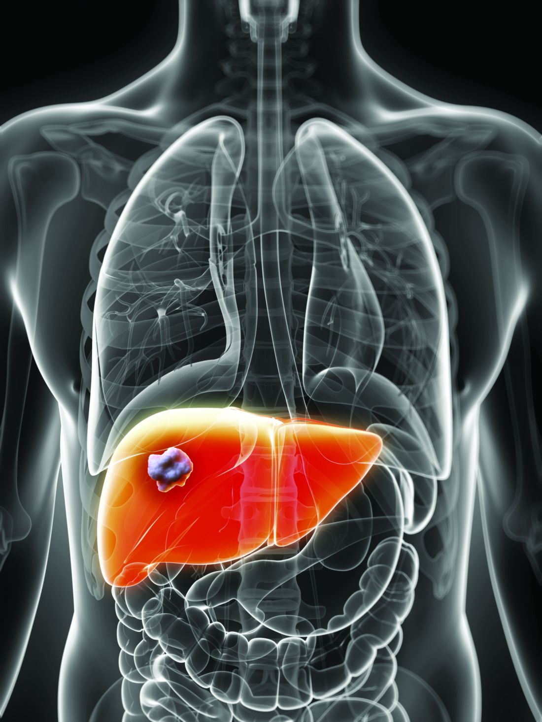
Compared with no treatment, DAA therapy cut risk of hepatocellular carcinoma by about one-third and all-cause mortality by about half in the study, which included about 10,000 adult patients with chronic hepatitis C virus (HCV) infection treated at 1 of 32 hepatology centers in France (NCT01953458).
There were no signs of increased risk of hepatocellular carcinoma during treatment with DAAs, providing more evidence refuting earlier, single-center reports that had suggested an increased incidence early after treatment. These findings also counterbalance a recent Cochrane review that could not confirm or reject a potential benefit of drugs on long-term morbidity and mortality.
Results of the study, published in the Lancet, are based on analysis of 9,895 patients, including 7,344 who started DAA treatment and 2,551 who remained untreated at a median follow-up of more than 31 months. The median patient age was 56 years, and 53% were men.
Treatment with DAAs reduced risk of hepatocellular carcinoma when compared with no DAA treatment, with a hazard ratio of 0.66 (95% confidence interval, 0.46-0.93), and reduced risk of all-cause mortality, with an HR of 0.48 (95% CI, 0.33-0.70), investigators reported in a multivariable analysis that adjusted for variables including age, sex, fibrosis score, HCV genotype, alcohol use, and more.
“These inverse associations persisted in the subgroup of patients who achieved a sustained virological response, whereas those who did not achieve a sustained virological response were a higher risk for hepatocellular carcinoma,” said the investigators, led by Fabrice Carrat, PhD, of Sorbonne Université, Institut National de la Santé et de la Recherche Médicale (INSERM), Paris.
Sustained virologic response was observed in 94% of patients who had known response status and sufficient follow-up, investigators said.
In patients with cirrhosis at baseline, DAA treatment had a similarly strong association with reduced hepatocellular carcinoma and mortality, with a sustained virologic response rate of 92% in those for whom sufficient data was available, they said.
There was no evidence for an increased risk of hepatocellular carcinoma on treatment, with an adjusted HR of 0.74 (95% CI, 0.49-1.13; P = 0.17), they added.
“Our results support urgent treatment of patients with advanced liver disease and extension of the follow-up of treated patients with less severe disease to assess the long-term clinical effect of direct-acting antiviral treatment,” Dr. Carrat and colleagues said in a commentary on their results.
However, the long-term effect of DAAs on liver decompensation has yet to be clarified, they added, noting that their study excluded patients with decompensated cirrhosis or a history of hepatocellular carcinoma.
Funding for the study came from INSERM, Agence Nationale de la Recherche, DGS (Direction Générale de la Santé), MSD, Janssen, Gilead, AbbVie, Bristol-Myers Squibb, and Roche. Dr. Carrat reported personal fees from Imaxio not related to the present study. Coauthors provided additional disclosures related to Gilead, AbbVie, Bristol-Myers Squibb, MSD, and Janssen, among others.
SOURCE: Carrat F et al. Lancet. 2019 Feb 11. doi: 10.1016/S0140-6736(18)32111-1
This study provides “substantive evidence” that curing hepatitis C virus with all-oral direct-acting antiviral regimens provides clinical benefits, according to Raymond T. Chung, MD, and his coauthors of a related editorial.
Investigators in this study provide the best evidence so far in support of guidelines that advise direct-acting antiviral (DAA) treatment for all patients with chronic hepatitis C virus (HCV) infection, the editorial’s authors stated.
Results of the French study provide a strong counterpoint to the findings of a recent Cochrane review of DAA trials that could not confirm or reject whether DAAs had effects on long-term morbidity and mortality related to HCV, added Dr. Chung and his coauthors. “Finally, they provide credence to the achievability of the goals set out by the World Health Organization (WHO), not only to eliminate HCV but also to substantially reduce its complications.”
The WHO targets were established in light of earlier evidence that sustained virologic responses are linked to reductions in hepatocellular carcinoma, liver transplantation, and mortality, they said.
“In view of the high sustained virological response and excellent tolerability achieved with DAAs, it seemed highly plausible to envision reductions in chronic HCV infection–related complications with these drugs,” they said in reference to the study by Carrat and colleagues.
This editorial appearing in the Lancet was authored by Jacinta A. Holmes, Stephanie M. Rutledge, and Raymond T. Chung of the Liver Center, Gastrointestinal Division, Massachusetts General Hospital, Boston. Dr. Chung provided disclosures related to AbbVie, Gilead, Merck, Bristol-Myers Squibb, Roche, Janssen, and Boehringer Ingelheim.
Direct-acting antivirals significantly decrease risk of hepatocellular carcinoma and mortality in persons with hepatitis C, according to results of the first prospective, longitudinal study to evaluate the effect of the drugs on complications related to the infection.
Compared with no treatment, DAA therapy cut risk of hepatocellular carcinoma by about one-third and all-cause mortality by about half in the study, which included about 10,000 adult patients with chronic hepatitis C virus (HCV) infection treated at 1 of 32 hepatology centers in France (NCT01953458).
There were no signs of increased risk of hepatocellular carcinoma during treatment with DAAs, providing more evidence refuting earlier, single-center reports that had suggested an increased incidence early after treatment. These findings also counterbalance a recent Cochrane review that could not confirm or reject a potential benefit of drugs on long-term morbidity and mortality.
Results of the study, published in the Lancet, are based on analysis of 9,895 patients, including 7,344 who started DAA treatment and 2,551 who remained untreated at a median follow-up of more than 31 months. The median patient age was 56 years, and 53% were men.
Treatment with DAAs reduced risk of hepatocellular carcinoma when compared with no DAA treatment, with a hazard ratio of 0.66 (95% confidence interval, 0.46-0.93), and reduced risk of all-cause mortality, with an HR of 0.48 (95% CI, 0.33-0.70), investigators reported in a multivariable analysis that adjusted for variables including age, sex, fibrosis score, HCV genotype, alcohol use, and more.
“These inverse associations persisted in the subgroup of patients who achieved a sustained virological response, whereas those who did not achieve a sustained virological response were a higher risk for hepatocellular carcinoma,” said the investigators, led by Fabrice Carrat, PhD, of Sorbonne Université, Institut National de la Santé et de la Recherche Médicale (INSERM), Paris.
Sustained virologic response was observed in 94% of patients who had known response status and sufficient follow-up, investigators said.
In patients with cirrhosis at baseline, DAA treatment had a similarly strong association with reduced hepatocellular carcinoma and mortality, with a sustained virologic response rate of 92% in those for whom sufficient data was available, they said.
There was no evidence for an increased risk of hepatocellular carcinoma on treatment, with an adjusted HR of 0.74 (95% CI, 0.49-1.13; P = 0.17), they added.
“Our results support urgent treatment of patients with advanced liver disease and extension of the follow-up of treated patients with less severe disease to assess the long-term clinical effect of direct-acting antiviral treatment,” Dr. Carrat and colleagues said in a commentary on their results.
However, the long-term effect of DAAs on liver decompensation has yet to be clarified, they added, noting that their study excluded patients with decompensated cirrhosis or a history of hepatocellular carcinoma.
Funding for the study came from INSERM, Agence Nationale de la Recherche, DGS (Direction Générale de la Santé), MSD, Janssen, Gilead, AbbVie, Bristol-Myers Squibb, and Roche. Dr. Carrat reported personal fees from Imaxio not related to the present study. Coauthors provided additional disclosures related to Gilead, AbbVie, Bristol-Myers Squibb, MSD, and Janssen, among others.
SOURCE: Carrat F et al. Lancet. 2019 Feb 11. doi: 10.1016/S0140-6736(18)32111-1
FROM THE LANCET
Key clinical point:
Major finding: DAAs reduced risk of hepatocellular carcinoma (HR, 0.66; 95% confidence interval, 0.46-0.93) and all-cause mortality (HR, 0.48; 95% CI, 0.33-0.70).
Study details: A prospective study including about 10,000 adults with chronic HCV infection enrolled at 1 of 32 centers in France.
Disclosures: Funding for the study came from INSERM, Agence Nationale de la Recherche, DGS (Direction Générale de la Santé), MSD, Janssen, Gilead, AbbVie, Bristol-Myers Squibb, and Roche. Dr. Carrat reported personal fees from Imaxio not related to the present study. Coauthors provided additional disclosures related to the study pharma sponsors among others.
Source: Carrat F et al. Lancet. 2019 Feb 11. doi: 10.1016/20S0140-6736(18)32111-1.
NASH: Fastest-growing cause of liver cancer in transplant candidates
Nonalcoholic steatohepatitis may soon supplant chronic hepatitis C as the leading cause of hepatocellular carcinoma among patients awaiting liver transplantation, according to the findings of a national longitudinal registry study.

The proportion of affected patients with nonalcoholic steatohepatitis (NASH) rose nearly 700% between 2002 and 2017 (P less than .0001), making NASH the only etiology to significantly rise in prevalence, reported Zobair Younossi, MD, MPH, of Inova Health System in Falls Church, Va., and his associates. Chronic hepatitis C remained the most common cause of liver cancer during the study period, but its prevalence fell by more than 10% in the last 3 years (2014-2017). These trends reflect the advent of “new, highly effective antiviral regimens” for hepatitis C, the global epidemic of obesity and metabolic syndrome, and the urgent need for effective, safe treatments for NASH, they wrote in Clinical Gastroenterology and Hepatology.
Historically, hepatocellular carcinoma is usually caused by chronic hepatitis C or B infection, but the global rise of obesity and type 2 diabetes mellitus has led to epidemic levels of NASH, a progressive form of nonalcoholic fatty liver disease that lacks useful predictive noninvasive biomarkers or safe treatments. This phenomenon, coupled with the advent of new, often-curative treatments for viral hepatitis, is making NASH a leading driver of both fibrosis and liver transplantation in the United States. To compare trends in liver cancer etiologies among transplant candidates, Dr. Younossi and his associates analyzed data on 158,347 adults who were wait-listed between 2002 and 2017 and captured by the national Scientific Registry of Transplant Recipients.
A total of 26,121 (16.5%) patients awaiting liver transplant had hepatocellular carcinoma. This proportion nearly quadrupled over the study period, from 6% to 23% (P less than .0001) and rose significantly (P less than .0001) for all liver cancer etiologies (hepatitis C and B, alcoholic liver disease, and NASH). However, the absolute rise in prevalence was far greater for NASH (1050%) than for chronic hepatitis C (more than 500%) or any other etiology.
Furthermore, while most (65%) liver cancer cases involved chronic hepatitis C, the proportion of cases involving NASH rose from 2% in 2002 to 18% in 2017 (P less than .0001). By 2017, NASH topped alcoholic liver disease, comorbid hepatitis C with alcoholic liver disease, and chronic hepatitis B as an etiology of hepatocellular carcinoma among patients listed for transplant. Conversely, by 2017, less than 50% of liver cancers were caused by hepatitis C – a more than 10% drop from 2014. Over the study period, NASH was the only etiology whose prevalence significantly increased among transplant-listed patients with hepatocellular carcinoma.
In this study, etiology of liver cancer did not seem to affect the likelihood of either death or transplantation. However, serious cardiovascular disease or late-stage cancer diagnosis might exclude many NASH patients from transplantation, the researchers wrote. “Thus, the population reported here actually may underestimate the true proportion of hepatocellular carcinoma cases related to nonalcoholic fatty liver disease and NASH in the United States. Because NASH is on a trajectory to become the most common cause of hepatocellular carcinoma in the United States, effective prevention strategies and treatment options are urgently needed for this currently underserved patient population.”
Minneapolis Medical Research Foundation is the contractor for the registry and supplied the data. Dr. Younossi reported ties to Bristol-Myers Squibb, Gilead Sciences, AbbVie, Intercept Pharmaceuticals, and GlaxoSmithKline.
SOURCE: Younossi Z et al. Clin Gastroenterol Hepatol. 2018 Jun 14. doi: 10.1016/j.cgh.2018.05.057.
Nonalcoholic steatohepatitis may soon supplant chronic hepatitis C as the leading cause of hepatocellular carcinoma among patients awaiting liver transplantation, according to the findings of a national longitudinal registry study.
The proportion of affected patients with nonalcoholic steatohepatitis (NASH) rose nearly 700% between 2002 and 2017 (P less than .0001), making NASH the only etiology to significantly rise in prevalence, reported Zobair Younossi, MD, MPH, of Inova Health System in Falls Church, Va., and his associates. Chronic hepatitis C remained the most common cause of liver cancer during the study period, but its prevalence fell by more than 10% in the last 3 years (2014-2017). These trends reflect the advent of “new, highly effective antiviral regimens” for hepatitis C, the global epidemic of obesity and metabolic syndrome, and the urgent need for effective, safe treatments for NASH, they wrote in Clinical Gastroenterology and Hepatology.
Historically, hepatocellular carcinoma is usually caused by chronic hepatitis C or B infection, but the global rise of obesity and type 2 diabetes mellitus has led to epidemic levels of NASH, a progressive form of nonalcoholic fatty liver disease that lacks useful predictive noninvasive biomarkers or safe treatments. This phenomenon, coupled with the advent of new, often-curative treatments for viral hepatitis, is making NASH a leading driver of both fibrosis and liver transplantation in the United States. To compare trends in liver cancer etiologies among transplant candidates, Dr. Younossi and his associates analyzed data on 158,347 adults who were wait-listed between 2002 and 2017 and captured by the national Scientific Registry of Transplant Recipients.
A total of 26,121 (16.5%) patients awaiting liver transplant had hepatocellular carcinoma. This proportion nearly quadrupled over the study period, from 6% to 23% (P less than .0001) and rose significantly (P less than .0001) for all liver cancer etiologies (hepatitis C and B, alcoholic liver disease, and NASH). However, the absolute rise in prevalence was far greater for NASH (1050%) than for chronic hepatitis C (more than 500%) or any other etiology.
Furthermore, while most (65%) liver cancer cases involved chronic hepatitis C, the proportion of cases involving NASH rose from 2% in 2002 to 18% in 2017 (P less than .0001). By 2017, NASH topped alcoholic liver disease, comorbid hepatitis C with alcoholic liver disease, and chronic hepatitis B as an etiology of hepatocellular carcinoma among patients listed for transplant. Conversely, by 2017, less than 50% of liver cancers were caused by hepatitis C – a more than 10% drop from 2014. Over the study period, NASH was the only etiology whose prevalence significantly increased among transplant-listed patients with hepatocellular carcinoma.
In this study, etiology of liver cancer did not seem to affect the likelihood of either death or transplantation. However, serious cardiovascular disease or late-stage cancer diagnosis might exclude many NASH patients from transplantation, the researchers wrote. “Thus, the population reported here actually may underestimate the true proportion of hepatocellular carcinoma cases related to nonalcoholic fatty liver disease and NASH in the United States. Because NASH is on a trajectory to become the most common cause of hepatocellular carcinoma in the United States, effective prevention strategies and treatment options are urgently needed for this currently underserved patient population.”
Minneapolis Medical Research Foundation is the contractor for the registry and supplied the data. Dr. Younossi reported ties to Bristol-Myers Squibb, Gilead Sciences, AbbVie, Intercept Pharmaceuticals, and GlaxoSmithKline.
SOURCE: Younossi Z et al. Clin Gastroenterol Hepatol. 2018 Jun 14. doi: 10.1016/j.cgh.2018.05.057.
FROM CLINICAL GASTROENTEROLOGY AND HEPATOLOGY
Key clinical point: Nonalcoholic steatohepatitis may soon become the leading cause of hepatocellular carcinoma among patients awaiting liver transplantation.
Major finding: The proportion of these patients with NASH rose nearly 700% between 2002 and 2017 (P less than .0001).
Study details: A longitudinal registry study of 26,121 patients listed for liver transplantation in the United States.
Disclosures: Minneapolis Medical Research Foundation is the contractor for the registry and supplied the data. Dr. Younossi reported ties to Bristol-Myers Squibb, Gilead Sciences, AbbVie, Intercept Pharmaceuticals, and GlaxoSmithKline.
Source: Younossi Z et al. Clin Gastroenterol Hepatol. 2018 Jun 14. doi: 10.1016/j.cgh.2018.05.057.
Residential HCV program improves veterans’ diagnosis and care
Integrating comprehensive and collaborative hepatitis C virus (HCV) care within a Veterans Affairs residential treatment program can substantially increase diagnosis and treatment of HCV-infected veterans with substance use disorder (SUD), according to the results of an evaluation study for the period from December 2014 to April 2018.

A total of 97.5% (582/597) of patient admissions to the program were screened for HCV infection, and 12.7% (74/582) of the cases were confirmed to be HCV positive. All of the positive cases were sent to an infectious disease (ID) clinic for further evaluation and, if appropriate, to begin HCV pharmacotherapy, according to the report, published in the Journal of Substance Abuse Treatment.
Of the HCV-positive cases, 78.4% (58/74) received pharmacotherapy, with a sustained virologic response rate of 82.8% (48/58), wrote Mary Jane Burton, MD, of the G.V. (Sonny) Montgomery VA Medical Center, Jackson, Miss., and her colleagues.
As part of the program, all veterans admitted to the SUD residential program were offered screening for HCV. Veterans with negative screening results received education about how to remain HCV negative via handouts and veterans who screened positive received brief supportive counseling and were referred to the ID clinic via a consult. Veterans confirmed to have chronic HCV infection receive education and evaluation in the HCV clinic while they attend the residential SUD program. Treatment for HCV is instituted as early as feasible and prescribing is in accordance with VA guidelines (Department of Veterans Affairs, 2018), with the goal of initiating pharmacotherapy treatment for HCV while the veteran is still in the residential program, according to the researchers.
Following discharge from the program, veterans on HCV treatment are scheduled for follow-up every 2 weeks in the HCV treatment clinic for the remainder of their pharmacotherapy, the researchers added.
Patient-level barriers to HCV treatment among the SUD population include reduced health literacy, low health care utilization, comorbid mental health conditions, and poor social support, according to the literature. Because multidisciplinary approaches to HCV treatment that mitigate these barriers have been shown to increase treatment uptake among these patients, the VA program was initiated, the researchers stated. Dr. Burton and her colleagues reported that 18.9% (14/74) of the HCV-positive cases were newly diagnosed and would have likely gone undetected without this program (J Substance Abuse Treatment. 2019;98:9-14).
“We have demonstrated that integrating a comprehensive HCV screening, education, referral, and treatment program within residential SUD treatment is feasible and effective in diagnosing previously unrecognized HCV infections, transitioning veterans into HCV care, and promoting treatment initiation,” the researchers concluded.
The Department of Veterans Affairs and the VA Center for Innovation supported the study. Dr. Burton reported research support from Merck Sharpe & Dohme.
Integrating comprehensive and collaborative hepatitis C virus (HCV) care within a Veterans Affairs residential treatment program can substantially increase diagnosis and treatment of HCV-infected veterans with substance use disorder (SUD), according to the results of an evaluation study for the period from December 2014 to April 2018.
A total of 97.5% (582/597) of patient admissions to the program were screened for HCV infection, and 12.7% (74/582) of the cases were confirmed to be HCV positive. All of the positive cases were sent to an infectious disease (ID) clinic for further evaluation and, if appropriate, to begin HCV pharmacotherapy, according to the report, published in the Journal of Substance Abuse Treatment.
Of the HCV-positive cases, 78.4% (58/74) received pharmacotherapy, with a sustained virologic response rate of 82.8% (48/58), wrote Mary Jane Burton, MD, of the G.V. (Sonny) Montgomery VA Medical Center, Jackson, Miss., and her colleagues.
As part of the program, all veterans admitted to the SUD residential program were offered screening for HCV. Veterans with negative screening results received education about how to remain HCV negative via handouts and veterans who screened positive received brief supportive counseling and were referred to the ID clinic via a consult. Veterans confirmed to have chronic HCV infection receive education and evaluation in the HCV clinic while they attend the residential SUD program. Treatment for HCV is instituted as early as feasible and prescribing is in accordance with VA guidelines (Department of Veterans Affairs, 2018), with the goal of initiating pharmacotherapy treatment for HCV while the veteran is still in the residential program, according to the researchers.
Following discharge from the program, veterans on HCV treatment are scheduled for follow-up every 2 weeks in the HCV treatment clinic for the remainder of their pharmacotherapy, the researchers added.
Patient-level barriers to HCV treatment among the SUD population include reduced health literacy, low health care utilization, comorbid mental health conditions, and poor social support, according to the literature. Because multidisciplinary approaches to HCV treatment that mitigate these barriers have been shown to increase treatment uptake among these patients, the VA program was initiated, the researchers stated. Dr. Burton and her colleagues reported that 18.9% (14/74) of the HCV-positive cases were newly diagnosed and would have likely gone undetected without this program (J Substance Abuse Treatment. 2019;98:9-14).
“We have demonstrated that integrating a comprehensive HCV screening, education, referral, and treatment program within residential SUD treatment is feasible and effective in diagnosing previously unrecognized HCV infections, transitioning veterans into HCV care, and promoting treatment initiation,” the researchers concluded.
The Department of Veterans Affairs and the VA Center for Innovation supported the study. Dr. Burton reported research support from Merck Sharpe & Dohme.
FROM THE JOURNAL OF SUBSTANCE ABUSE TREATMENT
mRECIST response to kinase inhibitors predicts survival in HCC
SAN FRANCISCO – Patients with hepatocellular carcinoma (HCC) who have a response to first-line kinase inhibitors based on modified RECIST (mRECIST) criteria live almost a year longer than counterparts who have stable or progressive disease, finds a retrospective post hoc analysis of the REFLECT trial.

“The inability to accurately evaluate response to targeted or locoregional therapies resulted in the adaptation of RECIST to mRECIST, guidelines specifically designed to evaluate response to treatment in HCC,” noted lead investigator Masatoshi Kudo, MD, PhD, of the department of gastroenterology and hepatology, Kindai University Faculty of Medicine, Osaka, Japan. “Based on recommendations from the EASL [European Association for the Study of the Liver] consensus conference in 2000, mRECIST was developed to assess response based on the reduction of viable tumor burden … rather than overall tumor shrinkage. However, EASL clinical practice guidelines also suggested additional studies are needed to validate this approach.”
The investigators analyzed data from REFLECT, a global phase 3 noninferiority, randomized, controlled trial that showed overall survival with the novel kinase inhibitor lenvatinib (Lenvima) was not inferior to that with the older kinase inhibitor sorafenib (Nexavar) in 954 patients with untreated, unresectable HCC (Lancet. 2018;391:1163-73).
The new analysis, reported at the 2019 GI Cancers Symposium, showed that, among all randomized patients, those having an objective response according to mRECIST criteria were 39% less likely to die after other factors were taken into account. The finding was consistent in a landmark analysis, which addresses the issue of lead-time bias.
“Objective response by mRECIST was an independent predictor of overall survival in patients with HCC regardless of treatment,” Dr. Kudo concluded, adding that the findings are consistent with those of several previous studies. “Thus, patients who achieve an objective response can potentially expect a longer overall survival. However, additional studies are needed to further validate the correlation between objective response and overall survival.”
Findings in context
Although the mRECIST criteria overcome some issues with the size-based RECIST criteria in assessing HCC, the former are not without their limitations, noted invited discussant Andrew X. Zhu, MD, PhD, of Harvard Medical School and Massachusetts General Hospital Cancer Center.

These limitations include, for example, the need for more stringent selection of target lesions and the potential for antiangiogenic agents to cause vasoconstriction, complicating measurement. “There is tremendous intraobserver and interobserver variability, and a tremendous learning curve,” he added, noting that in REFLECT the mRECIST overall response rate assessed by independent reviewers was considerably higher than that assessed by investigators.
To date, three other trials of targeted therapies have similarly found an association between mRECIST response and overall survival. But a fourth did not.
“In the era of immunotherapy being actively applied to HCC, it’s important to recognize that this association actually may be agent dependent,” Dr. Zhu said. “Even though the TKIs with antiangiogenic [activity] have shown an improved overall response [going] from RECIST to mRECIST, this has not been recapitulated, at least based on the current experience that we have, with checkpoint inhibitors.”
The REFLECT investigators’ conclusion “is certainly backed by the large dataset from a positive phase 3 trial. It has very sound statistical methodology, and it may actually provide the initial evidence that this biomarker may serve as a potentially relevant surrogate to predict overall survival,” he said.
At the same time, there is reason to be cautious given the post hoc, retrospective nature of the study; the lack of a comparison with conventional RECIST response; absence of analysis of the potential correlation of stable disease with overall survival; and the lumping together of two agents, among other issues.
“For these reasons, I do think additional studies are warranted for prospective validation of the findings,” Dr. Zhu concluded.
Study details
REFLECT patients had an investigator-assessed mRECIST objective response rate of 16.7% overall (24.1% with lenvatinib and 9.2% with sorafenib) and a median overall survival of 13.0 months (13.6 months with lenvatinib and 12.3 months with sorafenib).
Median overall survival was 22.4 months for mRECIST responders and 11.4 months for nonresponders (P less than .001), Dr. Kudo reported at the symposium, sponsored by the American Gastroenterological Association, the American Society for Clinical Oncology, the American Society for Radiation Oncology, and the Society of Surgical Oncology.
In a multivariate analysis, this difference translated to a 39% reduction in risk of death for responders (hazard ratio, 0.61; P less than .0001).
Moreover, the survival advantage of response was evident in landmark analyses, whether response was assessed at 2 months (HR, 0.75; P = .033), at 4 months (HR, 0.72; P = .009), or at 6 months (HR, 0.73; P = .010).
Dr. Kudo disclosed that he receives honoraria or has a consulting or advisory role with several pharmaceutical companies. The presentation was sponsored by Eisai and Merck.
SOURCE: Kudo M et al. GI Cancers Symposium, Abstract 186.
SAN FRANCISCO – Patients with hepatocellular carcinoma (HCC) who have a response to first-line kinase inhibitors based on modified RECIST (mRECIST) criteria live almost a year longer than counterparts who have stable or progressive disease, finds a retrospective post hoc analysis of the REFLECT trial.
“The inability to accurately evaluate response to targeted or locoregional therapies resulted in the adaptation of RECIST to mRECIST, guidelines specifically designed to evaluate response to treatment in HCC,” noted lead investigator Masatoshi Kudo, MD, PhD, of the department of gastroenterology and hepatology, Kindai University Faculty of Medicine, Osaka, Japan. “Based on recommendations from the EASL [European Association for the Study of the Liver] consensus conference in 2000, mRECIST was developed to assess response based on the reduction of viable tumor burden … rather than overall tumor shrinkage. However, EASL clinical practice guidelines also suggested additional studies are needed to validate this approach.”
The investigators analyzed data from REFLECT, a global phase 3 noninferiority, randomized, controlled trial that showed overall survival with the novel kinase inhibitor lenvatinib (Lenvima) was not inferior to that with the older kinase inhibitor sorafenib (Nexavar) in 954 patients with untreated, unresectable HCC (Lancet. 2018;391:1163-73).
The new analysis, reported at the 2019 GI Cancers Symposium, showed that, among all randomized patients, those having an objective response according to mRECIST criteria were 39% less likely to die after other factors were taken into account. The finding was consistent in a landmark analysis, which addresses the issue of lead-time bias.
“Objective response by mRECIST was an independent predictor of overall survival in patients with HCC regardless of treatment,” Dr. Kudo concluded, adding that the findings are consistent with those of several previous studies. “Thus, patients who achieve an objective response can potentially expect a longer overall survival. However, additional studies are needed to further validate the correlation between objective response and overall survival.”
Findings in context
Although the mRECIST criteria overcome some issues with the size-based RECIST criteria in assessing HCC, the former are not without their limitations, noted invited discussant Andrew X. Zhu, MD, PhD, of Harvard Medical School and Massachusetts General Hospital Cancer Center.
These limitations include, for example, the need for more stringent selection of target lesions and the potential for antiangiogenic agents to cause vasoconstriction, complicating measurement. “There is tremendous intraobserver and interobserver variability, and a tremendous learning curve,” he added, noting that in REFLECT the mRECIST overall response rate assessed by independent reviewers was considerably higher than that assessed by investigators.
To date, three other trials of targeted therapies have similarly found an association between mRECIST response and overall survival. But a fourth did not.
“In the era of immunotherapy being actively applied to HCC, it’s important to recognize that this association actually may be agent dependent,” Dr. Zhu said. “Even though the TKIs with antiangiogenic [activity] have shown an improved overall response [going] from RECIST to mRECIST, this has not been recapitulated, at least based on the current experience that we have, with checkpoint inhibitors.”
The REFLECT investigators’ conclusion “is certainly backed by the large dataset from a positive phase 3 trial. It has very sound statistical methodology, and it may actually provide the initial evidence that this biomarker may serve as a potentially relevant surrogate to predict overall survival,” he said.
At the same time, there is reason to be cautious given the post hoc, retrospective nature of the study; the lack of a comparison with conventional RECIST response; absence of analysis of the potential correlation of stable disease with overall survival; and the lumping together of two agents, among other issues.
“For these reasons, I do think additional studies are warranted for prospective validation of the findings,” Dr. Zhu concluded.
Study details
REFLECT patients had an investigator-assessed mRECIST objective response rate of 16.7% overall (24.1% with lenvatinib and 9.2% with sorafenib) and a median overall survival of 13.0 months (13.6 months with lenvatinib and 12.3 months with sorafenib).
Median overall survival was 22.4 months for mRECIST responders and 11.4 months for nonresponders (P less than .001), Dr. Kudo reported at the symposium, sponsored by the American Gastroenterological Association, the American Society for Clinical Oncology, the American Society for Radiation Oncology, and the Society of Surgical Oncology.
In a multivariate analysis, this difference translated to a 39% reduction in risk of death for responders (hazard ratio, 0.61; P less than .0001).
Moreover, the survival advantage of response was evident in landmark analyses, whether response was assessed at 2 months (HR, 0.75; P = .033), at 4 months (HR, 0.72; P = .009), or at 6 months (HR, 0.73; P = .010).
Dr. Kudo disclosed that he receives honoraria or has a consulting or advisory role with several pharmaceutical companies. The presentation was sponsored by Eisai and Merck.
SOURCE: Kudo M et al. GI Cancers Symposium, Abstract 186.
REPORTING FROM THE 2019 GI CANCERS SYMPOSIUM
Key clinical point: .
Major finding: Median overall survival was a respective 22.4 months and 11.4 months in patients who did and did not have a response according to mRECIST criteria (HR, 0.61; P less than .0001).
Study details: A retrospective post hoc analysis of a phase 3 randomized controlled trial of lenvatinib versus sorafenib among 954 patients with untreated, unresectable HCC (REFLECT trial).
Disclosures: Dr. Kudo disclosed that he receives honoraria or has a consulting or advisory role with several pharmaceutical companies. The presentation was sponsored by Eisai and Merck.
Source: Kudo M et al. 2019 GI Cancers Symposium, Abstract 186.
HBV, HCV, HIV testing of new cancer patients advised
Oncologists should consider testing all patients with newly diagnosed cancers for infection with the hepatitis B and C viruses, a multicenter team has recommended.
A prospective study of hepatitis B virus (HBV), hepatitis C virus (HCV), and HIV infections among 3,051 patients with newly diagnosed cancers showed that 6.5% of patients tested positive for previous HBV and 0.6% had chronic HBV infection. In addition, 2.4% of patients were positive for HCV, and 1.1% for HIV infections, reported Scott D. Ramsey, MD, PhD, from the Fred Hutchinson Cancer Research Center in Seattle, and colleagues.
“Many patients had no known risk factors for infection, suggesting that current risk-based models for screening may be insufficient. Thus, we believe our results warrant consideration of universal testing of patients with newly diagnosed cancer for HBV and HCV infection, particularly if such an approach is shown to be cost effective,” they wrote in JAMA Oncology.
The investigators noted that patients with undiagnosed hepatitis and/or HIV infections could transmit them to unsuspecting caregivers, adding that “with effective treatments available, not screening for these viruses misses an opportunity to reduce future morbidity associated with these infections and to avoid viral reactivation during treatment, with resulting morbidity and mortality.”
To estimate the prevalence of the infections in patients with newly diagnosed cancers, investigators looked at a cohort of 3,051 patients with a cancer diagnosis made within the previous 120 days at nine academic medical centers and nine community oncology centers representing a total of 41 cancer clinics affiliated with the SWOG Cancer Research Network (formerly the Southwest Oncology Group).
The median patient age was 60.6 years. Female patients constitute 60.4% of the sample; 18.1% were black, and 18.3% were of Hispanic heritage.
Of 3,050 patients for whom HBV testing results were available, 6.5% (197) were positive for previous HBV infection, compared with an estimated U.S. population prevalence of 4.7%. In addition, 0.6% (19 patients) were found to have chronic HBV, compared with an estimated 0.3% US population prevalence.
HCV infections were detected in 2.4% (71 of 2990 patients), compared with an estimated population prevalence of 1.3%, and HIV infections were detected in 1.1%, compared with a background estimated population prevalence of 0.3%.
In all, 32 patients were diagnosed with viral infections by testing performed for the study, including 8 patients with chronic HBV, 22 with HCV, and 2 with HIV.
Additionally, the authors found that 4 patients with chronic HBV, 23 with HCV, and 7 with HIV had no identifiable risk factors.
The highest prevalence of infections occurred among patients with liver cancer, nonliver and noncolorectal cancers of the gastrointestinal tract, head and neck cancers, lung cancers, and prostate cancer. A finding of viral positivity changed the treatment plan in only 8% of all infected patients, however.
“Given that most HIV-infected patients in our study knew their viral status, the yield of universal HIV testing among patients with newly diagnosed cancer may likely be low. Although age-directed screening is recommended for HIV and HCV, uptake rates in primary care are variable and low overall,” Dr. Ramsey and his colleagues wrote.
The study was supported by grants from the National Cancer Institute. Dr. Ramsey and several co-authors reported receiving NCI grants, and multiple co-authors reported grants and/or consulting fees from various companies.
SOURCE: Ramsey SD et al. JAMA Oncol. 2019 Jan 17. doi: 10.1001/jamaoncol.2018.6437.
FROM JAMA ONCOLOGY
Key clinical point: Patients with newly diagnosed cancers should be screened for viral infections that may pose a transmission risk or could be reactivated by cancer therapies.
Major finding: Infection rates of HBV, HCV, and HIV in patients with newly diagnosed cancers were 6.5%, 2.4%, and 1.1%, respectively.
Study details: Prospective study of viral infections in 3,051 patients with a diagnosis of cancer within the previous 120 days.
Disclosures: The study was supported by grants from the National Cancer Institute. Dr. Ramsey and several coauthors reported receiving NCI grants, and multiple coauthors reported grants and/or consulting fees from various companies.
Source: Ramsey SD et al. JAMA Oncology. 2019 Jan 17. doi: 10.1001/jamaoncol.2018.6437.
Perceptions of liver transplantation for ALD are evolving

“The findings suggest that early liver transplant for alcoholic hepatitis may be leading to broader acceptance of ALD for liver transplant,” Brian P. Lee, MD, of the University of California, San Francisco, and his colleagues wrote in JAMA Internal Medicine.
The researchers conducted a prospective cohort study of 9,438 patients with ALD who received a liver transplant from 2002 to 2016. Data were obtained from the United Network for Organ Sharing national database.
Study participants were evaluated for patterns, both nationally and regionally, related to liver transplant for the treatment of ALD. In addition, Dr. Lee and his colleagues completed a sensitivity analysis, which evaluated specific clinical parameters, including patient and graft survival, hepatocellular carcinoma (HCC), and hepatitis C viral (HCV) infection.
“Because there is no national policy regarding early liver transplant, we hypothesized that changes may vary regionally as liver transplant programs shifted their attitudes toward increased acceptance of early liver transplant for alcoholic hepatitis and ALD,” the researchers wrote.
After analysis, the researchers found that liver transplantation for patients with ALD increased proportionally from 24.2% to 36.7% from 2002 to 2016, respectively. With HCV-infected recipients included, the proportion of liver transplants rose from 15.3% to 30.6% over the same period, representing a twofold increase of transplants received for this indication.
The degree of increase was reported to vary based on geographic region and was linked with differences in patient-specific factors.
“There may be regional disparities in access to liver transplant for ALD; whether this is related to different attitudes toward ALD and requirements for sobriety is unknown,” they added.
The researchers acknowledged that a key limitation of the study was the use of registry data. As a result, Dr. Lee and his colleagues reported that all conclusions are not causal, but rather only by association.
The study was supported by the National Institute of Diabetes and Digestive and Kidney Diseases UCSF Liver Center. The authors reported no conflicts of interests.
SOURCE: Lee BP et al. JAMA Intern Med. 2019 Jan 22. doi: 10.1001/jamainternmed.2018.6536.
One of the most significant findings of the study by Brian P. Lee, MD, and his colleagues is the major shift in attitudes surrounding the eligibility criteria for patients with ALD to undergo liver transplantation.
More than 3 decades ago, a group of surgical experts gathered together to discuss evaluation criteria for candidacy of individuals to undergo liver transplantation. They recommended that patients with ALD be required to restrict alcohol consumption for 6 months prior to being listed eligible for surgery. The group presumed that a period of complete avoidance may induce some degree of disease remission, circumventing the need for transplant altogether.
However, these suggestions were given without the use of evidence, formed largely on the basis of opinion, and recent data dispute these recommendations. On the contrary, relapse rates for alcohol use disorder has been shown to be due to factors other than length of abstinence. While these findings have lessened bias surrounding ALD and liver transplantation, the assumption still remains prevalent in clinical practice today.
These results highlight the unanswered question of how to best approach treatment of individuals with ALD, and whether the recent rise of patients undergoing liver transplantation for ALD, without a continued duration of abstinence, should be a concern of clinicians.
Mack C. Mitchell, MD, is affiliated with the department of internal medicine at the University of Texas in Dallas. Dr. Mitchell reported having financial affiliations with the National Institute of Alcohol and Alcohol Abuse. These comments are adapted from his accompanying editorial (JAMA Intern Med. 2019 Jan 22. doi: 10.1001/jamainternmed.2018.6532 ).
“The findings suggest that early liver transplant for alcoholic hepatitis may be leading to broader acceptance of ALD for liver transplant,” Brian P. Lee, MD, of the University of California, San Francisco, and his colleagues wrote in JAMA Internal Medicine.
The researchers conducted a prospective cohort study of 9,438 patients with ALD who received a liver transplant from 2002 to 2016. Data were obtained from the United Network for Organ Sharing national database.
Study participants were evaluated for patterns, both nationally and regionally, related to liver transplant for the treatment of ALD. In addition, Dr. Lee and his colleagues completed a sensitivity analysis, which evaluated specific clinical parameters, including patient and graft survival, hepatocellular carcinoma (HCC), and hepatitis C viral (HCV) infection.
“Because there is no national policy regarding early liver transplant, we hypothesized that changes may vary regionally as liver transplant programs shifted their attitudes toward increased acceptance of early liver transplant for alcoholic hepatitis and ALD,” the researchers wrote.
After analysis, the researchers found that liver transplantation for patients with ALD increased proportionally from 24.2% to 36.7% from 2002 to 2016, respectively. With HCV-infected recipients included, the proportion of liver transplants rose from 15.3% to 30.6% over the same period, representing a twofold increase of transplants received for this indication.
The degree of increase was reported to vary based on geographic region and was linked with differences in patient-specific factors.
“There may be regional disparities in access to liver transplant for ALD; whether this is related to different attitudes toward ALD and requirements for sobriety is unknown,” they added.
The researchers acknowledged that a key limitation of the study was the use of registry data. As a result, Dr. Lee and his colleagues reported that all conclusions are not causal, but rather only by association.
The study was supported by the National Institute of Diabetes and Digestive and Kidney Diseases UCSF Liver Center. The authors reported no conflicts of interests.
SOURCE: Lee BP et al. JAMA Intern Med. 2019 Jan 22. doi: 10.1001/jamainternmed.2018.6536.
FROM JAMA INTERNAL MEDICINE
Key clinical point: Liver transplantation for alcohol-associated liver disease (ALD) is occurring earlier and more frequently than before.
Major finding: The proportion of patients undergoing liver transplantation for ALD has nearly doubled from 2002 to 2016.
Study details: A prospective cohort study of 9,438 patients who underwent liver transplant for ALD in the United States from 2002 to 2016.
Disclosures: The study was supported by the National Institute of Diabetes and Digestive and Kidney Diseases UCSF Liver Center. The authors reported no conflicts of interest.
Source: Lee BP et al. JAMA Intern Med. 2019 Jan 22. doi: 10.1001/jamainternmed.2018.6536.
Diet low in free sugars shows promise for adolescent NAFLD
after 8 weeks, compared with boys on a usual diet.
“Because of growing evidence implicating dietary sugars in NAFLD, well-controlled studies in children with NAFLD are needed to inform clinical practice and public policy,” wrote Jeffrey B. Schwimmer, MD, of the University of California, San Diego, La Jolla, and colleagues in JAMA.
The researchers randomized 40 boys aged 11-16 years with active NAFLD to a diet low in free sugars or their usual diet. The intervention diet involved personalized menu planning and provision of meals for the boys’ entire households that were designed to restrict free sugar intake to less than 3% of daily calories. Adherence to the diet was assessed by twice-weekly phone calls.
In the intervention group, hepatic steatosis decreased from an average of 25% at baseline to 17% after 8 weeks, compared with a change from 21% to 20% in the control group. The adjusted mean difference at 8 weeks was −6.23%, which was statistically significant (P less than .001).
The average age of the participants was 13 years, 95% were Hispanic. All 40 completed the study, and 18 of the 20 boys in the intervention group reported less than 3% of calories from free sugar during the study period. No adverse events were reported related to study participation.
The results were limited by several factors, including the small sample size and homogeneous population. In addition, neither hepatic steatosis or serum alanine aminotransferase (ALT) levels decreased enough to enter the normal range, the researchers noted. The findings, though preliminary, support the value of reducing free sugars, including glucose, fructose, and sucrose, to help manage NAFLD in adolescents, and “further research is required to assess long-term and clinical outcomes,” they said.
The study was supported by grants from multiple foundations and organizations, including the Nutrition Science Initiative, the University of California, San Diego, the National Institutes of Health, Children’s Healthcare of Atlanta and Emory University Pediatric Biostatistics Core, and the Georgia Clinical and Translational Science Alliance. Dr. Schwimmer reported receiving research support from Galmed and Intercept.
SOURCE: Schwimmer JB et al. JAMA. 2019;321(3):256-265.
FROM JAMA
Key clinical point: After 8 weeks, adolescent boys on a diet low in free sugars showed significant improvement in hepatic steatosis, compared with those on a usual diet.
Major finding: Hepatic steatosis decreased from an average of 25% at baseline to 17% after 8 weeks in the modified diet group, compared with a decrease from 21% to 20% in the usual diet group.
Study details: The data come from an open-label, randomized trial of 40 adolescent boys aged 11 to 16 years with active NAFLD.
Disclosures: The study was supported by grants from multiple foundations and organizations, including the Nutrition Science Initiative, the University of California, San Diego, the National Institutes of Health, Children’s Healthcare of Atlanta and Emory University Pediatric Biostatistics Core, and the Georgia Clinical and Translational Science Alliance. Dr. Schwimmer reported receiving research support from Galmed and Intercept.
Source: Schwimmer JB et al. JAMA. 2019;321(3):256-265.
FDA approves cabozantinib for previously treated HCC
The Food and Drug Administration has approved cabozantinib tablets (Cabometyx) for patients with hepatocellular carcinoma (HCC) who have been previously treated with sorafenib.

Approval was based on an improvement in overall survival over placebo seen in the phase 3 CELESTIAL trial for patients with advanced HCC who received prior sorafenib.
Median overall survival was 10.2 months with cabozantinib versus 8.0 months with placebo (hazard ratio, 0.76; 95% confidence interval, 0.63-0.92; P = .0049). Median progression-free survival was 5.2 months with cabozantinib and 1.9 months with placebo (HR, 0.44; 95% CI, 0.36-0.52; P less than .0001). Objective response rates were 4% with cabozantinib and 0.4% with placebo (P = .0086), Exelixis, makers of the drug, said in a press release.
The most common grade 3 or 4 adverse events in the patients who received cabozantinib, compared with those who received placebo, were palmar-plantar erythrodysesthesia (17% vs. 0%), hypertension (16% vs. 2%), increased aspartate aminotransferase (12% vs. 7%), fatigue (10% vs. 4%), and diarrhea (10% vs. 2%). Treatment-related grade 5 adverse events occurred in six patients in the cabozantinib group (hepatic failure, esophagobronchial fistula, portal vein thrombosis, upper gastrointestinal hemorrhage, pulmonary embolism, and hepatorenal syndrome) and in one patient in the placebo group (hepatic failure).
Cabozantinib is also approved to treat renal cell carcinoma and medullary thyroid cancer.
Checkpoint inhibitor pembrolizumab was granted accelerated approval for the same HCC indication – to treat patients who have been previously treated with sorafenib – in late 2018.
Exelixis and its partner Ipsen have launched a phase 3 trial of cabozantinib in combination with the checkpoint inhibitor atezolizumab versus sorafenib in previously untreated advanced HCC. The trial will also explore single-agent activity of cabozantinib in the first-line setting, the company said in the press release.
The Food and Drug Administration has approved cabozantinib tablets (Cabometyx) for patients with hepatocellular carcinoma (HCC) who have been previously treated with sorafenib.
Approval was based on an improvement in overall survival over placebo seen in the phase 3 CELESTIAL trial for patients with advanced HCC who received prior sorafenib.
Median overall survival was 10.2 months with cabozantinib versus 8.0 months with placebo (hazard ratio, 0.76; 95% confidence interval, 0.63-0.92; P = .0049). Median progression-free survival was 5.2 months with cabozantinib and 1.9 months with placebo (HR, 0.44; 95% CI, 0.36-0.52; P less than .0001). Objective response rates were 4% with cabozantinib and 0.4% with placebo (P = .0086), Exelixis, makers of the drug, said in a press release.
The most common grade 3 or 4 adverse events in the patients who received cabozantinib, compared with those who received placebo, were palmar-plantar erythrodysesthesia (17% vs. 0%), hypertension (16% vs. 2%), increased aspartate aminotransferase (12% vs. 7%), fatigue (10% vs. 4%), and diarrhea (10% vs. 2%). Treatment-related grade 5 adverse events occurred in six patients in the cabozantinib group (hepatic failure, esophagobronchial fistula, portal vein thrombosis, upper gastrointestinal hemorrhage, pulmonary embolism, and hepatorenal syndrome) and in one patient in the placebo group (hepatic failure).
Cabozantinib is also approved to treat renal cell carcinoma and medullary thyroid cancer.
Checkpoint inhibitor pembrolizumab was granted accelerated approval for the same HCC indication – to treat patients who have been previously treated with sorafenib – in late 2018.
Exelixis and its partner Ipsen have launched a phase 3 trial of cabozantinib in combination with the checkpoint inhibitor atezolizumab versus sorafenib in previously untreated advanced HCC. The trial will also explore single-agent activity of cabozantinib in the first-line setting, the company said in the press release.
Low-normal thyroid function tied to advanced fibrosis
Advanced fibrosis affected 5.9% of adults with low-normal thyroid function or subclinical hypothyroidism – more than double the prevalence among adults with strict-normal thyroid function (2.8%; P less than .001), according to the results of a large survey study.
Based on these findings, therapy to improve low thyroid function might help prevent advanced fibrosis secondary to nonalcoholic fatty liver disease, wrote Donghee Kim, MD, PhD, of Stanford University (Calif.), together with his associates in Clinical Gastroenterology and Hepatology.
Prior research has linked low-normal thyroid function with obesity, cardiometabolic diseases, and fractures. For this study, Dr. Kim and his coinvestigators analyzed data from 7,259 adults who lacked major etiologies of chronic liver disease and were included in the National Health and Nutrition Examination Survey between 2007 and 2012.
After accounting for demographic, socioeconomic, and clinical variables, the odds of biopsy-confirmed advanced fibrosis were 100% higher in adults with low-normal thyroid function or subclinical hypothyroidism, compared with adults with strict-normal thyroid function (odds ratio, 2.0; 95% confidence interval, 1.2-3.3). The prevalence and odds of advanced fibrosis was similar in each of these two subgroups. Furthermore, low thyroid function remained strongly linked with advanced fibrosis after accounting for insulin resistance using data from fasting subjects (OR, 2.3; 95% CI, 1.2-4.4).
Previously, Dr. Kim and his coinvestigators found a strong link between biopsy-proven advanced fibrosis and low-normal thyroid function or subclinical hypothyroidism among adults in Korea. “These [new] results are consistent with our previous observations in [an] Asian population, and show their generalizability to the Western world across all ethnicities.”
The researchers did not acknowledge external funding sources. They reported having no conflicts of interest.
SOURCE: Kim D et al. Clin Gastroenterol Hepatol. 2018 Nov 17. doi: 10.1016/j.cgh.2018.11.024.
FROM CLINICAL GASTROENTEROLOGY AND HEPATOLOGY
Key clinical point: Low-normal thyroid function correlates significantly with advanced fibrosis.
Major finding: In all, 5.9% of adults with low-normal thyroid function had advanced fibrosis, compared with 2.8% of individuals with strict-normal thyroid function (P less than .001).
Study details: A study of 7,259 adults from the National Health and Nutrition Examination Survey (2007-2012).
Disclosures: The investigators did not acknowledge funding sources. They reported having no conflicts of interest.
Source: Kim D et al. Clin Gastroenterol Hepatol. 2018 Nov 17. doi: 10.1016/j.cgh.2018.11.024.
Does reduced degradation of insulin by the liver cause type 2 diabetes?
LOS ANGELES –
That’s a hypothesis that Richard N. Bergman, PhD, and his colleagues are testing in his lab at the Sports Spectacular Diabetes and Obesity Wellness and Research Center at Cedars-Sinai Medical Center, Los Angeles.
“More than 50% of insulin secreted into the portal vein is degraded by the liver and never enters the systemic circulation,” Dr. Bergman said at the World Congress on Insulin Resistance, Diabetes & Cardiovascular Disease. “We have found that if you make an animal insulin resistant with a high fat diet, they degrade less of the insulin. Why is that? They deliver a higher fraction of the insulin into the systemic circulation. One of the answers is that the liver is a gateway for insulin delivery to the systemic circulation.” In fact, when he and his colleagues tested a population of normal dogs, they found wide variability in the ability of the liver to take up and degrade insulin (Diabetes. 2018 67[8]:1495-503).
“It ranged from 20% to 70%; I didn’t believe these data,” said Dr. Bergman, who is also chair in diabetes research at Cedars-Sinai. “We had to redo the study and the same thing was true. There’s a wide variation in what fraction of insulin that enters the liver is degraded. That led to the idea that this could be true in humans.”
To follow up on this concept, he and his colleagues used data from 100 African immigrants without diabetes to develop a model to estimate hepatic versus extrahepatic insulin clearance in humans (Diabetes. 2016;65[6]:1556-64). “This population was chosen because previous studies have suggested that individuals of African descent have reduced hepatic insulin clearance compared with Western subjects,” the authors wrote in the article. “Similarly, FSIGT [frequently sampled intravenous glucose tolerance test] data from two groups showed that African American women had much higher plasma insulin concentrations than European American women during periods of elevated endogenous secretion but not after intravenous insulin infusion, also suggesting reduced hepatic, but not extrahepatic, insulin clearance in African American subjects. Thus, this population was of special interest for applying a model that could quantify both hepatic and extrahepatic insulin clearance.”
The model was able to reproduce accurately the full plasma insulin profiles observed during the FSIGT and identify clear differences in parameter values among individuals. “The ability of the liver to degrade insulin is very variable across a normal human population,” Dr. Bergman said. “That means this may be a controlled variable.”
In a separate analysis of 23 African American and 23 European American women, Dr. Bergman, Francesca Piccinini, PhD, Barbara A. Gower, PhD, and colleagues found that hepatic but not extrahepatic insulin clearance is lower in the African American women, compared with their European American counterparts (Diabetes. 2017;66[10]:2564-70). Data from a cohort of children found the same thing (Diabetes Obes & Metab. 2018 Jul 18. doi: 10.1111/dom.13471).
“What does this mean that different ethnic groups have different clearance of insulin?” he asked. “It means that African Americans deliver a higher fraction of secreted insulin into the systemic circulation. We know that African Americans tend to be hyperinsulinemic. That isn’t necessarily due to oversecretion of insulin; it’s likely due primarily to reduced degradation of insulin. The question then is, can the reduced insulin clearance play a causal role in the pathogenesis of type 2 diabetes?”
He hypothesized that, in normal individuals, half of insulin secreted by the pancreas is exported into the systemic circulation and half is degraded. “We propose that in people at risk for diabetes, insulin is secreted by the pancreas but much less of it is degraded,” Dr. Bergman continued. “Insulin gets into the systemic circulation, so then you can get hyperinsulinemia, and insulin resistance. The resistance stresses the beta cells of the pancreas. Thus, the idea is that differences in clearance of insulin by the liver in some individuals may be pathogenic in the cause of diabetes.”
Dr. Bergman reported that he has done consulting/collaboration with Janssen, January, Novo Nordisk, and Zafgen. He has also received research grants from Astra Zeneca, Janssen, and the National Institutes of Health.
EXPERT ANALYSIS FROM WCIRDC 2018