User login
Crohn’s disease tied to anal canal high-risk HPV infection
Crohn’s disease was significantly associated with anal canal high-risk human papillomavirus (HPV) infection in a prospective, single-center study of patients undergoing colonoscopy for various indications.
High-risk HPV and HPV strain 16 were detected in 30% of patients with Crohn’s disease and 18% of patients without Crohn’s disease (P = .005), said Lucine Vuitton, MD, of University Hospital of Besançon (France) and her associates. “Increasing our knowledge of HPV infection of anal tissues could help physicians identify populations at risk and promote prophylaxis with vaccination and adequate screening,” the investigators wrote in the November issue of Clinical Gastroenterology and Hepatology.
Most anal cancers are squamous cell carcinomas, for which infection with high-risk HPV (especially high-risk HPV16) is a driving risk factor. Case studies and literature reviews have linked Crohn’s disease to increased rates of anal canal cancers, but population-based data were lacking, the researchers wrote. Therefore, they prospectively analyzed anal tissue samples from 467 consecutive patients undergoing colonoscopy at a tertiary care center in France. Median age was 54 years (interquartile range, 18-86 years), and 52% of patients were women. No patient had detectable macroscopic neoplastic lesions at the anal margin at baseline.
The researchers used the QIAamp DNA Blood minikit (Qiagen) for DNA extraction and the INNO-LiPA HPV Genotyping Extra kit (Fujirebio Diagnostics) for HPV DNA detection and genotyping. These methods identified HPV DNA in anal tissue samples from 34% of the patients and high-risk HPV DNA in 18% of patients. The most prevalent genotype was HPV16 (detected in 7% of samples), followed by HPV51, HPV52, and HPV39.
A total of 112 patients were receiving at least one immunosuppressive treatment for inflammatory bowel disease or another condition. Seventy patients had Crohn’s disease, and 29 patients had ulcerative colitis. The prevalence of anal canal high-risk HPV and HPV16 infection in patients with ulcerative colitis was similar to that seen in those without inflammatory bowel disease. However, patients with Crohn’s disease were more likely to have anal canal high-risk HPV infection (30%) and HPV16 infection (14%), compared with patients without Crohn’s disease (18% and 7%, respectively). Additionally, among 22 patients with Crohn’s disease and perianal involvement, 11 had HPV DNA in the anal canal versus 30% of other patients with inflammatory bowel disease.
Women were more likely to have anal canal high-risk HPV (23%) infection than were men (13%; P = .004). In a multivariable analysis of self-reported data and medical data, significant risk factors for high-risk HPV infection included female sex, a history of sexually transmitted infections, having more than 10 sexual partners over the life course, having at least one sexual partner during the past year, current smoking, and immunosuppressive therapy. The multivariable analysis also linked Crohn’s disease with anal canal high-risk HPV16 infection (odds ratio, 3.8), but the association did not reach statistical significance (95% confidence interval, 0.9-16.9).
Most patients with Crohn’s disease were on immunosuppressive therapy, “which markedly affected statistical power,” the researchers commented. Nonetheless, their findings support HPV vaccination for patients with Crohn’s disease, as well as efforts to target high-risk patients who could benefit from anal cancer screening, they said.
The work was funded by the APICHU research grant from Besançon (France) University Hospital and by the Région de Franche-Comté. Dr. Vuitton disclosed ties to AbbVie, Ferring, MSD, Hospira, Janssen, and Takeda. Three coinvestigators disclosed relationships with AbbVie, MSD, Hospira, Mayoli, and Roche.
SOURCE: Vuitton L et al. Clin Gastroenterol Hepatol. 2018 Nov. doi: 10.1016/j.cgh.2018.03.008.
FROM CLINICAL GASTROENTEROLOGY AND HEPATOLOGY
Key clinical point: Crohn’s disease was associated with high-risk human papillomavirus infection.
Major finding: High-risk HPV and HPV16 were detected in 30% of patients with Crohn’s disease versus 18% of those without Crohn’s disease (P = .005).
Study details: Analyses of anal tissue samples from 467 consecutive patients, including 70 with Crohn’s disease.
Disclosures: The work was funded by the APICHU research grant from Besançon (France) University Hospital and by the Région de Franche-Comté. Dr. Vuitton disclosed ties to AbbVie, Ferring, MSD, Hospira, Janssen, and Takeda. Three coinvestigators disclosed relationships with AbbVie, MSD, Hospira, Mayoli, and Roche.
Source: Vuitton L et al. Clin Gastroenterol Hepatol. 2018 Nov. doi: 10.1016/j.cgh.2018.03.008.
Length of stay, complications predict readmission for cirrhosis patients
PHILADELPHIA – Patients with cirrhosis have a higher risk of hospital readmission if their length of stay is less than 4 days, if they have cirrhosis-related complications, and if they are discharged to an extended-care facility or to home health care, according to a recent presentation at the annual meeting of the American College of Gastroenterology.
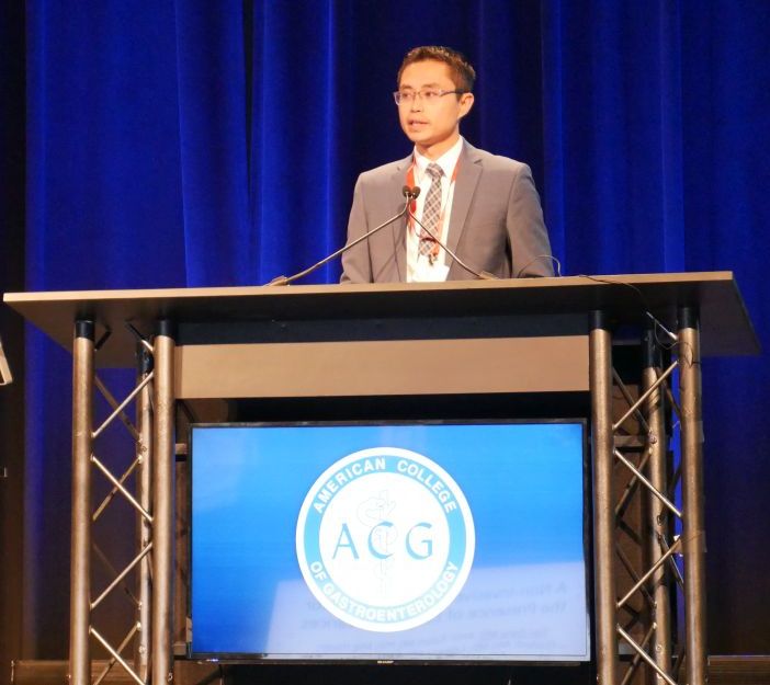
“The presence of cirrhosis-related complications is very strongly associated with readmissions,” Chandraprakash Umapathy, MD, MS, from the University of California, San Francisco, Fresno, said during his presentation. “Quality improvement efforts should focus on optimizing the management of complications of cirrhosis in the outpatient setting to reduce readmissions.”
In a retrospective cohort study, Dr. Umapathy and colleagues identified 230,036 patients from the Healthcare Cost and Utilization Project National Readmission Database for 2014 who had been discharged with a diagnosis of cirrhosis; of these patients, there were 185,737 index cases after excluding readmissions. Included patients had a mean age of 60.2 years and mean length of stay of 6.4 days, with 46% of patients having a length of stay longer than 4 days and mean total charges of $56,519. With regard to cirrhosis, 55% of patients displayed cirrhosis complications and 6.7% had more than three cirrhosis-related complications; the most common complication was ascites, in 32% of patients.
Overall, 11.09% of patients were readmitted at 30 days and 18.74% of patients were readmitted at 90 days, Dr. Umapathy said. Patients were more likely to be readmitted at 30 days if they were originally admitted on a weekend (adjusted prevalence ratio, 1.06; P = .001); into a medium (1.09; P = .009) or large (1.11; P less than .001) hospital; were admitted at a metropolitan teaching hospital (1.07; P less than .001); were insured by Medicaid (1.07; P less than .001); or were transferred to an extended care (1.51; P less than .001) facility or discharged to home health care (1.43; P less than .001).
Compared with patients who were not readmitted at 30 days, patients with 30-day readmission had a higher rate of alcoholic liver disease (43% vs. 46%; P less than .001), hepatitis C (28% vs. 32%; P less than .001), ascites (31% vs. 43%; P less than .001), hepatic encephalopathy (15% vs. 22%; P less than .001), hepatorenal syndrome (2.3% vs. 4.9%; P less than .001), hepatocellular cancer (5.1% vs. 5.7%; P = .001), presence of any cirrhosis complications (54% vs. 65%; P less than .001), and presence of more than three cirrhosis-related complications (6.3% vs. 10%; P less than .001). When adjusted in a multivariate analysis, association with readmission at 30 days for patients with cirrhosis-related complications such as ascites (1.42; P less than .001), hepatic encephalopathy (1.44; P less than .001), and hepatorenal syndrome (1.34; P less than .001) remained, Dr. Umapathy noted.
Length of stay longer than 4 days (0.84; P less than .001) and variceal hemorrhage (0.74; P = .002) were associated with reduced risk of readmissions at 30 days. “Focus on length of stay may result in patients being discharged prematurely, leading to higher early readmission,” Dr. Umapathy said.
Dr. Umapathy reports no relevant conflicts of interest.
SOURCE: Umapathy C et al. ACG 2018, Presentation 60
REPORTING FROM ACG 2018
Key clinical point: Cirrhosis-related complications and shorter length of stay were associated with higher rates of readmissions for patients with cirrhosis.
Major finding: 11.09% of patients were readmitted at 30 days and 18.74% of patients at 90 days, with the most common reasons for readmission including presence of cirrhosis complications and length of stay less than 4 days.
Study details: A retrospective cohort study of 185,737 index cases in the Healthcare Cost and Utilization Project National Readmission Database.
Disclosures: Dr. Umapathy reports no relevant conflicts of interest.
Source: Umapathy C et al. ACG 2018, Presentation 60.
Child-Pugh class does not affect HE recurrence in patients taking rifaximin
PHILADELPHIA – according to a recent presentation at the annual meeting of the American College of Gastroenterology.
Steven L. Flamm, MD, from Northwestern University, Chicago, and his colleagues examined results from a previous randomized, double-blinded trial of 140 patients receiving twice-daily rifaximin at 550 mg for 6 months in which the results showed rifaximin successfully maintained remission of hepatic encephalopathy (HE), compared with 159 patients receiving placebo.
“This pivotal study was published in March of 2010, but one of the post hoc assessments that was not performed was looking at various different phases of hepatic impairment as dictated by [Child-Pugh] class and each of those responses to this product,” Dr. Flamm said in his presentation.
Patients in the study were included if they had a Model For End-Stage Liver Disease score of 25 or less and two or more overt HE within 6 months (Conn score 1 or less) but were currently in remission. The researchers allowed the use of concomitant lactulose during the study period, which was used in 94.1% of rifaximin and 91.2% of placebo patients.
In the post hoc analysis, Dr. Flamm and his colleagues divided rifaximin and placebo patients into Child-Pugh class A (46 patients vs. 56 patients), class B (65 patients vs. 72 patients), and class C (17 patients vs. 14 patients) groups. For rifaximin and placebo patients, the mean age was 57.3 years and 57.2 years in the class A group, 54.4 years and 57.0 years in the class B group, and 56.1 years and 57.6 years in the class C group, respectively.
Overall, 8 of 46 rifaximin patients (17.4%) in the Child-Pugh class A and 15 of 65 rifaximin patients (23.1%) in the class B groups experienced an overt HE event during the 6-month study period, compared with 26 of 56 patients in the class A (46.4%) and 32 of 72 patients (44.4%) in the class B placebo groups; 5 of 17 rifaximin patients (29.4%) in the Child-Pugh class C group experienced their first overt HE event, compared with 9 of 14 (64.3%) patients in the placebo group.
With regard to first HE-related hospitalization, 5 of 46 patients (10.9%) in the rifaximin Child-Pugh class A group, 8 of 65 rifaximin patients (12.3%) in the class B group, and 4 of 17 rifaximin patients (23.5%) in the class C group experienced hospitalization because of HE, compared with 15 of 56 patients (23.2%) in the Child-Pugh class A group, 15 of 72 patients (20.8%) in the class B group, and 5 of 14 patients (35.7%) in the class C placebo group. The researchers noted lactulose use in the majority of patient cases in the study “provided further benefit” in reducing overt HE events.
“Although numeric differences were observed favoring rifaximin for the incidence of HE-related hospitalizations, a risk reduction versus placebo could not be firmly established, and presumably this was largely due to a lack of adequate power because of small sample size,” Dr. Flamm said.
This study and its analysis were supported by Salix Pharmaceuticals. Dr. Flamm and other coauthors report advisory committee membership, board membership, employment, or consultancy with AbbVie, Bristol-Myers Squibb, Gilead Sciences, Intercept Pharmaceuticals, Merck and Salix Pharmaceuticals. One coauthor reported no relevant conflicts of interest.
SOURCE: Flamm SL et al ACG 2018, Presentation 58.
REPORTING FROM ACG 2018
Key clinical point: Child-Pugh class does not significantly affect the overt hepatic encephalopathy recurrence rate in patients taking rifaximin, compared with placebo.
Major finding: A total of 17.4% of Child-Pugh class A, 23.1% of class B, and 29.4% class C patients taking rifaximin experienced overt hepatic encephalopathy, compared with 46.4% of Child-Pugh class A, 44.4% of class B, and 64.3% of class C patients receiving placebo.
Study details: A post hoc analysis of 299 patients receiving twice-daily rifaximin or placebo for 6 months.
Disclosures: This study and its analysis were supported by Salix Pharmaceuticals. Dr. Flamm and other coauthors reported advisory committee membership, board memberships, employment, or consultancy with AbbVie, Bristol-Myers Squibb, Gilead Sciences, Intercept Pharmaceuticals, Merck, and Salix Pharmaceuticals. One coauthor reported no relevant conflicts of interest.
Source: Flamm SL et al. ACG 2018, Presentation 58.
ACIP supports hepatitis A vaccine for homeless individuals
Homeless individuals aged 1 year and older should be vaccinated against hepatitis A, based on a unanimous vote at a meeting of the Centers for Disease Control and Prevention’s Advisory Committee on Immunization Practices.
“It is important that we take a national approach to vaccinating homeless” people, Noele Nelson, MD, PhD, MPH, of the CDC’s Division of Viral Hepatitis, said in a presentation prior to the vote, in which all 11 committee members voted in favor of hepatitis A vaccination for the homeless population.
Even limited vaccination will increase the herd immunity of the homeless population over time, she said.
Dr. Nelson presented data on the pros and cons of routine hepatitis A vaccination for homeless individuals aged 1 year and older. The Hepatitis Vaccines Work Group convened four meetings in advance of the October ACIP meeting and reached a consensus that homelessness is an independent indication for hepatitis A vaccination, she said.
If the hepatitis A vaccine is included as an ACIP recommendation, “it is more likely to be considered by homeless service providers,” noted Dr. Nelson. She also cited a low quality of evidence for adverse events associated with hepatitis A vaccination.
The work group considerations in the wake of a nationwide hepatitis A outbreak earlier in 2018 included the challenges of controlling outbreaks, which can spread quickly among the homeless population because of poor personal hygiene, limited sanitation, and tight living quarters. These factors make the homeless population more reliant on a vaccine for protection. An outbreak in San Diego, Calif., in particular, occurred largely in the homeless population.
“Routine vaccination is a more feasible approach to reach the homeless over time through regular homeless care providers,” Dr. Nelson said. As for costs, integrating vaccination into routine care for the homeless is cheaper and much less disruptive than the cost of responding to an outbreak.
The “cons” of recommending routine hepatitis A vaccination for the homeless population included the challenges of administrative record keeping. However, during the public comment period, Mae Morgan, MD, an internist who is medical director of Mercy Care Decatur Street & City of Refuge in Atlanta, emphasized that local homeless care organizations have procedures to manage routine vaccination. “If anyone is concerned that there is not a network in place, there are health centers to do this [that] would implement the vaccine.”
The ACIP committee members had no financial conflicts to disclose.
FROM AN ACIP MEETING
Aspirin cuts risk of liver and ovarian cancer
Regular long-term aspirin use may lower the risk of hepatocellular carcinoma (HCC) and ovarian cancer, adding to the growing evidence that aspirin may play a role as a chemopreventive agent, according to two new studies published in JAMA Oncology.
In the first study, led by Tracey G. Simon, MD, of Massachusetts General Hospital, Boston, the authors evaluated the associations between aspirin dose and duration of use and the risk of developing HCC. They conducted a population-based study, with a pooled analysis of two large prospective U.S. cohort studies: the Nurses’ Health Study and the Health Professionals Follow-up Study. The cohort included a total of 133,371 health care professionals who reported long-term data on aspirin use, frequency, dosage, and duration of use.
For the 87,507 female participants, reporting began in 1980, and for the 45,864 men, reporting began in 1986. The mean age for women was 62 years and was 64 years for men at the midpoint of follow-up (1996). Compared with nonaspirin users, those who used aspirin regularly tended to be older, former smokers, and regularly used statins and multivitamins. During the follow-up period, which was more than 26 years, there were 108 incident cases of HCC (65 women, 43 men; 47 with noncirrhotic HCC).
The investigators found that regular aspirin use was associated with a significantly lower HCC risk versus nonregular use (multivariable hazard ratio, 0.51; 95% confidence interval, 0.34-0.77), and estimates were similar for both sexes. Adjustments for regular NSAID use (for example, at least two tablets per week) did not change the data, and results were similar after further adjustment for coffee consumption and adherence to a healthy diet. The benefit also appeared to be dose related, as compared with nonuse, the multivariable-adjusted HR for HCC was 0.87 (95% CI, 0.51-1.48) for up to 1.5 tablets of standard-dose aspirin per week and 0.51 (95% CI, 0.30-0.86) for 1.5-5 tablets per week. The most benefit was for at least five tablets per week (HR, 0.49; 95% CI, 0.28-0.96; P = .006).
“Our findings add to the growing literature suggesting that the chemopreventive effects of aspirin may extend beyond colorectal cancer,” they wrote.
In the second study, Mollie E. Barnard, ScD, of the Harvard School of Public Health, Boston, and her colleagues looked at whether regular aspirin or NSAID use, as well as the patterns of use, were associated with a lower risk of ovarian cancer.
The data used were obtained from 93,664 women in the Nurses’ Health Study (NHS), who were followed up from 1980 to 2014, and 111,834 people in the Nurses’ Health Study II (NHSII), who were followed up from 1989 to 2015. For each type of agent, including aspirin, low-dose aspirin, nonaspirin NSAIDs, and acetaminophen, they evaluated the timing, duration, frequency, and number of tablets that were used. The mean age of participants in the NHS at baseline was 45.9 years and 34.2 years in the NHSII.
There were 1,054 incident cases of epithelial ovarian cancer identified during the study period. The authors did not detect any significant associations between aspirin and ovarian cancer risk when current users and nonusers were compared, regardless of dose (HR, 0.99; 95% CI, 0.83-1.19). But when low-dose (less than or equal to 100 mg) and standard-dose (325 mg) aspirin were analyzed separately, an inverse association for low-dose aspirin (HR, 0.77; 95% CI, 0.61-0.96) was observed. However, there was no association for standard-dose aspirin (HR, 1.17; 95% CI, 0.92-1.49).
In contrast, use of nonaspirin NSAIDs was positively associated with a higher risk of ovarian cancer when compared with nonuse (HR, 1.19; 95% CI, 1.00-1.41), and there were significant positive trends for duration of use (P = .02) and cumulative average tablets per week (P = .03). No clear associations were identified for acetaminophen use.
“Our results also suggest an increased risk of ovarian cancer among long-term, high-quantity users of nonaspirin analgesics, although this finding may reflect unmeasured confounding,” wrote Dr. Barnard and her coauthors. “Further exploration is warranted to evaluate the mechanisms by which heavy use of aspirin, nonaspirin NSAIDs, and acetaminophen may contribute to the development of ovarian cancer and to replicate our findings.”
The ovarian cancer study was supported by awards from the National Institutes of Health. Dr. Barnard was supported by awards from the National Cancer Institute, and her coauthors had no disclosures to report. The HCC study was funded by an infrastructure grant from the Nurses’ Health Study, an infrastructure grant from the Health Professionals Follow-up Study, and NIH grants to several of the authors. Dr. Chan has previously served as a consultant for Bayer on work unrelated to this article. No other disclosures were reported.
SOURCES: Barnard ME et al. JAMA Oncol. 2018 Oct 4. doi: 10.1001/jamaoncol.2018.4149; Simon TG et al. JAMA Oncol. 2018 Oct 4. doi: 10.1001/jamaoncol.2018.4154.
In an accompanying editorial published in JAMA Oncology, Victoria L. Seewaldt, MD, of the City of Hope Comprehensive Cancer Center in Duarte, Calif., asked if we “have arrived,” as these two studies are a critical step in realizing the potential of aspirin for cancer chemoprevention beyond colorectal cancer.
Aspirin use is very common in the United States, with almost half of adults aged between 45 and 75 years taking it regularly. Many regular users also believe that aspirin has potential to protect against cancer, and in a 2015 study – which was conducted prior to any formal cancer prevention guidelines – 18% of those taking aspirin on a regular basis reported doing so to prevent cancer.
Based on the strength of the association between aspirin use and colorectal cancer risk reduction, the U.S. Preventive Services Task Force recommended in 2015 that, among individuals aged between 50 and 69 years who have specific cardiovascular risk profiles, colorectal cancer prevention be included as part of the rationale for regular aspirin prophylaxis, Dr. Seewaldt noted. Aspirin became the first drug to be included in USPSTF recommendations for cancer chemoprevention in a “population not characterized as having a high risk of developing cancer.”
But it now appears aspirin may be able to go beyond colorectal cancer for chemoprevention. Ovarian cancer and hepatocellular carcinoma are in need of new prevention strategies and these findings provide important information that can help guide chemoprevention with aspirin.
These two studies “have the power to start to change clinical practice,” Dr. Seewaldt wrote, but more research is needed to better understand the underlying mechanism behind the appropriate dose and duration of use. Importantly, the authors of both studies cautioned that the potential benefits of aspirin must be weighed against the risk of bleeding, which is particularly important in patients with chronic liver disease.
“To reach the full promise of aspirin’s ability to prevent cancer, there needs to be better understanding of dose, duration, and mechanism,” she emphasized.
Dr. Seewaldt reported receiving grants from the National Institutes of Health/National Cancer Institute and is supported by the Prevent Cancer Foundation.
Regular long-term aspirin use may lower the risk of hepatocellular carcinoma (HCC) and ovarian cancer, adding to the growing evidence that aspirin may play a role as a chemopreventive agent, according to two new studies published in JAMA Oncology.
In the first study, led by Tracey G. Simon, MD, of Massachusetts General Hospital, Boston, the authors evaluated the associations between aspirin dose and duration of use and the risk of developing HCC. They conducted a population-based study, with a pooled analysis of two large prospective U.S. cohort studies: the Nurses’ Health Study and the Health Professionals Follow-up Study. The cohort included a total of 133,371 health care professionals who reported long-term data on aspirin use, frequency, dosage, and duration of use.
For the 87,507 female participants, reporting began in 1980, and for the 45,864 men, reporting began in 1986. The mean age for women was 62 years and was 64 years for men at the midpoint of follow-up (1996). Compared with nonaspirin users, those who used aspirin regularly tended to be older, former smokers, and regularly used statins and multivitamins. During the follow-up period, which was more than 26 years, there were 108 incident cases of HCC (65 women, 43 men; 47 with noncirrhotic HCC).
The investigators found that regular aspirin use was associated with a significantly lower HCC risk versus nonregular use (multivariable hazard ratio, 0.51; 95% confidence interval, 0.34-0.77), and estimates were similar for both sexes. Adjustments for regular NSAID use (for example, at least two tablets per week) did not change the data, and results were similar after further adjustment for coffee consumption and adherence to a healthy diet. The benefit also appeared to be dose related, as compared with nonuse, the multivariable-adjusted HR for HCC was 0.87 (95% CI, 0.51-1.48) for up to 1.5 tablets of standard-dose aspirin per week and 0.51 (95% CI, 0.30-0.86) for 1.5-5 tablets per week. The most benefit was for at least five tablets per week (HR, 0.49; 95% CI, 0.28-0.96; P = .006).
“Our findings add to the growing literature suggesting that the chemopreventive effects of aspirin may extend beyond colorectal cancer,” they wrote.
In the second study, Mollie E. Barnard, ScD, of the Harvard School of Public Health, Boston, and her colleagues looked at whether regular aspirin or NSAID use, as well as the patterns of use, were associated with a lower risk of ovarian cancer.
The data used were obtained from 93,664 women in the Nurses’ Health Study (NHS), who were followed up from 1980 to 2014, and 111,834 people in the Nurses’ Health Study II (NHSII), who were followed up from 1989 to 2015. For each type of agent, including aspirin, low-dose aspirin, nonaspirin NSAIDs, and acetaminophen, they evaluated the timing, duration, frequency, and number of tablets that were used. The mean age of participants in the NHS at baseline was 45.9 years and 34.2 years in the NHSII.
There were 1,054 incident cases of epithelial ovarian cancer identified during the study period. The authors did not detect any significant associations between aspirin and ovarian cancer risk when current users and nonusers were compared, regardless of dose (HR, 0.99; 95% CI, 0.83-1.19). But when low-dose (less than or equal to 100 mg) and standard-dose (325 mg) aspirin were analyzed separately, an inverse association for low-dose aspirin (HR, 0.77; 95% CI, 0.61-0.96) was observed. However, there was no association for standard-dose aspirin (HR, 1.17; 95% CI, 0.92-1.49).
In contrast, use of nonaspirin NSAIDs was positively associated with a higher risk of ovarian cancer when compared with nonuse (HR, 1.19; 95% CI, 1.00-1.41), and there were significant positive trends for duration of use (P = .02) and cumulative average tablets per week (P = .03). No clear associations were identified for acetaminophen use.
“Our results also suggest an increased risk of ovarian cancer among long-term, high-quantity users of nonaspirin analgesics, although this finding may reflect unmeasured confounding,” wrote Dr. Barnard and her coauthors. “Further exploration is warranted to evaluate the mechanisms by which heavy use of aspirin, nonaspirin NSAIDs, and acetaminophen may contribute to the development of ovarian cancer and to replicate our findings.”
The ovarian cancer study was supported by awards from the National Institutes of Health. Dr. Barnard was supported by awards from the National Cancer Institute, and her coauthors had no disclosures to report. The HCC study was funded by an infrastructure grant from the Nurses’ Health Study, an infrastructure grant from the Health Professionals Follow-up Study, and NIH grants to several of the authors. Dr. Chan has previously served as a consultant for Bayer on work unrelated to this article. No other disclosures were reported.
SOURCES: Barnard ME et al. JAMA Oncol. 2018 Oct 4. doi: 10.1001/jamaoncol.2018.4149; Simon TG et al. JAMA Oncol. 2018 Oct 4. doi: 10.1001/jamaoncol.2018.4154.
FROM JAMA ONCOLOGY
Key clinical point: Regular aspirin use was associated with a decreased risk of ovarian cancer and hepatocellular carcinoma.
Major finding: Low-dose aspirin was associated with a 23% lower risk of ovarian cancer and a 49% reduced risk of developing hepatocellular carcinoma.
Study details: The hepatocellular carcinoma study was a population-based study of two nationwide, prospective cohorts of 87,507 men and 45,864 women; the ovarian cancer study was a cohort study using data from two prospective cohorts, with 93,664 people in one and 111,834 in the other.
Disclosures: The ovarian cancer study was supported by awards from the National Institutes of Health. Dr. Barnard was supported by awards from the National Cancer Institute, and her coauthors had no disclosures to report. The hepatocellular carcinoma study was funded by an infrastructure grant from the Nurses’ Health Study, an infrastructure grant from the Health Professionals Follow-up Study, and NIH grants to several of the authors. Dr. Chan has previously served as a consultant for Bayer on work unrelated to this article. No other disclosures were reported.
Sources: Barnard ME et al. JAMA Oncol. 2018 Oct 4. doi: 10.1001/jamaoncol.2018.4149; Simon TG et al. JAMA Oncol. 2018 Oct 4. doi: 10.1001/jamaoncol.2018.4154.
Novel score predicts esophageal varices in patients with cirrhosis prior to EGD
PHILADELPHIA – A novel score accurately predicted the size and presence of esophageal varices in a noninvasive manner, which may help clinicians avoid unnecessary esophagogastroduodenoscopy in patients with cirrhosis, according to a recent award-winning presentation at the annual meeting of the American College of Gastroenterology.
Although there are two validated scores for predicting esophageal varices (EV), platelet count to spleen diameter ratio and Baveno VI criteria, they have drawbacks, Tien Dong, MD, from the University of California, Los Angeles said.
“The limitations to these existing scores and criteria are both of them require imaging to do, so because of that, they oftentimes are limited in national clinical use,” Dr. Dong said in his presentation of his team’s abstract, which won the ACG Governors Award for Excellence in Clinical Research. “The other thing is that, even though it’s recommended, sometimes spleen diameter on a normal ultrasound is not reported. Furthermore, elastography – even though it’s becoming more and more common – is still not yet readily available across the country.”
Dr. Dong and his colleagues sought to identify noninvasive clinical predictors of EV to create a predictive score to identify EV to overcome these drawbacks. They gathered endoscopy data from the Olive View–UCLA and West Los Angeles Veterans Affairs Hospital to create a discovery cohort (165 patients) and tested the score on patients from Ronald Reagan UCLA Medical Center in a validation cohort (73 patients).
They used a random forest classifier machine learning algorithm “to create a forest of decision trees so that it can tell us which variables it believes to be the most predictive of our outcomes of interest,” Dr. Dong said.
The algorithm identified several variables that appeared to be predictive of EV presence, such as international normalized ratio, aminotransferase, platelet mean, hemoglobin, albumin and blood urea nitrogen less than or equal to 3, which the researchers used to create an EV-endoscopy (EV-Endo) score.
In the discovery cohort, area under the curve (AUC) for the presence of EV was 0.81, compared with an AUC of 0.82 in the validation cohort, while there was an AUC of 0.77 in the discovery cohort and an AUC of 0.79 for small/absent vs. medium/large EV. Patients with Child-Pugh class A cirrhosis had an AUC of 0.81 for EV presence and an AUC of 0.77 for EV size. The researchers then created a cutoff score of 3.48 or less and 7.70 or more for EV presence, which had a sensitivity and specificity of 93.9% and 97.5%, respectively. EV-Endo score EV size cutoff scores were also 3.48 or less and 7.70 or more, with a sensitivity of 95.8% and specificity of 95.0%.
Dr. Dong reports no conflicts of interest.
SOURCE: Hauer M et al. ACG 2018. Presentation 31.
REPORTING FROM ACG 2018
Black patients present as sicker, more likely to receive liver transplant
PHILADELPHIA – Black patients are more likely to be put on a transplant list because of acute liver failure, be listed as status 1, and receive a liver transplant, compared with white patients, according to a recent presentation at the annual meeting of the American College of Gastroenterology.
Lauren D. Nephew, MD, MSCE, of Indiana University in Indianapolis, and her colleagues performed a retrospective cohort study of black and white patients with a minimum age of 18 years in the United Network of Organ Sharing database who were wait-listed for a liver transplantation during 2002-2016. They examined patient clinical characteristics, acute liver failure (ALF) etiologies, wait-list status, and posttransplant survival outcomes through Kaplan Meier analysis.
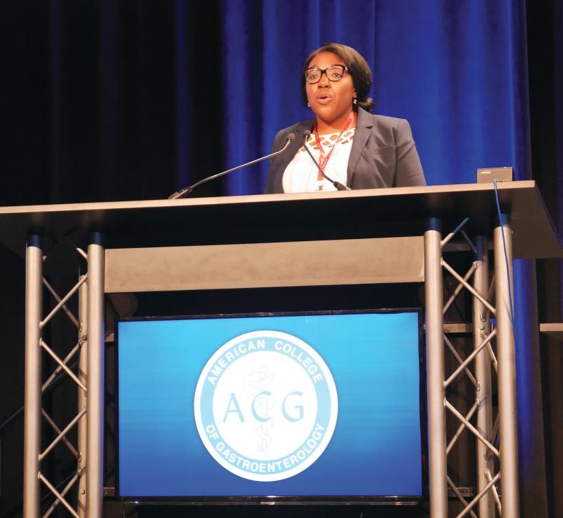
“We really wanted to explore this topic in patients with acute liver failure, some of the sickest patients that we see,” Dr. Nephew said in her presentation. “We wanted to really determine whether or not there were differences in clinical characteristics and etiologies of acute liver failure in patients by race who are listed for liver transplantation.”
“Then, we wanted to compare wait-list outcomes,” she added, such as “differences by race in liver transplantation or wait-list removal because of death or becoming too sick for transplant.”
There were 11,289 patients in the white ALF group and 2,112 patients in the black ALF group; 2,876 (25.5%) of patients in the white ALF and 790 (37.4%) in the black ALF group were listed as status 1, which indicated an expected survival of 7 days or less. There were similar clinical characteristics for the white and black ALF status 1 patients regarding age (34.2 years vs. 36.3 years), Model for End-Stage Liver Disease (MELD) score (34 vs. 36; P less than .001), international normalized ratio (INR) test (mean 4.5 vs. mean 5.0; P = .001), creatinine levels (2.1 mg/dL vs. 1.9 mg/dL; P less than .001), and percentage of patients who were hepatic encephalopathy grade 3 or 4 (60.0% vs. 63.2%; P = .10). However, Dr. Nephew noted significantly higher bilirubin levels in the black ALF status 1 cohort (17.9 mg/dL), compared with the white ALF status 1 cohort (11.3 mg/dL; P less than .001).
The causes for ALF in each group included drug-induced liver failure (white status 1 cohort, 34.1%; black status 1 cohort, 20.6%), autoimmune hepatitis (2.7% vs. 9.4%), Wilson’s disease (0.58% vs. 0.13%), unknown etiology (34.5% vs. 42.5%), and other etiology (22.9% vs. 17%). For patients who underwent liver transplant and wait-list removal, there were no significant differences in wait-list removal “despite black patients being sicker at presentation,” Dr. Nephew said. Black patients were more likely to be listed to status 1 and transplanted at 62% (490 patients), compared with white patients at 53% (1,524 patients). There were 713 white patients (24.8%) removed from the transplant list, compared with 114 (13.8%) of black patients.
“If you are transplanted and you don’t die, then you are likely removed from the list for other reasons, and the most common reason is that you improved and became well, and so white patients were significantly more likely to be removed from the wait-list because of improvement, compared with black patients,” Dr. Nephew said.
In a competing risk analysis, the researchers found the hazard ratio for white patients who were status 1 and removed from the wait-list because of death or becoming too sick was 1.04 (95% confidence interval, 0.89-1.21) and those white patients who were listed as status 1 and then transplanted was 1.2 (95% CI, 1.08-1.30). In a multivariate analysis, the hazard ratio for white patients who were listed as status 1 and transplanted, which contained bilirubin at transplant, was 1.08 (95% CI, 0.98-1.19). Kaplan Meier 1-year survival post-transplant was 82.8% in white patients and 79.6% in black patients (P = .09).
“I think the question that we’ve been asking ourselves is, is this because black patients are presenting later with their acute liver disease and are sicker at presentation, or do they just have worse liver disease inherently on presentation that drove these findings?” Dr. Nephew said.
Dr. Nephew reports no relevant conflicts of interest.
SOURCE: Nephew L et al. ACG 2018, Presentation 59.
REPORTING FROM ACG 2018
Key clinical point: Black patients are sicker than white patients when they present with acute liver failure and are awaiting liver transplantation.
Major finding: Black patients with acute liver failure were more likely to be wait-listed, listed as status 1, and have higher Model for End-Stage Liver Disease (MELD) scores, creatinine levels, and INR tests, compared with white patients.
Study details: A retrospective cohort analysis of patients with acute liver failure awaiting a liver transplant in the United Network of Organ Sharing database.
Disclosures: Dr. Nephew reports no relevant conflicts of interest.
Source: Nephew L et al. ACG 2018, Presentation 59.
Myeloperoxidase elevated in HCV-related liver cancer
Myeloperoxidase (MPO) expression was significantly higher in hepatitis C virus (HCV)–related hepatocellular carcinoma (HCC) cases when compared with cirrhotic patients, according to a study of 59 patients with HCV-related liver disease.
HCV is the main cause of liver disease, wrote Mohamed Abdel-Hamed, MD, of Minia University in Egypt, and his colleagues. In addition, HCV is associated with significant oxidative stress, which has been identified as a significant metabolic pathway culminating in hepatic cirrhosis, liver failure, and liver cancer. Thus the researchers studied the role of MPO, an oxidative enzyme released at sites of inflammation, in the possible etiology of HCV-related liver cancer.
The patients were divided into two groups, 25 with HCC and 34 with chronic liver diseases with cirrhosis. All patients were examined immunohistochemically to demonstrate the expression of myeloperoxidase, according to the report published in Meta Gene.
Subjects with HCC showed markedly increased MPO expression when compared with MPO expression in hepatocytes of subjects with liver cirrhosis, who mostly showed a mild degree of expression. In addition, no mild expression of MPO was detected in the subjects with HCC. These findings were highly statistically significant (P less than .0001), according to Dr. Abdel-Hamed and his colleagues.
“The present study showed that marked expression of MPO plays an important role in the pathogenesis of HCV-associated HCC,” the authors stated. “This study could provide valuable, predictive parameters that can be used clinically in the prognosis of HCC patients.”
The authors did not report any disclosures.
SOURCE: Abdel-Hamid M et al. Meta Gene. 2018 Dec;18:1-8.
FROM META GENE
Key clinical point:
Major finding: Hepatocellular carcinoma subjects showed a marked increase of myeloperoxidase expression when compared with subjects with liver cirrhosis (P less than .0001).
Study details: An immunohistochemical analysis of 59 patients infected with hepatitis C virus.
Disclosures: The authors did not report any disclosures.
Source: Abdel-Hamid M et al. Meta Gene. 2018 Dec;18:1-8.
MELD sodium score tied to better transplant outcomes
Factoring hyponatremic status into liver graft allocations led to significant reductions in wait-list mortality, researchers reported in the November issue of Gastroenterology.
Hyponatremic patients with low MELD scores benefited significantly from allocation based on the end-stage liver disease–sodium (MELD-Na) score, while its survival benefit was less evident among patients with higher scores, said Shunji Nagai, MD, PhD, of Henry Ford Hospital, Detroit, and his associates. “Therefore, liver allocation rules such as Share 15 and Share 35 need to be revised to fulfill the Final Rule under the MELD-Na based allocation,” they wrote.
The Share 35 rule offers liver grafts locally and regionally to wait-listed patients with MELD-Na scores of at least 35. Under the Share 15 rule, livers are offered regionally or nationally before considering local candidates with MELD scores under 15. The traditional MELD scoring system excluded hyponatremia, which has since been found to independently predict death from cirrhosis. Therefore, in January 2016, a modified MELD-Na score was implemented for patients with traditional MELD scores of at least 12. The MELD-Na score assigns patients between 1 and 11 additional points, and patients with low MELD scores and severe hyponatremia receive the most points. To assess the impact of this change, Dr. Nagai and his associates compared wait-list and posttransplantation outcomes during the pre and post–MELD-Na eras and the survival benefit of liver transplantation during the MELD-Na period. The study included all adults wait-listed for livers from June 2013, when Share 35 was implemented, through September 2017.
Mortality within 90 days on the wait list fell significantly during the MELD-Na era (hazard ratio, 0.74; P less than .001). Transplantation conferred a “definitive” survival benefit when MELD-Na scores were 21-23 (HR versus wait list, 0.34; P less than .001). During the traditional MELD period, the equivalent cutoff was 15-17 (HR, 0.36; P less than .001). “As such, the current rules for liver allocation may be suboptimal under the MELD-Na–based allocation and the criteria for Share 15 may need to be revisited,” the researchers wrote. They recommended raising the cutoff to 21.
The study also confirmed mild hyponatremia (130-134 mmol/L), moderate hyponatremia (125-129 mmol/L), and severe hyponatremia (less than 125 mmol/L) as independent predictors of wait-list mortality during the traditional MELD era. Hazard ratios were 1.4, 1.8, and 1.7, respectively (all P less than .001). The implementation of MELD-Na significantly weakened these associations, with HRs of 1.1 (P = .3), 1.3 (P = .02), and 1.4 (P = .04), respectively).
The probability of transplantation also rose significantly during the MELD-Na era (HR, 1.2; P less than .001), possibly because of the opioid epidemic, the researchers said. Although greater availability of liver grafts might have improved wait-list outcomes, all score categories would have shown a positive impact if this was the only reason, they added. Instead, MELD-Na most benefited patients with lower scores.
Finally, posttransplantation outcomes worsened during the MELD-Na era, perhaps because of transplant population aging. However, the survival benefit of transplant shifted to higher score ranges during the MELD-Na era even after the researchers controlled for this effect. “According to this analysis,” they wrote, “the survival benefit of liver transplant was definitive in patients with score category of 21-23, which could further validate our proposal to revise Share 15 rule to ‘Share 21.’ ”
The investigators reported having no external funding sources or conflicts of interest.
SOURCE: Nagai S et al. Gastroenterology. 2018 Jul 26. doi: 10.1053/j.gastro.2018.07.025.
FROM GASTROENTEROLOGY
Key clinical point: The implementation of the MELD sodium (MELD-Na) score for liver allocation was associated with significantly improved outcomes for wait-listed patients.
Major finding: During the MELD-Na era, mortality within 90 days on the liver wait list dropped significantly (HR, 0.74; P less than .001) while the probability of transplant rose significantly (HR, 1.2; P less than .001).
Study details: Comparison of 18,850 adult transplant candidates during the traditional MELD era versus 14,512 candidates during the MELD-Na era.
Disclosures: The investigators had no external funding sources or conflicts of interest.
Source: Nagai S et al. Gastroenterology. 2018 Jul 26. doi: 10.1053/j.gastro.2018.07.025.
Web-based lifestyle program aids in weight loss for NAFLD patients
A web-based intervention for management of nonalcoholic fatty liver disease – which researchers said may help reach busy or remote patients not able to attend in-person sessions – had a similar number of patients reach a target weight-loss goal of at least 10% of body weight, compared with a group-based intervention.
“The use of web education in the management of noncommunicable diseases has long been suggested, considering the huge number of cases at risk and patients’ needs,” wrote Arianna Mazzotti, MD, of the department of medical and surgical sciences, Alma Mater University, Bologna, Italy, and her colleagues. Their report was published in the Journal of Hepatology.
“The majority of cases are in an age range where job constraints make it difficult to implement a systematic face-to-face or group approach, whereas the eHealth procedures may keep the contact between patients and therapists without disrupting normal daily living.”
Dr. Mazzotti and her colleagues studied 716 patients with nonalcoholic fatty liver disease (NAFLD) at the university between January 2010 and December 2015 who attended either a web-based NAFLD intervention (278 patients) or an in-person, group-based lifestyle modification program (438 patients). Patients in the web-based intervention tended to be younger males with a higher education level, similar mean body mass index (33 kg/m2), and significantly lower blood pressure and rates of type 2 diabetes mellitus. The primary outcome included weight loss of at least 10%; secondary outcomes included changes in weight, changes in lifestyle, surrogate markers of steatosis and fibrosis, and alanine aminotransferase (ALT) within normal limits, researchers said.
The group-based program consisted of five 2-hour weekly sessions counseling patients on diet and physical activity, whereas the web-based intervention reproduced these sessions in addition to questionnaires, “highly interactive” slides, examples, and games as well as a mechanism to ask questions. Regardless of intervention, patients attended a 6-month in-person follow-up where they received treatment and reinforcement for comorbidities such as type 2 diabetes mellitus.
In the web-based intervention, 76% of patients attended the 6-month follow-up, 58% attended the 12-month follow-up, and 43% attended the 24-month follow-up, compared with 87%, 80%, and 69% of patients in the group-based intervention, respectively. Patients in the web-based intervention had a significantly decreased intake of calories after 6 months (273 kcal/day vs. 193 kcal/day; P = .006) compared with the group-based intervention. Physical activity significantly increased at 6 months for both groups, but there were no significant differences between groups.
Body weight decreased for the web-based intervention by 3.4% at 6 months, 4.9% at 12 months, and 5.5% at 24 months, compared with 3.1% at 6 months, 4.0% at 12 months, and 4.2% at 24 months in the group-based intervention. There was a nearly two-point reduction in body mass index for both groups, with 20% of web-based intervention patients and 15% of group-based intervention patients achieving the 10% weight-loss target; and, when the researchers performed a logistic regression analysis, the web-based intervention group was not associated with less short- and long-term 10% weight reduction after attrition rates and confounders were adjusted for.
At 24-month follow-up, the researchers found a decrease in ALT levels by an average of 22 ± 32 mU/mL, with the web-based intervention group having normalized ALT levels in 18% of cases at 6 months, 32% at 12 months, and 35% at 24 months, compared with 16% of cases at 6 months, 22% of cases at 12 months, and 29% of cases at 24 months in the group-based intervention. Compared with the group-based intervention, there was a higher reduction in fatty liver index scores at 12-month follow-up (71.3 vs. 78.0; P less than .001) and 24-month follow-up (68.9 vs. 76.3; P = .002) for the web-based intervention group. The researchers noted NAFLD fibrosis score and Fib-4 scores were reduced in both groups.
“The [web-based intervention] program might be extended to other units and/or general practitioners, increasing its impact in the community in prevention and treatment of progressive NAFLD,” Dr. Mazzotti and her colleagues wrote. “It might also be superimposed to drug treatment in the most severe cases, with possible additive effects.”
This study was supported by a grant from the European Community Seventh Framework Program. The authors report no relevant conflicts of interest.
*This story was updated on 10/4/2018.
SOURCE: Mazzotti A et al. J Hepatol. 2018 Oct 2. doi: 10.1016/j.jhep.2018.07.013.
FROM THE JOURNAL OF HEPATOLOGY
Key clinical point: A web-based lifestyle modification intervention was similarly effective as a group intervention in reducing weight for patients with nonalcoholic fatty liver disease.
Major finding: In the web-based intervention, body weight decreased by 3.4% at 6 months, 4.9% at 12 months, and 5.5% at 24 months, compared with 3.1% at 6 months, 4.0% at 12 months, and 4.2% at 24 months in the group-based intervention. A similar number of patients in both groups achieved the 10% weight-loss target.
Study details: An observational study of 716 patients participating in web-based or group nonalcoholic fatty liver disease interventions at the University of Bologna between January 2010 and December 2015.
Disclosures: The European Community Seventh Framework Program supported the study. The authors report no conflicts of interest.
Source: Mazzotti A et al. J Hepatol. 2018 Oct 2. doi: 10.1016/j.jhep.2018.07.013.

