User login
Preoperative variables can predict prolonged air leak
Prolonged air leak is a well-known complication after lung cancer surgery that can worsen patient outcomes and drive up costs, and while international authors have developed tools to calculate the risk of PAL, their use has been limited in the United States for various reasons. Researchers at the University of Pittsburgh have reported on a predictive model that uses easy-to-obtain patient factors, such as forced expiratory volume and smoking history, to help surgeons identify patients at greatest risk for complications and implement preventative measures.
Adam Attaar and his coauthors reported that their nomogram had an accuracy rate of 76%, with a 95% confidence interval, for predicting PAL after surgery (J Thorac Cardiovasc Surg. 2017 March;153[3]:690-9). “Using readily available candidate variables, our nomogram predicts increasing risk of prolonged air leak with good discriminatory ability,” noted Mr. Attaar, a student at University of Pittsburgh, and his coauthors.
Previously published reports put the incidence of PAL complications at 6%-18%, they noted. In the University of Pittsburgh series of 2,317 patients who had pulmonary resection for lung cancer or nodules from January 2009 to June 2014, the incidence was 8.6%.
In this series, patients with PAL were more likely to be older, men, and smokers, and to have a lower body mass index, peripheral vascular disease, chronic obstructive pulmonary disease, a history of steroid use, a high Zubrod score and lower forced expiratory volume.“They were less likely to have diabetes or to be hospitalized before surgery,” the researchers said. Surgical factors that characterized patients with PAL were resection for primary lung cancer rather than benign or metastatic tumors; lobectomy/segmentectomy or bilobectomy rather than wedge resection; a right-sided resection; thoracotomy; and a surgeon with higher annual caseloads.
Not all those factors made it into the nomogram, however. The nomogram scores each of these 10 variables to calculate the risk of PAL, in order of their weighting: lower forced expiratory volume, procedure type, BMI, right-sided thoracotomy, preoperative hospitalization, annual surgeon caseload, wedge resection by thoracotomy, reoperation, smoking history, and Zubrod score. A second nomogram drops out surgeon volume to make it more generalizable to other institutions.
In explaining higher surgeon volume as a risk factor for PAL, the researchers said that high-volume surgeons may be operating on patients with variables not accounted for in the Society of Thoracic Surgeons General Thoracic Surgery Database. “These unmeasured variables … could reveal modifiable technical factors to reduce the incidence of PAL and require further study,” the researchers said.
Fast-track discharge has gained acceptance in recent years as a way to spare patients a prolonged hospital stay and cut costs, but in this series the median hospital stay for patients with PAL was 10 days vs. 4 days for non-PAL patients (P less than 0.001).
“An accurate and generalizable PAL risk stratification tool could facilitate surgical decision making and patient-specific care” and aid in the design of trials to evaluate air-leak reduction methods such as sealants, buttressed staple lines, and pneumoperitoneum the researchers wrote.
Going forward, further development of the model would involve a multicenter study and inclusion of risk factors not accounted for in the thoracic surgery database, they noted.
The researchers had no relevant financial relationships to disclose.
The authors of this study “have performed a rigorous set of analyses to create this model,” Chi-Fu Jeffrey Yang, MD, of Duke University, Durham, N.C., noted in his invited commentary (J Thorac Cardiovasc Surg. 2017 March;53[3]:700-1). “The strengths of this study include its sound statistical analysis and study design,” Dr. Yang wrote. He gave the authors credit for using bootstrapping to internally validate the model.
However, Dr. Yang said that the database used by the researchers did not account for “numerous important variables,” including presence of pleural adhesions and emphysema status. The analysis also grouped lobectomy and segmentectomy together, and did not consider intraoperative variables such as sealant use, or postoperative management.
While Dr. Yang commended the study authors for developing a “reliable nomogram,” getting it implemented in the clinic is another hurdle. “It is commonly cited that it takes approximately 17 years for research evidence to translate into daily practice,” he said. To shorten that time line, he suggested the authors take a cue from various tech groups: Develop an app that surgeons can use.
Dr. Yang had no relevant financial relationships to disclose.
Prolonged air leak is a well-known complication after lung cancer surgery that can worsen patient outcomes and drive up costs, and while international authors have developed tools to calculate the risk of PAL, their use has been limited in the United States for various reasons. Researchers at the University of Pittsburgh have reported on a predictive model that uses easy-to-obtain patient factors, such as forced expiratory volume and smoking history, to help surgeons identify patients at greatest risk for complications and implement preventative measures.
Adam Attaar and his coauthors reported that their nomogram had an accuracy rate of 76%, with a 95% confidence interval, for predicting PAL after surgery (J Thorac Cardiovasc Surg. 2017 March;153[3]:690-9). “Using readily available candidate variables, our nomogram predicts increasing risk of prolonged air leak with good discriminatory ability,” noted Mr. Attaar, a student at University of Pittsburgh, and his coauthors.
Previously published reports put the incidence of PAL complications at 6%-18%, they noted. In the University of Pittsburgh series of 2,317 patients who had pulmonary resection for lung cancer or nodules from January 2009 to June 2014, the incidence was 8.6%.
In this series, patients with PAL were more likely to be older, men, and smokers, and to have a lower body mass index, peripheral vascular disease, chronic obstructive pulmonary disease, a history of steroid use, a high Zubrod score and lower forced expiratory volume.“They were less likely to have diabetes or to be hospitalized before surgery,” the researchers said. Surgical factors that characterized patients with PAL were resection for primary lung cancer rather than benign or metastatic tumors; lobectomy/segmentectomy or bilobectomy rather than wedge resection; a right-sided resection; thoracotomy; and a surgeon with higher annual caseloads.
Not all those factors made it into the nomogram, however. The nomogram scores each of these 10 variables to calculate the risk of PAL, in order of their weighting: lower forced expiratory volume, procedure type, BMI, right-sided thoracotomy, preoperative hospitalization, annual surgeon caseload, wedge resection by thoracotomy, reoperation, smoking history, and Zubrod score. A second nomogram drops out surgeon volume to make it more generalizable to other institutions.
In explaining higher surgeon volume as a risk factor for PAL, the researchers said that high-volume surgeons may be operating on patients with variables not accounted for in the Society of Thoracic Surgeons General Thoracic Surgery Database. “These unmeasured variables … could reveal modifiable technical factors to reduce the incidence of PAL and require further study,” the researchers said.
Fast-track discharge has gained acceptance in recent years as a way to spare patients a prolonged hospital stay and cut costs, but in this series the median hospital stay for patients with PAL was 10 days vs. 4 days for non-PAL patients (P less than 0.001).
“An accurate and generalizable PAL risk stratification tool could facilitate surgical decision making and patient-specific care” and aid in the design of trials to evaluate air-leak reduction methods such as sealants, buttressed staple lines, and pneumoperitoneum the researchers wrote.
Going forward, further development of the model would involve a multicenter study and inclusion of risk factors not accounted for in the thoracic surgery database, they noted.
The researchers had no relevant financial relationships to disclose.
FROM THE JOURNAL OF THORACIC AND CARDIOVASCULAR SURGERY
Key clinical point: Preoperative variables can be evaluated to determine patient risk for prolonged air leak (PAL) in lung resection for cancer.
Major finding: A nomogram demonstrated 76% discriminatory accuracy in predicting PAL after lung resection.
Data source: Analysis of 2,522 pulmonary resections performed at eight hospitals within the University of Pittsburgh health system from January 2009 to June 2014.
Disclosures: The researchers had no conflicts of interest to disclose.
EBUS scope, EUS-FNA similarly effective
When assessing a patient for lung cancer, a procedure involving the insertion of an EBUS scope in the esophagus – EUS-B-FNA – can achieve similarly accurate results as endoscopic ultrasound guided–fine-needle aspiration (EUS-FNA), according to a new study.
This finding could lead patients to choose EUS-B-FNA over EUS-FNA – the standard of care for analyzing potential metastasis of the left adrenal glands (LAGs) – resulting in both time and cost savings for patients.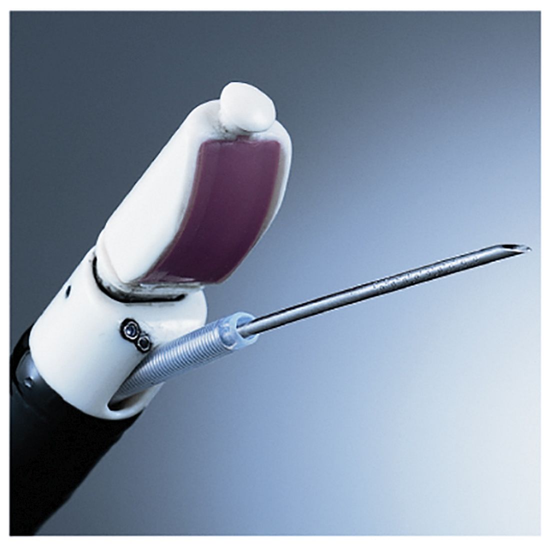
“A recent report showed that LAG visualization using the EBUS scope was possible in 85% of patients,” according to the authors of this study, including Prof. Jouke T. Annema, MD of the University of Amsterdam. Prior to this new research, it was unknown to what extent a single EBUS scope adequately assess and sample the LAGs and how its performance related to the use of a conventional endoscopic ultrasound–guided scope (Lung Cancer. 2017. doi: org/10.1016/j.lungcan.2017.02.011).
Dr. Annema and his coauthors recruited patients from four centers – three in the Netherlands, one in Poland – and followed them prospectively. Patients with “(suspected) lung cancer [who] had an indication for both mediastinal lymph node and LAG sampling” were recruited for the study. The researchers followed 44 patients through final diagnosis to determine if they ultimately had lung cancer.
Subjects first received complete mediastinal and hilar staging of lung cancer and any present tumors via an EBUS and EUS-B procedure. Following an EBUS examination of the mediastinum, the EBUS scope was retracted from the trachea and positioned into the esophagus for an examination of the mediastinal nodes. Then, the EBUS scope was advanced into the stomach for identification of the LAG. Afterward, the routine EUS-FNA was performed. LAG analysis across both methods involved visualizing the LAG and collecting an adequate tissue sample for testing.
“In short, in order to locate the LAG, a structured three step approach was used according to the EUS assessment tool (EUS-AT): identification of the liver, abdominal aorta, coeliac trunk, left kidney, and LAG,” the authors noted. “By turning the EBUS scope clockwise from the liver, the abdominal aorta and coeliac trun[k] are identified. By subsequently turning the EBUS scope gently in caudal direction, the left kidney and LAG are identified.”
Endoscopists then evaluated both procedures in each subject according to feasibility and practicability to determine if the findings of the experimental procedure were usable. Finally, a cytologic exam was conducted, using Giemsa or Papanicolaou staining to determine if any present cancer had metastasized, and a final diagnosis was made.
LAG analysis had a success rate of 89% (39/44; 95% confidence interval, 76-95%) for EUS-B-FNA, compared with 93% (41/44; 95% CI, 82-98%) for EUS-FNA. Similarly, when looking at the rate of sensitivity for LAG metastases, EUS-B had a rate of sensitivity for LAG metastases of at least 87% (95% CI, 65-97%), while EUS-FNA was found to be at least 83% (95% CI, 62-95%. Endoscopists were equally satisfied with both procedures in the “majority” of cases in this study.
“In [five] cases (11%), the EUS-B-FNA procedure was unsuccessful, due to the inability to make good contact of the ultrasound transducer and the stomach wall,” the authors explained. “The conventional EUS scope is more stable as a result of the increased tube diameter. Another advantage of the conventional echo-endoscope is its wider scanning angle. ... The conventional EUS scope is also longer than the EBUS scope, [but that] does not seem to be the limiting factor.”
No funding source was disclosed for this study. The authors reported no relevant financial disclosures.
FROM LUNG CANCER
Key clinical point:
Major finding: EUS-B-FNA had a success rate of 89%, versus 93% for EUS-FNA, while sensitivity for LAG metastases were 87% and 83%, respectively.
Data source: Multicenter, prospective study of 44 consecutive suspected lung cancer patients.
Disclosures: No funding source disclosed. Authors reported no relevant financial disclosures.
STAS predictive for lung SCC recurrence
First described in 2015, tumor spread through air spaces is a recently recognized form of invasion in lung carcinoma, but it has not been well described in lung squamous cell carcinoma. However, a study out of Memorial Sloan-Kettering Cancer Center reports spread through air spaces (STAS) is one of the most significant histologic findings in lung squamous cell carcinoma (SCC).
In multivariable models for any recurrence and lung cancer–specific death, the researchers found that STAS was a significant independent predictor for both outcomes (P = .034 and .016, respectively).
“We found that STAS in lung SCC was associated with p-stage, lymphatic and vascular invasion, necrosis, larger nuclear diameter, increased mitoses and high Ki-67 labeling index,” wrote lead author Shaohua Lu, MD, and coauthors (J Thorac Oncol. 2017 Feb;12[2]:223-34). Their findings are based on an analysis of 445 patients who had resection for stage I-III SCC over a 10-year period ending in 2009.
The Sloan-Kettering Group previously reported that STAS was a predictor of recurrence in stage I lung adenocarcinoma patients who had a limited resection (J Thorac Oncol. 2015;10[5]:806-14), and others reported STAS was a clinically significant finding in the disease. In the latest study, Dr. Lu and colleagues set out to determine if STAS is associated with tumor aggressiveness in lung SCC by using a large cohort of patients who had lung SCC resection. The lung resections they studied are from the aforementioned 2015 study that used immunohistochemistry to confirm squamous differentiation in otherwise poorly differentiated tumors.
Two pathologists reviewed tumor slides and used Ki-67 staining to confirm squamous differentiation. The study population comprised 98% former smokers and the median age was 71.3; 76% (336) were older than 65.
Dr. Lu and colleagues noted how STAS in lung SCC differs from its presentation in lung adenocarcinoma. “In contrast to lung adenocarcinoma, in which STAS can manifest as micropapillary clusters, solid nests or single cells, all STAS lesions in lung SCCs consist of solid tumor cell nests,” they wrote.
They found that STAS was associated with a higher risk of recurrence in SCC patients who had lobectomy, but not sublobar resection, whereas in patients with lung adenocarcinoma STAS was associated with a high risk of recurrence if they had sublobar resection.
The study observed STAS in 132 patients (30%). With a median follow-up of 3.4 years, 61% (273) of all patients died in that time. STAS tumors were more aggressive in nature than were non-STAS tumors. Pathologic features strongly associated with STAS were lymphatic invasion (40% for STAS vs. 19% for non-STAS patients); vascular invasion (36% vs. 22%); larger tumor size (median 4 cm vs. 3 cm); higher Ki-67 labeling index (32% vs. 13%); and higher tumor stage (23% with p-stage I, 35% p-stage II, and 43% p-stage III), all significant differences. Patients with STAS also had a higher 5-year cumulative incidence of any recurrence (39% vs. 26%) and lung cancer-specific death (30% vs. 14%), both significant differences.
STAS has an “insidious pattern of tumor invasion” that can be difficult for pathologists to detect and requires the gathering of specimens that include the adjacent lung parenchyma, Dr. Lu and colleagues said. They also dispelled the myth that STAS is an ex vivo artifact. “STAS is morphologically different from tissue floaters and contaminant or extraneous tissues that can lead to diagnostic errors,” they said.
And while the study showed that STAS is an independent predictor of recurrence and cancer-specific death, it was not predictive of overall survival – perhaps because most of the study population was over age 65 and were more likely to die from other causes rather than lung cancer. “We found a strong correlation between STAS and high-grade morphologic patterns such as nuclear size, nuclear atypia, mitotic count and Ki-67 labeling index, suggesting that STAS is associated with tumor proliferation,” Dr. Lu and coauthors said.
“Because we found STAS to show greater prognostic significance than lymphatic vascular, and visceral pleural invasion, all of which are histologic features recommended to be recorded in pathology reports for lung cancer specimens, in the future, STAS may be appropriate to add to this list,” the researchers noted.
Dr. Lu and coauthors had no financial relationships to disclose.
STAS (spread through air spaces) has emerged as a harbinger of poor clinical behavior in adenocarcinoma of the lung. In this new manuscript, a team from Memorial Sloan-Kettering Cancer Center demonstrates that this phenomenon is evident in squamous cell cancer of the lung as well.

While the study needs to be replicated in other datasets, it demonstrates the power of careful pathologic examination in predicting tumor biology. The age old concept deserves renewed emphasis in the current era of ‘Omics’ of various kinds.
Sai Yendamuri, MD, is professor and chair of the department of thoracic surgery at Roswell Park Cancer Institute in Buffalo, N.Y., and is an associate medical editor for Thoracic Surgery News. He has no relevant disclosures.
STAS (spread through air spaces) has emerged as a harbinger of poor clinical behavior in adenocarcinoma of the lung. In this new manuscript, a team from Memorial Sloan-Kettering Cancer Center demonstrates that this phenomenon is evident in squamous cell cancer of the lung as well.
While the study needs to be replicated in other datasets, it demonstrates the power of careful pathologic examination in predicting tumor biology. The age old concept deserves renewed emphasis in the current era of ‘Omics’ of various kinds.
Sai Yendamuri, MD, is professor and chair of the department of thoracic surgery at Roswell Park Cancer Institute in Buffalo, N.Y., and is an associate medical editor for Thoracic Surgery News. He has no relevant disclosures.
First described in 2015, tumor spread through air spaces is a recently recognized form of invasion in lung carcinoma, but it has not been well described in lung squamous cell carcinoma. However, a study out of Memorial Sloan-Kettering Cancer Center reports spread through air spaces (STAS) is one of the most significant histologic findings in lung squamous cell carcinoma (SCC).
In multivariable models for any recurrence and lung cancer–specific death, the researchers found that STAS was a significant independent predictor for both outcomes (P = .034 and .016, respectively).
“We found that STAS in lung SCC was associated with p-stage, lymphatic and vascular invasion, necrosis, larger nuclear diameter, increased mitoses and high Ki-67 labeling index,” wrote lead author Shaohua Lu, MD, and coauthors (J Thorac Oncol. 2017 Feb;12[2]:223-34). Their findings are based on an analysis of 445 patients who had resection for stage I-III SCC over a 10-year period ending in 2009.
The Sloan-Kettering Group previously reported that STAS was a predictor of recurrence in stage I lung adenocarcinoma patients who had a limited resection (J Thorac Oncol. 2015;10[5]:806-14), and others reported STAS was a clinically significant finding in the disease. In the latest study, Dr. Lu and colleagues set out to determine if STAS is associated with tumor aggressiveness in lung SCC by using a large cohort of patients who had lung SCC resection. The lung resections they studied are from the aforementioned 2015 study that used immunohistochemistry to confirm squamous differentiation in otherwise poorly differentiated tumors.
Two pathologists reviewed tumor slides and used Ki-67 staining to confirm squamous differentiation. The study population comprised 98% former smokers and the median age was 71.3; 76% (336) were older than 65.
Dr. Lu and colleagues noted how STAS in lung SCC differs from its presentation in lung adenocarcinoma. “In contrast to lung adenocarcinoma, in which STAS can manifest as micropapillary clusters, solid nests or single cells, all STAS lesions in lung SCCs consist of solid tumor cell nests,” they wrote.
They found that STAS was associated with a higher risk of recurrence in SCC patients who had lobectomy, but not sublobar resection, whereas in patients with lung adenocarcinoma STAS was associated with a high risk of recurrence if they had sublobar resection.
The study observed STAS in 132 patients (30%). With a median follow-up of 3.4 years, 61% (273) of all patients died in that time. STAS tumors were more aggressive in nature than were non-STAS tumors. Pathologic features strongly associated with STAS were lymphatic invasion (40% for STAS vs. 19% for non-STAS patients); vascular invasion (36% vs. 22%); larger tumor size (median 4 cm vs. 3 cm); higher Ki-67 labeling index (32% vs. 13%); and higher tumor stage (23% with p-stage I, 35% p-stage II, and 43% p-stage III), all significant differences. Patients with STAS also had a higher 5-year cumulative incidence of any recurrence (39% vs. 26%) and lung cancer-specific death (30% vs. 14%), both significant differences.
STAS has an “insidious pattern of tumor invasion” that can be difficult for pathologists to detect and requires the gathering of specimens that include the adjacent lung parenchyma, Dr. Lu and colleagues said. They also dispelled the myth that STAS is an ex vivo artifact. “STAS is morphologically different from tissue floaters and contaminant or extraneous tissues that can lead to diagnostic errors,” they said.
And while the study showed that STAS is an independent predictor of recurrence and cancer-specific death, it was not predictive of overall survival – perhaps because most of the study population was over age 65 and were more likely to die from other causes rather than lung cancer. “We found a strong correlation between STAS and high-grade morphologic patterns such as nuclear size, nuclear atypia, mitotic count and Ki-67 labeling index, suggesting that STAS is associated with tumor proliferation,” Dr. Lu and coauthors said.
“Because we found STAS to show greater prognostic significance than lymphatic vascular, and visceral pleural invasion, all of which are histologic features recommended to be recorded in pathology reports for lung cancer specimens, in the future, STAS may be appropriate to add to this list,” the researchers noted.
Dr. Lu and coauthors had no financial relationships to disclose.
FROM THE JOURNAL OF THORACIC ONCOLOGY
Key clinical point: Spread through air spaces (STAS) is a prognostic histologic finding in lung squamous cell carcinoma.
Major finding: STAS was observed in 30% of patients and frequency increased with age.
Data source: Retrospective analysis of 445 resections for solitary stage I-III lung squamous cell carcinoma at Memorial Sloan-Kettering Cancer Center between 1999 and 2009.
Disclosure: Dr. Lu and coauthors reported having no relevant financial disclosures.

Decision support tool appears to safely reduce CSF use
ORLANDO – A decision support tool safely reduces use of colony-stimulating factors (CSFs) in patients undergoing chemotherapy for lung cancer, suggests a retrospective claims-based cohort study of nearly 3,500 patients across the country.
The rate of CSF use fell among patients treated in the nine states that implemented the tool – a library of chemotherapy regimens and their expected FN risk that uses preauthorization and an algorithm to promote risk-appropriate, guideline-adherent use – but it remained unchanged in the 39 states and the District of Columbia, where usual practice continued, investigators reported at a symposium on quality care sponsored by the American Society of Clinical Oncology and simultaneously published (J Oncol Pract. 2017 March 4. doi: 10.1200/JOP.2017.020867). The adjusted difference was nearly 9%.
“Decision support programs like the one highlighted here could be one way, definitely not the only way, of achieving guideline-adherent CSF use and reducing practice variation across the country,” commented coinvestigator Abiy Agiro, PhD, associate director of payer and provider research at HealthCore, a subsidiary of Anthem, in Wilmington, Delaware.
“Such efforts could also have unintended consequences, so it’s important to study relevant patient outcomes,” he added. “In this case, although it appears that the incidence of febrile neutropenia rising does not seem to relate with the program, the study does not establish the safety of CSF use reduction in lung cancer patients receiving chemotherapy. So, we should take the results with that caveat.”
Parsing the findings
Although the United States makes up just 4% of the world’s population, it uses nearly 80% of CSFs sold by a leading manufacturer, according to invited discussant Thomas J. Smith, MD, a professor of oncology and palliative medicine at Johns Hopkins University in Baltimore.
“When we rewrote the ASCO [American Society of Clinical Oncology] guidelines on CSF use in 2015, there were some specific indications: dose-intense chemo for adjuvant breast cancer and uroepithelial cancer and ... when the risk of febrile neutropenia is about 20% and dose reduction is not an appropriate strategy. We were quick to point out that most regimens have a risk of febrile neutropenia much less than that,” he noted.
Dr. Agiro and his colleagues’ findings are valid, real, and reproducible, Dr. Smith maintained. However, it is unclear to what extent the observed levels of CSF use represented overuse.
“In lung cancer, there are very few regimens that have a febrile neutropenia rate close to 20%,” he elaborated. “What we don’t know is how much of this [use] was actually justified. I would suspect it is 10% or 15%, rather than 40%.”
CSF use, as guided by the new tool, “might not support increased dose density [of chemotherapy], but I would challenge anybody in the audience to show me data in normal solid tumor patients that [show that] dose density maintained by CSFs makes a difference in overall survival,” he said.
Questions yet to be addressed include the difficulty and cost of using the decision support tool and the possible negative impact on practices’ finances, according to Dr. Smith.
“When ESAs [erythropoiesis-stimulating agents] came off being used so much, some of my friends’ practices took a 15% to 20% drop in their revenue, and this is an important source of revenue for a lot of practices,” he explained. “So, I hope that when we take this revenue away, that we are cognizant of that and realize that it’s just another stress on practices, many of which are under significant stress already.”
Study details
An estimated 26% of uses of CSFs in patients with lung cancer are not in accordance with the ASCO practice guidelines, according to Dr. Agiro. “Such variations from recommendations are sometimes the reason why different stakeholders take actions” to improve care, such as ASCO’s Quality Oncology Practice Initiative (QOPI) and the American Board of Internal Medicine’s Choosing Wisely initiative (J Oncol Pract. 2015;11:338-43).
The decision support tool evaluated in the study uses preauthorization before delivery of care and, therefore, differs from point-of-care interventions, he noted.
“The tool allows access to a library of chemotherapy regimens and their associated, expected febrile neutropenia risk based on the myelotoxicity of the planned regimens as indicated in published trials. The tool is accessible online and provides real-time recommendations that are tailored based on disease- and patient-specific factors for either the use of CSF or not,” he elaborated.
According to the tool’s algorithm, use is recommended for patients who are given a regimen with a high risk of febrile neutropenia (greater than 20%) and is not recommended for those given a low-risk regimen (less than 10%). It is tailored according to the presence of additional risk factors for the intermediate-risk group.
Oncologists use the tool only for patients starting a new chemotherapy and only in the first cycle, when the risk of febrile neutropenia is highest, according to Dr. Agiro. “Once the approval is given in the first cycle, it remains in effect for the next 6 months, so they don’t have to use it again and again in additional cycles,” he explained.
The decision support tool was implemented in nine states starting in July 2014. In the study, which was funded by Anthem, the investigators analyzed administrative claims data from commercially insured adult patients starting chemotherapy for lung cancer, assessing changes in outcomes between a preimplementation period (April 2013 to Dec. 2013) and a postimplementation period (July 2014 to March 2015).
Analyses were based on 1,857 patients in the states that implemented the tool and 1,610 patients in the states that did not.
The percentage of patients receiving CSFs in the 6 months after starting chemotherapy fell in states that implemented the decision support tool (from 48.4% to 35.6%) but remained stable in states that did not (43.2% and 44.4%), Dr. Agiro reported. The adjusted difference in differences was –8.7% (P less than .001).
Meanwhile, the percentage of patients admitted for febrile neutropenia or experiencing this outcome while hospitalized increased in both states implementing the tool (from 2.8% to 4.3%) and those not implementing it (from 3.1% to 5.1%). Although the magnitude of increase was smaller in the former (+1.5% vs. +2.0%), the difference was not significant. Findings were essentially the same among the subset of patients aged 65 years and older.
“It’s important to study both intended and unintended consequences of such interventions,” Dr. Agiro noted. “Our study goes beyond financial considerations by looking at unintended outcomes: in this case, focusing on the incidence of febrile neutropenia, an outcome that is of prime interest to patients and oncologists and payers alike.”
The study may have missed some cases of febrile neutropenia, he acknowledged. “Also, there are other important outcomes of concern. For example, were there any delays in chemotherapy administration or immune recovery that could have been triggered by the implementation of the decision support program?”
The impact, both intended and unintended, on practices warrants evaluation as well, he further noted. “An important question could be, ‘Does it take less time to use this decision support tool compared to the time taken with normal care processes?’ ”
Dr. Agiro disclosed that he is employed by, has stock or other ownership interests in, and receives research funding from Anthem.
AT THE QUALITY CARE SYMPOSIUM
Key clinical point:
Major finding: The percentage of patients receiving CSFs fell in states that used the tool, versus those that did not (difference in differences, –8.7%), but changes in admissions for febrile neutropenia did not differ significantly.
Data source: A retrospective cohort study of 3,467 patients from 48 states starting chemotherapy for lung cancer.
Disclosures: Dr. Agiro disclosed that he is employed by, has stock or other ownership interests in, and receives research funding from Anthem. The study was funded by Anthem.
Lung cancer pathways reduce cost of care without compromising outcomes
ORLANDO – Implementation of clinical pathways aimed at improving appropriate, evidence-based care for patients with metastatic non–small-cell lung cancer (NSCLC) reduces costs without negatively affecting survival, the Dana-Farber Cancer Institute’s experience suggests.
“At Dana-Farber ... we have looked toward pathways as a potential tool to help manage complexity and resource utilization,” senior author David M. Jackman, MD, explained at a symposium on quality care sponsored by the American Society of Clinical Oncology. “We see pathways as a patient-centered platform that provides real-time decision-making support across the continuum of cancer care. We think that these should be based on preemptive decision making, reflect current standards of care, incorporate feedback from which we can learn from our practice patterns, and support clinical research.”
After the customized Dana-Farber Lung Pathways were implemented in 2014, the cost of outpatient care per patient in the first year after diagnosis fell by about $17,000, or 25%, primarily driven by reduced use of antineoplastic agents, according to data reported at the symposium and simultaneously published (J Oncol Pract. 2017 Mar 4. doi: 10.1200/JOP.2017.021741). Meanwhile, median survival remained at about 11 months, even trending slightly upward.
“Frankly, I’d like to think that we were delivering reasonable and expert care prior to 2014, so I did not anticipate that we were going to see a major change in terms of improvement in survival. But it is important for us to make sure that as we implemented Pathways, there was certainly no decrease in such care,”said Dr. Jackman, medical director of Clinical Pathways at Dana-Farber and an assistant professor of medicine, Harvard Medical School, Boston.
He and his colleagues plan to expand Pathways to cover the full spectrum of cancer care at their center, encompassing medical, radiation, and surgical oncology, he said.
“We also think that pathways can have a major impact on things like symptom management and survivorship care,” he added. “And as we work to embed all of our trials within our Pathways system, and as we push to have our trials in our satellites and in our network affiliates, we hope that this combination of activity can help move us from being not just a good care network, but also a research network.”
The pathways will still have to address some of the thornier issues related to the value of care, Dr. Jackman acknowledged “It’s incredibly easy for us to look at two equivalent therapies in terms of toxicity and efficacy and pick the cheaper one. The harder conversations are to come, that is, what if something is x dollars more expensive and only improves things by a small number of months, is it really worth it?
“Finally, we hope that pathways can be an area for innovation, not used solely to manage costs and to make decisions based on yesteryear, but also to help us move forward and to be the watering hole where everybody comes, as we build out our system that is looking granularly at genomics in order to help match patients with trial opportunities, and for researchers, to help them find specific patients for their trials,” he said. “Pathways can potentially be the nexus where everyone comes and where doctors are informed in real time about opportunities for their patients.”
More evidence of benefit
The Dana-Farber study adds to others showing that the benefits of pathways are real and reproducible, according to invited discussant Thomas J. Smith, MD, professor of oncology and palliative medicine at Johns Hopkins Medicine in Baltimore.

“More importantly, I think, for patients, who are getting hit with these bills and might have a 20% copay, it’s going to reduce their copays and for all the right reasons,” Dr. Smith concluded.
Pathways development
In developing the pathways, Dana-Farber began with lung cancer in part because the center sees a high volume of patients with the disease. In addition, decision making for this malignancy is complex, and there was considerable variation in oncologists’ practices.
“Our platform exists as an independent web-based system that currently lives outside of our EMR. Physicians can access this in real time, in the clinic room with the patient if they so choose,” Dr. Jackman explained. “From our EMR, we are flagged every time a provider orders a new start [of therapy], whether it’s IV chemo, oral chemo, or hormonal therapy. From our vendor, we receive granular treatment decision information made within the pathways system – information about the provider and site, information about the patients, their disease, and the line of therapy, as well as other important factors that drive decision making. Finally, from our clinical trials system interface, we can confirm trial enrollment data.”
Oncologists are free to leave the suggested pathway if their clinical judgment favors an alternate course, according to Dr. Jackman.
“We always want our physicians to feel comfortable treating the patients in front of them however they see best fit. If that means an off-pathway therapy, we want them to have the freedom to do that,” he said. “But we think one of the major tools of the pathways is to help capture the reasons why. So if they think it’s warranted and appropriate, go ahead, go off pathway, but tell us why you are doing it so we can learn from it.”
Using Pathways has not proved burdensome, according to Dr. Jackman. Navigating through the system requires about a minute or two, and use is required only when a patient is starting a new therapy, which typically occurs less than once per half-day clinic session.
Study details
In the study, he and colleagues compared costs of care in the first year after diagnosis of stage IV NSCLC between 160 patients treated at Dana-Farber in 2012 (before Pathways implementation) and 210 patients treated there in 2014 (after Pathways implementation).
“It should be noted that because we are a free-standing outpatient cancer center, all of the costs that we were able to gather are intramural and therefore related only to outpatient activities,” he pointed out.
The total annual costs of care per patient, adjusted for potential confounders (age, sex, race, distance to the institute, clinical trial enrollment, and EGFR and ALK status) fell by $17,085 after implementation of Pathways, from $69,122 to $52,037 (P = .01), he reported.
The largest source of cost savings by far, accounting for 73% of the total, was reduced use of antineoplastic agents (chemotherapy, biologics, and other anticancer agents). Cost for this component fell from $44,237 per patient to $31,846 (P less than .01).
“The majority of this savings came through a reduction in the use of what we considered unwarranted use of combination chemotherapy,” Dr. Jackman said. “In the first-line setting, we specifically went after the regimen of carboplatin, pemetrexed, and bevacizumab; based on our interpretation of the PointBreak study, we felt that that regimen did not bring additional efficacy but did essentially double drug costs. In going after that, we reduced not only use of that but also the subsequent use of pemetrexed plus bevacizumab maintenance. In the second-line setting, with the implementation of Pathways, we saw a decrease in the use of inappropriate platinum-based doublet therapy in those patients who had previously progressed on a platinum-based doublet.”
Median overall survival did not decrease and in fact increased slightly, from 10.7 months before Pathways implementation to 11.2 months afterward (P = .08). Corresponding 1-year rates of survival were 52% and 64%.
“We stand on the shoulders of those who came before us, who have also shown savings associated with implementation of pathways,” concluded Dr. Jackman. “But we hope that we add our voice and our data to this argument that pathways, I think, are a reasonable tool as we try to manage complexity and resource utilization. In addition, we do so without impinging upon clinical outcomes.”
The study was limited by its inclusion of only outpatient costs at Dana-Farber, he acknowledged. “You and we would be very interested in being able to know whether our Pathways implementation affected ED [emergency department] visits or hospitalizations. To that end, we are working with some of our regional payers to try to transparently share data around outcomes, costs, and usage, so that we can learn more in this regard.”
Dr. Jackman disclosed that he is an adviser or consultant to Bayer, Celgene, CVS Caremark, Genentech, and Lilly.
ORLANDO – Implementation of clinical pathways aimed at improving appropriate, evidence-based care for patients with metastatic non–small-cell lung cancer (NSCLC) reduces costs without negatively affecting survival, the Dana-Farber Cancer Institute’s experience suggests.
“At Dana-Farber ... we have looked toward pathways as a potential tool to help manage complexity and resource utilization,” senior author David M. Jackman, MD, explained at a symposium on quality care sponsored by the American Society of Clinical Oncology. “We see pathways as a patient-centered platform that provides real-time decision-making support across the continuum of cancer care. We think that these should be based on preemptive decision making, reflect current standards of care, incorporate feedback from which we can learn from our practice patterns, and support clinical research.”
After the customized Dana-Farber Lung Pathways were implemented in 2014, the cost of outpatient care per patient in the first year after diagnosis fell by about $17,000, or 25%, primarily driven by reduced use of antineoplastic agents, according to data reported at the symposium and simultaneously published (J Oncol Pract. 2017 Mar 4. doi: 10.1200/JOP.2017.021741). Meanwhile, median survival remained at about 11 months, even trending slightly upward.
“Frankly, I’d like to think that we were delivering reasonable and expert care prior to 2014, so I did not anticipate that we were going to see a major change in terms of improvement in survival. But it is important for us to make sure that as we implemented Pathways, there was certainly no decrease in such care,”said Dr. Jackman, medical director of Clinical Pathways at Dana-Farber and an assistant professor of medicine, Harvard Medical School, Boston.
He and his colleagues plan to expand Pathways to cover the full spectrum of cancer care at their center, encompassing medical, radiation, and surgical oncology, he said.
“We also think that pathways can have a major impact on things like symptom management and survivorship care,” he added. “And as we work to embed all of our trials within our Pathways system, and as we push to have our trials in our satellites and in our network affiliates, we hope that this combination of activity can help move us from being not just a good care network, but also a research network.”
The pathways will still have to address some of the thornier issues related to the value of care, Dr. Jackman acknowledged “It’s incredibly easy for us to look at two equivalent therapies in terms of toxicity and efficacy and pick the cheaper one. The harder conversations are to come, that is, what if something is x dollars more expensive and only improves things by a small number of months, is it really worth it?
“Finally, we hope that pathways can be an area for innovation, not used solely to manage costs and to make decisions based on yesteryear, but also to help us move forward and to be the watering hole where everybody comes, as we build out our system that is looking granularly at genomics in order to help match patients with trial opportunities, and for researchers, to help them find specific patients for their trials,” he said. “Pathways can potentially be the nexus where everyone comes and where doctors are informed in real time about opportunities for their patients.”
More evidence of benefit
The Dana-Farber study adds to others showing that the benefits of pathways are real and reproducible, according to invited discussant Thomas J. Smith, MD, professor of oncology and palliative medicine at Johns Hopkins Medicine in Baltimore.
“More importantly, I think, for patients, who are getting hit with these bills and might have a 20% copay, it’s going to reduce their copays and for all the right reasons,” Dr. Smith concluded.
Pathways development
In developing the pathways, Dana-Farber began with lung cancer in part because the center sees a high volume of patients with the disease. In addition, decision making for this malignancy is complex, and there was considerable variation in oncologists’ practices.
“Our platform exists as an independent web-based system that currently lives outside of our EMR. Physicians can access this in real time, in the clinic room with the patient if they so choose,” Dr. Jackman explained. “From our EMR, we are flagged every time a provider orders a new start [of therapy], whether it’s IV chemo, oral chemo, or hormonal therapy. From our vendor, we receive granular treatment decision information made within the pathways system – information about the provider and site, information about the patients, their disease, and the line of therapy, as well as other important factors that drive decision making. Finally, from our clinical trials system interface, we can confirm trial enrollment data.”
Oncologists are free to leave the suggested pathway if their clinical judgment favors an alternate course, according to Dr. Jackman.
“We always want our physicians to feel comfortable treating the patients in front of them however they see best fit. If that means an off-pathway therapy, we want them to have the freedom to do that,” he said. “But we think one of the major tools of the pathways is to help capture the reasons why. So if they think it’s warranted and appropriate, go ahead, go off pathway, but tell us why you are doing it so we can learn from it.”
Using Pathways has not proved burdensome, according to Dr. Jackman. Navigating through the system requires about a minute or two, and use is required only when a patient is starting a new therapy, which typically occurs less than once per half-day clinic session.
Study details
In the study, he and colleagues compared costs of care in the first year after diagnosis of stage IV NSCLC between 160 patients treated at Dana-Farber in 2012 (before Pathways implementation) and 210 patients treated there in 2014 (after Pathways implementation).
“It should be noted that because we are a free-standing outpatient cancer center, all of the costs that we were able to gather are intramural and therefore related only to outpatient activities,” he pointed out.
The total annual costs of care per patient, adjusted for potential confounders (age, sex, race, distance to the institute, clinical trial enrollment, and EGFR and ALK status) fell by $17,085 after implementation of Pathways, from $69,122 to $52,037 (P = .01), he reported.
The largest source of cost savings by far, accounting for 73% of the total, was reduced use of antineoplastic agents (chemotherapy, biologics, and other anticancer agents). Cost for this component fell from $44,237 per patient to $31,846 (P less than .01).
“The majority of this savings came through a reduction in the use of what we considered unwarranted use of combination chemotherapy,” Dr. Jackman said. “In the first-line setting, we specifically went after the regimen of carboplatin, pemetrexed, and bevacizumab; based on our interpretation of the PointBreak study, we felt that that regimen did not bring additional efficacy but did essentially double drug costs. In going after that, we reduced not only use of that but also the subsequent use of pemetrexed plus bevacizumab maintenance. In the second-line setting, with the implementation of Pathways, we saw a decrease in the use of inappropriate platinum-based doublet therapy in those patients who had previously progressed on a platinum-based doublet.”
Median overall survival did not decrease and in fact increased slightly, from 10.7 months before Pathways implementation to 11.2 months afterward (P = .08). Corresponding 1-year rates of survival were 52% and 64%.
“We stand on the shoulders of those who came before us, who have also shown savings associated with implementation of pathways,” concluded Dr. Jackman. “But we hope that we add our voice and our data to this argument that pathways, I think, are a reasonable tool as we try to manage complexity and resource utilization. In addition, we do so without impinging upon clinical outcomes.”
The study was limited by its inclusion of only outpatient costs at Dana-Farber, he acknowledged. “You and we would be very interested in being able to know whether our Pathways implementation affected ED [emergency department] visits or hospitalizations. To that end, we are working with some of our regional payers to try to transparently share data around outcomes, costs, and usage, so that we can learn more in this regard.”
Dr. Jackman disclosed that he is an adviser or consultant to Bayer, Celgene, CVS Caremark, Genentech, and Lilly.
AT THE QUALITY CARE SYMPOSIUM
Key clinical point:
Major finding: The annual cost of outpatient care per patient fell by $17,085, mainly because of reduced use of antineoplastic agents, whereas median survival remained at about 11 months.
Data source: A cohort study among patients with newly diagnosed metastatic NSCLC, comparing 160 treated before and 210 treated after pathways implementation.
Disclosures: Dr. Jackman disclosed that he is an adviser or consultant to Bayer, Celgene, CVS Caremark, Genentech, and Lilly.
Cancers in patients deemed lowest risk under Lung-RADS
A reporting system for lung cancer screening with low-dose computed tomography may underemphasize important abnormal findings other than nodules, researchers say, potentially leading to missed malignancies.
The American College of Radiology Lung Imaging Reporting and Data System, or Lung-RADS, was introduced in 2014 to standardize reporting for low-dose CT findings and also to reduce false-positive rates, by applying tighter criteria that was used in the National Lung Screening Trial.
Lung-RADS does not have specific reporting categories for patients with isolated hilar and mediastinal adenopathy or pleural effusion in the absence of lung nodules, even though these can indicate malignancy. It does allow for the inclusion of what is called an “S” code to indicate clinically significant findings other than nodules.
In the March 2017 issue of CHEST, Hiren Mehta, MD, and his colleagues at the University of Florida in Gainesville, report on four cases from their center in which patients with these pathologies had their scans read as Lung-RADS category 1, indicating a less than 1% likelihood of malignancy. No S codes were added to their reports. Subsequent testing in these patients revealed cancers (CHEST. 2017 March;151[3]:525-26).
The four cases were:
- A 56-year-old male with hilar and mediastinal adenopathy who was recommended for repeat screening at 12 months. The patient presented 6 months later with pneumonia; biopsy revealed large cell lung cancer.
- A 76-year-old male with paratracheal lymph nodes and a solitary subcarinal lymph node. A subsequent biopsy revealed adenocarcinoma.
- A 67-year-old male whose scan showed bulky hilar and mediastinal adenopathy. Subsequent testing revealed Hodgkin’s lymphoma.
- A 75-year-old female whose scan showed a small pleural effusion and no nodules. Repeat scanning at 1 year showed enlargement of the effusion and lung adenocarcinoma.
Dr. Mehta and colleagues noted in their analysis that Lung-RADS has not been studied prospectively in real practice settings and that the four cases – two of which involved delayed diagnosis – reveal “a significant limitation” of Lung-RADS.
“Based on our experience, we believe that particular caution should be exercised in reporting Lung-RADS 1 category for patients with adenopathy/pleural effusion with no lung nodules, as a majority of the lung cancer screening scans will be ordered by [primary care providers] ... [As] with any new system, an ongoing evaluation of the performance of Lung-RADS should be conducted so that the sensitivity and mortality benefit seen in the [National Lung Screening Trial] is not compromised.”
We strongly believe, based on our experience with these 4 cases that the new version of Lung-RADS 2.0 should [account for shortcomings of the current Lung-RADS] and have a separate category for findings that are highly suspicious for malignancy but do not have an accompanying lung nodule,” they wrote.
The investigators did not disclose outside funding or conflicts of interest related to their findings.
*This story was updated March 16, 2017, with the correct journal source.
The performance of lung cancer screening does not absolve the interpreter from pointing out clinically important findings whether or not they are related to lung cancer. Review of the entire examination for other potentially significant findings should be performed and reported in accordance with applicable standards, says The Joint American College of Radiology and Society of Thoracic Radiology practice parameter for the performance and reporting of lung cancer screening thoracic CT. In addition to adenopathy and pleural effusion, detection of abnormalities such as severe coronary artery calcifications, aortic aneurysms, severe emphysema and suspicious masses in the upper abdomen should be called out not just in the body of the report, but also in the final impression so that it is easily available to the reader of the report.
Lung-RADS recognizes the importance of incidental findings with an additional coding letter, the “S” code. The letter “S” should be attached any time there is an abnormality considered clinically important that is not a pulmonary nodule. For the cases presented in this study, the appropriate code for the subjects should have been Lung-RADS 1S with a specific recommendation for the management of the “S” findings. It is incumbent on individuals interpreting these examinations to appropriately account for and report all significant findings, not simply lung nodules, and to be familiar with and understand Lung-RADS. Judicious use of the Lung-RADS “S” code along with specific discussion of the report’s final impression is recommended as a means of improving communication.
James Ravenel, MD; Nichole Tanner, MD, MSCR, FCCP; and Gerard Silvestri, MD, MS, FCCP, are with the Medical University of South Carolina, Charleston. Dr. Tanner also is with the Ralph H. Johnson Veterans Affairs Hospital, Charleston.
These comments have been modified from an editorial accompanying Dr. Mehta and his colleagues’ study in CHEST (Chest. 2017 March;151[3]:539-43). The authors disclosed no conflicts of interest related to their editorial.
A reporting system for lung cancer screening with low-dose computed tomography may underemphasize important abnormal findings other than nodules, researchers say, potentially leading to missed malignancies.
The American College of Radiology Lung Imaging Reporting and Data System, or Lung-RADS, was introduced in 2014 to standardize reporting for low-dose CT findings and also to reduce false-positive rates, by applying tighter criteria that was used in the National Lung Screening Trial.
Lung-RADS does not have specific reporting categories for patients with isolated hilar and mediastinal adenopathy or pleural effusion in the absence of lung nodules, even though these can indicate malignancy. It does allow for the inclusion of what is called an “S” code to indicate clinically significant findings other than nodules.
In the March 2017 issue of CHEST, Hiren Mehta, MD, and his colleagues at the University of Florida in Gainesville, report on four cases from their center in which patients with these pathologies had their scans read as Lung-RADS category 1, indicating a less than 1% likelihood of malignancy. No S codes were added to their reports. Subsequent testing in these patients revealed cancers (CHEST. 2017 March;151[3]:525-26).
The four cases were:
- A 56-year-old male with hilar and mediastinal adenopathy who was recommended for repeat screening at 12 months. The patient presented 6 months later with pneumonia; biopsy revealed large cell lung cancer.
- A 76-year-old male with paratracheal lymph nodes and a solitary subcarinal lymph node. A subsequent biopsy revealed adenocarcinoma.
- A 67-year-old male whose scan showed bulky hilar and mediastinal adenopathy. Subsequent testing revealed Hodgkin’s lymphoma.
- A 75-year-old female whose scan showed a small pleural effusion and no nodules. Repeat scanning at 1 year showed enlargement of the effusion and lung adenocarcinoma.
Dr. Mehta and colleagues noted in their analysis that Lung-RADS has not been studied prospectively in real practice settings and that the four cases – two of which involved delayed diagnosis – reveal “a significant limitation” of Lung-RADS.
“Based on our experience, we believe that particular caution should be exercised in reporting Lung-RADS 1 category for patients with adenopathy/pleural effusion with no lung nodules, as a majority of the lung cancer screening scans will be ordered by [primary care providers] ... [As] with any new system, an ongoing evaluation of the performance of Lung-RADS should be conducted so that the sensitivity and mortality benefit seen in the [National Lung Screening Trial] is not compromised.”
We strongly believe, based on our experience with these 4 cases that the new version of Lung-RADS 2.0 should [account for shortcomings of the current Lung-RADS] and have a separate category for findings that are highly suspicious for malignancy but do not have an accompanying lung nodule,” they wrote.
The investigators did not disclose outside funding or conflicts of interest related to their findings.
*This story was updated March 16, 2017, with the correct journal source.
FROM CHEST*
Key clinical point: The current Lung-RADS system for classing low-dose CT results may not adequately capture cancer risk in patients with adenopathy or pleural effusion
Major finding: Four patients with adenopathy or pleural effusion in the absence of nodules were found to have lung cancer despite first scans classed as negative
Data source: Case reports from a university based center using Lung-RADS 1.0 in its lung cancer screening program.
Disclosures: The investigators did not disclose outside funding or conflicts of interest related to their findings.
Watch and wait often better than resecting in ground-glass opacities
Three years of follow-up is adequate for partially solid ground-glass opacity lesions that do not progress, while pure ground-glass opacity lesions that show no progression may require further follow-up care, a study suggests.
The results of the study strengthen the argument for taking a “watch and wait” approach, and raise the question of whether patient outcomes can be improved without more precise diagnostic criteria, said study author Shigei Sawada, MD, PhD, a researcher at the Shikoku Cancer Center in Matsuyama, Japan, and his colleagues. They drew these conclusions from performing a long-term outcome investigation of 226 patients with pure or mixed ground-glass opacity lesions shown by CT imaging to be 3 cm or less in diameter.
Once established that the disease has stabilized in a pure or mixed ground-glass opacity lesion, “the frequency of CT examinations could probably be reduced or ... discontinued,” the investigators wrote. The study is published online in Chest (2017;151[2]:308-15).
Because ground-glass opacities often can remain unchanged for years, reflexively choosing resection can result in a patient’s being overtreated. Meanwhile, the use of increasingly accurate imaging technology likely means detection rates of such lesions will continue to increase, leaving clinicians to wonder about optimal management protocols, particularly since several guidance documents include differing recommendations on the timing of surveillance CTs for patients with stable disease.
The study includes 10-15 years of follow-up data on the 226 patients, registered between 2000 and 2005. Across the study, there were nearly twice as many women as men, all with an average age of 61 years. About a quarter had multiple ground-glass opacities; about a quarter also had partially consolidated lesions. Of the 124 patients who’d had resections, all but one was stage IA. The most prominent histologic subtype was adenocarcinoma in situ in 63 patients, followed by 39 patients with minimally invasive adenocarcinomas, and 19 with lepidic predominant adenocarcinomas. Five patients had papillary-predominant adenocarcinomas.
Roughly one-quarter of the cohort did not receive follow-up examinations after 68 months, as their lesions either remained stable or were shown to have reduced in size. Another 45 continued to undergo follow-up examinations.
After initial detection of a pure ground-glass opacity, the CT examination schedule was every 3, 6, and 12 months, and then annually. After detection of a mixed ground-glass opacity, a CT examination was given every 3 months for the first year, then reduced to every 6 months thereafter. In patients with stable disease, the individual clinicians determined whether to obtain additional CT follow-up imaging.
A ground-glass lesion was determined to have progressed if the diameter increased, as it did in about a third of patients; or, if there was new or increased consolidation, as there was in about two-thirds of patients. The table of consolidation/tumor ratios (CTR) used included CTR zero, also referred to as a pure ground-glass lesion; CTR 1-25; CTR 26-50; and CTR equal to or greater than 51. When there were multiple lesions, the largest one detected was the target.
All cases of patients with a CTR of more than zero were identified within 3 years, while 13.6% of patients with a CTR of zero required more than 3 years to identify tumor growth. Aggressive cancer was detected in 4% of patients with a CTR of zero and in 70% of those with a CTR greater than 25% (P less than .001). Aggressive cancer was seen in 46% of those with consolidation/tumor ratios that increased during follow-up and in 8% of those whose tumors increased in diameter (P less than .007). After about 10 years of follow-up after resection, 1.6% of cancers recurred.
There were two deaths from lung cancer among the study’s patients. The first, a 54-year-old man, had an acinar-predominant adenocarcinoma, 5 mm in diameter with a consolidation/tumor ratio of 0.75 that increased during follow-up. The recurrence developed in the mediastinal lymph nodes 51 months after resection surgery. The second patient had a papillary-predominant adenocarcinoma appearing as a pure ground-glass opacity 27 mm in diameter. The consolidation/tumor ratio also increased during follow-up, with recurrences in the bone and mediastinal lymph nodes at 30 months post resectioning.
Neither patient was re-biopsied, and both were diagnosed according to CT imaging alone. There were 13 other patient deaths from non–lung cancer related causes.
Given the 3-year timespan necessary to detect tumor growth in all but the CTR zero group, and the study’s size and long-term nature, the investigators concluded that a follow-up period of 3 years for patients with part-solid lesions “should be adequate.”
By contrast, CHEST recommends CT scans be done for at least 3 years in patients with pure ground-glass lesions and between 3 and 5 years in the other CTR groups with nodules measuring 8 mm or less. The National Comprehensive Cancer Network guideline advises low-dose CT scanning until a patient is no longer eligible for definitive treatment.
Dr. Sawada and his colleagues did not use an exact criterion for tumor growth in their study, such as a precise ratio of increase in size or consolidation, in part because at the time of the study the most common form of CT evaluation was visual inspection; they reported that tumors exhibiting growth most commonly increased between 2 and 3 mm in either size or consolidation. “Evaluations based on visual inspections can be imprecise, and different physicians may arrive at different judgments,” the investigators wrote. “However, [the use of] computer-aided diagnosis systems are not yet commonly applied in clinical practice.”
Although imaging should have guided the decision to resect, according to Dr. Sawada and his coauthors, two-thirds of patients in the study were given the procedure even though their lesions were not shown by CT scans to have progressed. This was done either at the patient’s request, or per the clinical judgment of a physician.

Also becoming more specific about changing CTRs would be helpful in developing management protocols, according to Dr. Detterbeck. “In my opinion, we need to start factoring in the rate of change. A gradual 2 mm increase in size over a period of 5 years may not be an appropriate trigger for resection.”
Neither the investigators nor the editorial writer had any relevant disclosures.
Eric Gartman, MD, FCCP, comments: This study provides further support that the biology of ground-glass and part-solid nodules is different than fully solid nodules – and we should not be in a rush to resect these lesions. While the recommendations are likely to evolve over time as more information becomes available, this conservative approach toward nonsolid nodules is currently adopted in the Lung-RADS guidelines.
Eric Gartman, MD, FCCP, comments: This study provides further support that the biology of ground-glass and part-solid nodules is different than fully solid nodules – and we should not be in a rush to resect these lesions. While the recommendations are likely to evolve over time as more information becomes available, this conservative approach toward nonsolid nodules is currently adopted in the Lung-RADS guidelines.
Eric Gartman, MD, FCCP, comments: This study provides further support that the biology of ground-glass and part-solid nodules is different than fully solid nodules – and we should not be in a rush to resect these lesions. While the recommendations are likely to evolve over time as more information becomes available, this conservative approach toward nonsolid nodules is currently adopted in the Lung-RADS guidelines.
Three years of follow-up is adequate for partially solid ground-glass opacity lesions that do not progress, while pure ground-glass opacity lesions that show no progression may require further follow-up care, a study suggests.
The results of the study strengthen the argument for taking a “watch and wait” approach, and raise the question of whether patient outcomes can be improved without more precise diagnostic criteria, said study author Shigei Sawada, MD, PhD, a researcher at the Shikoku Cancer Center in Matsuyama, Japan, and his colleagues. They drew these conclusions from performing a long-term outcome investigation of 226 patients with pure or mixed ground-glass opacity lesions shown by CT imaging to be 3 cm or less in diameter.
Once established that the disease has stabilized in a pure or mixed ground-glass opacity lesion, “the frequency of CT examinations could probably be reduced or ... discontinued,” the investigators wrote. The study is published online in Chest (2017;151[2]:308-15).
Because ground-glass opacities often can remain unchanged for years, reflexively choosing resection can result in a patient’s being overtreated. Meanwhile, the use of increasingly accurate imaging technology likely means detection rates of such lesions will continue to increase, leaving clinicians to wonder about optimal management protocols, particularly since several guidance documents include differing recommendations on the timing of surveillance CTs for patients with stable disease.
The study includes 10-15 years of follow-up data on the 226 patients, registered between 2000 and 2005. Across the study, there were nearly twice as many women as men, all with an average age of 61 years. About a quarter had multiple ground-glass opacities; about a quarter also had partially consolidated lesions. Of the 124 patients who’d had resections, all but one was stage IA. The most prominent histologic subtype was adenocarcinoma in situ in 63 patients, followed by 39 patients with minimally invasive adenocarcinomas, and 19 with lepidic predominant adenocarcinomas. Five patients had papillary-predominant adenocarcinomas.
Roughly one-quarter of the cohort did not receive follow-up examinations after 68 months, as their lesions either remained stable or were shown to have reduced in size. Another 45 continued to undergo follow-up examinations.
After initial detection of a pure ground-glass opacity, the CT examination schedule was every 3, 6, and 12 months, and then annually. After detection of a mixed ground-glass opacity, a CT examination was given every 3 months for the first year, then reduced to every 6 months thereafter. In patients with stable disease, the individual clinicians determined whether to obtain additional CT follow-up imaging.
A ground-glass lesion was determined to have progressed if the diameter increased, as it did in about a third of patients; or, if there was new or increased consolidation, as there was in about two-thirds of patients. The table of consolidation/tumor ratios (CTR) used included CTR zero, also referred to as a pure ground-glass lesion; CTR 1-25; CTR 26-50; and CTR equal to or greater than 51. When there were multiple lesions, the largest one detected was the target.
All cases of patients with a CTR of more than zero were identified within 3 years, while 13.6% of patients with a CTR of zero required more than 3 years to identify tumor growth. Aggressive cancer was detected in 4% of patients with a CTR of zero and in 70% of those with a CTR greater than 25% (P less than .001). Aggressive cancer was seen in 46% of those with consolidation/tumor ratios that increased during follow-up and in 8% of those whose tumors increased in diameter (P less than .007). After about 10 years of follow-up after resection, 1.6% of cancers recurred.
There were two deaths from lung cancer among the study’s patients. The first, a 54-year-old man, had an acinar-predominant adenocarcinoma, 5 mm in diameter with a consolidation/tumor ratio of 0.75 that increased during follow-up. The recurrence developed in the mediastinal lymph nodes 51 months after resection surgery. The second patient had a papillary-predominant adenocarcinoma appearing as a pure ground-glass opacity 27 mm in diameter. The consolidation/tumor ratio also increased during follow-up, with recurrences in the bone and mediastinal lymph nodes at 30 months post resectioning.
Neither patient was re-biopsied, and both were diagnosed according to CT imaging alone. There were 13 other patient deaths from non–lung cancer related causes.
Given the 3-year timespan necessary to detect tumor growth in all but the CTR zero group, and the study’s size and long-term nature, the investigators concluded that a follow-up period of 3 years for patients with part-solid lesions “should be adequate.”
By contrast, CHEST recommends CT scans be done for at least 3 years in patients with pure ground-glass lesions and between 3 and 5 years in the other CTR groups with nodules measuring 8 mm or less. The National Comprehensive Cancer Network guideline advises low-dose CT scanning until a patient is no longer eligible for definitive treatment.
Dr. Sawada and his colleagues did not use an exact criterion for tumor growth in their study, such as a precise ratio of increase in size or consolidation, in part because at the time of the study the most common form of CT evaluation was visual inspection; they reported that tumors exhibiting growth most commonly increased between 2 and 3 mm in either size or consolidation. “Evaluations based on visual inspections can be imprecise, and different physicians may arrive at different judgments,” the investigators wrote. “However, [the use of] computer-aided diagnosis systems are not yet commonly applied in clinical practice.”
Although imaging should have guided the decision to resect, according to Dr. Sawada and his coauthors, two-thirds of patients in the study were given the procedure even though their lesions were not shown by CT scans to have progressed. This was done either at the patient’s request, or per the clinical judgment of a physician.
Also becoming more specific about changing CTRs would be helpful in developing management protocols, according to Dr. Detterbeck. “In my opinion, we need to start factoring in the rate of change. A gradual 2 mm increase in size over a period of 5 years may not be an appropriate trigger for resection.”
Neither the investigators nor the editorial writer had any relevant disclosures.
FROM CHEST
Key clinical point:
Major finding: Of 226 patients with ground-glass opacity lesions 3 cm or less in size, 124 had resection, 57 required no further follow-up, and 45 continue to receive follow-up.
Data source: Long-term study of 226 patients with pure or mixed ground-glass opacities of 3 cm or less given regular CT imaging between 2000 and 2005.
Disclosures: Neither the investigators nor the editorial writer had any relevant disclosures.

Osimertinib helps NSCLC that progresses despite EGFR-TKIs
Osimertinib, an epidermal growth factor receptor tyrosine kinase inhibitor selective for both EGFR and T790M mutations that render cancers resistant to EGFR-TKIs, yielded a high overall response rate, “encouraging” progression-free survival, and a durable treatment response in advanced non–small cell lung cancer (NSCLC) that had progressed despite EGFR-TKI therapy, according to a report published online Feb. 21 in the Journal of Clinical Oncology.
In a manufacturer-sponsored, open-label phase II trial, 198 patients in 10 countries took 80 mg of oral osimertinib once daily for a median duration of 13.2 months (range, 1-18 months). The overall response rate was 62%, and the median duration of response was 15.2 months. The disease control rate was 90%, said James Chih-Hsin Yang, MD, PhD, of National Taiwan Hospital, Taipei, and his associates.
The median progression-free survival was 12.3 months, and the treatment benefit was generally consistent across all subgroups of patients regardless of age, smoking status, previous therapies, and duration of treatment. Questionnaire responses showed that patients “had consistent and sustained improvements in key lung cancer symptoms including dyspnea, cough, chest pain, and pain in the arm or shoulder,” as well as in global health status and physical functioning. This is particularly noteworthy because some patients had received “many (up to 11) lines of cancer therapy before osimertinib,” the investigators reported (J Clin Oncol. 2017 Feb 21. doi: 10.1200/jco.2016.70.3223).
“We also report encouraging systemic progression-free survival with osimertinib in patients with CNS metastases, and a high CNS response rate (64%) in those with measurable CNS lesions,” they wrote. This finding is particularly important “because new pharmacologic strategies are needed to treat brain metastases, given the long-term complications of brain radiation,” they added.
Osimertinib was generally well tolerated, with 21% of patients having adverse effects leading to dose interruptions and 5% to dose reductions. Nine patients (3%) discontinued the agent because of adverse effects, which included interstitial lung disease (3 fatal cases), QT prolongation, a reduced neutrophil count, and severe vomiting and diarrhea.
This trial was sponsored by AstraZeneca. Dr. Yang and his associates reported ties to numerous industry sources.
FROM THE JOURNAL OF CLINICAL ONCOLOGY
Key clinical point: Osimertinib yielded a high overall response rate, “encouraging” progression-free survival, and a durable treatment response in advanced non–small cell lung cancer that had progressed despite EGFR-TKI therapy.
Major finding: The overall response rate was 62%, and the median duration of response was 15.2 months.
Data source: An international manufacturer-sponsored, open-label phase II trial involving 198 patients treated for a mean of 13.2 months.
Disclosures: This trial was sponsored by AstraZeneca. Dr. Yang and his associates reported ties to numerous industry sources.
Can a nomogram foretell invasive pulmonary adenocarcinoma?
The diagnosis of solitary peripheral subsolid nodule carries with it an undefined risk of invasive pulmonary carcinoma, but clinicians have not had a tool that can help guide their planning for surgery. However, researchers in China have developed a nomogram that they said may aid clinicians to predict the risk of invasive pulmonary adenocarcinoma in these patients.
“Validation by the use of bootstrap resampling revealed optimal discrimination and calibration, indicating that the nomogram may have clinical utility,” said Chenghua Jin, MD, and Jinlin Cao, MD, of Zhejiang University, Hangzhou, China, and coauthors. They reported their findings in the February issue of the Journal of Thoracic and Cardiovascular Surgery (2017;153:42-9).
The nomogram accounts for the following factors: computed tomography attenuation; nodule size; spiculation; signs of vascular convergence; pleural tags; and solid proportion. “The nomogram showed a robust discrimination with an area under the receiver operating characteristic curve of 0.894,” Dr. Jin and coauthors reported. An area under the curve of 1 is equivalent to 100%, so the area under the curve this study reported shows close to 90% accuracy.
The study involved a retrospective analysis of 273 consecutive patients who had resection of a solitary peripheral subsolid nodule at Zhejiang University School of Medicine from January 2013 to December 2014. Subsolid pulmonary nodules include pure ground-glass nodules and part-solid nodules that feature both solid and ground-glass components. “The optimal management of patients with a subsolid nodule is of growing clinical concern, because the most common diagnosis for resected subsolid nodules is lung adenocarcinoma,” Dr. Jin and colleagues indicated.
Of the study population, 58% were diagnosed with invasive pulmonary adenocarcinoma. Other diagnoses within the group were benign (13%), atypical adenomatous hyperplasia (1%), adenocarcinoma in situ (6.5%) and minimally invasive adenocarcinoma (21%).
Results of the multivariable analyses showed that invasive pulmonary adenocarcinoma correlated with the following characteristics: lesion size; spiculation; vascular convergence; and pleural tag. Factors that were not significant included age, family history of lung cancer, CT attenuation, and solid proportion. However, the researchers did include CT attenuation, along with solid proportion, in the final regression analysis based on their contributions to the statistical analysis.
For the model, CT attenuation of –500 to –200 Hounsfield units carried an odds ratio of 1.690 (P = .228) while CT attenuation greater than –200 HU had an OR of 1.791 (P = .645). Positive spiculation had an OR of 3.312 (no P value given) and negative vascular convergence an OR of 0.300 (no P value given).
While a number of prediction models have been devised and validated to evaluate the likelihood of malignancy in pulmonary nodules, they have not given subsolid nodules “specific or detailed consideration,” Dr. Jin and and coauthors said. “To our knowledge, this study was the first to construct a quantitative nomogram to predict the probability of invasive pulmonary adenocarcinoma in patients with subsolid nodules,” the researchers wrote.
One limitation of the study is its selection bias toward patients with a greater probability of having a malignancy. Also, validation of the nomogram requires external analysis with additional databases from other countries and with more diverse ethnic groups. Another shortcoming is the retrospective nature of the study and a small number of patients who had positron emission tomography. “Further data collection, wider geographic recruitment, and incorporation of positron emission tomography results and some molecular factors could improve this model for future use,” Dr. Jin and coauthors concluded.
Dr. Jin and Dr. Cao had no relevant financial disclosures. The study received funding from the Zhejiang Province Science and Technology Plan.
The nomogram Dr. Jin and coauthors present can be a valuable tool for determining the extent of resection of subsolid pulmonary nodules and to distinguish invasive from preinvasive disease where preoperative needle biopsy and intraopertiave frozen section typically cannot, Bryan Burt, MD, of Baylor College of Medicine, Houston, said in his invited commentary (J Thorac Cardiovasc Surg. 2017;153:460-1).
“However,” Dr. Burt added, “as the accuracy of frozen section for this disease improves, as it has in select centers, the clinical utility of such a nomogram will diminish.”
Use of the nomogram relies on experienced chest radiologists to aid in scoring variables and a validation methodology that a retrospective trial cannot meet, Dr. Burt said. “Of note, this nomogram was constructed from a dataset composed of only surgically resected lesions, and it will be imperative to validate these methods among a larger cohort of individuals with subsolid pulmonary nodules treated both surgically and nonsurgically, ideally in a prospective trial,” Dr. Burt concluded.
Dr. Burt had no relevant financial disclosures.
The diagnosis of solitary peripheral subsolid nodule carries with it an undefined risk of invasive pulmonary carcinoma, but clinicians have not had a tool that can help guide their planning for surgery. However, researchers in China have developed a nomogram that they said may aid clinicians to predict the risk of invasive pulmonary adenocarcinoma in these patients.
“Validation by the use of bootstrap resampling revealed optimal discrimination and calibration, indicating that the nomogram may have clinical utility,” said Chenghua Jin, MD, and Jinlin Cao, MD, of Zhejiang University, Hangzhou, China, and coauthors. They reported their findings in the February issue of the Journal of Thoracic and Cardiovascular Surgery (2017;153:42-9).
The nomogram accounts for the following factors: computed tomography attenuation; nodule size; spiculation; signs of vascular convergence; pleural tags; and solid proportion. “The nomogram showed a robust discrimination with an area under the receiver operating characteristic curve of 0.894,” Dr. Jin and coauthors reported. An area under the curve of 1 is equivalent to 100%, so the area under the curve this study reported shows close to 90% accuracy.
The study involved a retrospective analysis of 273 consecutive patients who had resection of a solitary peripheral subsolid nodule at Zhejiang University School of Medicine from January 2013 to December 2014. Subsolid pulmonary nodules include pure ground-glass nodules and part-solid nodules that feature both solid and ground-glass components. “The optimal management of patients with a subsolid nodule is of growing clinical concern, because the most common diagnosis for resected subsolid nodules is lung adenocarcinoma,” Dr. Jin and colleagues indicated.
Of the study population, 58% were diagnosed with invasive pulmonary adenocarcinoma. Other diagnoses within the group were benign (13%), atypical adenomatous hyperplasia (1%), adenocarcinoma in situ (6.5%) and minimally invasive adenocarcinoma (21%).
Results of the multivariable analyses showed that invasive pulmonary adenocarcinoma correlated with the following characteristics: lesion size; spiculation; vascular convergence; and pleural tag. Factors that were not significant included age, family history of lung cancer, CT attenuation, and solid proportion. However, the researchers did include CT attenuation, along with solid proportion, in the final regression analysis based on their contributions to the statistical analysis.
For the model, CT attenuation of –500 to –200 Hounsfield units carried an odds ratio of 1.690 (P = .228) while CT attenuation greater than –200 HU had an OR of 1.791 (P = .645). Positive spiculation had an OR of 3.312 (no P value given) and negative vascular convergence an OR of 0.300 (no P value given).
While a number of prediction models have been devised and validated to evaluate the likelihood of malignancy in pulmonary nodules, they have not given subsolid nodules “specific or detailed consideration,” Dr. Jin and and coauthors said. “To our knowledge, this study was the first to construct a quantitative nomogram to predict the probability of invasive pulmonary adenocarcinoma in patients with subsolid nodules,” the researchers wrote.
One limitation of the study is its selection bias toward patients with a greater probability of having a malignancy. Also, validation of the nomogram requires external analysis with additional databases from other countries and with more diverse ethnic groups. Another shortcoming is the retrospective nature of the study and a small number of patients who had positron emission tomography. “Further data collection, wider geographic recruitment, and incorporation of positron emission tomography results and some molecular factors could improve this model for future use,” Dr. Jin and coauthors concluded.
Dr. Jin and Dr. Cao had no relevant financial disclosures. The study received funding from the Zhejiang Province Science and Technology Plan.
EXPERT ANALYSIS FROM THE JOURNAL OF THORACIC AND CARDIOVASCULAR SURGERY
Key clinical point: Investigators developed a nomogram that may help predict the risk of invasive pulmonary adenocarcinoma for patients with a solitary peripheral subsolid nodule.
Major finding: This nomogram may help clinicians individualize each patient’s prognosis for invasive pulmonary adenocarcinoma and develop treatment plans accordingly.
Data source: Retrospective analysis of 273 consecutive patients who had surgery to remove a solitary peripheral subsolid nodule at a single center.
Disclosure: The investigators received support from the Zhejiang Province Science and Technology Plan. Dr. Jin and Dr. Cao reported having no relevant financial disclosures.
Study finds community-hospital lung cancer screening feasible
HOUSTON – Implementing a lung cancer screening program in a community hospital setting is feasible and saves lives, but implementation comes with its share of challenges, results from a single-center analysis demonstrated.
“Deaths from lung cancer surpass mortality of all other malignancies,” Simran Randhawa, MD, said during a press briefing at the annual meeting of the Society of Thoracic Surgeons. “The majority of lung cancers are found at an advanced stage, and the 5-year overall survival has only marginally improved over the past 40 years, and is approximately 17% according to recent data.
“Most people who die from lung cancer are former smokers,” said Dr. Randhawa, of the Einstein Healthcare Network, Philadelphia. “Former smokers cannot benefit from primary prevention, so this is where lung cancer screening comes into action.”
In 2011, the National Lung Screening Trial (NLST) showed a 20% relative reduction in lung cancer death with annual low-dose CT of the chest in high-risk patients. The reduction in mortality by any cause was by 6.7%.
“The number needed to be screened to prevent one death is 320, which may seem like a lot, but it is a very competitive number when compared to mammography or colonoscopy,” Dr. Randhawa said. “Lung cancer screening is also cost effective. It costs about $73,000 per quality adjusted life year, which can be further improved if offered with smoking cessation intervention.”
In October 2013, Einstein Healthcare Network offered a free lung cancer screening program. The purpose of the current study, which was led by Doraid Jarrar, MD, at the University of Pennsylvania, Philadelphia, was to evaluate the feasibility of establishing a lung cancer screening program in a community hospital system, to identify barriers to adoption, and to benchmark their experience with NLST results.
They promoted the screening program through flyers, radio programs, face-to-face information sessions, and a multidisciplinary lung symposium, prospectively collected data over 12 months, and decided patient eligibility based on NLST criteria. Results were reported using the Lung CT Screening Reporting and Data System (Lung-RADS).
Dr. Randhawa reported results from a total of 278 patients. Their average age was 64 years, 62% were female, 65% were African American, and the average number of pack years was 43.
Most (88%) were diagnosed with Lung-RADS 1 or 2 (negative or benign appearance), 7% were Lung-RADS 3 (probably benign but requiring close follow-up with CT scan), and 5% were Lung-RADS 4 (suspicious with a chance of malignancy).
Of the 11 patients who were diagnosed with Lung-RADS 4, 4 underwent lifesaving lung resection surgery for stage 1 disease, 1 patient was diagnosed with stage 4 disease, and the rest were either benign on follow-up or lost to follow-up.
“On further investigation, we found that 60% of the patients who showed up for the lung cancer screening were referred to us by their primary care physician, 22% had heard about our program through flyers as well as radio advertisements, 2% via Web search, and 1% through newsletters,” Dr. Randhawa said.
At the end of the study period, the researchers distributed a survey to all primary care and referring physicians. About 42% said they referred more than 10 patients in the last year, while 16% made no such referrals. When asked if they knew that lung cancer screening is recommended by the U.S. Preventive Services Task Force on par with mammography and colonoscopy, 74% said that they knew, but 26% indicated that they were not aware of that fact.
“When asked about any barriers they may have encountered, one physician commented on the lack of time to counsel his patients,” Dr. Randhawa said. “Most of the physicians expressed concerns about precertification [delays] and requirement for prior authorization for lung cancer screening.”
She acknowledged certain limitations of the study, including its small sample size and the fact that the researchers interviewed primary care physicians catering to an underserved population in the community, “which is not comparable to the NLST population,” she said.
“However, [our results] could be potentially more generalizable,” Dr. Randhawa noted. “In the future, we hope that there is evolving technology used for CT screening with reduction in radiation dose, and more accurate biomarkers will be developed to identify patients at highest risk for lung cancer. We aim to save lives through early detection of lung cancer with responsible CT lung screening.”
One of the study authors, Tracy Kane, MD, disclosed being a member of the speakers bureau for AstraZeneca and receiving honoraria from the company. The other researchers reported having no financial disclosures.
Key clinical point:
Major finding: Of patients who participated in a community screening program over the course of 1 year, 88% were diagnosed with Lung-RADS 1 or 2, 7% were Lung-RADS 3, and 5% were Lung-RADS 4.
Data source: A 12-month prospective study of 278 patients at risk for lung cancer.
Disclosures: One of the study authors, Tracy Kane, MD, disclosed being a member of the speakers bureau for AstraZeneca and receiving honoraria from the company. The other researchers reported having no financial disclosures.