User login
Chemotherapy drugs recalled in US

Photo by Bill Branson
The pharmaceutical company Mylan is conducting a US-wide recall of several injectable chemotherapy drugs.
Testing of retention samples revealed foreign particulate matter in lots of gemcitabine, carboplatin, methotrexate, and cytarabine. So Mylan issued a
recall of these lots to the hospital/user level.
To date, Mylan has not received any reports of adverse events related to this recall. However, administering injectables that contain foreign particulates can have severe consequences.
Intrathecal administration could result in a life-threatening adverse event or permanent impairment of a body function. Intravenous administration has the potential to damage and/or obstruct blood vessels, which could induce emboli, particularly in the lungs. Intravenous injection can also result in local inflammation, phlebitis, allergic response, and/or embolization in the body and infection.
Intra-arterial administration could result in damage to blood vessels in the distal extremities or organs. And intramuscular administration could result in foreign-body inflammatory response, with local pain, swelling, and possible long-term granuloma formation.
Recall details
The following drugs are included in this recall:
- Gemcitabine for Injection, USP 200 mg; 10 mL; NDC number: 67457-464-20; Lot number: 7801396; Expiration date: 08/2016
- Gemcitabine for Injection, USP 200 mg; 10 mL; NDC number: 67457-464-20; Lot number: 7801401; Expiration date: 08/2016
- Gemcitabine for Injection, USP 200 mg; 10 mL; NDC number: 0069-3857-10; Lot number: 7801089; Expiration date: 07/2015
- Gemcitabine for Injection, USP 2 g; 100 mL; NDC number: 67457-463-02; Lot number: 7801222; Expiration date: 03/2016
- Gemcitabine for Injection, USP 1 g; 50 mL; NDC number: 67457-462-01; Lot number: 7801273; Expiration date: 05/2016
- Carboplatin Injection 10 mg/mL; 100 mL; NDC number: 67457-493-46; Lot number: 7801312; Expiration date: 06/2015
- Methotrexate Injection, USP 25 mg/mL; 2 mL (5 x 2 mL); NDC number: 0069-0146-02; Lot number: 7801082; Expiration date: 07/2015
- Cytarabine Injection 20 mg/mL; 5 mL (10 x 5mL); NDC number: 0069-0152-02; Lot number: 7801050; Expiration date: 05/2015.
Gemcitabine for Injection, USP 200 mg is an intravenously administered product indicated for the treatment of ovarian cancer, breast cancer, non-small cell lung cancer, and pancreatic cancer. These lots were distributed in the US between February 18, 2014, and December 19, 2014, and were manufactured and packaged by Agila Onco Therapies Limited, a Mylan company. Lot 7801089 is packaged with a Pfizer Injectable label.
Carboplatin Injection 10 mg/mL is an intravenously administered product indicated for the treatment of advanced ovarian carcinoma. The lot was distributed in the US between August 11, 2014, and October 7, 2014, and was packaged by Agila Onco Therapies Limited, a Mylan company, with a Mylan Institutional label.
Methotrexate Injection, USP 25 mg/mL can be administered intramuscularly, intravenously, intra-arterially, or intrathecally and is indicated for certain neoplastic diseases, severe psoriasis, and adult rheumatoid arthritis. The lot was distributed in the US between January 16, 2014, and March 25, 2014, and was packaged by Agila Onco Therapies Limited, a Mylan company, with a Pfizer Injectables label.
Cytarabine Injection can be administered intravenously or intrathecally and in combination with other approved anticancer drugs. Cytarabine is indicated for remission induction in acute non-lymphocytic leukemia in adults and pediatric patients. The lot was distributed in the US between May 02, 2014, and July 24, 2014, and was manufactured and packaged by Agila Onco Therapies Limited, a Mylan company located in Bangalore, India, and is packaged with a Pfizer Injectables label.
Mylan is notifying its distributors and customers by letter and is arranging for the return of all recalled products. Distributors, retailers, hospitals, clinics, and physicians with the recalled products should stop using them and return them to the place of purchase.
Consumers with questions regarding this recall can contact Mylan Customer Relations at 1-800-796-9526 or customer.service@mylan.com, Monday through Friday from 8 am to 5 pm EST.
Consumers should contact their physicians or healthcare providers if they have experienced any problems that may be related to using these drugs.
Adverse reactions or quality problems related to the use of these product may be reported to the US Food and Drug Administration’s MedWatch Adverse Event Reporting Program. ![]()
‘Watch and wait’ may be inadvisable for CLL

PHILADELPHIA—Withholding treatment from chronic lymphocytic leukemia (CLL) patients because they are of advanced age and have comorbidities may not be in their best interest, according to research presented at the AACR Annual Meeting 2015.
Most of the patients in this prospective, single-center study had 2 or more comorbidities, and their median age was 63.
But less than a quarter of the patients died of comorbidities, and none of them died of old age.
Most patients died of CLL progression or conditions possibly related to CLL.
Paolo Strati, MD, of the Mayo Clinic in Rochester, Minnesota, and his colleagues presented these findings in a poster at the meeting (abstract 5267).
The researchers evaluated 1174 CLL patients, starting within 9 months of CLL diagnosis, who consented to be studied between January 2002 and November 2014.
The patients’ median age was 63 (range, 23-89), 67% were male, and 98% were Caucasian. Fifty-two percent had a Rai stage of 0, 44% had stage I-II, and 4% had stage III-IV disease. Forty-four percent of patients were IGHV-unmutated, 40% had del13q, 9% had del11q, 5% had del17p.
“The baseline characteristics are what you generally see in a CLL population,” Dr Strati noted. “Most patients did have some form of other medical condition aside from CLL. In particular, 82% of patients, at the time of CLL diagnosis, had 2 or more comorbidities.”
Comorbidities included rheumatologic conditions (42%), hyperlipidemia (41%), hypertension (40%), genitourinary conditions (35%), gastrointestinal disorders (33%), obesity (32%), cardiac conditions (28%), other cancers (20%), respiratory conditions (18%), psychiatric diseases (17%), endocrine disorders (14%), diabetes (10%), substance abuse (5%), stroke (3%), venous thromboembolism (3%), and sexually transmitted infections (3%).
“If you are an average physician of CLL patients and see that they are old, with 2 or more comorbidities, you are very tempted not to do anything,” Dr Strati said. “You are assuming the patients are going to die of something other than CLL, and that’s actually an assumption across several countries.”
But Dr Strati and his colleagues found that was not the case for most of the patients they studied.
The researchers were able to determine the cause of death in 135 patients. Fifty-one percent of those patients died of progressive CLL, and an additional 26% died of causes potentially related to CLL, such as infections (5%) and second cancers (21%). Only 22% of patients died of comorbidities.
“We also looked into whether there was any association between baseline characteristics, baseline comorbidities, and causes of death, but there was not,” Dr Strati said, noting that this reinforces the idea that CLL patients are most likely to die of CLL progression.
Dr Strati and his colleagues are still investigating the influence of other comorbidities and clinical factors at diagnosis—such as smoking and the Charlson Comorbidity Index—on survival and the ultimate cause of death in CLL patients. The team plans to present these data at iwCLL 2015.
Still, Dr Strati said the data the researchers have collected thus far suggest physicians should consider treating CLL patients despite their advanced age and the presence of comorbidities, perhaps using biological agents if patients are unable to receive chemotherapy.
In addition, he said this research suggests patients should not be excluded from clinical trials due to advanced age or comorbidities. And he hopes these data will lead to a study comparing the outcomes of treating and not treating this patient population. ![]()
miR expression may predict long-term prognosis in DLBCL

Photo courtesy of NIH
MicroRNA (miR) expression may help us predict long-term prognosis in diffuse large B-cell lymphoma (DLBCL), according to a study published in
Investigators identified 8 miRs that were differently expressed in DLBCL patients with poor prognosis and patients with favorable prognosis.
However, many of the miRs that have been linked to DLBCL prognosis in previous studies were not associated with prognosis in this study.
“Our data are in agreement with previous findings showing that miR signature is predictive of prognosis for patients with DLBCL, although with different miRs achieving statistical significance,” said study author Meir Lahav, MD, of Tel Aviv University in Israel.
Dr Lahav and his colleagues analyzed miR signatures from tissue biopsies taken from 83 patients with DLBCL who were treated between 1995 and 2003.
Patients who relapsed within 9 months from the start of treatment were defined as poor prognosis (n=43), and patients with disease-free survival of at least 5 years were defined as good prognosis (n=40).
The investigators analyzed RNA using microarrays developed by Rosetta Genomics. To validate the microarray results, the team used quantitative real-time polymerase chain reaction (qRT-PCR) and an independent set of 13 samples.
They found that 4 miRs were upregulated in the poor-prognosis group compared to the good-prognosis group: hsa-miR-17-5p, hsa-miR-19b-3p, hsa-miR-20a-5p, and hsa-miR-106a-5p.
And 4 miRs were downregulated in the poor-prognosis group compared to the good-prognosis group: hsa-miR-150-5p, hsa-miR-342-3p, hsa-miR-181a-5p, and hsa-miR-140-3p.
The investigators said the strongest and most consistent correlation was for miR-342-3p and miR-150-5p, which discriminated between the 2 prognostic groups in the microarray analysis, qRT-PCR, and the independent validation set.
Several miRs that were found to have prognostic value in previous studies did not differentiate the prognostic groups in this study. These were miR-155-5p, miR-21-5p, miR-18a-5p, miR-221-3p, and miR-222-3p. However, one miR—miR-181a-5p—had prognostic value in a previous study and the current study.
The investigators said the differences in miRs might be explained by the fact that this study had a larger sample size and longer follow-up than previous studies.
The differences might also reflect prognostic changes with rituximab treatment, as the patients in this study did not receive rituximab (only CHOP).
Either way, the investigators said these results suggest that analyzing miR expression can potentially improve our ability to predict prognosis in DLBCL and may therefore have a significant clinical impact. ![]()
Device can test multiple cancer drugs in tumors

Image courtesy of
Presage Biosciences
A device that tests multiple cancer drugs in living tumor tissue could guide treatment selection in patients with lymphoma and other cancers, according to researchers.
They also believe the device, called CIVO, could help speed up drug development by testing the efficacy of candidate drugs in very small doses while sparing patients side effects.
CIVO is a handheld microinjection platform that can deliver small doses of up to 8 drugs or combinations of drugs into a tumor.
The device proved effective for testing multiple cancer drugs in xenograft mouse models, dogs, and humans with lymphoma.
Richard Klinghoffer, PhD, of Presage Biosciences in Seattle, Washington, and his colleagues described their research with CIVO in Science Translational Medicine. The research was funded by Presage Biosciences, the National Institutes of Health, and Seattle Children’s Hospital Neuro-Oncology Fund.
About CIVO
CIVO is designed for tumors near the skin surface, such as lymphoma or skin and breast cancer.
The technology enables the placement of multiple columns of drugs directly into the tumor along the needle axis, spanning the full depth of the tumor. This makes it possible to assess drug effects with multiple biomarkers and in multiple regions along the injection axis to capture the heterogeneity of response within the tumor.
Later (typically 24 to 72 hours after injection), the tumor is resected for subsequent analysis, and responses are measured with multiple immunohistochemistry-based assays and high-resolution scanning.
Results in mice
In xenograft lymphoma models, CIVO microinjection of well-characterized anticancer agents (vincristine, doxorubicin, mafosfamide, and prednisolone) induced spatially defined cellular changes around sites of drug exposure, specific to the known mechanisms of each drug. And the observed localized responses predicted responses to the same drugs systemically delivered in animals.
In pair-matched drug-resistant and drug-sensitive lymphoma models, CIVO correctly demonstrated tumor resistance to doxorubicin and vincristine.
The researchers also identified an unexpected enhanced sensitivity to the active form of cyclophosphamide in multidrug-resistant lymphomas compared with chemotherapy-naïve lymphomas.
And a CIVO-enabled in vivo screen of oncology agents revealed that a novel mTOR pathway inhibitor exhibits significantly increased tumor-killing activity in the drug-resistant setting compared with chemotherapy-naïve tumors.
Results in dogs and humans
Dogs with lymphoma showed no toxicity when injected with drugs via CIVO. And the researchers said they observed robust, easily tracked, drug-specific responses in the animals.
For lymphoma patients, the researchers used CIVO to inject microdoses of vincristine into the tumors in patients’ lymph nodes. Cells surrounding the injections died, and there were no serious adverse events, although patients did report mild discomfort.
“This analysis creates a comprehensive portrait of drug response that has never been seen before this early in the drug development process,” Dr Klinghoffer said. “Using this technology, we can assess how drugs, both as single agents and in combinations, impact the biology of tumor cells in the context of the native tumor microenvironment.”
“[T]ranslation of CIVO to the clinical setting has enabled assessment on all aspects of tumor biology, including drug effects on tumor-infiltrating immune cells. This sets the stage for a new type of pre-phase 1 clinical study in which multiple drugs or drug combinations can be tested simultaneously, directly in a patient’s own tumor, without toxicity associated with systemic drug delivery.” ![]()
CCSs more likely to claim social security support

Photo courtesy of
Huntsman Cancer Institute
A new study indicates that childhood cancer survivors (CCSs) are more likely than individuals without a cancer history to enroll on federal programs that provide disability benefits.
CCSs diagnosed between 1970 and 1986 were about 2 to 5 times as likely as control subjects to utilize such a program.
“The long-term impact of cancer can affect other issues besides health outcomes,” said study author Anne Kirchhoff, PhD, of the Huntsman Cancer Institute at the University of Utah.
“We need to do a better job of helping people function throughout their lives, not just when they’re finishing their cancer therapy.”
Dr Kirchhoff and her colleagues conducted this research and detailed the results in the Journal of the National Cancer Institute.
The researchers looked at health insurance surveys completed in 2011 and 2012 by a random sample of 698 CCSs who were diagnosed between the ages of 0 and 20 years. Today, they range in age from 20s to early 60s.
The patients are part of a National Cancer Institute initiative, called the Childhood Cancer Survivor Study, which has followed more than 14,000 children and adolescents since 1994 who were diagnosed with cancer and survived at least 5 years after diagnosis. A comparison group of 210 siblings without cancer also responded to the survey and were used as controls.
Dr Kirchhoff and her colleagues looked at current or former enrollment on 2 federal disability programs:
- Supplemental security income (SSI), which is for people with limited income who have no prior work history
- Social security disability insurance (SSDI), which pays disability benefits to adults ages 18 years and older who have worked and paid social security taxes.
In all, 13.5% of CCSs reported being enrolled on SSI in the past or present, and 10% of survivors reported being enrolled on SSDI at some point. This was substantially higher than for the comparison group, in which 2.6% of patients reported SSI enrollment and 5.4% reported SSDI enrollment.
In addition, CCSs reported current enrollment in SSI more frequently than the US population, at rates of 7.3% and 2.5%, respectively.
Dr Kirchoff and her colleagues also identified survivor socio-demographic and treatment characteristics that were associated with a higher rate of enrollment in federal support programs.
“Survivors that were younger at diagnosis, age 4 or under, were about 7 times more likely to be on SSI than we see with survivors that were diagnosed in their adolescence,” she said.
SSI enrollment was more likely for female CCSs and for survivors with a history of cranial radiation treatment as well.
Dr Kirchhoff noted that, over the years, research on CCSs has caused hospitals to rethink how to better care for cancer survivors.
“There’s really a growing strategy to support survivors in the long-term,” she said. “For example, here at Huntsman Cancer Institute, we have a pediatric cancer late-effects clinic, which helps manage issues that might come up with childhood cancer survivors in the long term, including health-management support, health-behavior support, and access to providers to help them with other issues.” ![]()
PET scans could prevent unnecessary RT in HL
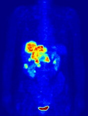
Image by Jens Langner
Performing PET scans immediately after chemotherapy may reveal which Hodgkin lymphoma (HL) patients need radiotherapy (RT).
A study published in NEJM showed similar rates of progression-free survival in HL patients who werePET-negative after chemotherapy, whether they received subsequent RT or not.
However, the investigators said longer follow-up is needed to determine if eliminating RT in PET-negative patients will lead to fewer late effects and improved overall survival.
The 602 patients who agreed to take part in this trial, known as RAPID, had a PET scan performed after chemotherapy. Patients who tested positive received RT.
Those who tested negative were divided into 2 groups. One group of 211 patients received no further treatment, and the other group of 209 patients had the standard RT.
At a median of 60 months of follow-up, the proportion of patients who were alive and disease-free was 94.6% in the RT group and 90.8% in the group that hadn’t received further treatment.
Eight patients in the RT group progressed, and 8 died (3 with disease progression, 1 of whom died from HL). Five of the deaths occurred in patients who did not ultimately receive RT.
In the untreated group, 20 patients progressed, and 4 patients died (2 with disease progression and none from HL).
“This research is an important step forward,” said study author John Radford, of The University of Manchester and The Christie NHS Foundation Trust in the UK.
“The results of RAPID show that, in early stage Hodgkin lymphoma, radiotherapy after initial chemotherapy marginally reduces the recurrence rate, but this is bought at the expense of exposing to radiation all patients with negative PET findings, most of whom are already cured.” ![]()
Forgoing radiotherapy after chemo in early Hodgkin’s is close call
For patients with early-stage Hodgkin’s lymphoma, forgoing radiotherapy after three cycles of chemotherapy when PET scans show negative findings – known as a PET-directed or response-adapted approach – was not found to be noninferior to routine consolidation radiotherapy in extending progression-free survival, according to a report published online April 23 in the New England Journal of Medicine.
The PET-directed technique is intended to spare the estimated 9 out of 10 patients who are cured by the chemotherapy from having to receive unnecessary radiotherapy, which carries late toxic effects such as secondary cancers and premature cardiovascular disease. To determine whether this approach caused an unacceptable increase in the relapse rate, researchers performed a randomized, controlled, phase III noninferiority trial in 602 previously untreated patients aged 16-75 years (median age, 34 years) who had stage IA or IIA Hodgkin’s lymphoma with no mediastinal bulk and no night sweats, unexplained fever, or weight loss, reported Dr. John Radford of the Institute of Cancer Sciences, University of Manchester (England), and his associates.
The study participants, enrolled and treated at 94 medical centers across the United Kingdom, had three cycles of doxorubicin, bleomycin, vinblastine, and dacarbazine therapy and then underwent PET scanning. The 420 who had negative findings on PET then were randomly assigned to receive either 30 Gy of involved-field radiotherapy (209 patients) or no further treatment (211 patients).
After a median of 62 months of follow-up, both groups had excellent outcomes. Three-year progression-free survival was 94.6% with radiotherapy and 90.8% without it; overall survival was 97.1% with radiotherapy and 99.0% without it. However, the modest advantage conveyed by radiotherapy in the 3-year progression-free survival rate – 3.8 percentage points in the intention-to-treat analysis and 6.3 percentage points in the per-protocol analysis – was enough to negate a finding of noninferiority for forgoing radiotherapy, the investigators wrote (N. Engl. J. Med. 2015 April 23 [doi:10.1056/NEJMoa1408648]).
It is important to note that this marginal survival advantage “is bought at the expense of exposing all patients to radiation, most of whom will not benefit and some of whom will be harmed,” Dr. Radford and his associates wrote.
This report addressed medium-term outcomes. Continued follow-up in this ongoing study will determine whether the response-adapted approach leads to fewer second cancers, less cardiovascular disease, and superior survival in the long term, they added.
The report by Radford et al. raises important questions. Is a 4 percentage-point difference in the rate of relapse worth the added risks of radiation therapy? And should 100 patients be exposed to radiation to keep 4 from relapsing, with no evidence of long-term benefit?
When patients are fully informed of the risks and benefits, some will choose the additional radiotherapy because they cannot abide any increase in the medium-term risk of relapse. But others will elect to minimize their long-term risks and trust that they are among the 90% of patients who have been cured by chemotherapy and can forgo radiation.
Dr. Dan L. Longo is a deputy editor of NEJM and professor of medicine at Harvard and the Dana-Farber Cancer Institute, both in Boston. He reported having no relevant financial disclosures. Dr. James O. Armitage is in the division of hematology-oncology at the University of Nebraska Medical Center, Omaha. He reported receiving personal fees from Celgene, Conatus, Coherus, GlaxoSmithKline, Roche, Spectrum, TESARO, and Ziopharm. Dr. Longo and Dr. Armitage made these remarks in an editorial accompanying Dr. Radford’s report (N. Engl. J. Med. 2015 April 23 [doi:10.1056/NEJMe1502888]).
For patients with early-stage Hodgkin’s lymphoma, forgoing radiotherapy after three cycles of chemotherapy when PET scans show negative findings – known as a PET-directed or response-adapted approach – was not found to be noninferior to routine consolidation radiotherapy in extending progression-free survival, according to a report published online April 23 in the New England Journal of Medicine.
The PET-directed technique is intended to spare the estimated 9 out of 10 patients who are cured by the chemotherapy from having to receive unnecessary radiotherapy, which carries late toxic effects such as secondary cancers and premature cardiovascular disease. To determine whether this approach caused an unacceptable increase in the relapse rate, researchers performed a randomized, controlled, phase III noninferiority trial in 602 previously untreated patients aged 16-75 years (median age, 34 years) who had stage IA or IIA Hodgkin’s lymphoma with no mediastinal bulk and no night sweats, unexplained fever, or weight loss, reported Dr. John Radford of the Institute of Cancer Sciences, University of Manchester (England), and his associates.
The study participants, enrolled and treated at 94 medical centers across the United Kingdom, had three cycles of doxorubicin, bleomycin, vinblastine, and dacarbazine therapy and then underwent PET scanning. The 420 who had negative findings on PET then were randomly assigned to receive either 30 Gy of involved-field radiotherapy (209 patients) or no further treatment (211 patients).
After a median of 62 months of follow-up, both groups had excellent outcomes. Three-year progression-free survival was 94.6% with radiotherapy and 90.8% without it; overall survival was 97.1% with radiotherapy and 99.0% without it. However, the modest advantage conveyed by radiotherapy in the 3-year progression-free survival rate – 3.8 percentage points in the intention-to-treat analysis and 6.3 percentage points in the per-protocol analysis – was enough to negate a finding of noninferiority for forgoing radiotherapy, the investigators wrote (N. Engl. J. Med. 2015 April 23 [doi:10.1056/NEJMoa1408648]).
It is important to note that this marginal survival advantage “is bought at the expense of exposing all patients to radiation, most of whom will not benefit and some of whom will be harmed,” Dr. Radford and his associates wrote.
This report addressed medium-term outcomes. Continued follow-up in this ongoing study will determine whether the response-adapted approach leads to fewer second cancers, less cardiovascular disease, and superior survival in the long term, they added.
Key clinical point: Foregoing radiotherapy after chemotherapy was not found “noninferior” to undergoing radiotherapy for early-stage Hodgkin’s lymphoma.
Major finding: Three-year progression-free survival was 94.6% with radiotherapy and 90.8% without it, and overall survival was 97.1% with radiotherapy and 99.0% without it.
Data source: A randomized, controlled phase III noninferiority trial involving 420 adolescents and adults at 94 medical centers in the United Kingdom followed for a median of 5 years.
Disclosures: This study was supported by Leukemia and Lymphoma Research, the Lymphoma Research Trust, Teenage Cancer Trust, and the U.K. Department of Health; no commercial support was provided. Dr. Radford reported having no relevant financial disclosures; two of his associates reported ties to numerous industry sources.
Check sweat glands, hair follicles in mycosis fungoides
SAN FRANCISCO – Check for syringotropism and folliculotropism in biopsies when managing mycosis fungoides, based on data from an ongoing observational, prospective study at Thomas Jefferson University in Philadelphia.
The presence of syringotropism and folliculotropism indicates the need for more aggressive treatment, according to lead investigator Dr. Joya Sahu, of the department of dermatology at the university.
Mycosis fungoides – the most common form of cutaneous T-cell lymphoma – is usually thought to favor the epidermis, but investigators at Thomas Jefferson University have found that it often works its tentacles deeper into the skin to attack hair follicles (folliculotropism) or eccrine glands (syringotropism), Dr. Sahu said at the annual meeting of the American Academy of Dermatology.
The researchers checked biopsy samples to see how common those variants were in 34 new patients with mycosis fungoides (most with stage 1 disease). Overall, 18 (52.9%) had folliculotropism, 22 (64.7%) had syringotropism, and 15 (44.1%) had both.
Not surprisingly, deeper penetration indicated worse disease, Dr. Sahu said. On the modified Severity Weighted Assessment tool (mSWAT) – a measure of surface area involvement and lesion severity – the mean scores were 57.51 in patients with folliculotropism, 59.4 in patients with syringotropism, and 66.4 in patients with both. The higher mSWAT scores also correlated with more severe pruritus and the likelihood that the patient had tried four or more treatments. By contrast, the nine patients without folliculotropism or syringotropism, who had a mean mSWAT score of 16.85, had tried only one or two treatments.
Almost all of the cases presented classically; two had head and neck lesions or other signs of folliculotropic disease, and both of these patients had folliculotropism and syringotropism on biopsy. None of the patients had a syringotropic presentation.
“The majority of patients studied exhibited either folliculotropism or syringotropism, implying greater prevalence,” Dr. Sahu said. “These presentations also have findings indicative of more severe disease. We propose that histopathology reports on patients with suspected [mycosis fungoides] should document the presence of folliculotropism and syringotropism as they may aid in diagnosis and in predicting severity and progression risk,” she noted.
She cautioned, however, that her clinic is a tertiary referral center, and as such might see patients with more severe disease, compared with other clinics.
The patients were otherwise typical of the mycosis fungoides population, she said. About two-thirds were men, and the average age was 63 years.
Dr. Sahu said she had no relevant financial conflicts of interest.
AT THE AAD ANNUAL MEETING
Key clinical point: Routinely check mycosis fungoides patients for sweat gland and hair follicle involvement.
Major finding: Among 34mycosis fungoides patients with mostly stage 1 disease, 18 (52.9%) had folliculotropism, 22 (64.7%) had syringotropism, and 15 (44.1%) had both on biopsy.
Data source: An observational, prospective study of 34 adults with mycosis fungoides.
Disclosures: The lead investigator declared no relevant financial conflicts.
Discovery could aid treatment of leukemia, lymphoma

Photo courtesy of IRCM
Researchers say they have uncovered a mechanism that could aid the development of therapies for lymphomas and leukemias.
The group’s research shed new light on a mechanism affecting activation-induced deaminase (AID), an enzyme that has proven crucial for immune response.
Javier Di Noia, PhD, of Institut de Recherches Cliniques de Montreal (IRCM) in Quebec, Canada, and his colleagues described this mechanism in The Journal of Experimental Medicine.
Dr Di Noia noted that, although AID is crucial for an efficient antibody response, high levels of the enzyme can have harmful effects and lead to cancer-causing mutations.
“The objective is to find the perfect level of AID activity to maximize the protection it provides to the body while reducing the risk of damage it can cause to cells,” he said.
Dr Di Noia and his colleagues previously found that heat-shock protein 90 (Hsp90) maintains the levels of AID by stabilizing it while it is still immature. In fact, they discovered that inhibiting Hsp90 significantly reduces the levels of AID in the cell.
“Through this new study, we identified another mechanism, controlled by the protein eEF1a [elongation factor eukaryotic elongation factor 1 α], that has the opposite effect,” said Stephen P. Methot, a PhD student in Dr Di Noia’s lab.
“The protein eEF1a retains AID in the cell’s cytoplasm, away from the genome. However, unlike Hsp90, it maintains AID in a ready-to-act state. We discovered that blocking the interaction between AID and eEF1a helps AID access the cell nucleus and thereby boosts AID activity. As a result, this could increase immune response and help fight infections, for instance.”
“We found the eEF1a mechanism is necessary to restrict AID activity in the cell. It acts as a buffer by allowing the cell to accumulate enough AID to be efficient but limits its activity to prevent the oncogenic or toxic effects that could result if too much AID is in continuous contact with the genome.”
The researchers also identified 2 existing drugs that can act on the eEF1a mechanism to release AID into the cell. The team said these drugs could potentially be used to boost AID activity and, thus, immune responses.
“With this discovery, we now understand mechanisms that can both reduce and increase the activity of AID by targeting different proteins,” Dr Di Noia said.
“This knowledge could eventually lead to new treatments to boost the immune system and help our aging population fight influenza, for example, as AID activity in our cells decreases with age. On the other hand, therapies could also be developed to lower toxic levels of AID in certain cancers such as B-cell lymphoma and leukemia.” ![]()
Photo courtesy of IRCM
Researchers say they have uncovered a mechanism that could aid the development of therapies for lymphomas and leukemias.
The group’s research shed new light on a mechanism affecting activation-induced deaminase (AID), an enzyme that has proven crucial for immune response.
Javier Di Noia, PhD, of Institut de Recherches Cliniques de Montreal (IRCM) in Quebec, Canada, and his colleagues described this mechanism in The Journal of Experimental Medicine.
Dr Di Noia noted that, although AID is crucial for an efficient antibody response, high levels of the enzyme can have harmful effects and lead to cancer-causing mutations.
“The objective is to find the perfect level of AID activity to maximize the protection it provides to the body while reducing the risk of damage it can cause to cells,” he said.
Dr Di Noia and his colleagues previously found that heat-shock protein 90 (Hsp90) maintains the levels of AID by stabilizing it while it is still immature. In fact, they discovered that inhibiting Hsp90 significantly reduces the levels of AID in the cell.
“Through this new study, we identified another mechanism, controlled by the protein eEF1a [elongation factor eukaryotic elongation factor 1 α], that has the opposite effect,” said Stephen P. Methot, a PhD student in Dr Di Noia’s lab.
“The protein eEF1a retains AID in the cell’s cytoplasm, away from the genome. However, unlike Hsp90, it maintains AID in a ready-to-act state. We discovered that blocking the interaction between AID and eEF1a helps AID access the cell nucleus and thereby boosts AID activity. As a result, this could increase immune response and help fight infections, for instance.”
“We found the eEF1a mechanism is necessary to restrict AID activity in the cell. It acts as a buffer by allowing the cell to accumulate enough AID to be efficient but limits its activity to prevent the oncogenic or toxic effects that could result if too much AID is in continuous contact with the genome.”
The researchers also identified 2 existing drugs that can act on the eEF1a mechanism to release AID into the cell. The team said these drugs could potentially be used to boost AID activity and, thus, immune responses.
“With this discovery, we now understand mechanisms that can both reduce and increase the activity of AID by targeting different proteins,” Dr Di Noia said.
“This knowledge could eventually lead to new treatments to boost the immune system and help our aging population fight influenza, for example, as AID activity in our cells decreases with age. On the other hand, therapies could also be developed to lower toxic levels of AID in certain cancers such as B-cell lymphoma and leukemia.”
Epigenomic findings may help predict relapse in DLBCL

Photo by Rhoda Baer
Epigenomic heterogeneity at diagnosis may predict relapse in diffuse large B-cell lymphoma (DLBCL), according to research published in Nature Communications.
Investigators made this connection by reviewing biopsies taken from DLBCL patients before and after treatment.
The epigenome in these patients’ cancer cells changed greatly after treatment, and the global epigenome of pretreatment biopsies was substantially different in patients who relapsed and those who did not. There was more cell-to-cell heterogeneity in patients who relapsed.
“This is the first study I know of in cancer that looks at changes in the epigenome before and after treatment, and what we found could ultimately make traditional treatments much more effective,” said study author Olivier Elemento, PhD, of Weill Cornell Medical College in New York, New York.
To uncover the role of epigenetic involvement in DLBCL, Dr Elemento and his colleagues analyzed banked biopsies from patients. In each sample set, the investigators looked at sites in the epigenome where a methyl group was added or removed after DLBCL recurred.
They found a change in methylation that occurred between 39,808 and 1,035,960 specific methylation sites, depending on the sample. In addition, they identified between 78 and 13,162 differently methylated regions in the epigenome in relapsed disease.
“These are massive changes, given that the epigenome has 20 million methylation sites,” Dr Elemento said. “Our study shows that, in some cases, up to one-twentieth of the entire epigenome is changed after treatment. There are many more epigenetic changes than there are altered genes in DLBCL.”
“Once you have changes in methylation, the end result is an imbalanced expression of proteins,” added Giorgio Inghirami, MD, also of Weill Cornell.
“The tumor after chemotherapy is not the same as the tumor before treatment. This why it is so critical to have biopsies before any treatment of [primary or relapsed] lesions.”
The investigators hope this work will ultimately allow clinicians and researchers to predict treatment resistance in individual patients.