User login
FDA grants priority review to CAR T-cell therapy for DLBCL
The Food and Drug Administration has granted a priority review for the CAR T-cell therapy tisagenlecleucel suspension, formerly CTL019, for the treatment of adult patients with relapsed or refractory diffuse large B-cell lymphoma who are ineligible for or relapsed after autologous stem cell transplant.

The current application is based on a 6-month primary analysis from the single-arm, phase 2 JULIET clinical trial in adult patients with relapsed or refractory diffuse large B-cell lymphoma. According to results presented at ASH 2017, among 81 patients followed for at least 3 months before data cutoff, best overall response rate was 53%, and 40% had a complete response. Cytokine release syndrome (all grades) occurred in 58% of infused patients. Other grade 3 or 4 adverse events included neurologic toxicities, cytopenias lasting more than 28 days, infections, and febrile neutropenia.
Tisagenlecleucel suspension is marketed as Kymriah by Novartis.
The Food and Drug Administration has granted a priority review for the CAR T-cell therapy tisagenlecleucel suspension, formerly CTL019, for the treatment of adult patients with relapsed or refractory diffuse large B-cell lymphoma who are ineligible for or relapsed after autologous stem cell transplant.
The current application is based on a 6-month primary analysis from the single-arm, phase 2 JULIET clinical trial in adult patients with relapsed or refractory diffuse large B-cell lymphoma. According to results presented at ASH 2017, among 81 patients followed for at least 3 months before data cutoff, best overall response rate was 53%, and 40% had a complete response. Cytokine release syndrome (all grades) occurred in 58% of infused patients. Other grade 3 or 4 adverse events included neurologic toxicities, cytopenias lasting more than 28 days, infections, and febrile neutropenia.
Tisagenlecleucel suspension is marketed as Kymriah by Novartis.
Drug’s label updated to include risk of allergic reactions

The US Food and Drug Administration (FDA) and Tesaro, Inc., have updated the prescribing information for Varubi® (rolapitant) injectable emulsion to include a new warning about the risk of allergic reactions.
Varubi injectable emulsion is a substance P/neurokinin receptor antagonist approved to prevent delayed nausea and vomiting associated with chemotherapy in adults.
Since Varubi injectable emulsion gained FDA approval, there have been reports of anaphylaxis, anaphylactic shock, and other serious hypersensitivity reactions to the drug, some of which required hospitalization.
Now, the labeling for Varubi injectable emulsion has been changed to include information about these events. The changes include modifications to the CONTRAINDICATIONS, WARNINGS and PRECAUTIONS, and ADVERSE REACTIONS sections of the label.
Since Varubi injectable emulsion was introduced to the US market in late November 2017, at least 7000 doses of the drug have been administered to patients receiving emetogenic chemotherapy in the US, according to Tesaro.
Anaphylaxis, anaphylactic shock, and other serious hypersensitivity reactions have occurred during or soon after the infusion of Varubi. Most reactions have occurred within the first few minutes of administration.
The FDA has advised that patients who are hypersensitive to any component of Varubi injectable emulsion (including soybean oil) do not receive the drug. And patients with known allergies to legumes or other related allergens should be monitored closely.
The FDA said healthcare professionals should be vigilant for signs of hypersensitivity or anaphylaxis in all patients receiving Varubi injectable emulsion, both during administration and afterward.
Symptoms of anaphylaxis can include wheezing, difficulty breathing, swelling of the face or throat, hives, flushing, itching, abdominal cramping, abdominal pain, vomiting, back pain, chest pain, hypotension, and shock.
If anaphylaxis or any other serious hypersensitivity/infusion reaction occurs, Varubi injectable emulsion should be stopped immediately and permanently. The patient should receive appropriate medical management, including epinephrine and/or antihistamines.
To ensure patients and healthcare professionals are aware of the label update to Varubi injectable emulsion, Tesaro has issued a Dear Healthcare Professional letter. In addition, the updated prescribing information has been posted on the Varubi website.
For any questions about the use of Varubi injectable emulsion or to report adverse events related to the drug, contact Tesaro’s medical information department at 1-844-4-TESARO (1-844-483-7276).
Adverse events related to Varubi should also be reported to the FDA’s MedWatch Safety Information and Adverse Event Reporting Program. ![]()
Generic bortezomib available in US
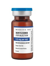
Fresenius Kabi has introduced its generic version of Velcade, Bortezomib for Injection, to the US market.
This is the first intravenous alternative to Velcade available in the US.
Bortezomib for Injection is available as a single dose vial containing 3.5 mg of lyophilized powder.
The product is approved to treat patients with multiple myeloma and patients with mantle cell lymphoma who have received at least 1 prior therapy.
For details, see the prescribing information for Bortezomib for Injection.
Velcade is a registered trademark of Millennium Pharmaceuticals, Inc. ![]()
Survival differences among AYAs with blood cancers

A new report has revealed differences in survival among adolescents and young adults (AYAs) with hematologic malignancies.
The report includes information on AYAs—ages 15 to 39—living in Los Angeles County who were diagnosed with common cancers between 1988 and 2014.
The data showed differences in 5-year survival rates according to sex, race, age, and socioeconomic status (SES).
For example, lymphoma survival rates were lower for males, African Americans (AAs), older AYAs, and patients with low socioeconomic status (SES).
For AYAs with leukemias, there was no survival difference according to sex, but AAs had worse survival than patients of other races. And the impact of age and SES varied according to leukemia type.
“Cancer survival data are poorly understood for 15- to 39-year-olds,” noted Amie Hwang, PhD, of the University of Southern California Keck School of Medicine in Los Angeles.
That is why she and her colleagues created the report, “Cancer in Los Angeles County: Survival Among Adolescents and Young Adults 1988-2014.”
According to the authors, this is the first report to break down cancer survival rates for AYAs into segments on race/ethnicity, sex, age group, SES, and cancer stage.
Survival data for patients with hematologic malignancies were as follows.
Acute lymphoblastic leukemia
There were 1137 cases of acute lymphoblastic leukemia in the AYA population in Los Angeles County during the period studied. This included 752 males and 385 females.
Five-year survival was similar between males (43%) and females (41%).
Younger AYAs had better survival than older AYAs (48% for ages 15-24, 35% for ages 25-34, and 32% for ages 35-39).
Survival was highest among non-Latino whites (NLWs, 56%), followed by Asian/Pacific Islanders (APIs, 52%), patients of other/unknown races (51%), Latino whites (LWs, 38%), and AAs (29%).
Survival declined with SES (55% for high, 42% for middle, and 36% for low SES).
Acute myeloid leukemia
There were 1195 cases of acute myeloid leukemia—641 males and 554 females.
Five-year survival was similar for males (40%) and females (43%) as well as for the different age groups (45% for ages 15-24 vs 40% for the older age groups).
Survival was highest among NLWs (44%), followed by LWs (43%), APIs (40%), other/unknown (33%), and AAs (25%).
Survival declined somewhat with SES (49% for high, 39% for middle, and 41% for low SES).
Chronic myeloid leukemia
There were 655 cases of chronic myeloid leukemia—408 males and 247 females.
Five-year survival was similar for males (70%) and females (71%), but it was slightly higher for older AYAs (69% for ages 15-24, 68% for ages 25-34, and 76% for ages 35-39).
Survival was highest among patients in the “other/unknown” race category (76%), followed by LWs (73%), NLWs/APIs (both 72%), and AAs (57%).
Survival declined somewhat with SES (76% for high, 67% for middle, and 68% for low SES).
Hodgkin lymphoma
There were 2993 AYAs diagnosed with Hodgkin lymphoma—1553 males and 1440 females.
The 5-year survival rate was higher in females (93%) than males (86%) and in younger AYAs (93% for ages 15-24, 89% for ages 25-34, and 85% for ages 35-39).
Survival was highest among patients in the “other/unknown” race category (96%), followed by APIs/NLWs (both 91%), LWs (88%), and AAs (83%).
Survival declined with SES (95% for high, 89% for middle, and 83% for low SES).
And survival was lower for patients with advanced-stage disease (93% localized, 94% regional, and 83% distant).
Non-Hodkgin lymphoma
There were 4485 AYAs diagnosed with non-Hodgkin lymphoma during the study period—3064 males and 1421 females.
The 5-year survival rate was higher in females (75%) than males (46%) and in younger AYAs (69% for ages 15-24, 51% for ages 25-34, and 52% for ages 35-39).
Survival was highest among patients in the “other/unknown” race category (88%), followed by APIs (68%), LWs/NLWs (both 53%), and AAs (50%).
Survival declined with SES (68% for high, 54% for middle, and 45% for low SES).
And survival was lower for patients with advanced-stage disease (61% localized, 66% regional, and 46% distant).
“Adolescents and young adults go to the doctor less often because they have this superhero mentality, like they’re invincible,” said author Dennis Deapen, DrPH, of the University of Southern California Keck School of Medicine.
“Once they do go to a health professional, their cancer diagnosis can be delayed because cancer isn’t the first concern doctors have for this age group. It comes as no surprise that patients diagnosed with late-stage cancer have reduced survival rates.” ![]()
Marine animals aid development of cytotoxicity assay

Researchers have looked to deep-sea creatures with the goal of creating a better cytotoxicity assay.
The team harnessed the power of enzymes responsible for marine animal bioluminescence to create the “Matador assay,” which can be used to determine whether cellular and immune-therapeutic agents are actually killing target cells.
The researchers said the Matador assay is quick and simple as well as “highly sensitive,” with the ability to detect cytotoxicity induced by several types of therapies.
Preet M. Chaudhary, MD, PhD, of the University of Southern California Keck School of Medicine in Los Angeles, and his colleagues described the assay in Scientific Reports.
“One of the most promising areas in cancer research is immunotherapy. . .,” Dr Chaudhary said. “It is also one of the most difficult because the methods for testing immunotherapies are not ideal.”
“Radioactive chromium release assay is the gold standard for testing whether an immunotherapy kills cancer cells. This method is expensive, complicated, and requires special disposal practices. Other available methods also suffer from limitations and don’t allow scientists to rapidly screen immunotherapeutic agents to find the best candidates.”
Dr Chaudhary and his colleagues set out to develop a simple, precise, and inexpensive cytotoxicity assay based on marine animal luciferases, the enzymes responsible for bioluminescence.
The team used a group of small crustaceans and deep-sea shrimp, which were selected for their bright bioluminescence. Their luciferases became the basis of the Matador assay.
Engineered to get trapped inside cells, the luciferases leak out of cells when they die, causing a visible glow. The level of luminescence can then be measured with a luminometer.
To test the Matador assay’s effectiveness at measuring cell death, the researchers used several types of cancer cells, including chronic myelogenous leukemia, acute myelogenous leukemia, Burkitt lymphoma, and solid tumor cells.
The team treated these cells with a variety of therapies, including chimeric antigen receptor (CAR) T cells, bispecific T-cell engagers, monoclonal antibodies, and natural killer cells.
Results showed the Matador assay could detect the death of a single cell, a level of sensitivity superior to that of existing cytotoxicity assays.
The researchers also pointed out that the Matador assay is fast, inexpensive, and can be performed in a 384-well plate format, saving time and reagents.
“In our hands, the Matador assay can detect cell death in as little as 30 minutes, which can ultimately translate to more expedient treatments for patients getting cellular immunotherapies such as CAR T cells,” Dr Chaudhary said.
In fact, Dr Chaudhary’s lab has developed more than 75 cancer cell lines expressing the marine luciferases and used them with the Matador assay to develop next-generation CAR T cells.
Dr Chaudhary believes the Matador assay has many potential applications in biomedical research and cellular therapy manufacturing.
“It could potentially play a role in screening other types of anticancer agents or even measuring environmental toxins,” he said. ![]()
Overcoming resistance to ibrutinib in CLL

New research appears to explain why ibrutinib may be less effective in certain patients with chronic lymphocytic leukemia (CLL).
It seems the Bruton’s tyrosine kinase (BTK) inhibitor has a diminished capacity to delocalize and kill tumor cells expressing an adhesive protein called CD49d.
But combining ibrutinib with drugs that block CD49d activation could prevent CLL cells from sheltering in lymphoid organs.
Valter Gattei, MD, of CRO Aviano National Cancer Institute in Aviano, Italy, and his colleagues reported these findings in the Journal of Experimental Medicine.
The team noted that CD49d, the α chain of the CD49d/CD29 integrin heterodimer very late antigen 4 (VLA-4), is expressed in about 40% of CLL cases.
These patients tend to have poorer outcomes than patients who do not express CD49d, but the role of VLA-4 in CLL was unclear.
With this study, researchers found that B-cell receptor (BCR) signaling can activate VLA-4 in CD49d-expressing CLL cells, thereby enhancing the cells’ adhesiveness.
Even though ibrutinib treatment impaired BCR signaling in these cells, it was unable to fully prevent the pathway from activating VLA-4 and enhancing cell adhesion.
The researchers analyzed 3 cohorts of CLL patients and found that patients expressing higher levels of CD49d had reduced responses to ibrutinib.
The BTK inhibitor appeared less able to displace tumor cells from lymph nodes into the blood, resulting in decreased lymph node shrinkage and shorter progression-free survival times.
“Our results suggest that VLA-4-expressing CLL cells residing in the secondary lymphoid organs can receive BCR-mediated stimuli that can activate VLA-4 even in the presence of ibrutinib,” said study author Antonella Zucchetto, ScD, also of CRO Aviano National Cancer Institute.
“This activation leads to enhanced retention of VLA-4-positive CLL cells in tissue sites, thereby affecting patient outcome.”
Fortunately, the researchers found a way around this obstacle. Inhibiting BTK and phosphatidylinositide 3-kinase (PI3K) simultaneously completely blocked VLA-4 activation in CLL cells.
The researchers treated CLL cells with ibrutinib, the PI3K inhibitor idelalisib, or a combination of both.
Neither drug alone was able to fully block anti-IgM-induced VLA-4 activation. However, the team found that simultaneous inhibition of BTK and PI3K “completely abolished the integrin response to BCR triggering.”
The researchers also added idelalisib to ibrutinib-treated CLL cells (collected from patients at day 30 on ibrutinib) and observed a complete upset of anti-IgM–induced VLA-4 activation.
“Our data suggest that evaluation of CD49d expression in patients initiating ibrutinib therapy may identify those cases that would benefit from combination therapy approaches designed to completely block VLA-4 activation and VLA-4-mediated retention of leukemic cells in protective tissue compartments,” Dr Gattei said. ![]()
Denosumab indication now includes multiple myeloma, Amgen announces
The Food and Drug Administration has expanded the indications for denosumab (Xgeva), previously indicated for the prevention of skeletal-related events in patients with bone metastases from solid tumors, to include patients with multiple myeloma, according to a press release from Amgen, the manufacturer of Xgeva.
“Up to 40% of [multiple myeloma] patients remain untreated for the prevention of bone complications, and the percentage is highest among patients with renal impairment at the time of diagnosis. Denosumab, which is not cleared through the kidneys, offers multiple myeloma patients bone protection with a convenient subcutaneous administration, providing patients with a novel treatment option,” Dr. Noopur Raje, director of the Center for Multiple Myeloma, Massachusetts General Hospital Cancer Center, Boston, said in the press release.
Adverse events in multiple myeloma patients were broadly similar to the known safety profile of denosumab. The most common adverse events were diarrhea, nausea, anemia, back pain, thrombocytopenia, peripheral edema, hypocalcemia, upper respiratory tract infection, rash, and headache. The most common adverse event resulting in discontinuation of treatment was osteonecrosis of the jaw.
Find the full press release on the Amgen website.
Continue to opt for HDT/ASCT for multiple myeloma
High-dose therapy with melphalan followed by autologous stem cell transplant (HDT/ASCT) is still the best option for multiple myeloma even after almost 2 decades with newer and highly effective induction agents, according to a recent systematic review and two meta-analyses.
Given the “unprecedented efficacy” of “modern induction therapy with immunomodulatory drugs and proteasome inhibitors (also called ‘novel agents’),” investigators “have sought to reevaluate the role of HDT/ASCT,” wrote Binod Dhakal, MD, of the Medical College of Wisconsin, and his colleagues. The report is in JAMA Oncology.
To solve the issue, they analyzed five randomized controlled trials conducted since 2000 and concluded that HDT/ASCT is still the preferred treatment approach.
Despite a lack of demonstrable overall survival benefit, there is a significant progression-free survival (PFS) benefit, low treatment-related mortality, and potential high minimal residual disease-negative rates conferred by HDT/ASCT in newly-diagnosed multiple myeloma, the researchers noted.
The combined odds for complete response were 1.27 (95% confidence interval, 0.97-1.65, P = .07) with HDT/ASCT, compared with standard-dose therapy (SDT). The combined hazard ratio (HR) for PFS was 0.55 (95% CI, 0.41-0.7, P less than .001) and 0.76 for overall survival (95% CI, 0.42-1.36, P = .20) in favor of HDT.
PFS was best with tandem HDT/ASCT (HR, 0.49, 95% CI, 0.37-0.65) followed by single HDT/ASCT with bortezomib, lenalidomide, and dexamethasone consolidation (HR, 0.53, 95% CI, 0.37-0.76) and single HDT/ASCT alone (HR, 0.68, 95% CI, 0.53-0.87), compared with SDT. However, none of the HDT/ASCT approaches had a significant impact on overall survival.
Meanwhile, treatment-related mortality with HDT/ASCT was minimal, at less than 1%.
“The achievement of high [minimal residual disease] rates with HDT/ASCT may render this approach the ideal platform for testing novel approaches (e.g., immunotherapy) aiming at disease eradication and cures,” the researchers wrote.
The researchers reported relationships with a number of companies, including Takeda, Celgene, and Amgen, that make novel induction agents.
SOURCE: Dhakal B et al. JAMA Oncol. 2018 Jan 4. doi: 10.1001/jamaoncol.2017.4600.
FROM JAMA ONCOLOGY
Key clinical point:
Major finding: The combined odds for complete response were 1.27 (95% CI 0.97-1.65, P = .07) with HDT/ASCT, compared with standard-dose therapy (SDT).
Study details: A systematic review and two meta-analyses examining five phase 3 clinical trials reported since 2000.
Disclosures: The researchers reported relationships with a number of companies, including Takeda, Celgene, and Amgen, that make novel induction agents.
Source: Dhakal B et al. JAMA Oncol. 2018 Jan 4. doi: 10.1001/jamaoncol.2017.4600.
How to manage cytokine release syndrome
ATLANTA – Closely monitoring for cytokine release syndrome (CRS) and starting anticytokine therapy early can prevent life-threatening organ toxicities in recipients of chimeric antigen receptor (CAR) T-cell therapy, according to Daniel W. Lee III, MD.
There’s no evidence that early anticytokine therapy impairs antitumor response, he noted. “If you wait to give anticytokine therapy until a patient is intubated, it’s too late,” Dr. Lee said at the annual meeting of the American Society of Hematology. “If you wait until a patient has been hypotensive for days, it’s probably too late. If you intervene earlier, you can avoid intubation.”
Treating CRS is a clinical decision that shouldn’t hinge on cytokine levels, according to Dr. Lee. He and his colleagues base treatment on their revised severity grading assessment, which spans mild, moderate, severe, and life-threatening syndromes (Blood. 2014;124:188-95).
Using a consistent CRS severity grading system enables physicians to treat rationally across trials and CAR T-cell therapies, he said. His system defines grade 1 CRS as flu-like symptoms and fever up to 41.5 degrees Celsius. Patients with grade 1 CRS should receive antipyretics and analgesia as needed and should be monitored closely for infections and fluid imbalances, Dr. Lee said.
Hypotension signifies progression beyond grade 1 CRS. Affected patients should receive no more than two to three IV fluid boluses and then should “quickly move on to vasopressors,” such as norepinephrine, he emphasized.
His and his team implemented this important change after one of their patients, a 14-year-old boy with severe hypotensive grade 4 CRS, died of a cardiovascular event after receiving multiple IV fluid boluses. “We had not appreciated the extent of this patient’s ventricular strain,” Dr. Lee said. The patient was heavily pretreated and had an “extremely high disease burden” (more than 99% marrow involvement, hepatosplenomegaly, and pronounced blastic leukocytosis), which increased his risk of severe CRS, he noted. “Admittedly, we pushed the envelope a little bit, and we learned you should start anticytokine therapies much earlier. Earlier seems to be better, although we do not yet know if prophylactic tocilizumab or corticosteroids can prevent CRS symptoms before they start.”
For hypotensive patients on pressors, Dr. Lee recommends vigilant supportive care and daily echocardiograms to monitor ejection fraction and ventricular wall mobility. His system defines grade 2 CRS as hypotension responsive to one low-dose pressor or to fluid therapy and hypoxia responsive to less than 40% oxygen therapy. Patients with grade 2 CRS who also have comorbidities should receive tocilizumab – with or without corticosteroids – both of which “remain the standard of care for managing CRS,” he said. Severe CRS often stems from supraphysiologic release of interleukin 6, which induces not only classic IL-6 signaling but also proinflammatory trans-signaling across many cell types. Tocilizumab reverses this process by binding and blocking the IL-6 receptor, Dr. Lee noted.
Patients with grade 3 CRS have hypotension requiring multiple or high-dose pressors and hypoxia requiring at least 40% oxygen therapy. These patients have grade 3 organ toxicity and grade 4 transaminitis, Dr. Lee said. Even if they lack comorbidities, they need vigilant supportive care, tocilizumab, and possibly corticosteroids, he added. The ultimate goal is to avoid grade 4 CRS, he said, which involves grade 4 organ toxicity, requires mechanical ventilation, and yields a poor prognosis despite vigilant supportive care, tocilizumab, and corticosteroids.
Dr. Lee reported having no relevant conflicts of interest.
EXPERT ANALYSIS FROM ASH 2017
DLBCL survivors at greater risk of autoimmune, infectious diseases
ATLANTA—A population-based study indicates that, compared to other cancer survivors, patients who survive diffuse large B-cell lymphoma (DLBCL) have an increased risk of autoimmune and infectious diseases.
For example, investigators found the risk of being diagnosed with impaired humoral immunity was 16.2 times higher in female DLBCL survivors than in breast cancer survivors, 14.8 times higher in male DLBCL survivors than in prostate cancer survivors, and 12.5 times higher in all DLBCL survivors than in survivors of head and neck cancer.
“Most of the treatments that we give for lymphoma have profound effects on the immune system, either directly or indirectly, including many of the T-cell-directed therapies,” said Tanaya Shree, MD, PhD, of Stanford University Medical Center in California.
“There have been studies on many of the effects suffered by lymphoma survivors, but very little is known about their immune health.”
Dr Shree and her colleagues undertook this study to determine how the immune system fares in lymphoma survivors. The investigators limited their analysis to survivors of DLBCL.
Dr Shree presented the findings at the 2017 ASH Annual Meeting (abstract 198*).
Study design
Investigators pulled data from the California Cancer Registry for patients with DLBCL as their first primary cancer diagnosed between 1991 and 2012. Patients had to be 18 or older at diagnosis and have survived more than a year after diagnosis.
“Importantly, we counted only diagnoses [of autoimmune and infectious diseases] that first appeared between 1 and 10 years after cancer diagnosis,” Dr Shree explained. “So any diagnosis we saw that had also been seen prior to cancer diagnosis or even up to 1 year post-cancer diagnosis, we considered to be pre-existing and were excluded from the analysis in order to really focus on new incident cases during survivorship.”
Investigators used the same criteria for the comparator cohorts.
The survivor data was linked to statewide discharge databases, and investigators performed the incidence analysis based on ICD-9 codes.
Investigators used Poisson regression analysis to obtain incident ratios and adjusted the models for age, race, and year of diagnosis.
They graphed the incident rate ratios for all the diagnoses that were significantly different between the DLBCL cohort and the comparator cohorts.
“[W]e considered a P value of less than 0.0005 to be significant,” Dr Shree clarified.
Survivor characteristics
The cohorts comprised 802,255 survivors of DLBCL (n=21,690), breast cancer (n=337,591), prostate cancer (n=325,533), melanoma (n=73,196), and head and neck cancer (n=44,245).
“At least 75% of patients in each cohort were aged 40 to 79,” Dr Shree noted, “with a good representation of elderly patients.”
The median follow-up time was 6.1 years for DLBCL patients and ranged from 5.7 years for head and neck cancer survivors to 8.3 years for prostate cancer survivors.
About three-quarters of patients in each cohort had hospitalization data within 1 to 10 years from cancer diagnosis.
DLBCL vs breast cancer
“Interestingly, we found some familiar names amongst the top-scoring diagnoses,” Dr Shree said.
Deficiency of humoral immunity (16.2-fold), autoimmune hemolytic anemia (9.9-fold), Sicca syndrome (6.9-fold), and immune thrombocytopenia (3.1-fold) were higher in female DLBCL survivors than breast cancer survivors.
“All of these have known associations with lymphoma,” Dr Shree said. “But we also found, surprisingly, increased rates of fungal [6.0-fold] and viral pneumonia [3.3-fold], and many other codes associated with respiratory infections. We also found a 3-fold increased rate of meningitis.”
“The only diagnosis statistically more common amongst breast cancer patients was cervicitis and endocervicitis, and this likely relates to the fact that many of these patients are undergoing hormone therapy.”
DLBCL vs prostate cancer
“We saw some of the same diagnoses come up as top-scoring hits, including viral [4.5-fold] and fungal pneumonia [8.2-fold], and meningitis [3.9-fold], and, in this case, Staphylococcal meningitis [8.6-fold],” Dr Shree said.
Deficiency of humoral immunity (14.8-fold), autoimmune hemolytic anemia (8.9-fold), Sicca syndrome (8.6-fold), and immune thrombocytopenia (4.8-fold) were also higher in the male DLBCL survivors than in prostate cancer survivors.
“No diagnoses were statistically more common in the prostate cancer survivors [than in male DLBCL survivors],” Dr Shree noted.
DLBCL vs head and neck cancer
“Again, the top 4 hits were the same 4 diagnoses we have been seeing repeatedly,” Dr Shree said.
Deficiency of humoral immunity (12.5-fold), autoimmune hemolytic anemia (9.3-fold), Sicca syndrome (5.5-fold), and immune thrombocytopenia (4.5-fold) were increased for DLBCL survivors compared to survivors of head and neck cancer.
DLBCL survivors also had an increased risk of respiratory infections, especially viral (4.4-fold) and fungal pneumonias (4.0-fold), meningitis (3.0-fold), and chronic lymphocytic thyroiditis (2.8-fold), also known as Hashimoto’s thyroiditis.
On the other hand, bacterial pneumonias and skin infections were more common in the head and neck cancer survivors than in DLBCL survivors.
DLBCL vs melanoma
“Interestingly, we did not see an increased risk for immune thrombocytopenias [in DLBCL survivors] compared to melanoma survivors in this comparison, which we had in all the other comparisons,” Dr Shree noted.
“But we did see [an increased risk for] the other diagnoses that we had been tracking, including, again, fungal pneumonia [6.9-fold], viral pneumonia [4.7-fold], and miscellaneous viral infections [2.6-fold].”
The only diagnosis that was statistically more common among melanoma survivors than DLBCL survivors was vitiligo.
Risks persist over time
The investigators assessed whether the elevated risks were static over the 1- to 10-year analysis period.
They took the top diagnoses—humoral deficiency, autoimmune hemolytic anemia, Sicca syndrome, and immune thrombocytopenia—and reviewed them for all cohorts to determine the rate of new cases.
“[F]or 3 out of these 4 diagnoses [humoral deficiency, autoimmune hemolytic anemia, and Sicca syndrome], increased incident rates are highest in the first 1 to 3 years after diagnosis in the lymphoma patients,” Dr Shree said.
“But even at 5 to 10 years out, these patients continue to have increased incidence of these diagnoses compared to the other cohorts, suggesting that these risks really do remain elevated over some time.”
The investigators repeated the analysis using broader categories of diagnoses with each category encompassing many ICD-9 codes.
“[I]n 12 out of 18 broad categories that we looked at, we can still find statistically significant differences in the incident rates for these diagnoses, and they were all increased in the lymphoma patients compared to the other cohorts,” Dr Shree explained.
“[T]hese increases were seen across multiple comparisons, suggesting that this phenomenon seems to be really lymphoma-specific and not specific to any of the individual comparisons we had chosen to perform.”
The findings, she said, have a lot of implications.
“We are particularly interested in which features of patients’ treatment contribute most to these elevated risks,” Dr Shree said. “And, of course, we want to know what to be able to tell our patients and how to follow them during survivorship.”
The investigators are currently validating their findings with further analysis of the Stanford lymphoma survivors cohort of approximately 3500 patients. ![]()
*Data in the abstract differ from the presentation.