User login
Study examines utility of repeat outpatient electrodiagnostic testing
AUSTIN, TEX. – During a 3-year period, 5.7% of patients referred to an electromyography laboratory returned for at least one additional electrodiagnostic study, according to research presented at the annual meeting of the American Association of Neuromuscular and Electrodiagnostic Medicine. A preliminary analysis suggests that repeat testing for the same indication does not change symptom or disease management in about one-third of cases.

Physicians may request repeat electrodiagnostic studies to monitor previous, new, or progressing symptoms in the same or different body segments. “While the utility of [electrodiagnostic] studies for clinical care has been established, testing is associated with some patient risk, time, and cost,” the researchers wrote. “To date, there have been no known studies investigating the utility of repeat [electrodiagnostic] testing in the outpatient setting.”
To study referral patterns and outcomes following repeat electrodiagnostic testing, Aimee K. Boegle, MD, PhD, an instructor in neurology at Beth Israel Deaconess Medical Center in Boston, and colleagues examined all outpatient electromyography and nerve conduction studies performed between 2015 and 2017 in the neurology department at their institution. The investigators excluded patients who underwent inpatient electrodiagnostic studies from their analysis.
Approximately 4,800 patients underwent electrodiagnostic testing, 276 of whom underwent testing more than once.
Among patients who underwent two studies, 55% were referred by a different physician for the second study. Median neuropathy was the most common referring and final diagnosis among patients who underwent repeat electrodiagnostic testing, Dr. Boegle said. This finding was not surprising because carpal tunnel syndrome is among the most common reasons for referral overall.
Median neuropathy was the referring diagnosis in 31% and the final diagnosis in 30%, cervical radiculopathy in 15% and 14%, ulnar neuropathy in 14% and 17%, lumbosacral radiculopathy in 12% and 10%, and polyneuropathy in 8% and 10%.
The neurology and orthopedics departments made the most referrals for repeat electrodiagnostic studies (49.4% and 29.3%, respectively), followed by primary care physicians/internal medicine (13%).
About 24% of the returning patients underwent testing for the same indication as their initial referral.
A preliminary analysis of 26 patients who underwent a repeat study for the same indication found no change in treatment in 34%. When a study prompted intervention, a conservative course of management such as a splint or physical therapy was used in 42%. About 8% received a pharmacologic intervention, such as a medication change or steroid injections. Another 8% received a surgical intervention and about 8% received further work-up.
The researchers had no relevant disclosures.
SOURCE: Boegle AK et al. AANEM 2019, Abstract 85.
AUSTIN, TEX. – During a 3-year period, 5.7% of patients referred to an electromyography laboratory returned for at least one additional electrodiagnostic study, according to research presented at the annual meeting of the American Association of Neuromuscular and Electrodiagnostic Medicine. A preliminary analysis suggests that repeat testing for the same indication does not change symptom or disease management in about one-third of cases.
Physicians may request repeat electrodiagnostic studies to monitor previous, new, or progressing symptoms in the same or different body segments. “While the utility of [electrodiagnostic] studies for clinical care has been established, testing is associated with some patient risk, time, and cost,” the researchers wrote. “To date, there have been no known studies investigating the utility of repeat [electrodiagnostic] testing in the outpatient setting.”
To study referral patterns and outcomes following repeat electrodiagnostic testing, Aimee K. Boegle, MD, PhD, an instructor in neurology at Beth Israel Deaconess Medical Center in Boston, and colleagues examined all outpatient electromyography and nerve conduction studies performed between 2015 and 2017 in the neurology department at their institution. The investigators excluded patients who underwent inpatient electrodiagnostic studies from their analysis.
Approximately 4,800 patients underwent electrodiagnostic testing, 276 of whom underwent testing more than once.
Among patients who underwent two studies, 55% were referred by a different physician for the second study. Median neuropathy was the most common referring and final diagnosis among patients who underwent repeat electrodiagnostic testing, Dr. Boegle said. This finding was not surprising because carpal tunnel syndrome is among the most common reasons for referral overall.
Median neuropathy was the referring diagnosis in 31% and the final diagnosis in 30%, cervical radiculopathy in 15% and 14%, ulnar neuropathy in 14% and 17%, lumbosacral radiculopathy in 12% and 10%, and polyneuropathy in 8% and 10%.
The neurology and orthopedics departments made the most referrals for repeat electrodiagnostic studies (49.4% and 29.3%, respectively), followed by primary care physicians/internal medicine (13%).
About 24% of the returning patients underwent testing for the same indication as their initial referral.
A preliminary analysis of 26 patients who underwent a repeat study for the same indication found no change in treatment in 34%. When a study prompted intervention, a conservative course of management such as a splint or physical therapy was used in 42%. About 8% received a pharmacologic intervention, such as a medication change or steroid injections. Another 8% received a surgical intervention and about 8% received further work-up.
The researchers had no relevant disclosures.
SOURCE: Boegle AK et al. AANEM 2019, Abstract 85.
REPORTING FROM AANEM 2019
Neurologists consider flu shot safe in most patients with autoimmune neuromuscular disorders
AUSTIN, TEX. – (CIDP), according to a survey presented at the annual meeting of the American Association of Neuromuscular and Electrodiagnostic Medicine. They are more conservative in recommending immunization for patients with a history of Guillain-Barré syndrome, however. Temporally associated disease relapses may be a risk factor for relapse with subsequent immunization, according to the investigators.

Influenza vaccination of patients with autoimmune neuromuscular disorders such as myasthenia gravis, CIDP, or Guillain-Barré syndrome is controversial, and no clear guideline helps clinicians to decide whether vaccination for such patients is appropriate. Tess Litchman, a medical student at Yale University, New Haven, Conn., and colleagues conducted a web-based survey of neurologists throughout the United States to examine current practices for recommending influenza vaccination for patients with myasthenia gravis, CIDP, and Guillain-Barré syndrome.
The researchers received 184 survey responses, with the highest proportions of responses coming from California (8.8%), Connecticut (8.8%), and Texas (8.3%). On average, respondents had been in practice for 15.5 years. Their reported practice specialties were neuromuscular medicine in 50%, general neurology in 20%, mixed specialties in 20%, and other in 10%.
Across practice settings, neurologists followed 6,448 patients with myasthenia gravis, 2,310 patients with CIDP, and 1,907 patients with Guillain-Barré syndrome. Approximately 83% of respondents reported recommending influenza vaccination for all of their patients with myasthenia gravis, 59% reported recommending vaccination for all of their patients with CIDP, and 43% of respondents reported recommending vaccination for all of their patients with Guillain-Barré syndrome. About 2%, 8%, and 15% of respondents reported that they do not recommend influenza vaccination for any of their patients with myasthenia gravis, CIDP, and Guillain-Barré syndrome, respectively.
A temporal association between disease relapse and influenza vaccination was reported in 1.5% of patients with myasthenia gravis, 3.7% of patients with CIDP, and 8.7% of patients with Guillain-Barré syndrome. Recurrent relapses occurred in 87% (26 of 30) of patients with myasthenia gravis, 92% (23 of 25) of patients with CIDP, and 74% (26 of 35) of patients with Guillain-Barré syndrome who received another influenza vaccination.
“According to existing guidelines per the Centers for Disease Control and Prevention Advisory Committee on Immunization Practices, all patients with myasthenia gravis and CIDP should be vaccinated, and patients with Guillain-Barré syndrome who did not develop the syndrome due to a flu shot should be vaccinated,” said Richard J. Nowak, MD, director of the program in clinical and translational neuromuscular research at Yale and one of the senior investigators on the study. “This survey demonstrates that clearer guidelines and education from a professional academic neurology society is an unmet need and would be helpful to better inform the neurology community about the possible risks and benefits of immunization in myasthenia gravis, CIDP, and Guillain-Barré syndrome patients. We hope to utilize these initial results to stimulate a larger scale study, and identify whether this topic represents a knowledge gap in the community or an area in which we can improve on the best-practice standard.”
Dr. Nowak had no relevant disclosures. The study was supported by the department of neurology at Yale University; there was no external funding.
SOURCE: Litchman T et al. AANEM 2019, Abstract 16.
AUSTIN, TEX. – (CIDP), according to a survey presented at the annual meeting of the American Association of Neuromuscular and Electrodiagnostic Medicine. They are more conservative in recommending immunization for patients with a history of Guillain-Barré syndrome, however. Temporally associated disease relapses may be a risk factor for relapse with subsequent immunization, according to the investigators.
Influenza vaccination of patients with autoimmune neuromuscular disorders such as myasthenia gravis, CIDP, or Guillain-Barré syndrome is controversial, and no clear guideline helps clinicians to decide whether vaccination for such patients is appropriate. Tess Litchman, a medical student at Yale University, New Haven, Conn., and colleagues conducted a web-based survey of neurologists throughout the United States to examine current practices for recommending influenza vaccination for patients with myasthenia gravis, CIDP, and Guillain-Barré syndrome.
The researchers received 184 survey responses, with the highest proportions of responses coming from California (8.8%), Connecticut (8.8%), and Texas (8.3%). On average, respondents had been in practice for 15.5 years. Their reported practice specialties were neuromuscular medicine in 50%, general neurology in 20%, mixed specialties in 20%, and other in 10%.
Across practice settings, neurologists followed 6,448 patients with myasthenia gravis, 2,310 patients with CIDP, and 1,907 patients with Guillain-Barré syndrome. Approximately 83% of respondents reported recommending influenza vaccination for all of their patients with myasthenia gravis, 59% reported recommending vaccination for all of their patients with CIDP, and 43% of respondents reported recommending vaccination for all of their patients with Guillain-Barré syndrome. About 2%, 8%, and 15% of respondents reported that they do not recommend influenza vaccination for any of their patients with myasthenia gravis, CIDP, and Guillain-Barré syndrome, respectively.
A temporal association between disease relapse and influenza vaccination was reported in 1.5% of patients with myasthenia gravis, 3.7% of patients with CIDP, and 8.7% of patients with Guillain-Barré syndrome. Recurrent relapses occurred in 87% (26 of 30) of patients with myasthenia gravis, 92% (23 of 25) of patients with CIDP, and 74% (26 of 35) of patients with Guillain-Barré syndrome who received another influenza vaccination.
“According to existing guidelines per the Centers for Disease Control and Prevention Advisory Committee on Immunization Practices, all patients with myasthenia gravis and CIDP should be vaccinated, and patients with Guillain-Barré syndrome who did not develop the syndrome due to a flu shot should be vaccinated,” said Richard J. Nowak, MD, director of the program in clinical and translational neuromuscular research at Yale and one of the senior investigators on the study. “This survey demonstrates that clearer guidelines and education from a professional academic neurology society is an unmet need and would be helpful to better inform the neurology community about the possible risks and benefits of immunization in myasthenia gravis, CIDP, and Guillain-Barré syndrome patients. We hope to utilize these initial results to stimulate a larger scale study, and identify whether this topic represents a knowledge gap in the community or an area in which we can improve on the best-practice standard.”
Dr. Nowak had no relevant disclosures. The study was supported by the department of neurology at Yale University; there was no external funding.
SOURCE: Litchman T et al. AANEM 2019, Abstract 16.
REPORTING FROM AANEM 2019
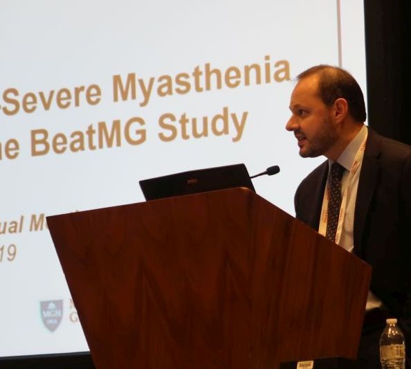
Placebo response in negative rituximab BeatMG trial provides important lessons
AUSTIN, TEX. – Among patients with acetylcholine receptor (AChR) antibody-positive generalized myasthenia gravis, rituximab and placebo may have a similar glucocorticoid-sparing effect regardless of disease severity, according to research presented at the annual meeting of the American Association of Neuromuscular and Electrodiagnostic Medicine.
B Cell Targeted Treatment In Myasthenia Gravis (BeatMG) was a 52-week, randomized, double-blind, placebo-controlled clinical trial. The phase 2 study’s primary outcomes were safety and the glucocorticoid-sparing effect assessed by the percentage of patients who reduced their mean daily prednisone dose by at least 75% and maintained clinical stability. Secondary outcomes were change in Myasthenia Gravis Composite (MGC) score and change in Quantitative Myasthenia Gravis (QMG) score from baseline to 52 weeks.
Investigators randomized 52 participants 1:1 to rituximab (Rituxan) or placebo. Patients were taking at least 15 mg/day of prednisone and were a mean of about age 50 years. About two-thirds were treated with glucocorticoids alone at baseline, and nearly two-thirds had a Myasthenia Gravis Foundation of America (MGFA) clinical classification of II. “It was a mildly symptomatic group of individuals in terms of disease severity,” said Richard Nowak, MD, assistant professor of neurology and director of the Yale Myasthenia Gravis Clinic in New Haven, Conn.
During the study, 60% of patients who received rituximab had a 75% or greater reduction in their mean daily prednisone dose and maintained clinical stability. “However, what surprised us is that we had a significantly high placebo response rate of 56%,” he said. The difference between groups was not significant.
Patients who received rituximab had “directionally favorable reductions” in MGC and QMG scores over 52 weeks, compared with patients who received placebo. Nevertheless, “after correcting for baseline differences, there was no significant difference between the two groups,” Dr. Nowak said.
Rituximab had good safety and tolerability, and the placebo group had a threefold higher rate of clinical relapse requiring IV immunoglobulin or plasmapheresis. “While the placebo arm did achieve a similar rate of reduction in their steroid dose, at 52 weeks the patients may have been doing less well, reflected by the higher rate of relapse,” he said.
Subgroup analysis
To explore whether rituximab might benefit patients who were treatment resistant or had more symptomatic disease, Dr. Nowak and his colleagues conducted a post hoc subgroup analysis of 20 participants – 10 in the rituximab arm and 10 in the placebo arm – who were MGFA class III-IV. In each group, 70% were on glucocorticoid treatment alone, and 90% were MGFA class III.
As in the overall study, the glucocorticoid-sparing effect was not significant (60% of the rituximab group vs. 50% of the placebo group).
Mean change in QMG score was –3.9 in the rituximab group and –0.5 in the placebo group, and mean change in MGC score was –7.0 in the rituximab group and –4.8 in the placebo group. These secondary outcomes again show “directional favorability” with rituximab, Dr. Nowak said. The researchers saw similar trends for scores that assess quality of life and activities of daily living.
In the subgroup analysis, myasthenia gravis relapses requiring rescue therapy occurred in 20% of patients in the rituximab arm and in 30% of the placebo arm. Overall, the researchers did not see a difference in treatment response between those with moderate to severe disease and those with mild disease, Dr. Nowak said.
Suggestions from the data
The post hoc subgroup analysis should be interpreted with caution, and the study does not provide firm conclusions, Dr. Nowak noted. In addition, the trial population does not reflect all patients with myasthenia gravis. Dr. Nowak routinely uses rituximab in patients with muscle-specific kinase antibody-positive disease, he said. For acetylcholine receptor antibody-positive generalized myasthenia gravis, which has approved therapies available, Dr. Nowak presents rituximab as an option for some patients. Further research may clarify where B-cell depletion therapy may fit into the treatment paradigm.
Nonetheless, BeatMG and the subgroup analysis may help physicians better understand the disease and the role of various therapies. Investigators successfully lowered prednisone dose “at a pretty high rate in the placebo arm,” he said. “It suggests that many of our patients potentially may be on higher than required prednisone doses.”
Finally, the BeatMG findings emphasize the need for placebo-controlled trials to understand potential therapies. “We need to pause when we see a lot of retrospective and uncontrolled studies that are very promising,” Dr. Nowak said.
The study was supported by the National Institute of Neurological Disorders and Stroke. Genentech provided the study drug and placebo through an investigator-sponsored study agreement. Dr. Nowak has received research support from Alexion Pharmaceuticals, Genentech, Grifols, and Ra Pharmaceuticals. He has served as a paid consultant for Alexion, Momenta, Ra, Roivant, Shire, Grifols, and CSL Behring.
SOURCE: Nowak R et al. AANEM 2019. Unnumbered Abstract: Rituximab in patients with moderate to severe myasthenia gravis: a subgroup analysis of the BeatMG study
REPORTING FROM AANEM 2019
Congenital myasthenic syndrome diagnosed best with repetitive stimulation and jitter analysis
AUSTIN, TEX. – suggests newly presented research.
“In case RS is negative, SFEMG [single fiber electromyography] alone is not very specific and cannot distinguish CMS from mitochondrial myopathies, even in the presence of impulse blocking,” Vitor Marques Caldas, MD, a neurologist at the Syrian Libanes Hospital in Brasilia, Brazil, and a PhD student at the University of São Paulo, told attendees at the annual meeting of the American Association for Neuromuscular and Electrodiagnostic Medicine. “An isolated SFEMG test can lead to a misdiagnosis of myasthenia syndrome if not interpreted in the right clinical context.”
The researchers sought to understand the relative sensitivity and specificity of low-frequency RS versus jitter analysis using disposable concentric needle electrodes (CNE).
The study involved 69 patients, of whom 19 had mitochondrial myopathy, 18 had congenital myopathy, 18 had CMS, and 14 were asymptomatic controls. The control group all tested normal with both RS and jitter analysis.
The 18 participants with CMS, average age 24 years, received low-frequency RS in at least six different muscles: two distal muscles (abductor digiti minimi and tibialis anterior), two proximal muscles (deltoid and trapezius) and two facial muscles (nasalis and orbicularis oculi). They also underwent jitter analysis of their orbicularis oculi muscle under voluntary activation using CNE.
These patients had heterogeneous genetic profiles: 11 had the CHRNE gene mutation, 2 had the RAPSN gene mutation, 2 had the COLQ gene mutation, 2 had the DOK-7 gene mutation, and 1 had the COL13A1 mutation.
All but two patients with congenital CMS tested positive (88.9%) with RS: one female with CHRNE mutation and one male with RAPSN mutation. Using mean jitter, all but one patient tested positive (94.4%): a female with DOK-7 mutation who had tested abnormal on RS.
All patients with CMS tested positive with at least one of the two tests, but only 83.3% tested positive with both tests, resulting in a sensitivity of 83.3%, a specificity of 100%, and overall accuracy of 95.6% using both tests.
Among the 19 patients with mitochondrial myopathy, 5 had abnormal jitter analysis.
When the researchers looked only at participants with abnormal jitter analysis but normal RS, two of these were patients with CMS, but another seven had congenital or mitochondrial myopathies. Using abnormal jitter alone therefore resulted in a sensitivity of 100% but a specificity of only 86%, for overall 86.5% accuracy.
“It’s important to notice that if you have an abnormal jitter, we have to look at the clinical symptoms of the patients,” Dr. Marques Caldas said in an interview. “Jitter abnormalities are not enough to distinguish between myasthenic disorder and a myopathic disorder.”
The research used no external funding, and Dr. Marques Caldas had no disclosures.
SOURCE: Caldas VM et al. AANEM 2019. Unnumbered Abstract: Sensitivity of neurophysiologic tests regarding the neuromuscular junction in patients with congenital myasthenic syndromes.
REPORTING FROM AANEM 2019
Late response to eculizumab may occur in minority of myasthenia gravis patients
AUSTIN – according to a secondary analysis presented at the annual meeting of the American Association of Neuromuscular and Electrodiagnostic Medicine.
Evidence for the sustained effectiveness of eculizumab, a terminal complement inhibitor, in adult patients with antiacetylcholine receptor antibody-positive refractory generalized myasthenia gravis was provided by the 6-month, double-blind, placebo-controlled REGAIN study and its open-label extension. James F. Howard Jr., MD, distinguished professor of neuromuscular disease at the University of North Carolina in Chapel Hill and colleagues sought to analyze response profiles in REGAIN and its open-label extension.
The findings raise the possibility that complement inhibition with eculizumab should not be abandoned rapidly. “We accept that [eculizumab] works quickly,” Dr. Howard said. “There is an impression that if you don’t respond within the first 3 months, you’re not going to respond. I think these data would suggest otherwise.”
The investigators analyzed participants’ Myasthenia Gravis–Activities of Daily Living (MG-ADL) and Quantitative Myasthenia Gravis (QMG) scores, which had been recorded throughout REGAIN and the extension. They defined early and late responses as improvement in MG-ADL score (i.e., a decrease of three or more points) or QMG score (i.e., a decrease of five or more points) occurring at 12 weeks or earlier or after 12 weeks, respectively, after initiation of eculizumab therapy. Patients randomized to eculizumab in REGAIN initially were treated with an IV induction dose of 900 mg/week before receiving 1,200 mg every 2 weeks thereafter.
Dr. Howard and colleagues included 98 patients in their analysis. Approximately 32% of patients achieved their first response within the first week of treatment, and 15% responded at week 2. About 16% of patients had a late response.
Responses to treatment on the MG-ADL scale had occurred in 67.3% by week 12 and in 84.7% by the end of the extension. Treatment with eculizumab resulted in QMG responses in 56.1% by week 12 and 71.4% by the end of the extension. The investigators observed response over multiple consecutive assessments for the vast majority of patients.
At week 130, the least-squares mean percentage changes from baseline in MG-ADL score were −61.9% and −47.5% in early and late MG-ADL responders, respectively. The least-squares mean percentage changes from baseline in QMG score were −40.8% and −55.5% in early and late QMG responders, respectively.
The investigators observed significant baseline differences between early versus late QMG responders in mean duration of myasthenia gravis (10.46 years vs. 5.46 years) and mean QMG score (18.6 vs. 15.1).
“I can’t explain [this finding]. It may simply be due to the low numbers of patients,” Dr. Howard said. “Whether this is going to hold up in postmarketing analysis remains to be seen. I’m not convinced that that is meaningful.”
Study funding was provided by Alexion Pharmaceuticals (the developer of eculizumab), the Centers for Disease Control and Prevention, and the National Institutes of Health. Dr. Howard reported receiving research support and consulting fees or honoraria from Alexion and several other pharmaceutical companies. Several other authors reported financial relationships with Alexion and other pharmaceutical companies; two authors are employees of Alexion.
Howard J et al. AANEM 2019. Unnumbered Abstract.
REPORTING FROM AANEM 2019
Neurologists publish consensus statement on stridor in MSA
The statement was published Oct. 1 in Neurology. In addition to reviewing the literature on the topic and providing recommendations, the authors described several areas for future research.
MSA is a rare neurodegenerative disorder that entails autonomic failure, cerebellar ataxia, and parkinsonism. Laryngeal stridor has a high positive predictive value in the diagnosis of MSA, but consensus about its definition and clinical implications had not been established previously. The Istituto di Ricovero e Cura a Carattere Scientifico (IRCCS) delle Scienze Neurologiche di Bologna (Italy) convened a consensus conference of experts in 2017 to determine diagnostic criteria for stridor in MSA, define its prognostic value, suggest treatment options, and indicate subjects for future research. The neurologists reviewed studies of any design that reported original data. They based their statements on 34 published articles, most of which were class III or IV.
The authors defined stridor in MSA as “a strained, high-pitched, harsh respiratory sound, mainly inspiratory, caused by laryngeal dysfunction leading to narrowing of the rima glottidis.” Stridor may occur exclusively during sleep or during sleep and wakefulness. It may be recognized during a clinical examination, through witness report, or through an audio recording. Neurologists may consider laryngoscopy to exclude mechanical lesions or functional vocal cord abnormalities related to other neurologic conditions, wrote the authors. Drug-induced sleep endoscopy and video polysomnography also may be considered.
Whether stridor, or certain features of stridor, affects survival in MSA is uncertain. “Stridor within 3 years of motor or autonomic symptom onset may shorten survival,” according to the statement. “However, identification of stridor onset may be difficult.” Moreover, stridor during wakefulness is considered to reflect a more advanced stage of disease, compared with stridor during sleep. Although stridor can be distressing for the patient and his or her caregivers, its influence on health-related quality of life has yet to be determined, according to the statement.
Continuous positive airway pressure (CPAP) during sleep can be a useful symptomatic treatment and should be considered a first-line therapy for stridor, wrote the authors. Tracheostomy, another effective symptomatic treatment, bypasses upper-airway obstruction at the larynx. “Persistent and severe stridor may require tracheostomy,” according to the statement. It is not certain whether CPAP improves survival in patients with MSA and stridor, and tracheostomy may improve survival. The literature contains insufficient evidence about whether minimally invasive procedures or botulinum toxin injections are effective symptomatic treatments for stridor, wrote the authors.
During their review of the literature, the authors identified what they considered to be several research gaps. The diagnosis of stridor remains challenging, and investigators should develop a questionnaire for detecting stridor, they wrote. A smartphone application also could be developed to recognize stridor automatically. “The relationship between stridor and other breathing disorders (i.e., central apneas and breathing rate abnormalities) and their respective contributions to disease prognosis and survival should be determined through a multicenter prospective study,” according to the statement. Finally, randomized controlled trials comparing CPAP and tracheostomy for various degrees of stridor could guide physicians’ choice of treatment.
The IRCCS funded the study. One of the authors is a section editor for Neurology, and other authors reported receiving honoraria from various companies such as Novartis, Sanofi, and UCB.
FROM NEUROLOGY
Viral cause of acute flaccid myelitis eludes detection
A study of 305 cases of acute flaccid myelitis has found further evidence of a viral etiology but is yet to identify a single pathogen as the primary cause.
Writing in Pediatrics, researchers published an analysis of patients presenting with acute flaccid limb weakness from January 2015 to December 2017 across 43 states.
A total of 25 cases were judged as probable for acute flaccid myelitis (AFM) because they met clinical criteria and had a white blood cell count above 5 cells per mm3 in cerebrospinal fluid, while 193 were judged as confirmed cases based on the additional presence of spinal cord gray matter lesions on MRI.
Overall, 83% of patients had experienced fever, cough, runny nose, vomiting, and/or diarrhea for a median of 5 days before limb weakness began. Two-thirds of patients had experienced a respiratory illness, 62% had experienced a fever, and 29% had experienced gastrointestinal illness.
Overall, 47% of the 193 patients who had specimens tested at a Centers for Disease Control and Prevention or non-CDC laboratory had a pathogen found at any site, 10% had a pathogen detected from a sterile site such as cerebrospinal fluid or sera, and 42% had a pathogen detected from a nonsterile site.
Among 72 patients who had serum specimens tested at the CDC, 2 were positive for enteroviruses. Among the 90 patients who had upper respiratory specimens tested, 36% were positive for either enteroviruses or rhinoviruses.
A number of stool specimens were also tested; 15% were positive for enteroviruses or rhinoviruses and one was positive for parechovirus.
Cerebrospinal fluid was tested in 170 patients, of which 4 were positive for enteroviruses. The testing also found adenovirus, Epstein-Barr virus, human herpesvirus 6, and mycoplasma in six patients. Sera testing of 123 patients found 9 were positive for enteroviruses, West Nile virus, mycoplasma, and coxsackievirus B.
“In our summary of national AFM surveillance from 2015 to 2017, we demonstrate that cases were widely distributed across the United States, the majority of cases occurred in late summer or fall, children were predominantly affected, there is a spectrum of clinical severity, and no single pathogen was identified as the primary cause of AFM,” wrote Tracy Ayers, PhD, from the National Center for Immunization and Respiratory Diseases, and coauthors. “We conclude that symptoms of a viral syndrome within the week before limb weakness, detection of viral pathogens from sterile and nonsterile sites from almost half of patients, and seasonality of AFM incidence, particularly during the 2016 peak year, strongly suggest a viral etiology, including [enteroviruses].”
The authors of an accompanying editorial noted that the clinical syndrome of acute flaccid paralysis caused by myelitis in the gray matter of the spinal cord has previously been associated with a range of viruses, including poliovirus, enteroviruses, and flaviviruses, so a single etiology to explain all cases would not be expected.
“The central question remains: What is driving seasonal biennial nationwide outbreaks of AFM since 2014?” wrote Kevin Messaca, MD, and colleagues from the University of Colorado at Denver, Aurora.
Two authors declared consultancies, grants, and research contracts with the pharmaceutical sector. No other conflicts of interest were declared. One editorial author declared funding from the National Institute of Allergy and Infectious Diseases.
SOURCE: Ayers T et al. Pediatrics. 2019 Oct 7. doi: 10.1542/peds.2019-1619.
*Updated 10/14/2019.
FROM PEDIATRICS
Key clinical point: Acute flaccid myelitis shows a strong suggestion of viral etiology but a single causal virus is not identified.
Major finding: Patients with acute flaccid myelitis show infection with a range of viruses including enteroviruses.
Study details: A study of 305 cases of acute flaccid myelitis in the United States.
Disclosures: Two authors declared consultancies, grants, and research contracts with the pharmaceutical sector. No other conflicts of interest were declared.
Source: Ayers T et al. Pediatrics. 2019 Oct 7. doi: 10.1542/peds.2019-1619.
Coming soon!
Check back later this month for top news from the 2019 AANEM Annual Meeting in Austin.
Check back later this month for top news from the 2019 AANEM Annual Meeting in Austin.
Check back later this month for top news from the 2019 AANEM Annual Meeting in Austin.
Can a novel steroidal anti-inflammatory drug benefit patients with Duchenne muscular dystrophy?

Daily treatment with vamorolone at doses of 2.0 mg/kg per day and 6.0 mg/kg per day suggested possible efficacy in a 24-week study, researchers said. The exploratory study included 48 boys who had completed a phase 2a trial.
The treatment was safe and well tolerated, and patients who received 2.0 mg/kg per day had significantly improved muscle function, as assessed by time to stand, compared with natural history controls, according to the results, which were published in Neurology.
In addition, the novel drug may reduce “safety concerns typically seen with traditional glucocorticoids,” wrote Eric P. Hoffman, PhD, and coauthors. Dr. Hoffman is president and CEO of ReveraGen BioPharma in Rockville, Md., which is developing the drug, and associate dean for research in the school of pharmacy and pharmaceutical sciences at Binghamton (N.Y.) University.
In preclinical studies, vamorolone retained anti-inflammatory efficacy while reducing adverse effects, compared with prednisolone, in a manner that is “consistent with vamorolone blocking [nuclear factor-kappa beta]–associated proinflammatory signals as a ligand/receptor monomeric state instead of the traditional molecular models of ligand/receptor dimeric complexes,” the authors said.
Phase 1 and phase 2a studies suggest that the drug may have an improved safety profile. To assess possible efficacy and define optimal doses, the investigators conducted the 24-week extension study. Participants were boys aged 4 years to younger than 7 years who had never been treated with glucocorticoids. They received 0.25, 0.75, 2.0, or 6.0 mg/kg per day vamorolone in an oral suspension formulation. Twelve boys received each dose level.
“Vamorolone was well tolerated ... with no adverse events leading to reduction of drug dosing or withdrawal from the trial,” they said. “The [timed stand from supine] primary outcome measure in vamorolone-treated patients with DMD supports efficacy of the 2.0-mg/kg/d dose ... at 24 weeks,” they said. A secondary outcome measure, the 6-minute walk test, supports efficacy at this dose at 12 and 24 weeks of treatment.
Furthermore, the data indicate that the 2.0-mg/kg per day dose may be associated with less weight gain and improved bone turnover and insulin resistance biomarkers, relative to prednisone therapy. “There was evidence of adrenal suppression in a subset of boys with DMD treated with 2.0 mg/kg/d vamorolone, with 18% of patients showing reduced morning cortisol levels,” the authors said. “Future studies of vamorolone will include adrenocorticotropic hormone–challenge tests to further explore adrenal function.”
A double-blind, placebo-controlled trial of vamorolone is underway. Investigators are testing two doses of vamorolone (2.0 and 6.0 mg/kg per day) versus placebo and prednisone (0.75 mg/kg per day). Researchers plan to enroll 120 patients, with 30 patients in each arm.
ReveraGen BioPharma received funds for the present study from Actelion Pharmaceuticals, U.S. and European government agencies, and nonprofit foundations. Dr. Hoffman and some of his collaborators are cofounders of ReveraGen. Other coauthors received support from the company.
SOURCE: Hoffman EP et al. Neurology. 2019 Aug 26. doi: 10.1212/WNL.0000000000008168.
FROM NEUROLOGY
Peripheral nervous system events have lasting impact on SLE patients
Peripheral nervous system disease, predominantly neuropathies, constitutes a substantial proportion of the manifestations of neuropsychiatric systemic lupus erythematosus (SLE) and has a lasting negative impact on health-related quality of life, John G. Hanly, MD, of Queen Elizabeth II Health Sciences Center and Dalhousie University, Halifax, N.S., and associates reported in Arthritis & Rheumatology.
According to the study of 1,827 SLE patients who had been recently diagnosed and enrolled in the Systemic Lupus International Collaborating Clinics (SLICC) network at sites in Europe, Asia, and North America during 1999-2011, 161 peripheral nervous system (PNS) events occurred in 139 of the patients (8%) over a mean 7.6 years of follow-up.
Using the seven American College of Rheumatology case definitions for PNS disease in neuropsychiatric SLE, most of the events were peripheral neuropathy (41%), mononeuropathy (27%), and cranial neuropathy (24%). For 110 with peripheral neuropathy or mononeuropathy who underwent electrophysiologic testing, axonal damage was often present (42%), followed by demyelination (22%).
The PNS events were attributed to SLE in about 58%-75% of the patients. Based on these data the investigators estimated that after 10 years the cumulative incidence of any PNS event regardless of its attribution was about 9%, and it was nearly 7% for events attributed to SLE.
The probability that the neuropathies would not resolve over time was estimated at about 43% for peripheral neuropathy, 29% for mononeuropathy, and 30% for cranial neuropathy. Resolution of neuropathy was most rapid for cranial neuropathy, followed by mononeuropathy and peripheral neuropathy.
Patients with PNS events had significantly lower physical and mental health component scores on the 36-item Short Form Health Survey than did patients without a neuropsychiatric event up to the study assessment, and these differences persisted for 10 years of follow-up.
These “findings provide a benchmark for the assessment of future treatment modalities,” the investigators concluded.
SOURCE: Hanly JG et al. Arthritis Rheumatol. 2019 Aug 7. doi: 10.1002/art.41070.
REPORTING FROM ARTHRITIS & RHEUMATOLOGY