User login
Doug Brunk is a San Diego-based award-winning reporter who began covering health care in 1991. Before joining the company, he wrote for the health sciences division of Columbia University and was an associate editor at Contemporary Long Term Care magazine when it won a Jesse H. Neal Award. His work has been syndicated by the Los Angeles Times and he is the author of two books related to the University of Kentucky Wildcats men's basketball program. Doug has a master’s degree in magazine journalism from the S.I. Newhouse School of Public Communications at Syracuse University. Follow him on Twitter @dougbrunk.
Don’t discount the role of patient satisfaction in successful outcomes
DENVER – In the opinion of Dr. Steven R. Feldman, key elements of the therapeutic process include making the right diagnosis, prescribing the right treatment, and getting patients to use prescribed medication.
"These three elements can be considered technical aspects of care, but patient satisfaction is also critical in its own right, because it determines the outcomes of care," Dr. Feldman said at the annual meeting of the American Academy of Dermatology.
Getting patients to use prescribed medication, for example, "is critical to treatment success and depends on the quality of the interaction between patients and clinicians," he said. "You want that interaction to be good. Not only is it good for business and reduces malpractice risk, it is essential for getting patients well."

In dermatology, medication compliance among patients with acne and psoriasis is notoriously poor, but physicians can make an impact by focusing on trust, motivation, and understanding, said Dr. Feldman, professor of dermatology, pathology, and public health sciences at Wake Forest University, Winston-Salem, N.C. "These factors are just as important as the right diagnosis and the right treatment, and they’re largely under our control," he said.
Dr. Feldman, who founded the online patient satisfaction survey service www.DrScore.com, listed seven traits that patients identify in outstanding physicians: access, communication (including listening, forming partnership with the patient, and giving information); a personality/demeanor that projects empathy; medical care (including technical competence, timely diagnoses, treatment, and thoroughness); follow-up (including test results and referrals); facilities; and office/staff coordination.
"Traits patients don’t like include poor access, poor communications, poor follow-up, and a lack of interpersonal skills," said Dr. Feldman, who also is a member of the AAD Outcome Study Workgroup. "In good medical practice you want to make the right diagnosis, prescribe the right treatment, communicate and follow up, and project the appearance of empathy. Think about how you are going to pay attention to this particular patient just before you enter the room."
He noted that, when it comes to patient satisfaction, projecting the appearance of empathy is actually more important than being empathetic.
"I assume all doctors care deeply about their patients," Dr. Feldman said. "But if the patient doesn’t realize the doctor cares, then the patient will not be satisfied, won’t be trusting, and is at risk of poor adherence and poor treatment outcome."
Patient satisfaction studies suggest that patients care more about having a caring/friendly physician than the physician’s age, gender, or office wait time.
"It’s not just the doctor’s behavior that’s important," he continued. "Attend to warmth and fuzziness in your entire practice as though it matters, because it does. Our beliefs are strongly influenced by context."
Simple ways to foster positive physician-patient interaction include using images to communicate risks in perspective, providing written instruction for care, and giving your cell phone number to patients. That gesture alone "is a powerful statement of how much you care about the patient," Dr. Feldman said. "Leave your patients with the clear realization that you care about them."
Counseling patients about the potential side effects of medications is also important, "because fear of [side effects] can reduce compliance," he said. "For acne patients on spironolactone, you might say something like, ‘This drug is a diuretic. In addition to its effects on your acne, you may also notice some weight loss.’ For patients with scalp psoriasis, you might tell them that their recommended treatment may sting. The stinging is a sign that it’s working."
Dr. Feldman disclosed that, in addition to founding DrScore.com and the adherence company Causa Research, he has received grants and/or research funding from numerous pharmaceutical companies.
DENVER – In the opinion of Dr. Steven R. Feldman, key elements of the therapeutic process include making the right diagnosis, prescribing the right treatment, and getting patients to use prescribed medication.
"These three elements can be considered technical aspects of care, but patient satisfaction is also critical in its own right, because it determines the outcomes of care," Dr. Feldman said at the annual meeting of the American Academy of Dermatology.
Getting patients to use prescribed medication, for example, "is critical to treatment success and depends on the quality of the interaction between patients and clinicians," he said. "You want that interaction to be good. Not only is it good for business and reduces malpractice risk, it is essential for getting patients well."
In dermatology, medication compliance among patients with acne and psoriasis is notoriously poor, but physicians can make an impact by focusing on trust, motivation, and understanding, said Dr. Feldman, professor of dermatology, pathology, and public health sciences at Wake Forest University, Winston-Salem, N.C. "These factors are just as important as the right diagnosis and the right treatment, and they’re largely under our control," he said.
Dr. Feldman, who founded the online patient satisfaction survey service www.DrScore.com, listed seven traits that patients identify in outstanding physicians: access, communication (including listening, forming partnership with the patient, and giving information); a personality/demeanor that projects empathy; medical care (including technical competence, timely diagnoses, treatment, and thoroughness); follow-up (including test results and referrals); facilities; and office/staff coordination.
"Traits patients don’t like include poor access, poor communications, poor follow-up, and a lack of interpersonal skills," said Dr. Feldman, who also is a member of the AAD Outcome Study Workgroup. "In good medical practice you want to make the right diagnosis, prescribe the right treatment, communicate and follow up, and project the appearance of empathy. Think about how you are going to pay attention to this particular patient just before you enter the room."
He noted that, when it comes to patient satisfaction, projecting the appearance of empathy is actually more important than being empathetic.
"I assume all doctors care deeply about their patients," Dr. Feldman said. "But if the patient doesn’t realize the doctor cares, then the patient will not be satisfied, won’t be trusting, and is at risk of poor adherence and poor treatment outcome."
Patient satisfaction studies suggest that patients care more about having a caring/friendly physician than the physician’s age, gender, or office wait time.
"It’s not just the doctor’s behavior that’s important," he continued. "Attend to warmth and fuzziness in your entire practice as though it matters, because it does. Our beliefs are strongly influenced by context."
Simple ways to foster positive physician-patient interaction include using images to communicate risks in perspective, providing written instruction for care, and giving your cell phone number to patients. That gesture alone "is a powerful statement of how much you care about the patient," Dr. Feldman said. "Leave your patients with the clear realization that you care about them."
Counseling patients about the potential side effects of medications is also important, "because fear of [side effects] can reduce compliance," he said. "For acne patients on spironolactone, you might say something like, ‘This drug is a diuretic. In addition to its effects on your acne, you may also notice some weight loss.’ For patients with scalp psoriasis, you might tell them that their recommended treatment may sting. The stinging is a sign that it’s working."
Dr. Feldman disclosed that, in addition to founding DrScore.com and the adherence company Causa Research, he has received grants and/or research funding from numerous pharmaceutical companies.
AT THE AAD ANNUAL MEETING

Novel agent for papulopustular rosacea found safe, effective in phase III trials
DENVER – Ivermectin 1% cream applied once daily was effective and safe in treating patients with moderate to severe papulopustular rosacea, based on data from a pair of phase III studies.
Ivermectin 1% cream, a novel agent being developed by Galderma Laboratories, is of interest to rosacea researchers because it possesses unique anti-inflammatory and anti-parasitic properties, Dr. Linda Stein Gold said at the annual meeting of the American Academy of Dermatology.

Dr. Stein Gold of the department of dermatology at Henry Ford Medical Center, Detroit, explained that Demodex folliculorum, which is typically found on the human face, may trigger an immune response in rosacea patients. Ivermectin 1% is anti-parasitic against Demodex, providing an "innovative treatment" option in this patient population, she said. The investigational agent has not yet been approved by the Food and Drug Administration.
Dr. Stein Gold and her associates set out to assess the efficacy and safety of 1% ivermectin cream vs. a control vehicle cream in patients with moderate to severe papulopustular rosacea. The design included two identical, double-blind, parallel group, 12-week studies.
In both studies, a total of 910 patients were randomized 2:1 to receive 1% ivermectin cream or a control cream once daily. There were two co-primary endpoints: the percentage of patients who achieved a "clear" or "almost clear" score on the Investigator’s Global Assessment (IGA) scale at week 12, and the change in inflammatory lesion counts from baseline to week 12. The secondary efficacy endpoint assessment was the percentage change in inflammatory lesion count from baseline to week 12. The researchers also assessed safety endpoints by examining adverse events.
The mean age of patients was 50 years, 96% were white, and 67% were women. Patients had about 30 lesions each; 76-82% were classified as having moderate rosacea based on IGA score, and the rest had severe disease. More than 90% of patients in each arm completed the study through week 12.
At week 12 in both studies, a significantly higher percentage of patients in the treatment group achieved treatment success compared with those in the control group (38-40% vs. 12-19%, respectively; P less than .001). That difference was seen as early as week four.
Both studies also demonstrated that treatment with ivermectin 1% was significantly superior to the control vehicle in reducing lesion counts, with significance seen as early as week two and continuing for the duration of the study. Patients in the treatment groups in both studies showed an average reduction of more than 20 lesions, while controls in the two studies had reductions of 12 and 13.4 lesions, respectively.
Fewer treatment-related adverse events were reported in the ivermectin group, compared with the control group (3.4% vs. 7.2%, respectively). The most common treatment-related adverse event in the first study was sensation of skin burning (1.8% in the ivermectin group vs. 2.6% in controls) while the most common adverse event in the second study were pruritus and dry skin (0.7% and 0.9%, respectively). "This drug was exceptionally well tolerated, very safe as well as efficacious," Dr. Stein Gold said.
In an interview, she acknowledged certain limitations of the study, including the fact that "we did not look at maintenance of efficacy when a patient stops the drug. However, that is being looked at in other studies."
The study was funded by Galderma R&D. Dr. Stein Gold disclosed that she is a consultant for Galderma.
DENVER – Ivermectin 1% cream applied once daily was effective and safe in treating patients with moderate to severe papulopustular rosacea, based on data from a pair of phase III studies.
Ivermectin 1% cream, a novel agent being developed by Galderma Laboratories, is of interest to rosacea researchers because it possesses unique anti-inflammatory and anti-parasitic properties, Dr. Linda Stein Gold said at the annual meeting of the American Academy of Dermatology.
Dr. Stein Gold of the department of dermatology at Henry Ford Medical Center, Detroit, explained that Demodex folliculorum, which is typically found on the human face, may trigger an immune response in rosacea patients. Ivermectin 1% is anti-parasitic against Demodex, providing an "innovative treatment" option in this patient population, she said. The investigational agent has not yet been approved by the Food and Drug Administration.
Dr. Stein Gold and her associates set out to assess the efficacy and safety of 1% ivermectin cream vs. a control vehicle cream in patients with moderate to severe papulopustular rosacea. The design included two identical, double-blind, parallel group, 12-week studies.
In both studies, a total of 910 patients were randomized 2:1 to receive 1% ivermectin cream or a control cream once daily. There were two co-primary endpoints: the percentage of patients who achieved a "clear" or "almost clear" score on the Investigator’s Global Assessment (IGA) scale at week 12, and the change in inflammatory lesion counts from baseline to week 12. The secondary efficacy endpoint assessment was the percentage change in inflammatory lesion count from baseline to week 12. The researchers also assessed safety endpoints by examining adverse events.
The mean age of patients was 50 years, 96% were white, and 67% were women. Patients had about 30 lesions each; 76-82% were classified as having moderate rosacea based on IGA score, and the rest had severe disease. More than 90% of patients in each arm completed the study through week 12.
At week 12 in both studies, a significantly higher percentage of patients in the treatment group achieved treatment success compared with those in the control group (38-40% vs. 12-19%, respectively; P less than .001). That difference was seen as early as week four.
Both studies also demonstrated that treatment with ivermectin 1% was significantly superior to the control vehicle in reducing lesion counts, with significance seen as early as week two and continuing for the duration of the study. Patients in the treatment groups in both studies showed an average reduction of more than 20 lesions, while controls in the two studies had reductions of 12 and 13.4 lesions, respectively.
Fewer treatment-related adverse events were reported in the ivermectin group, compared with the control group (3.4% vs. 7.2%, respectively). The most common treatment-related adverse event in the first study was sensation of skin burning (1.8% in the ivermectin group vs. 2.6% in controls) while the most common adverse event in the second study were pruritus and dry skin (0.7% and 0.9%, respectively). "This drug was exceptionally well tolerated, very safe as well as efficacious," Dr. Stein Gold said.
In an interview, she acknowledged certain limitations of the study, including the fact that "we did not look at maintenance of efficacy when a patient stops the drug. However, that is being looked at in other studies."
The study was funded by Galderma R&D. Dr. Stein Gold disclosed that she is a consultant for Galderma.
AT THE AAD ANNUAL MEETING
Major Finding: At week 12, a significantly higher percentage of rosacea patients in the ivermectin 1% cream treatment group achieved scores of "clear" or "almost clear" on the Investigator’s Global Assessment compared with those in the control group (38-40% vs. 12-19%, respectively; P less than .001).
Data Source: Two pivotal phase 3 studies in which 910 patients with moderate to severe papulopustular rosacea were randomized 2:1 to receive 1% ivermectin cream or a vehicle cream (control) once daily for 12 weeks.
Disclosures: The study was funded by Galderma R&D. Dr. Stein Gold disclosed that she is a consultant for Galderma.

VIDEO: Ivermectin shows promise for treating papulopustular rosacea
DENVER – A limited number of safe and effective topical medications are currently available to treat chronic papulopustular (inflammatory) rosacea. In a video interview at the annual meeting of the American Academy of Dermatology, Dr. Linda Stein Gold highlighted results of two pivotal phase 3 trials of ivermectin 1%, an investigational drug being evaluated for the treatment of the disorder. Both studies of ivermectin 1% cream met their co-primary efficacy endpoints of treatment success as defined by the Investigator’s Global Assessment (IGA) rating of clear skin and change in inflammatory lesion count.
DENVER – A limited number of safe and effective topical medications are currently available to treat chronic papulopustular (inflammatory) rosacea. In a video interview at the annual meeting of the American Academy of Dermatology, Dr. Linda Stein Gold highlighted results of two pivotal phase 3 trials of ivermectin 1%, an investigational drug being evaluated for the treatment of the disorder. Both studies of ivermectin 1% cream met their co-primary efficacy endpoints of treatment success as defined by the Investigator’s Global Assessment (IGA) rating of clear skin and change in inflammatory lesion count.
DENVER – A limited number of safe and effective topical medications are currently available to treat chronic papulopustular (inflammatory) rosacea. In a video interview at the annual meeting of the American Academy of Dermatology, Dr. Linda Stein Gold highlighted results of two pivotal phase 3 trials of ivermectin 1%, an investigational drug being evaluated for the treatment of the disorder. Both studies of ivermectin 1% cream met their co-primary efficacy endpoints of treatment success as defined by the Investigator’s Global Assessment (IGA) rating of clear skin and change in inflammatory lesion count.
AT THE AAD ANNUAL MEETING

Nanotherapies make inroads in wound regeneration
DENVER – Move over, gauze, bandages, and moist wound-healing techniques. Nanomaterials are making significant inroads in wound regeneration.
Dr. Adam Friedman, director of dermatologic research at the Montefiore-Einstein College of Medicine, New York, highlighted four nanotherapies in the field of wound care.

• Antimicrobial nano-based dressings. The antibacterial properties of silver have been documented since 1000 B.C., said Dr. Friedman of the departments of dermatology and of physiology and biophysics at the medical school. Silver ions are believed to directly disrupt pathogen cell walls/membranes and suppress respiratory enzymes and electron transport components. Silver has been used commercially for decades, with demonstrated antimicrobial effects (Crede’s 1% silver nitrate eyedrops were used to prevent mother to child transmission of gonococcal eye infection); anti-inflammatory properties (silver nitrate is used in pleurodesis); and infection protection (silver nanoparticle–impregnated wound dressings prevent infection and enhance wound healing).
Silver nanoparticles have an "increased likelihood of directly interacting with the target bacteria or virus," Dr. Friedman said in an interview in advance of the annual meeting of the American Academy of Dermatology. "They bind to and/or disturb bacterial cell membrane activity as well as release silver ions much more readily then their bulk counterparts." Silver dressings currently on the market include Silvercel, Aquacel, and Acticoat.
Similarly to nanometals, the biological activity of curcumin (a water-insoluble polyphenolic compound derived from tumeric) can be used in the nanoform and has been shown to both effectively clear methicillin-resistant Staphylococcus aureus in burn wound infections and accelerate the healing of thermal burn wounds.
• Immunomodulating antimicrobial nanoparticles. One of the most promising immunomodulators in wound healing is the gaseous molecule nitric oxide (NO). This potential has yet to be realized, Dr. Friedman said, as NO is highly unstable and its site of action is often microns from its source of generation. Nanotechnology can allow for controlled and sustained release of this evasive biomolecule and make therapeutic translation a reality. "At Einstein, we are utilizing NO-generating nanoparticles to accelerate wound healing and eradicate multidrug resistant pathogens," he said. "But, because NO is integral to so many biological processes, it can do so much more. This technology is also being studied for the treatment of cardiovascular disease and even the topical treatment of erectile dysfunction."
• Gene modifying/silencing technologies. RNA interference is an endogenous mechanism to control gene expression in a variety of organisms. "We can take advantage of this process using siRNA (small interfering RNA) to manipulate limitless biological processes," Dr. Friedman explained. "While a hot area, translation to the bedside has been difficult as siRNA are very unstable and have a difficult time reaching their targets inside cells. Nanoparticles have been shown to overcome these limitations." Dr. Friedman noted how siRNA encapsulate nanoparticles targeting fidgetin-like 2 (an ATPase that cleaves microtubules), can knock down this gene in vivo, resulting in accelerated epithelial cell migration and hastened wound closure in both excisional and burn wound mouse models.
• Growth factor–releasing nanoparticles. Growth factors have been found to speed the healing of acute and chronic wounds in humans, including Regranex, a analogue of platelet-derived growth factor that is FDA-approved for treating leg and foot ulcers in diabetic patients, epidermal growth factor (donor-site wounds), fibroblast growth factor (burn wounds), and growth hormone (donor sites in burned children). "Nanomaterials offer many advantages here, from allowing for temporal release depending on the wound environment to delivering multiple factors at the same or different times, to even serving as a structural foundation for wound healing while releasing factors simultaneously," Dr. Friedman said. "The possibilities are limitless."
Dr. Friedman characterized nanomedicine as "a newborn branch of science. These promising and innovative tools should help us overcome limitations of traditional wound care, improve direct intervention on phases of wound healing, and provide better solutions for wound dressing that induce favorable wound healing environments. More work – and funding – is needed."
Dr. Friedman disclosed that he is a consultant and/or a member of the scientific advisory board for SanovaWorks, Prodigy, Oakstone Institute, Liquidia Technologies, L’Oréal, Amgen, Onset, Aveeno, GSK, MicroCures, and Nano Bio-Med. He is also a speaker for Onset and Amgen.
This article was updated March 24, 2014.
DENVER – Move over, gauze, bandages, and moist wound-healing techniques. Nanomaterials are making significant inroads in wound regeneration.
Dr. Adam Friedman, director of dermatologic research at the Montefiore-Einstein College of Medicine, New York, highlighted four nanotherapies in the field of wound care.
• Antimicrobial nano-based dressings. The antibacterial properties of silver have been documented since 1000 B.C., said Dr. Friedman of the departments of dermatology and of physiology and biophysics at the medical school. Silver ions are believed to directly disrupt pathogen cell walls/membranes and suppress respiratory enzymes and electron transport components. Silver has been used commercially for decades, with demonstrated antimicrobial effects (Crede’s 1% silver nitrate eyedrops were used to prevent mother to child transmission of gonococcal eye infection); anti-inflammatory properties (silver nitrate is used in pleurodesis); and infection protection (silver nanoparticle–impregnated wound dressings prevent infection and enhance wound healing).
Silver nanoparticles have an "increased likelihood of directly interacting with the target bacteria or virus," Dr. Friedman said in an interview in advance of the annual meeting of the American Academy of Dermatology. "They bind to and/or disturb bacterial cell membrane activity as well as release silver ions much more readily then their bulk counterparts." Silver dressings currently on the market include Silvercel, Aquacel, and Acticoat.
Similarly to nanometals, the biological activity of curcumin (a water-insoluble polyphenolic compound derived from tumeric) can be used in the nanoform and has been shown to both effectively clear methicillin-resistant Staphylococcus aureus in burn wound infections and accelerate the healing of thermal burn wounds.
• Immunomodulating antimicrobial nanoparticles. One of the most promising immunomodulators in wound healing is the gaseous molecule nitric oxide (NO). This potential has yet to be realized, Dr. Friedman said, as NO is highly unstable and its site of action is often microns from its source of generation. Nanotechnology can allow for controlled and sustained release of this evasive biomolecule and make therapeutic translation a reality. "At Einstein, we are utilizing NO-generating nanoparticles to accelerate wound healing and eradicate multidrug resistant pathogens," he said. "But, because NO is integral to so many biological processes, it can do so much more. This technology is also being studied for the treatment of cardiovascular disease and even the topical treatment of erectile dysfunction."
• Gene modifying/silencing technologies. RNA interference is an endogenous mechanism to control gene expression in a variety of organisms. "We can take advantage of this process using siRNA (small interfering RNA) to manipulate limitless biological processes," Dr. Friedman explained. "While a hot area, translation to the bedside has been difficult as siRNA are very unstable and have a difficult time reaching their targets inside cells. Nanoparticles have been shown to overcome these limitations." Dr. Friedman noted how siRNA encapsulate nanoparticles targeting fidgetin-like 2 (an ATPase that cleaves microtubules), can knock down this gene in vivo, resulting in accelerated epithelial cell migration and hastened wound closure in both excisional and burn wound mouse models.
• Growth factor–releasing nanoparticles. Growth factors have been found to speed the healing of acute and chronic wounds in humans, including Regranex, a analogue of platelet-derived growth factor that is FDA-approved for treating leg and foot ulcers in diabetic patients, epidermal growth factor (donor-site wounds), fibroblast growth factor (burn wounds), and growth hormone (donor sites in burned children). "Nanomaterials offer many advantages here, from allowing for temporal release depending on the wound environment to delivering multiple factors at the same or different times, to even serving as a structural foundation for wound healing while releasing factors simultaneously," Dr. Friedman said. "The possibilities are limitless."
Dr. Friedman characterized nanomedicine as "a newborn branch of science. These promising and innovative tools should help us overcome limitations of traditional wound care, improve direct intervention on phases of wound healing, and provide better solutions for wound dressing that induce favorable wound healing environments. More work – and funding – is needed."
Dr. Friedman disclosed that he is a consultant and/or a member of the scientific advisory board for SanovaWorks, Prodigy, Oakstone Institute, Liquidia Technologies, L’Oréal, Amgen, Onset, Aveeno, GSK, MicroCures, and Nano Bio-Med. He is also a speaker for Onset and Amgen.
This article was updated March 24, 2014.
AT THE AAD ANNUAL MEETING
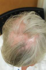
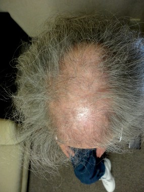
A consistent approach drives optimal scarring alopecia treatment
DENVER – To limit the progression of scarring alopecia, Dr. Jeff Donovan makes it a point to ask his patients about symptoms and shedding, and he always performs a thorough scalp examination to record the affected sites and signs of the condition.
"Everything on the history potentially may be important, but always ask about symptoms of itching, burning, pain, tenderness, and shedding," Dr. Donovan of the department of dermatology at the University of Toronto advised at the annual meeting of the American Academy of Dermatology.

Upon examination, he continued, document sites and signs by considering the following questions: Where is the hair loss – frontal, top, or occipital? Can you still see the follicular ostia? Is there erythema of the scalp? Is there perifollicular erythema or scale, crusting, pustules, or loss of eyebrow or body hair?
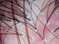
"When you perform dermoscopy of the normal scalp, one can see that the hairs are similar in ‘caliber’ (no miniaturization suggestive of androgenetic alopecia), and there are no changes around the hair follicles or between the hair follicles," Dr. Donovan said. "In scarring alopecia, a variety of findings may be present which help point to the correct diagnosis."
A 4-mm punch biopsy is helpful to confirm the diagnosis and is recommended in areas of early active disease, including areas that may have primary morphologic features, areas with a positive pull test (if possible), or areas that are symptomatic (if needed). "Diagnosing a hair disease with a biopsy requires a hair to be present in the biopsy," he noted. "Biopsies of completely scarred areas are not helpful." In scarring alopecias, inflammatory infiltrates are found in the upper parts of the hair follicle, which destroys hair follicle stem cells. "It’s this destruction of stem cells which ultimately leads to permanent hair loss," Dr. Donovan said.
Lichen planopilaris, a common form of scarring alopecia, typically occurs in middle age and is twice as common in women as in men. It most often affects the central scalp but may be present in other sites in up to half of cases. Key symptoms of lichen planopilaris (LPP) include hair loss, scalp pruritus, and pain/tenderness, often a burning sensation at the site of hair loss. On dermoscopy, most LPP cases appear as reduced hair density with scalp erythema and perifollicular scale, also called peripilar casts.
The goal of LPP treatment is to reduce symptoms and shedding and to stop the disease from occurring in new sites. "Regrowth is not possible in most scarring alopecias," said Dr. Donovan, who leads the University of Toronto’s program in hair transplantation and hair loss. "Treatments help to halt the underlying disease process. Disease activity may recur."
Treatment options for localized/limited LPP include intralesional triamcinolone acetonide and/or several treatments at home, including 0.05% clobetasol propionate lotion or foam, clobetasol propionate shampoo to help decrease itching and burning, fluocinolone acetonide oil one time per week to help with removal of scales, and topical 0.1% tacrolimus ointment (or compounded lotion) as needed.
Systemic treatment of LPP is also an option, and he said he relies on the dermatopathology report to guide his treatment decisions. If biopsy reveals minimal lymphocytic infiltrate, Dr. Donovan said he recommends doxycycline 100 mg b.i.d. as his first-line approach. If biopsy reveals moderate lymphocytic infiltrate, he turns to hydroxychloroquine 6 mg/kg.
His recommended second-line systemic treatment is mycophenolate mofetil 500 mg b.i.d. for 1 month, then 1,000 mg b.i.d. thereafter. Third-line systemic treatment options include cyclosporine 3-5 mg/kg per day and retinoids such as isotretinoin, but fewer than 20% of patients benefit from retinoids, he said. Once the disease becomes quiet, hair transplant surgery can sometimes be an option to restore hair density.
Dr. Donovan disclosed that he is the cofounder of Okavana Laboratories, a privately held company devoted to hair.
DENVER – To limit the progression of scarring alopecia, Dr. Jeff Donovan makes it a point to ask his patients about symptoms and shedding, and he always performs a thorough scalp examination to record the affected sites and signs of the condition.
"Everything on the history potentially may be important, but always ask about symptoms of itching, burning, pain, tenderness, and shedding," Dr. Donovan of the department of dermatology at the University of Toronto advised at the annual meeting of the American Academy of Dermatology.
Upon examination, he continued, document sites and signs by considering the following questions: Where is the hair loss – frontal, top, or occipital? Can you still see the follicular ostia? Is there erythema of the scalp? Is there perifollicular erythema or scale, crusting, pustules, or loss of eyebrow or body hair?
"When you perform dermoscopy of the normal scalp, one can see that the hairs are similar in ‘caliber’ (no miniaturization suggestive of androgenetic alopecia), and there are no changes around the hair follicles or between the hair follicles," Dr. Donovan said. "In scarring alopecia, a variety of findings may be present which help point to the correct diagnosis."
A 4-mm punch biopsy is helpful to confirm the diagnosis and is recommended in areas of early active disease, including areas that may have primary morphologic features, areas with a positive pull test (if possible), or areas that are symptomatic (if needed). "Diagnosing a hair disease with a biopsy requires a hair to be present in the biopsy," he noted. "Biopsies of completely scarred areas are not helpful." In scarring alopecias, inflammatory infiltrates are found in the upper parts of the hair follicle, which destroys hair follicle stem cells. "It’s this destruction of stem cells which ultimately leads to permanent hair loss," Dr. Donovan said.
Lichen planopilaris, a common form of scarring alopecia, typically occurs in middle age and is twice as common in women as in men. It most often affects the central scalp but may be present in other sites in up to half of cases. Key symptoms of lichen planopilaris (LPP) include hair loss, scalp pruritus, and pain/tenderness, often a burning sensation at the site of hair loss. On dermoscopy, most LPP cases appear as reduced hair density with scalp erythema and perifollicular scale, also called peripilar casts.
The goal of LPP treatment is to reduce symptoms and shedding and to stop the disease from occurring in new sites. "Regrowth is not possible in most scarring alopecias," said Dr. Donovan, who leads the University of Toronto’s program in hair transplantation and hair loss. "Treatments help to halt the underlying disease process. Disease activity may recur."
Treatment options for localized/limited LPP include intralesional triamcinolone acetonide and/or several treatments at home, including 0.05% clobetasol propionate lotion or foam, clobetasol propionate shampoo to help decrease itching and burning, fluocinolone acetonide oil one time per week to help with removal of scales, and topical 0.1% tacrolimus ointment (or compounded lotion) as needed.
Systemic treatment of LPP is also an option, and he said he relies on the dermatopathology report to guide his treatment decisions. If biopsy reveals minimal lymphocytic infiltrate, Dr. Donovan said he recommends doxycycline 100 mg b.i.d. as his first-line approach. If biopsy reveals moderate lymphocytic infiltrate, he turns to hydroxychloroquine 6 mg/kg.
His recommended second-line systemic treatment is mycophenolate mofetil 500 mg b.i.d. for 1 month, then 1,000 mg b.i.d. thereafter. Third-line systemic treatment options include cyclosporine 3-5 mg/kg per day and retinoids such as isotretinoin, but fewer than 20% of patients benefit from retinoids, he said. Once the disease becomes quiet, hair transplant surgery can sometimes be an option to restore hair density.
Dr. Donovan disclosed that he is the cofounder of Okavana Laboratories, a privately held company devoted to hair.
AT THE AAD ANNUAL MEETING
‘Culture of Safety’ Best Defense Against Sharps Injury
DENVER – Of all the procedures and behaviors that place dermatologists at risk for occupational exposure, needlestick injuries rank at the top.
According to 1999 data from the National Institute for Occupational Safety and Health, one in five health care workers sustains a needlestick injury each year, putting them at risk for acquiring pathogens such as HIV and hepatitis B and C viruses. "Conjunctival transmission of blood-borne pathogens can occur, and there are at least two cases of documented transmission of HIV via splashes," Dr. Joseph F. Sobanko said at the annual meeting of the American Academy of Dermatology. "Operating with exposed, nonintact skin also poses a risk of transmission of bloodborne pathogens."
Dr. Sobanko, a Mohs and reconstructive surgeon and director of dermatologic surgery education at the Hospital of the University of Pennsylvania, Philadelphia, emphasized that all health care employees are at risk of sharps injury. "Those physicians with the least experience are the most likely to receive an occupational exposure," he said. "When trainees and students receive injury, it is a risk factor for receiving future injuries, perhaps because of improper training early on."
A pilot study of sharps-related injuries in California health care facilities found that 49% occurred among nurses, followed by physicians (10%), phlebotomists (8%), and housekeeping and laundry personnel (7%), while the remainder occurred among a variety of other health care workers (Infect. Control Hosp. Epidemiol. 2003;24:113-21).
A separate survey study of needlestick injuries among 699 recent medical school graduates enrolled in a surgery residency at one of 17 medical centers in the United States found that 59% had a stick as a student, most commonly from suturing (42%), passing the needle (17%), and loading the needle (12%) (Academic Med. 2009;84:1815-21).
"Other studies have found that needlestick injuries commonly occur during device use and after device use during disposal," Dr. Sobanko noted.
Most occupational exposures are self-inflicted, and most sharps injuries tend to affect the left hand and digits, he continued. "When suturing, the injury that is self inflicted often happens on the nondominant hand," he said. "However, when not suturing but passing instruments (which shouldn’t be done), health care professionals are more likely to be injured on the dominant hand, because they accept the instrument with this hand. This is why ‘surgical neutral zones’ should be created to transfer instruments and eliminate this particular form of injury."
Occupational exposure is especially high in dermatology. One survey of 452 dermatologists queried in November of 2009 found a 90% injury rate (J. Am. Acad. Dermatol. 2011;65:648-50), while a separate, more recent survey of 336 dermatologists found an 85% injury rate 40% of the injuries had occurred within the year before the survey (Dermatol. Surg. 2013;39:1813-21). More than two-thirds of those same respondents (64%) reported having ever had a sharps injury that went unreported.
Procedures and behaviors that place dermatologists at highest risk for occupational exposure include drawing up solution, setting up a tray, injection, excision, biopsy, obtaining hemostasis, suturing, and disposal of sharps.
"Shortcuts, lack of focus, and improper training lead to avoidable accidents," Dr. Sobanko said. "Fostering a culture of safety can help reduce the risk of future injuries."
His recommended technique for uncapping a needle, for example, involves anchoring the top hand to the bottom hand, as in a golfer’s grip. Gentle extension releases the cap. His recommended technique for drawing solution involves resting the syringe on the hypothenar eminence of the left hand while holding the barrel with the thumb and index finger. This allows for safe placement of the bottle onto the needle. "This technique eliminates the risk of recoil injury if a cap is simply just pulled off a syringe at chest level, analogous to stretching a rubber band," he explained.
To avoid injuries while injecting, Dr. Sobanko advises ensuring that the hand or finger stabilizing the skin stays behind the path of the needle.
Dermatologists can keep themselves safe during office procedures by using protective sharps, eye protection, and gloves and by transferring instruments by implementing a neutral zone on the surgical tray. One review of seven studies of needle protective devices demonstrated an average 71% reduction in needlestick injuries (J. Hosp. Infect. 2003;53:237-42).
Dr. Sobanko described a safe needle device as one that is "easy to use and requires minimal effort to activate by the user. If activation is required, it must be a single-handed technique. The safety feature should click or be clear that it has activated, and the safety feature should remain protective throughout disposal."
Mental preparedness and motor repetition also play a role in protecting yourself. Dr. Sobanko’s five strategies for mental preparedness involve not rushing, knowing the pertinent anatomy for each case, having a proper tray set-up, having a proper preoperative plan, and not operating until an assistant is available.
Dr. Sobanko disclosed that he is a coeditor with Dr. Jacob Levitt of the forthcoming Springer book, "Atlas of Safe Practices in Office-Based Surgery."
AT THE AAD ANNUAL MEETING

‘Culture of Safety’ best defense against sharps injury
DENVER – Of all the procedures and behaviors that place dermatologists at risk for occupational exposure, needlestick injuries rank at the top.
According to 1999 data from the National Institute for Occupational Safety and Health, one in five health care workers sustains a needlestick injury each year, putting them at risk for acquiring pathogens such as HIV and hepatitis B and C viruses. "Conjunctival transmission of blood-borne pathogens can occur, and there are at least two cases of documented transmission of HIV via splashes," Dr. Joseph F. Sobanko said at the annual meeting of the American Academy of Dermatology. "Operating with exposed, nonintact skin also poses a risk of transmission of bloodborne pathogens."

Dr. Sobanko, a Mohs and reconstructive surgeon and director of dermatologic surgery education at the Hospital of the University of Pennsylvania, Philadelphia, emphasized that all health care employees are at risk of sharps injury. "Those physicians with the least experience are the most likely to receive an occupational exposure," he said. "When trainees and students receive injury, it is a risk factor for receiving future injuries, perhaps because of improper training early on."
A pilot study of sharps-related injuries in California health care facilities found that 49% occurred among nurses, followed by physicians (10%), phlebotomists (8%), and housekeeping and laundry personnel (7%), while the remainder occurred among a variety of other health care workers (Infect. Control Hosp. Epidemiol. 2003;24:113-21).
A separate survey study of needlestick injuries among 699 recent medical school graduates enrolled in a surgery residency at one of 17 medical centers in the United States found that 59% had a stick as a student, most commonly from suturing (42%), passing the needle (17%), and loading the needle (12%) (Academic Med. 2009;84:1815-21).
"Other studies have found that needlestick injuries commonly occur during device use and after device use during disposal," Dr. Sobanko noted.
Most occupational exposures are self-inflicted, and most sharps injuries tend to affect the left hand and digits, he continued. "When suturing, the injury that is self inflicted often happens on the nondominant hand," he said. "However, when not suturing but passing instruments (which shouldn’t be done), health care professionals are more likely to be injured on the dominant hand, because they accept the instrument with this hand. This is why ‘surgical neutral zones’ should be created to transfer instruments and eliminate this particular form of injury."
Occupational exposure is especially high in dermatology. One survey of 452 dermatologists queried in November of 2009 found a 90% injury rate (J. Am. Acad. Dermatol. 2011;65:648-50), while a separate, more recent survey of 336 dermatologists found an 85% injury rate 40% of the injuries had occurred within the year before the survey (Dermatol. Surg. 2013;39:1813-21). More than two-thirds of those same respondents (64%) reported having ever had a sharps injury that went unreported.
Procedures and behaviors that place dermatologists at highest risk for occupational exposure include drawing up solution, setting up a tray, injection, excision, biopsy, obtaining hemostasis, suturing, and disposal of sharps.
"Shortcuts, lack of focus, and improper training lead to avoidable accidents," Dr. Sobanko said. "Fostering a culture of safety can help reduce the risk of future injuries."
His recommended technique for uncapping a needle, for example, involves anchoring the top hand to the bottom hand, as in a golfer’s grip. Gentle extension releases the cap. His recommended technique for drawing solution involves resting the syringe on the hypothenar eminence of the left hand while holding the barrel with the thumb and index finger. This allows for safe placement of the bottle onto the needle. "This technique eliminates the risk of recoil injury if a cap is simply just pulled off a syringe at chest level, analogous to stretching a rubber band," he explained.
To avoid injuries while injecting, Dr. Sobanko advises ensuring that the hand or finger stabilizing the skin stays behind the path of the needle.
Dermatologists can keep themselves safe during office procedures by using protective sharps, eye protection, and gloves and by transferring instruments by implementing a neutral zone on the surgical tray. One review of seven studies of needle protective devices demonstrated an average 71% reduction in needlestick injuries (J. Hosp. Infect. 2003;53:237-42).
Dr. Sobanko described a safe needle device as one that is "easy to use and requires minimal effort to activate by the user. If activation is required, it must be a single-handed technique. The safety feature should click or be clear that it has activated, and the safety feature should remain protective throughout disposal."
Mental preparedness and motor repetition also play a role in protecting yourself. Dr. Sobanko’s five strategies for mental preparedness involve not rushing, knowing the pertinent anatomy for each case, having a proper tray set-up, having a proper preoperative plan, and not operating until an assistant is available.
Dr. Sobanko disclosed that he is a coeditor with Dr. Jacob Levitt of the forthcoming Springer book, "Atlas of Safe Practices in Office-Based Surgery."
DENVER – Of all the procedures and behaviors that place dermatologists at risk for occupational exposure, needlestick injuries rank at the top.
According to 1999 data from the National Institute for Occupational Safety and Health, one in five health care workers sustains a needlestick injury each year, putting them at risk for acquiring pathogens such as HIV and hepatitis B and C viruses. "Conjunctival transmission of blood-borne pathogens can occur, and there are at least two cases of documented transmission of HIV via splashes," Dr. Joseph F. Sobanko said at the annual meeting of the American Academy of Dermatology. "Operating with exposed, nonintact skin also poses a risk of transmission of bloodborne pathogens."
Dr. Sobanko, a Mohs and reconstructive surgeon and director of dermatologic surgery education at the Hospital of the University of Pennsylvania, Philadelphia, emphasized that all health care employees are at risk of sharps injury. "Those physicians with the least experience are the most likely to receive an occupational exposure," he said. "When trainees and students receive injury, it is a risk factor for receiving future injuries, perhaps because of improper training early on."
A pilot study of sharps-related injuries in California health care facilities found that 49% occurred among nurses, followed by physicians (10%), phlebotomists (8%), and housekeeping and laundry personnel (7%), while the remainder occurred among a variety of other health care workers (Infect. Control Hosp. Epidemiol. 2003;24:113-21).
A separate survey study of needlestick injuries among 699 recent medical school graduates enrolled in a surgery residency at one of 17 medical centers in the United States found that 59% had a stick as a student, most commonly from suturing (42%), passing the needle (17%), and loading the needle (12%) (Academic Med. 2009;84:1815-21).
"Other studies have found that needlestick injuries commonly occur during device use and after device use during disposal," Dr. Sobanko noted.
Most occupational exposures are self-inflicted, and most sharps injuries tend to affect the left hand and digits, he continued. "When suturing, the injury that is self inflicted often happens on the nondominant hand," he said. "However, when not suturing but passing instruments (which shouldn’t be done), health care professionals are more likely to be injured on the dominant hand, because they accept the instrument with this hand. This is why ‘surgical neutral zones’ should be created to transfer instruments and eliminate this particular form of injury."
Occupational exposure is especially high in dermatology. One survey of 452 dermatologists queried in November of 2009 found a 90% injury rate (J. Am. Acad. Dermatol. 2011;65:648-50), while a separate, more recent survey of 336 dermatologists found an 85% injury rate 40% of the injuries had occurred within the year before the survey (Dermatol. Surg. 2013;39:1813-21). More than two-thirds of those same respondents (64%) reported having ever had a sharps injury that went unreported.
Procedures and behaviors that place dermatologists at highest risk for occupational exposure include drawing up solution, setting up a tray, injection, excision, biopsy, obtaining hemostasis, suturing, and disposal of sharps.
"Shortcuts, lack of focus, and improper training lead to avoidable accidents," Dr. Sobanko said. "Fostering a culture of safety can help reduce the risk of future injuries."
His recommended technique for uncapping a needle, for example, involves anchoring the top hand to the bottom hand, as in a golfer’s grip. Gentle extension releases the cap. His recommended technique for drawing solution involves resting the syringe on the hypothenar eminence of the left hand while holding the barrel with the thumb and index finger. This allows for safe placement of the bottle onto the needle. "This technique eliminates the risk of recoil injury if a cap is simply just pulled off a syringe at chest level, analogous to stretching a rubber band," he explained.
To avoid injuries while injecting, Dr. Sobanko advises ensuring that the hand or finger stabilizing the skin stays behind the path of the needle.
Dermatologists can keep themselves safe during office procedures by using protective sharps, eye protection, and gloves and by transferring instruments by implementing a neutral zone on the surgical tray. One review of seven studies of needle protective devices demonstrated an average 71% reduction in needlestick injuries (J. Hosp. Infect. 2003;53:237-42).
Dr. Sobanko described a safe needle device as one that is "easy to use and requires minimal effort to activate by the user. If activation is required, it must be a single-handed technique. The safety feature should click or be clear that it has activated, and the safety feature should remain protective throughout disposal."
Mental preparedness and motor repetition also play a role in protecting yourself. Dr. Sobanko’s five strategies for mental preparedness involve not rushing, knowing the pertinent anatomy for each case, having a proper tray set-up, having a proper preoperative plan, and not operating until an assistant is available.
Dr. Sobanko disclosed that he is a coeditor with Dr. Jacob Levitt of the forthcoming Springer book, "Atlas of Safe Practices in Office-Based Surgery."
AT THE AAD ANNUAL MEETING

Asthma May Increase Risk of Cardiovascular Events
SAN DIEGO – Having asthma appears to significantly increase your risk for cardiovascular events, while having allergic rhinitis appears to protect your risk for such events, results from a large cohort study demonstrated.
Studies of mouse models have suggested that Th1 inflammation "is associated with atherosclerosis and plaque development, while the Th2 or general allergic response seems to be protective against atherosclerosis," Dr. Angelina Crans Yoon said during a press briefing at the annual meeting of the American Academy of Allergy, Asthma, and Immunology. At the same time, results from human studies regarding the association between allergic rhinitis and cardiovascular events are mixed, said Dr. Crans Yoon, a first-year allergy fellow at Kaiser Permanente Los Angeles Medical Center.

In an effort to assess the relationship between cardiovascular disease and allergic rhinitis, she and her associates used the Kaiser Permanente Southern California regional database and ICD-9 codes to compare the incidence of cardiovascular and cerebrovascular events and all-cause mortality in a cohort of 109,229 allergic rhinitis patients and 92,775 asthma patients who were seen between Jan. 1, 1995, and Dec. 31, 2012. The cohorts were matched by age, sex, and ethnicity to reference cohorts and followed for a median of 8 years.
Dr. Crans Yoon reported that patients with allergic rhinitis had significantly lower risk for myocardial infarction (hazard ratio, 0.75), cerebrovascular disease (HR, 0.81), and all-cause mortality (HR, 0.51), yet their risk of all cardiovascular events was equal to that of the control cohort (HR, 0.97). At the same time, patients with asthma had a significantly higher risk of all cardiovascular disease (HR, 1.36), yet no significantly higher risk of cerebrovascular disease (HR, 1.03) or all-cause mortality (HR, 1.00).
The findings "led us to think of more questions," Dr. Crans Yoon said. "Why is there this decreased risk of events in patients with allergic rhinitis? What explains the risk of cardiovascular events in patients with asthma? Is atopy related to these differences? We started some secondary analyses looking at medication use. It looks like if you use any medications for allergic rhinitis or asthma, you have a decreased risk of some of these events, except for long-acting beta-agonists, which is consistent with previous reports. We’re also starting to look at specific IgE data on these patients. It looks like positive IgE testing may be associated with a decreased risk of all these events."
She speculated that asthma physiology may explain why patients with asthma had significantly higher risk of cardiovascular disease but not cerebrovascular disease. "The interesting point is that potentially, atopic asthmatics may not have the same increased risk," she said.
Dr. Crans Yoon said that she had no relevant financial conflicts to disclose.
SAN DIEGO – Having asthma appears to significantly increase your risk for cardiovascular events, while having allergic rhinitis appears to protect your risk for such events, results from a large cohort study demonstrated.
Studies of mouse models have suggested that Th1 inflammation "is associated with atherosclerosis and plaque development, while the Th2 or general allergic response seems to be protective against atherosclerosis," Dr. Angelina Crans Yoon said during a press briefing at the annual meeting of the American Academy of Allergy, Asthma, and Immunology. At the same time, results from human studies regarding the association between allergic rhinitis and cardiovascular events are mixed, said Dr. Crans Yoon, a first-year allergy fellow at Kaiser Permanente Los Angeles Medical Center.
In an effort to assess the relationship between cardiovascular disease and allergic rhinitis, she and her associates used the Kaiser Permanente Southern California regional database and ICD-9 codes to compare the incidence of cardiovascular and cerebrovascular events and all-cause mortality in a cohort of 109,229 allergic rhinitis patients and 92,775 asthma patients who were seen between Jan. 1, 1995, and Dec. 31, 2012. The cohorts were matched by age, sex, and ethnicity to reference cohorts and followed for a median of 8 years.
Dr. Crans Yoon reported that patients with allergic rhinitis had significantly lower risk for myocardial infarction (hazard ratio, 0.75), cerebrovascular disease (HR, 0.81), and all-cause mortality (HR, 0.51), yet their risk of all cardiovascular events was equal to that of the control cohort (HR, 0.97). At the same time, patients with asthma had a significantly higher risk of all cardiovascular disease (HR, 1.36), yet no significantly higher risk of cerebrovascular disease (HR, 1.03) or all-cause mortality (HR, 1.00).
The findings "led us to think of more questions," Dr. Crans Yoon said. "Why is there this decreased risk of events in patients with allergic rhinitis? What explains the risk of cardiovascular events in patients with asthma? Is atopy related to these differences? We started some secondary analyses looking at medication use. It looks like if you use any medications for allergic rhinitis or asthma, you have a decreased risk of some of these events, except for long-acting beta-agonists, which is consistent with previous reports. We’re also starting to look at specific IgE data on these patients. It looks like positive IgE testing may be associated with a decreased risk of all these events."
She speculated that asthma physiology may explain why patients with asthma had significantly higher risk of cardiovascular disease but not cerebrovascular disease. "The interesting point is that potentially, atopic asthmatics may not have the same increased risk," she said.
Dr. Crans Yoon said that she had no relevant financial conflicts to disclose.
AT THE 2014 AAAAI ANNUAL MEETING

Asthma may increase risk of cardiovascular events
SAN DIEGO – Having asthma appears to significantly increase your risk for cardiovascular events, while having allergic rhinitis appears to protect your risk for such events, results from a large cohort study demonstrated.
Studies of mouse models have suggested that Th1 inflammation "is associated with atherosclerosis and plaque development, while the Th2 or general allergic response seems to be protective against atherosclerosis," Dr. Angelina Crans Yoon said during a press briefing at the annual meeting of the American Academy of Allergy, Asthma, and Immunology. At the same time, results from human studies regarding the association between allergic rhinitis and cardiovascular events are mixed, said Dr. Crans Yoon, a first-year allergy fellow at Kaiser Permanente Los Angeles Medical Center.
In an effort to assess the relationship between cardiovascular disease and allergic rhinitis, she and her associates used the Kaiser Permanente Southern California regional database and ICD-9 codes to compare the incidence of cardiovascular and cerebrovascular events and all-cause mortality in a cohort of 109,229 allergic rhinitis patients and 92,775 asthma patients who were seen between Jan. 1, 1995, and Dec. 31, 2012. The cohorts were matched by age, sex, and ethnicity to reference cohorts and followed for a median of 8 years.
Dr. Crans Yoon reported that patients with allergic rhinitis had significantly lower risk for myocardial infarction (hazard ratio, 0.75), cerebrovascular disease (HR, 0.81), and all-cause mortality (HR, 0.51), yet their risk of all cardiovascular events was equal to that of the control cohort (HR, 0.97). At the same time, patients with asthma had a significantly higher risk of all cardiovascular disease (HR, 1.36), yet no significantly higher risk of cerebrovascular disease (HR, 1.03) or all-cause mortality (HR, 1.00).
The findings "led us to think of more questions," Dr. Crans Yoon said. "Why is there this decreased risk of events in patients with allergic rhinitis? What explains the risk of cardiovascular events in patients with asthma? Is atopy related to these differences? We started some secondary analyses looking at medication use. It looks like if you use any medications for allergic rhinitis or asthma, you have a decreased risk of some of these events, except for long-acting beta-agonists, which is consistent with previous reports. We’re also starting to look at specific IgE data on these patients. It looks like positive IgE testing may be associated with a decreased risk of all these events."
She speculated that asthma physiology may explain why patients with asthma had significantly higher risk of cardiovascular disease but not cerebrovascular disease. "The interesting point is that potentially, atopic asthmatics may not have the same increased risk," she said.
Dr. Crans Yoon said that she had no relevant financial conflicts to disclose.
AT THE 2014 AAAAI ANNUAL MEETING
Major Finding: Compared with matched controls, patient’s asthma had a significantly higher risk of all cardiovascular disease (HR, 1.36) while patients with allergic rhinitis had a significantly lower risk for myocardial infarction (HR, 0.75).
Data Source: A study of 109,229 patients with allergic rhinitis and 92,775 patients with asthma who were treated at Kaiser Permanente Southern California and followed for a median of 8 years.
Disclosures: Dr. Crans Yoon said that she had no relevant financial conflicts to disclose.

Recurrent wheeze associated with antibiotic use early in life
SAN DIEGO – Antibiotic use prior to 6 months of age was associated with recurrent wheeze but not with allergic sensitization or other clinical allergic outcomes, results from a large birth cohort study demonstrated.
"In the medical literature, some but not all studies suggest that antibiotic use early in life is related to later childhood asthma and allergic disease," Kyra Jones, M.Ed., said at the annual meeting of the American Academy of Allergy, Asthma, and Immunology.
"It is well known that antibiotics affect the gut microbiome," she added. "What may not be as clear is the impact of the effect and its relationship to allergic outcomes. Since antibiotics affect the gut microbiome, we hypothesized that early antibiotic use would be associated with allergic outcomes."

In an effort to determine whether antibiotic use in the first 6 months of life is associated with allergic sensitization or clinical allergic outcomes at age 2-3 years, Ms. Jones, an epidemiologist in the department of public health sciences at Henry Ford Hospital, Detroit, and her associates obtained data from 1,258 pregnant women who participated in the WHEALS (Wayne County Health, Environment, Allergy, and Asthma Longitudinal Study) prospective birth cohort.
The women, who ranged in age from 21 to 49 years, were recruited between 2003 and 2007 at one of five Henry Ford Health System obstetric clinics in the western Wayne County suburbs or the city of Detroit. They were interviewed at 1, 6, 12, and 24 months following the birth of their child. Next, their children were brought into the clinic at 2-3 years of age and assessed for atopic dermatitis. The researchers analyzed the children’s blood for total IgE and specific IgE to milk, egg, peanut, dog, cat, dust mite, timothy grass, and cockroach and performed skin prick tests for the same allergens.
The primary exposure was defined as the mother reporting that her child had taken one or more of the following antibiotics within the first 6 months of life: cephalosporins, macrolides, penicillin, sulfonamides, and topical antibiotics. Outcomes of interest included total IgE, specific IgE, skin prick test positivity, history of physician diagnosis of atopic dermatitis, and parental report of wheezing at least twice in the first 2-3 years of life – including at least once in the preceding year. The researchers used logistic and linear regression models to assess associations between antibiotic exposure and outcomes.
Ms. Jones reported data from 680 children. More than half (58%) were African American and 51% were male. Slightly more than half (51.2%) had a positive specific IgE, 24.5% had a positive skin prick test, 22.7% had atopic dermatitis, and 15.6% had recurrent wheezing.
At 2 years, most outcomes were similar between children who used antibiotics and those who did not, but 22% of children who took antibiotics had recurrent wheeze, compared with 14% of those who did not, a difference that reached statistical significance (P = 0.03). Multivariate models revealed similar findings (odds ratio of 1.85 for recurrent wheeze in children who received antibiotics vs. those who did not; P = 0.019).
Strengths of the study, Ms. Jones said, include a geographically based, diverse population, while a key limitation was the fact that the children were examined at 2-3 years of age. "This is a little early to diagnose asthma," she noted. "These children have not fully developed their asthmatic and atopic characteristics, so we have to look at them when they’re a little older. Another limitation is the possibility that some of the antibiotics were given for respiratory conditions that were actually early symptoms of asthma."
Ms. Jones said that she had no relevant financial disclosures.
SAN DIEGO – Antibiotic use prior to 6 months of age was associated with recurrent wheeze but not with allergic sensitization or other clinical allergic outcomes, results from a large birth cohort study demonstrated.
"In the medical literature, some but not all studies suggest that antibiotic use early in life is related to later childhood asthma and allergic disease," Kyra Jones, M.Ed., said at the annual meeting of the American Academy of Allergy, Asthma, and Immunology.
"It is well known that antibiotics affect the gut microbiome," she added. "What may not be as clear is the impact of the effect and its relationship to allergic outcomes. Since antibiotics affect the gut microbiome, we hypothesized that early antibiotic use would be associated with allergic outcomes."
In an effort to determine whether antibiotic use in the first 6 months of life is associated with allergic sensitization or clinical allergic outcomes at age 2-3 years, Ms. Jones, an epidemiologist in the department of public health sciences at Henry Ford Hospital, Detroit, and her associates obtained data from 1,258 pregnant women who participated in the WHEALS (Wayne County Health, Environment, Allergy, and Asthma Longitudinal Study) prospective birth cohort.
The women, who ranged in age from 21 to 49 years, were recruited between 2003 and 2007 at one of five Henry Ford Health System obstetric clinics in the western Wayne County suburbs or the city of Detroit. They were interviewed at 1, 6, 12, and 24 months following the birth of their child. Next, their children were brought into the clinic at 2-3 years of age and assessed for atopic dermatitis. The researchers analyzed the children’s blood for total IgE and specific IgE to milk, egg, peanut, dog, cat, dust mite, timothy grass, and cockroach and performed skin prick tests for the same allergens.
The primary exposure was defined as the mother reporting that her child had taken one or more of the following antibiotics within the first 6 months of life: cephalosporins, macrolides, penicillin, sulfonamides, and topical antibiotics. Outcomes of interest included total IgE, specific IgE, skin prick test positivity, history of physician diagnosis of atopic dermatitis, and parental report of wheezing at least twice in the first 2-3 years of life – including at least once in the preceding year. The researchers used logistic and linear regression models to assess associations between antibiotic exposure and outcomes.
Ms. Jones reported data from 680 children. More than half (58%) were African American and 51% were male. Slightly more than half (51.2%) had a positive specific IgE, 24.5% had a positive skin prick test, 22.7% had atopic dermatitis, and 15.6% had recurrent wheezing.
At 2 years, most outcomes were similar between children who used antibiotics and those who did not, but 22% of children who took antibiotics had recurrent wheeze, compared with 14% of those who did not, a difference that reached statistical significance (P = 0.03). Multivariate models revealed similar findings (odds ratio of 1.85 for recurrent wheeze in children who received antibiotics vs. those who did not; P = 0.019).
Strengths of the study, Ms. Jones said, include a geographically based, diverse population, while a key limitation was the fact that the children were examined at 2-3 years of age. "This is a little early to diagnose asthma," she noted. "These children have not fully developed their asthmatic and atopic characteristics, so we have to look at them when they’re a little older. Another limitation is the possibility that some of the antibiotics were given for respiratory conditions that were actually early symptoms of asthma."
Ms. Jones said that she had no relevant financial disclosures.
AT 2014 AAAAI ANNUAL MEETING
Major Finding: 22% of children who took antibiotics in the first 6 months of life had recurrent wheeze, compared with 14% of those who did not, a difference that reached statistical significance (P = 0.03).
Data Source: A study of 680 children in the Wayne County, Mich., area that set out to determine whether antibiotic use in the first 6 months of life is associated with allergic sensitization or clinical allergic outcomes at age 2-3 years.
Disclosures: Ms. Jones said that she had no relevant financial disclosures.
