User login
Bringing you the latest news, research and reviews, exclusive interviews, podcasts, quizzes, and more.
Resection, neurostimulation combo found successful in eloquent cortical regions
NEW ORLEANS – Concurrent surgical resection and implanted strip electrodes eliminated refractory focal seizures in two patients with focal cortical dysplasia and reduced them by 62% in a third patient, according a report presented at the annual meeting of the American Epilepsy Society.
None of the patients had been considered surgical candidates because their seizure foci were in eloquent cortical regions; if fully resected, patients would have experienced marked neurologic deficits. But the combination procedure of flanking the incomplete resected foci with implanted electrodes allowed neurosurgeons to remove less tissue, preserving function while effectively treating previously untreatable seizures, Emily Mirro said at the meeting.
The two-in-one technique makes good surgical sense for these patients, she said in an interview. “If we simply performed the resection and closed without implanting the electrodes, just waiting to see if seizures develop or not, then going back to implant the electrodes, the surgery is riskier and more difficult,” said Ms. Mirro, director of field clinical engineering for NeuroPace, which makes the stimulator system.
At the meeting, she presented three case studies on behalf of primary authors Lawrence Shuer, MD, and Babak Razavi, MD, PhD, both of Stanford (Calif.) University.
The first patient was a 26-year-old with a focal cortical dysplasia in the right parietal region, causing about six seizures each month. At the time of surgery, surgeons flanked the resected region with four cortical strip leads over sensory cortex. The RNS System detected the first postsurgical seizure 1 month afterward. Five months later, the system was enabled at 0.5 milliamps. For the next year, the patient received about 100 stimulations per day, amounting to a total daily stimulation time of about 20 seconds. Electrographic seizures did return, at which point the system increased neurostimulation to about 2,000 per day (a total stimulation time of about 7 minutes per day). At 1.3 years, the patient remains seizure free.
Patient two was a 20-year-old with a left frontal transmantle cortical dysplasia that involved the inferior frontal sulcus. The baseline seizure frequency was about two per day. Surgeons removed the dysplastic area with a 2.0 cm x 0.5 cm resection; the deficit was flanked with two left-front cortical strip leads. In the following 9 days, the patient experienced eight seizures. At 14 days out, the system was enabled at 1 milliamp. This patient became seizure free and remains so at 1.3 years, with about 100 stimulations per day to suppress electrographic abnormalities.
The third patient, also 20 years old, had a left-parietal resection to the margin of the motor cortex. The baseline seizure frequency was up to 150 nocturnal events per month and several seizures during each day as well. The resection was flanked by one strip lead over the motor cortex; one depth lead implanted into it. Immediately after surgery, the patient experienced both electrographic and clinical seizures. The stimulator was enabled a week after surgery at 0.5 milliamps; this was titrated to 3 milliamps over 1.4 years. At last follow-up, the patient had about a 62% reduction in seizure frequency; all are now nocturnal.
None of the patients experienced any peri- or postoperative surgical complications.
Ms. Mirro is an employee of NeuroPace.
SOURCE: Razavi B et al. AES 2018, Abstract 2.315
REPORTING FROM AES 2018
Key clinical point:
Major finding: Two patients became seizure free and one had a 62% reduction in seizures.
Study details: A three-patient case series.
Disclosures: NeuroPace makes the neurostimulator used in the study. The presenter is an employee of NeuroPace.
Source: Razavi B et al. AES 2018, Abstract 2.315.
Comorbid TBI & PTSD raise the risk for sleep disturbances, pain
Veterans living with comorbid traumatic brain injury (TBI) and posttraumatic stress disorder were at increased risk for worse pain and sleep disturbances, reported Nadir M. Balba and colleagues at the VA Portland (Ore.) Health Care System.
The authors conducted a retrospective review of medical records at the VA Portland Health Care System (VAPORHCS) that evaluated 639 veterans who were referred to the VAPORHCS Sleep Disorders Clinic between May 2015 and November 2016. They wrote, “The purpose of this study was to determine whether Veterans with comorbid TBI and PTSD exhibit a higher prevalence of sleep disturbances (determined via self-report and objective polysomnography) and pain compared to Veterans with only TBI or PTSD.”
Patients were recruited to participate in the cross-sectional study, which included participation in an overnight sleep clinic as well as patient self-reported sleep quality, pain, and TBI and PTSD symptom severity. Sleep disturbances included insomnia, nightmares, sleep fragmentation, obstructive sleep apnea, and parasomnias. The survey tools used in the study included the Rivermead Post Concussion Questionnaire (RPCQ), the PTSD Checklist DSM-5 (PSTD-5), the Insomnia Severity Index (ISI), and the Functional Outcomes of Sleep Questionnaire-10 (FOSQ-10). Sleep studies were recorded using Polysmith version 9.0 and sleep staging was performed by a certified sleep technician and verified by a board-certified sleep medicine physician.
Patients were grouped into one of four trauma exposure classifications based on their prior history of trauma, including neither (n = 383), TBI (n = 67), PTSD (n = 126), and TBI+PTSD (n = 63).
Self-reported sleep disturbance, which was the worst among those with PTSD and those with comorbid TBI and PTSD, indicated that PTSD plays a more significant role in the occurrence of disturbed sleep than TBI, the researchers noted. “Participants in the TBI+PTSD and PTSD groups had significantly worse ISI scores (i.e., higher scores) compared to both the TBI and neither groups (P less than .001). Furthermore, participants in the TBI+PTSD and PTSD groups had significantly worse FOSQ-10 scores (ie, lower scores) compared to both the TBI and neither groups (P less than .001),” they wrote.
In terms of pain, patients with comorbid TBI and PTSD reported the greatest severity of pain, including more frequent headaches and worse photo and phono sensitivities. The TBI and PTSD groups, however, both scored significantly higher in their pain reports than those in the neither group, which suggests “that each of these conditions independently contributes to increased pain,” the authors observed. Ultimately, they cited multiple linear regression models, which attributed sleep disturbances and TBI symptom severity as the primary contributors to pain presentation.
“It is well established that sleep disturbances and pain are inextricably linked,” they said. The results of this study serve to validate that connection “but also suggest this link may be even stronger in those with comorbid TBI and PTSD,” they added.
The researchers cited self-report data as a possible study limitation. They also conceded that comorbid depression and substance use disorder could both play a role in further exacerbating sleep disturbance and pain.
Future research should evaluate how TBI and PTSD, along with other unidentified comorbid conditions, may work together in exacerbating symptoms so that more effective treatment interventions can be developed to address sleep and pain disturbance following multiple traumas.
The authors had no relevant financial disclosures to report.
SOURCE: Balba N et al. J Clin Sleep Med. 2018;14(11):1865-78.
FROM THE JOURNAL OF CLINICAL SLEEP MEDICINE
Key clinical point: The combination of traumatic brain injury and posttraumatic stress disorder is linked to a higher risk for sleep disturbances, pain, and light and noise sensitivities.
Major finding: Participants with comorbid TBI and PTSD and PTSD only had higher Insomnia Severity Index scores than those with TBI only or neither condition (P less than .001).
Study details: Retrospective medical record review.
Disclosures: The authors noted no relevant financial disclosures.
Source: Balba N et al. J Clin Sleep Med. 2018;14(11):1865-78.
Prenatal, postnatal neuroimaging IDs most Zika-related brain injuries
Prenatal ultrasound can identify most abnormalities in fetuses exposed to Zika virus during pregnancy, and neuroimaging after birth can detect infant exposure in cases that appeared normal on prenatal ultrasound, according to research published in JAMA Pediatrics.
“Absence of prolonged maternal viremia did not have predictive associations with normal fetal or neonatal brain imaging,” Sarah B. Mulkey, MD, PhD, from the division of fetal and transitional medicine at Children’s National Health System, in Washington, and her colleagues wrote. “Postnatal imaging can detect changes not seen on fetal imaging, supporting the current CDC [Centers for Disease Control and Prevention] recommendation for postnatal cranial [ultrasound].”
Dr. Mulkey and her colleagues performed a prospective cohort analysis of 82 pregnant women from Colombia and the United States who had clinical evidence of probable exposure to the Zika virus through travel (U.S. cases, 2 patients), physician referral, or community cases during June 2016-June 2017. Pregnant women underwent fetal MRI or ultrasound during the second or third trimesters between 4 weeks and 10 weeks after symptom onset, with infants undergoing brain MRI and cranial ultrasound after birth.
Of those 82 pregnancies, there were 80 live births, 1 case of termination because of severe fetal brain abnormalities, and 1 near-term fetal death of unknown cause. There was one death 3 days after birth and one instance of neurosurgical intervention from encephalocele. The researchers found 3 of 82 cases (4%) displayed fetal abnormalities from MRI, which consisted of 2 cases of heterotopias and malformations in cortical development and 1 case with parietal encephalocele, Chiari II malformation, and microcephaly. One infant had a normal ultrasound despite abnormalities displayed on fetal MRI.
After birth, of the 79 infants with normal ultrasound results, 53 infants underwent a postnatal brain MRI and Dr. Mulkey and her associates found 7 cases with mild abnormalities (13%). There were 57 infants who underwent cranial ultrasound, which yielded 21 cases of lenticulostriate vasculopathy, choroid plexus cysts, germinolytic/subependymal cysts, and/or calcification; these were poorly characterized by MRI.
“Normal fetal imaging had predictive associations with normal postnatal imaging or mild postnatal imaging findings unlikely to be of significant clinical consequence,” they said.
Nonetheless, “there is a need for long-term follow-up to assess the neurodevelopmental significance of these early neuroimaging findings, both normal and abnormal; such studies are in progress,” Dr. Mulkey and her colleagues said.
The researchers noted the timing of maternal infections and symptoms as well as the Zika testing, ultrasound, and MRI performance, technique during fetal MRI, and incomplete prenatal testing in the cohort as limitations in the study.
This study was funded in part by Children’s National Health System and by a philanthropic gift from the Ikaria Healthcare Fund. Dr. Mulkey received research support from the Thrasher Research Fund and is supported by awards from the National Institutes of Health National Center for Advancing Translational Sciences. The other authors reported no relevant conflicts of interest.
SOURCE: Mulkey SB et al. JAMA Pediatr. 2018 Nov. 26. doi: 10.1001/jamapediatrics.2018.4138.
While the study by Mulkey et al. adds to the body of evidence of prenatal and postnatal brain abnormalities, there are still many unanswered questions about the Zika virus and how to handle its unique diagnostic and clinical challenges, Margaret A. Honein, PhD, MPH, and Denise J. Jamieson, MD, MPH, wrote in a related editorial.
For example, Centers for Disease Control and Prevention recommendations state that infants with possible Zika exposure should receive an ophthalmologic and ultrasonographic examination at 1 month, and if the hearing test used otoacoustic emissions methods only, an automated auditory brainstem response test should be administered. While Mulkey et al. examined brain abnormalities in utero and in infants, it is not clear whether all CDC guidelines were followed in these cases.
In addition, because there is no reliable way to determine whether infants acquired Zika virus through the mother or through vertical transmission, assessing the proportion of congenitally infected infants or vertical-transmission infected infants who have neurodevelopmental disabilities and defects is not possible, they said. More longitudinal studies are needed to study the effects of the Zika virus and to prepare for the next outbreak.
“Zika was affecting pregnant women and their infants years before its teratogenic effect was recognized, and Zika will remain a serious risk to pregnant women and their infants until we have a safe vaccine that can fully prevent Zika virus infection during pregnancy,” they said. “Until then, ongoing public health efforts are essential to protect mothers and babies from this threat and ensure all disabilities associated with Zika virus infection are promptly identified, so that timely interventions can be provided.”
Dr. Honein is from the National Center on Birth Defects and Developmental Disabilities at the Centers for Disease Control and Prevention, and Dr. Jamieson is from the department of gynecology & obstetrics at Emory University School of Medicine, Atlanta. These comments summarize their editorial in response to Mulkey et al. (JAMA Pediatr. 2018 Nov. 26. doi: 10.1001/jamapediatrics.2018.4164). They reported no relevant conflicts of interest.
Prenatal ultrasound can identify most abnormalities in fetuses exposed to Zika virus during pregnancy, and neuroimaging after birth can detect infant exposure in cases that appeared normal on prenatal ultrasound, according to research published in JAMA Pediatrics.
“Absence of prolonged maternal viremia did not have predictive associations with normal fetal or neonatal brain imaging,” Sarah B. Mulkey, MD, PhD, from the division of fetal and transitional medicine at Children’s National Health System, in Washington, and her colleagues wrote. “Postnatal imaging can detect changes not seen on fetal imaging, supporting the current CDC [Centers for Disease Control and Prevention] recommendation for postnatal cranial [ultrasound].”
Dr. Mulkey and her colleagues performed a prospective cohort analysis of 82 pregnant women from Colombia and the United States who had clinical evidence of probable exposure to the Zika virus through travel (U.S. cases, 2 patients), physician referral, or community cases during June 2016-June 2017. Pregnant women underwent fetal MRI or ultrasound during the second or third trimesters between 4 weeks and 10 weeks after symptom onset, with infants undergoing brain MRI and cranial ultrasound after birth.
Of those 82 pregnancies, there were 80 live births, 1 case of termination because of severe fetal brain abnormalities, and 1 near-term fetal death of unknown cause. There was one death 3 days after birth and one instance of neurosurgical intervention from encephalocele. The researchers found 3 of 82 cases (4%) displayed fetal abnormalities from MRI, which consisted of 2 cases of heterotopias and malformations in cortical development and 1 case with parietal encephalocele, Chiari II malformation, and microcephaly. One infant had a normal ultrasound despite abnormalities displayed on fetal MRI.
After birth, of the 79 infants with normal ultrasound results, 53 infants underwent a postnatal brain MRI and Dr. Mulkey and her associates found 7 cases with mild abnormalities (13%). There were 57 infants who underwent cranial ultrasound, which yielded 21 cases of lenticulostriate vasculopathy, choroid plexus cysts, germinolytic/subependymal cysts, and/or calcification; these were poorly characterized by MRI.
“Normal fetal imaging had predictive associations with normal postnatal imaging or mild postnatal imaging findings unlikely to be of significant clinical consequence,” they said.
Nonetheless, “there is a need for long-term follow-up to assess the neurodevelopmental significance of these early neuroimaging findings, both normal and abnormal; such studies are in progress,” Dr. Mulkey and her colleagues said.
The researchers noted the timing of maternal infections and symptoms as well as the Zika testing, ultrasound, and MRI performance, technique during fetal MRI, and incomplete prenatal testing in the cohort as limitations in the study.
This study was funded in part by Children’s National Health System and by a philanthropic gift from the Ikaria Healthcare Fund. Dr. Mulkey received research support from the Thrasher Research Fund and is supported by awards from the National Institutes of Health National Center for Advancing Translational Sciences. The other authors reported no relevant conflicts of interest.
SOURCE: Mulkey SB et al. JAMA Pediatr. 2018 Nov. 26. doi: 10.1001/jamapediatrics.2018.4138.
FROM JAMA PEDIATRICS
Key clinical point:
Major finding: In 82 pregnant women, prenatal neuroimaging identified fetal abnormalities in 3 cases, while postnatal neuroimaging in 53 of the remaining 79 cases yielded an additional 7 cases with mild abnormalities.
Study details: A prospective longitudinal cohort study of 82 pregnant women with clinical evidence of probable Zika infection in Colombia and the United States.
Disclosures: This study was funded in part by Children’s National Health System and by a philanthropic gift from the Ikaria Healthcare Fund. Dr Mulkey received research support from the Thrasher Research Fund and is supported by awards from the National Institutes of Health National Center for Advancing Translational Sciences. The other authors reported no relevant conflicts of interest.
Source: Mulkey SB et al. JAMA Pediatr. 2018 Nov. 26; doi: 10.1001/jamapediatrics.2018.4138.
Cure for Sickle Cell?
Apixaban edges other direct acting anticoagulants for octogenarians, heavy menstrual bleeding in teens is often linked to bleeding disorders, and tanning use disorder should be added to the diagnostic and statistical manual of mental disorders.
Amazon Alexa
Apple Podcasts
Google Podcasts
Spotify
Apixaban edges other direct acting anticoagulants for octogenarians, heavy menstrual bleeding in teens is often linked to bleeding disorders, and tanning use disorder should be added to the diagnostic and statistical manual of mental disorders.
Amazon Alexa
Apple Podcasts
Google Podcasts
Spotify
Apixaban edges other direct acting anticoagulants for octogenarians, heavy menstrual bleeding in teens is often linked to bleeding disorders, and tanning use disorder should be added to the diagnostic and statistical manual of mental disorders.
Amazon Alexa
Apple Podcasts
Google Podcasts
Spotify
New and established AEDs have similar tolerability
NEW ORLEANS – according to an analysis presented at the annual meeting of the American Epilepsy Society. Approximately one-third of patients with epilepsy discontinue their AEDs because of adverse drug reactions, according to the researchers. An increasing number of concomitant AEDs is associated with decreasing tolerability.
Previous research by Patrick Kwan, MBBChir, PhD, chair of neurology at the University of Melbourne and his colleagues indicated that the introduction of AEDs with new mechanisms of action in the past two decades has not changed seizure outcome overall in newly diagnosed epilepsy. Researchers had not studied the long-term tolerability of AEDs, however.
Dr. Kwan, Zhibin Chen, PhD, a biostatistician at the University of Melbourne, and their colleagues examined AED-induced adverse drug reactions over a 30-year period. They analyzed data for adults who were newly treated with AEDs at the epilepsy unit of the Western Infirmary in Glasgow during July 1, 1982–Oct. 31, 2012. All patients were followed prospectively until April 30, 2016, or death. The researchers systematically reviewed patient-reported adverse drug reactions and categorized them with the Medical Dictionary for Regulatory Activities. They defined adverse reactions that resulted in AED discontinuation as intolerable.
The investigators included 1,527 patients in their analysis. Approximately 56% of the sample was male, and the median age was 37 years. Participants tried a total of 2,766 AED regimens, including 2,028 (73%) as monotherapy and 738 (27%) as combination therapy. Among the monotherapies, 927 (46%) were established AEDs, and 1,101 (54%) were newer AEDs.
In all, 675 (44%) patients reported adverse drug reactions. These reports included 391 (26%) patients with nervous system disorders (e.g., tremor, sedation, and headaches), 272 (18%) with general disorders (e.g., fatigue, ataxia, and irritability), and 136 (9%) with psychiatric disorders (e.g., aggression, depression, and mood swings). A total of 498 (33%) patients had at least one intolerable adverse drug reaction.
The established and newer AEDs, when taken as monotherapy, had similar rates of intolerable adverse drug reactions (odds ratio, 1.09).The crude rate of intolerable adverse drug reactions appeared to increase for each additional AED regimen tried. Multivariable analysis indicated that women were more likely to report intolerable adverse drug reactions than men.
Compared with patients taking monotherapy, patients taking two AEDs had 1.67 times the risk of developing an intolerable adverse drug reaction, after data adjustments for number of previous AED regimens tried, previous intolerable adverse drug reaction, age, sex, pretreatment psychiatric comorbidity, and epilepsy type. The odds increased further in patients on three AEDs (OR, 2.38) and four AEDs (OR, 5.24). Patients who had intolerable adverse drug reactions to previous AED regimens had much greater odds of experiencing a further event (OR, 22.7).
After considering all the above factors, the researchers found that the odds of intolerable adverse drug reactions decreased for each additional AED regimen. When analyzing the 642 patients who took more than one AED regimen, they found that those who failed the first AED because of adverse drug reactions were more likely to develop intolerable adverse drug reactions to subsequent regimens (OR, 5.09). The odds of drug withdrawal because of adverse drug reaction increased 12-fold for each additional previous intolerable adverse drug reaction (OR, 13.3).
The investigators received no funding for this study.
This article was updated 12/4/18.
SOURCE: Alsfouk B et al. AES 2018, Abstract 2.275.
REPORTING FROM AES 2018
Key clinical point: Patients are no more likely to tolerate newer AEDs than established AEDs.
Major finding: One-third of patients discontinue AEDs because of adverse drug reactions.
Study details: A retrospective analysis of prospectively collected data for 1,527 patients with epilepsy.
Disclosures: The investigators received no funding.
Source: Alsfouk et al. AES 2018, Abstract 2.275.
Infertility appears to be increased among women with epilepsy
NEW ORLEANS – based on a retrospective study presented at the annual meeting of the American Epilepsy Society.
Data recorded in the 2010-2014 Epilepsy Birth Control Registry indicates a 9.2% infertility rate and a 22.5% impaired fecundity rate among American women with epilepsy. Both rates are higher than the general population infertility rate of 6.0% and the 12.1% rate of impaired fecundity cited by the Centers for Disease Control and Prevention.
However, differences between the study of women with epilepsy and the study of the general population may limit the validity of this comparison, said Devon B. MacEachern, clinical and research coordinator at Neuroendocrine Associates in Wellesley Hills, Mass.
It is likewise uncertain whether use of antiepileptic drugs (AEDs) affects women’s fertility or fecundity.
The Epilepsy Birth Control Registry collected data from an Internet-based survey of 1,144 community-dwelling women with epilepsy aged 18-47 years. Participants provided information about demographics, epilepsy, AEDs, reproduction, and contraception.
The researchers focused on rates of infertility, impaired fecundity, and live birth or unaborted pregnancy among 978 American women, and additionally examined whether these outcomes were related to AED use.
Infertility was defined as the percentage of participants who had unprotected sex but did not become pregnant by 1 year. Impaired fecundity was the percentage of participants who were infertile or did not carry a pregnancy to live birth. The study excluded from the impaired fecundity analysis the 41 respondents whose only outcomes were induced abortions. The 18% of pregnancies that terminated as induced abortions were excluded from the live birth rate analysis.
In all, 373 registry participants had 724 pregnancies and 422 births between 1981 and 2013. The women had an average of 2.15 pregnancies at a mean age of 24.9 years (range, 13-44 years). In addition, 38 women (9.2%) tried to conceive, but were infertile. Of 306 women with a first pregnancy, 222 (72.5%) had a live birth. Among 292 women with two pregnancies, 260 (89.0%) had at least one live birth, and 180 (61.6%) had two live births.
Of the 373 women, 84 (22.5%) with pregnancies had impaired fecundity. The risk of impaired fecundity tended to be higher among women on AED polytherapy than among women on no AED (risk ratio, 1.74).
The ratio of live births to pregnancy (71.0%) was similar among women on no AEDs (71.3%), those on AED monotherapy (71.8%), and those on polytherapy (69.7%). The live birth rate was 67.5% for women taking enzyme-inducing AEDs, 89.1% for women taking glucuronidated AEDs, 72.8% for women taking nonenzyme-inducing AEDs, 63.3% for women taking enzyme-inhibiting AEDs, and 69.7% for women on polytherapy. Lamotrigine use was associated with the highest ratio of live births to pregnancies at 89.1%; valproate use was associated with the lowest ratio of live births to pregnancies at 63.3%.
The investigation was funded by the Epilepsy Foundation and Lundbeck.
SOURCE: MacEachern DB et al. AES 2018, Abstract 1.426.
REPORTING FROM AES 2018
Key clinical point: Women with epilepsy may have more difficulty conceiving or carrying a pregnancy to term than women without epilepsy.
Major finding: The rate of infertility is 9.2% and the rate of impaired fecundity is 22.5% among women with epilepsy.
Study details: A retrospective analysis of 373 participants in the Epilepsy Birth Control Registry.
Disclosures: The investigation was funded by the Epilepsy Foundation and Lundbeck.
Source: MacEachern DB et al. AES 2018, Abstract 1.426.
Patients with PNES have increased mortality
NEW ORLEANS – according to data presented at the annual meeting of the American Epilepsy Society. Patients with PNES have a mortality rate comparable to that of patients with drug-resistant epilepsy.
“This [finding] emphasizes the importance of correct diagnosis and identification of relevant pathologies in order to avoid preventable deaths in an important group of patients, where medical attention is often inappropriately directed to a dramatic but ultimately irrelevant clinical feature of the condition,” said Russell Nightscales, a first-year medical student at the University of Melbourne.*
Although PNES sometimes is mistaken for epilepsy and treated accordingly, it is a form of conversion disorder. The elevated risk of death among patients with epilepsy is understood, but few researchers have studied mortality in patients with PNES.
Mr. Nightscales and his colleagues conducted a retrospective cohort study of patients who had been admitted for a comprehensive epilepsy evaluation to one of two tertiary hospital video EEG monitoring (VEM) units in Melbourne between Jan. 1, 1995, and Dec. 31, 2015. The investigators ascertained mortality and cause of death by linking patient data to the Australian National Death Index (NDI). When a coroner’s report was available, they refined the cause of death using information from the National Coronial Information System. Each patient’s diagnosis was based on the consensus opinion of experienced epileptologists at the Comprehensive Epilepsy Meeting following a review of the clinical history, VEM data, and investigations. The researchers compared mortality in patients with PNES, epilepsy, or both conditions. They extracted clinical data through medical record review. Finally, they determined lifetime history of psychiatric disorders through review of neuropsychiatric reports.
Of 3,152 patients who underwent VEM, the investigators included 2,076 patients in their analyses. Of this population, 631 patients had PNES, 1,339 had epilepsy, and 106 had both. The standardized mortality ratio (SMR) among patients with PNES was 2.6 times greater than among the general population. Patients with PNES between ages 30 and 39 had a ninefold higher risk of death, compared with the general population. The SMR of patients with epilepsy was 3.2. The investigators found no significant difference in the rate of mortality between any of the patient groups after excluding 17 patients with epilepsy and a known brain tumor at the time of VEM, who had a malignant neoplasm of the brain listed as their primary cause of death.
Death resulted from external causes in 20% of all deaths among patients with PNES and in 53% of deaths with a known cause among patients who died below the age of 50. Suicide accounted for 24% of deaths among patients with PNES in this age group. Neoplasia and cardiorespiratory causes were responsible for 51% of deaths with a known cause across all ages and 67% of those between ages 50 and 69. Among people with epilepsy, external causes accounted for 7% of all deaths. Neoplasia and cardiorespiratory causes were observed in 42% of people with epilepsy. Epilepsy was responsible for 28% of deaths with a known cause among patients with epilepsy
The research was funded by Australia’s National Health and Medical Research Council and the RMH Neuroscience Foundation.
SOURCE: O’Brien TJ et al. AES 2018, Abstract 1.139.
*Correction 12/4/18: An earlier version of this article misstated the name of the presenter. Russell Nightscales presented this study.
REPORTING FROM AES 2018
Key clinical point: Mortality among patients with PNES is similar to that among patients with drug-resistant epilepsy.
Major finding: The standardized mortality ratio of patients with PNES is 2.6, compared with that of the general population.
Study details: A retrospective cohort study of 2,076 patients.
Disclosures: The research was funded by Australia’s National Health and Medical Research Council and the RMH Neuroscience Foundation.
Source: O’Brien TJ et al. AES 2018, Abstract 1.139.
Transdermal CBD gel decreases recalcitrant focal seizures
NEW ORLEANS – A synthetic, transdermal, cannabidiol gel reduced the rate of seizures by half in a group of adults with treatment-resistant focal seizures who were participating in an open-label, long-term extension trial.
A twice-daily, 390-mg dose of the gel, dubbed ZYN002 (Zynerba) for now, was consistently effective in the 24-month STAR 2 extension trial, John Messenheimer, MD, said at the annual meeting of the American Epilepsy Society.
ZYN002 provided continuing coverage for patients who had used the active compound in the randomized phase, and quickly reduced seizures in those who entered on placebo, said Dr. Messenheimer, a consultant neurologist from Moncure, N.C.
The synthetically produced cannabidiol (CBD) transdermal gel ZYN002 is formulated to be applied twice a day to the shoulder. In addition to incompletely controlled focal epilepsies, ZYN002 is also being investigated for fragile X syndrome, developmental and epileptic encephalopathies.
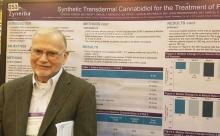
STAR 2 is the extension of STAR 1 (Synthetic Transdermal Cannabidiol for the Treatment of Epilepsy), a 12-week, phase 2a study of the gel. It randomized 181 patients to placebo or to 195 mg or 390 mg CBD gel twice daily.
Patients were a mean of about 40 years old. They had incompletely controlled focal epilepsies, experiencing about 10 seizures per month despite taking a median of three antiepileptic drugs (AEDs). The most commonly used AEDs were levetiracetam (45%), carbamazepine (41%), lamotrigine (33%), lacosamide (28%), and valproate (22%).
By the end of STAR 1, there was an median 18% reduction in seizures from baseline in the 195-mg group, and the 390-mg group experienced a 14% reduction. However, neither of these findings were statistically significant compared with placebo. Dr. Messenheimer said an unusually high 25% placebo response rate contributed to the nonsignificant findings.
Still, patients remained committed to the study, Dr. Messenheimer pointed out: 171 of the 174 STAR trial completers entered the STAR 2 extension. The entire cohort started on the 390-mg dose, and at month 5, they could titrate up to 585 mg or 780 mg daily, or reduce the does to 195 mg twice daily.
At the 18-month point, 76 patients remained in the study. Five discontinued because of an adverse event. Sixty stopped because the gel was ineffective, and the rest exited the study on the decision of an investigator. Dr. Messenheimer presented a responder analysis on 63 of the remaining subjects with full data, as well as an intent-to-treat analysis on the entire STAR 2 cohort.
Among the entire cohort, continued treatment appeared to confer increasing benefit, he said in an interview. By 3 months, the median seizure reduction rate was 25%; it increased to 40% by 6 months and 48% by 9 months. For the next 9 months, the seizure reduction rate stayed steady, hovering at around 55%.
“Among all the patients, we saw an increase in efficacy over 18 months. Half of the patients stayed on 390 mg, and of the half that titrated to higher doses. Most of these went up to 780 mg, but we really didn’t see that the higher doses conferred much benefit over the 390.”
The 63-patient cohort could be viewed as a responder-only analysis, Dr. Messenheimer said, since most of the dropouts occurred in the first few months of the study. Nevertheless, the response rates in the entire 171-person cohort were quite similar, with a 49% reduction by 3 months that increased to a median 55% reduction by 18 months.
The gel was generally well tolerated, although Dr. Messenheimer pointed out three serious adverse events that were probably drug related: two cases of anxiety and one case of increased seizures. Other events that occurred in significantly more of the CBD groups were headaches (12%), upper respiratory infection (11%), lacerations (9%), and fatigue (6%).
There were no liver enzyme abnormalities.
Zynerba sponsored the study; Dr. Messenheimer is a paid consultant for Zynerba.
SOURCE: O’Brien TJ et al. AES 2018, Abstract 2.253
REPORTING FROM AES 2018
Key clinical point:
Major finding: The gel reduced uncontrolled focal seizures by a median of about 50%.
Study details: The open-label extension study comprised 171 subjects.
Disclosures: Zynerba sponsored the study; Dr. Messenheimer is a paid consultant for Zynerba.
Source: O’Brien TJ et al. AES 2018, Abstract 2.253
Single-item scale effective for assessing sleep quality
The single-item sleep quality scale (SQS) produced favorable results comparable to other complex, time-intensive assessment tools, according to findings published in the Journal of Clinical Sleep Medicine.
In a study of 70 insomnia patients and 651 depression patients, concurrent criterion validity analysis yielded strong correlations between the SQS and the morning-questionnaire insomnia (MQI) and Pittsburgh Sleep Quality Index (PSQI) in patients with insomnia and depression, respectively. The investigators wrote, “The single-item format enables a patient-reported rating of sleep quality over a 7-day recall period without greatly increasing the patient’s burden. The use of a discretizing visual analog scale (VAS) increases the potential for a more sensitive measurement.” The SQS is a quick but accurate self-reported assessment of sleep quality.
The SQS was validated based on two studies. Eligible patients in the 4-week, randomized, multicenter insomnia study were aged 30-75 years and were receiving a Food and Drug Administration–approved hypnotic agent as usual treatment for insomnia based on criteria from the Diagnostic and Statistical Manual of Mental Disorders, Fourth Edition (DSM-IV). The MQI was used daily for the duration of the study, wrote Ellen Snyder, PhD, of Merck & Co., in Kenilworth, N.J., and her coauthors.
The depression study was a randomized, double-blind, parallel-group, 12-month international trial evaluating the safety of the substance P antagonist aprepitant, compared with paroxetine hydrochloride. Patients were aged 18 years or older, with a DSM-IV diagnosis of major depressive disorder. Patients completed the SQS and PSQI at baseline, week 1, and week 8.
In insomnia patients, a Pearson correlation of –.76 was found at week 1 for the SQS in relation to the MQI.
In patients with depression, Goodman-Kruskal correlation coefficients for the SQS in relation to the Pittsburgh Sleep Quality Index (PSQI) were –.87, –.88, and –.92 at baseline, week 1, and week 8, respectively.
Correlations were negative because “better sleep quality is associated with a lower score on the MQI and PSQI, but a higher score on the SQS,” the authors noted.
The results support the use of the SQS as a “practical sleep measure that can effectively gauge sleep quality without significantly increasing the burden of clinical trial participants,” compared with lengthier assessments such as the MQI and PSQI, they added.
Funding for the study was provided by Merck Sharp & Dohme.
SOURCE: Snyder E et al. J Clin Sleep Med. 2018;14(11):1849-57.
FROM THE JOURNAL OF CLINICAL SLEEP MEDICINE
Key clinical point: The
Major finding: Week 1 Pearson correlation was –0.76 between the SQS and the morning-questionnaire insomnia (MQI); week 8 Goodman-Kruskal correlation between SQS and the Pittsburgh Sleep Quality Index (PSQI) was –0.92.
Study details: An analysis of SQS versus other measures in 70 insomnia patients and 651 depression patients.
Disclosures: Funding for the study was provided by Merck Sharp & Dohme.
Source: Snyder E et al. J Clin Sleep Med. 2018;14(11):1849-57
CDC: Acute flaccid myelitis on the decline for 2018
, according to the Centers for Disease Control and Prevention.
Through Nov. 30, 134 cases of AFM in 33 states have been confirmed out of the 299 reported to the CDC. That represents “an increase of 18 confirmed cases from the previous week, but most of the latest confirmed AFM cases occurred in September and October,” the CDC reported Dec. 3.
There has been a pattern of increased AFM cases every other year for the previous 4 years: 120 cases in 2014, 22 cases in 2015, 149 cases in 2016, and 33 cases in 2017. “Most cases are reported between August and October, and a marked reduction in cases is seen in November. That pattern appears to be repeating in 2018 because states have reported fewer [persons under investigation] over the past couple of weeks. CDC expects this decline to continue,” the statement said.
The 16 confirmed cases in Texas are the most for any state this year, followed by Colorado with 15; Ohio with 10; and Illinois, New Jersey, and Washington with 9 each. California and Florida have not had any confirmed cases as of Nov. 30. Since 2014, over 90% of all confirmed AFM cases have occurred in children, the CDC noted.
More information on AFM is available at a CDC website for health care professionals.
