User login
Cardiology News is an independent news source that provides cardiologists with timely and relevant news and commentary about clinical developments and the impact of health care policy on cardiology and the cardiologist's practice. Cardiology News Digital Network is the online destination and multimedia properties of Cardiology News, the independent news publication for cardiologists. Cardiology news is the leading source of news and commentary about clinical developments in cardiology as well as health care policy and regulations that affect the cardiologist's practice. Cardiology News Digital Network is owned by Frontline Medical Communications.
Anticoagulation Shows No Benefit in Preventing Second Stroke
BOSTON — Patients who have had a stroke are thought to be at a higher risk for another one, but oral anticoagulation with edoxaban led to no discernible reduction in the risk for a second stroke, and the risk for major bleeding was more than quadruple the risk with no anticoagulation, a subanalysis of a major European trial has shown.
“There is no interaction between prior stroke or TIA [transient ischemic attack] and the treatment effect, and this is true for the primary outcome and the safety outcome,” Paulus Kirchoff, MD, director of cardiology at the University Heart and Vascular Center in Hamburg, Germany, said during his presentation of a subanalysis of the NOAH-AFNET 6 trial at the annual meeting of the Heart Rhythm Society (HRS) 2024. However, “there is a signal for more safety events in patients randomized to anticoagulation with a prior stroke.”
The subanalysis involved 253 patients who had had a stroke or TIA and who had device-detected atrial fibrillation (AF) from the overall NOAH-AFNET 6 population of 2536 patients, which enrolled patients 65 years and older with at least one additional CHA2DS-VASc risk factor and patients 75 years and older with device-detected subclinical AF episodes of at least 6 minutes. Patients were randomized to either edoxaban or no anticoagulation, but 53.9% of the no-anticoagulation group was taking aspirin at trial enrollment. Anticoagulation with edoxaban was shown to have no significant impact on stroke rates or other cardiovascular outcomes.
Subanalysis Results
In the subanalysis, a composite of stroke, systemic embolism, and cardiovascular death — the primary outcome — was similar in the edoxaban and no-anticoagulation groups (14/122 patients [11.5%] vs 16/131 patients [12.2%]; 5.7% vs 6.3% per patient-year).
The rate of recurrent stroke was also similar in the edoxaban and no-anticoagulation groups (4 of 122 patients [3.3%] vs 6 of 131 patients [4.6%]; 1.6% vs 2.3% per patient-year). And there were eight cardiovascular deaths in each group.
However, edoxaban patients had significantly higher rates of major bleeding.
“This is a subanalysis, so what we see in terms of the number of patients with events is not powered for a definitive answer, but we do see that there were 10 major bleeds in the group of patients with a prior stroke or TIA in NOAH,” Dr. Kirchoff reported. “Eight of those 10 major bleeds occurred in patients randomized to edoxaban.”
Results from the NOAH-AFNET 6 trial have been compared with those from the ARTESiA trial, which compared apixaban anticoagulation with aspirin in patients with subclinical AF and was also presented at HRS 2024. ARTESiA showed that apixaban significantly lowered the risk for stroke and systemic embolism.
“In ARTESiA, everyone was on aspirin when they were randomized to no anticoagulation; in NOAH, only about half were on aspirin,” Dr. Kirchoff said.
Both studies had similar outcomes for cardiovascular death in the anticoagulation and no-anticoagulation groups. “It’s not significant; it may be chance, but it’s definitely not the reduction in death that we have seen in the anticoagulant trials,” Dr. Kirchoff said. “When you look at the meta-analyses of the early anticoagulation trials, there’s a one third reduction in death, and here we’re talking about a smaller reduction.”
This research points to a need for a better way to evaluate stroke risk. “We need new markers,” Dr. Kirchoff said. “Some of them may be in the blood or imaging, genetics maybe, and one thing that really emerges from my perspective is that we now have the first evidence to suggest that patients with a very low atrial fibrillation burden have a low stroke rate.”
More research is needed to better understand AF characteristics and stroke risk, he said.
AF Care Enters a ‘Gray Zone’
The NOAH-AFNET 6 results, coupled with those from ARTESiA, are changing the paradigm for anticoagulation in patients with stroke, said Taya Glotzer, MD, an electrophysiologist at the Hackensack University Medical Center in Hackensack, New Jersey, who compiled her own analysis of the studies’ outcomes.
“In ARTESiA, the stroke reduction was only 0.44% a year, with a number needed to treat of 250,” she said. “In the NOAH-AFNET 6 main trial, the stroke reduction was 0.2%, with the number needed to treat of 500, and in the NOAH prior stroke patients, there was a 0.7% reduction, with a number needed to treat of 143.”
None of these trials would meet the standard for a class 1 recommendation for anticoagulation with a reduction of even 1%-2% per year, she noted, but they do show that the stroke rate “is very, very low” in prior patients with stroke.
“Prior to 2024, we knew what was black and white; we knew who to anticoagulate and who not to anticoagulate. And now we are in a gray zone, trying to balance the risk of stroke and bleeding. We have to individualize or hope for substudies, perhaps using the CHA2DS-VASc score or other information about the left atrium, to help us make decisions in these patients. It’s not just going to be black and white,” she said.
Dr. Kirchoff had no relevant financial relationships to disclose. Dr. Glotzer disclosed financial relationships with Medtronic, Abbott, Boston Scientific, and MediaSphere Medical.
A version of this article first appeared on Medscape.com.
FROM HRS 2024
Delays After Tests for Suspected Heart Failure ‘a Scandal’
LISBON, PORTUGAL — Few people with suspected heart failure and elevated N-terminal prohormone of brain natriuretic peptide (NT-proBNP) levels are receiving a diagnosis after a year, reported investigators, who say high rates of hospitalization are common.
Presenting here at the Heart Failure Association of the European Society of Cardiology (HFA-ESC) 2024, researchers shared results from the REVOLUTION-HF study involving almost 8000 people who consulted outpatient primary and secondary care over a 5-year period.
The outcomes were even worse in patients with high NT-proBNP levels.
Patients with suspected heart failure are “waiting far too long to see a specialist, and that results in a delay to guideline-directed medical therapy, despite the fact that we’re perfectly happy to slap them all on diuretics,” said study presenter Lisa Anderson, MD, PhD, Cardiovascular Clinical Academic Group, Molecular and Clinical Sciences Research Institute, St George’s Hospital, University of London, England.
“We need to rethink our management of heart failure patients presenting in the community,” she said.
A big gap exists internationally between presentation with heart failure, an elevated NT-proBNP, and confirmatory specialist assessment, she explained.
“It’s a scandal that patients are coming to the GP with signs and symptoms of heart failure, they get tested for natriuretic peptides, and nothing happens,” said co-author Antoni Bayés-Genís, MD, PhD, Heart Institute director, Hospital Universitari Germans Trias i Pujol Catedràtic, Barcelona, Spain.
“These patients may receive an echo, or not, in the coming 12 months,” and “during these 12 months, there is a huge number of heart failure hospitalizations and deaths that could probably be prevented.”
Why the Reluctance to Diagnose?
Many issues get in the way of early diagnosis, Dr. Bayés-Genís said. “Inertia, comorbidities, ageism.”
A lot of patients with heart failure are elderly women with some degree of weight gain, he said. “And they come to the clinic with fatigue, so we tell them, ‘Well, that’s normal.”
But “it may not be normal,” he added. “This is a very important topic that we, as a society, need to address.”
There are several “misconceptions” about heart failure, said Ileana L. Piña, MD, MPH, the Robert Stein Chair for Quality and Safety, Sidney Kimmel Medical College, Thomas Jefferson University, Philadelphia, who was not involved in the study.
For example, “we’re all convinced that guideline-directed medical therapy works,” but the evidence is only for patients “with a diagnosis.” In addition, “millions of patients get tested” for heart failure, but they already have a “known diagnosis.”
“When we study these drugs, we’re studying them on patients with manifest disease,” who are only then randomized, Dr. Piña said. “But we seldom see them while they’re developing heart failure. And it’s a process; it doesn’t happen overnight.”
Patients initially often think they may have asthma, and so what follows is an extended period of “uncertainty” and “important time lost” before they finally undergo the assessments that show that they have heart failure, she said.
However, “uncertainty” often lands a patient “in the emergency room or with an unscheduled office visit, where NT-proBNP might get ordered and there’s a long lineup for an echo.”
There are several strengths of the current study, Dr. Piña said, including the fact that 50% of the study population were women, and they were older than a typical trial population. Nevertheless, the results were “eye-opening but not surprising” and, in the end, “disappointing.”
“I agree, we need a revolution, Dr. Anderson,” Dr. Piña said. “The revolution of paying attention to the NT-proBNP when you get it and it’s elevated” and then following through with echocardiography and starting “guideline-directed medical therapy early.”
The diagnosis of heart failure “relies on the presentation of patients with nonspecific signs and symptoms,” such as dyspnea and peripheral edema, “but initiation of guideline-directed medical therapy — life-saving treatment — has to wait until we have a formal echocardiography and specialist clinician assessment,” Dr. Anderson said.
The latest clinical consensus statement from the Heart Failure Association “proposes both rule-in and rule-out NT-proBNP levels for heart failure diagnosis, and obviously we all recognize that it’s important to treat patients as soon as they’re diagnosed,” she explained.
REVOLUTION-HF
To examine the risk profile for patients presenting to outpatient care with suspected heart failure, the researchers conducted REVOLUTION-HF, which leveraged nationwide Swedish linked data from general practices, specialists, pharmacies, hospitals, and cause of death registers.
“Really impressively, most of these NT-proBNP tests were coming back within a day,” Dr. Anderson said, “so a really, really good turnaround.”
Individuals were excluded if they had an inpatient admission, echocardiography, or heart failure diagnosis between presentation and the NT-proBNP measurement.
These people were then compared with those presenting to primary or secondary outpatient care for any reason and matched for age, sex, care level, and index year. Both groups were followed up for 1 year.
“Despite this really impressive, almost immediate NT-proBNP testing,” the waiting times to undergo echocardiography were “really disappointing,” Dr. Anderson said.
The median time to first registered echocardiography was 40 days, and only 29% of patients with suspected heart failure received a diagnosis within a year of the index presentation date, which she described as “inadequately slow.”
“And how does this translate to medical therapy?” she asked.
Heart Failure Drugs
After the index presentation, the rate of loop diuretic use quadrupled among individuals suspected of having heart failure, but there was a “muted response” when it came to the prescribing of beta-blockers and the other pillars of heart failure therapy, which Dr. Anderson called “very disappointing.”
For outcomes after the index presentation, the rate of hospitalization was much higher in the group with suspected heart failure than in the control group (16.1 vs 2.2 events per 100 person-years). And all-cause mortality occurred more often in the group with suspected heart failure than in the control group (10.3 vs 6.5 events per 100 person-years).
Among patients with NT-proBNP levels of 2000 ng/L, there was a “rapid” onset of hospitalization “within the first few days” of the index presentation, which was tracked by a more linear rise in all-cause deaths, Dr. Anderson reported.
In the United Kingdom, “we are very proud of our 2- and 6-week pathways,” which stipulate that suspected heart failure patients with NT-proBNP levels between 400 and 2000 ng/L are to have a specialist assessment and transthoracic echocardiography within 6 weeks; for those with levels > 2000 ng/L, that interval is accelerated to 2 weeks, she said.
The current results show that “2 weeks is too slow.” And looking at the rest of the cohort with lower NT-proBNP levels, “patients have already been admitted and died” by 6 weeks, she said.
When patients are stratified by age, “you get exactly what you would expect,” Dr. Anderson said. “The older patients are the most at risk” for both hospitalization and all-cause mortality.
A version of this article appeared on Medscape.com.
FROM HFA-ESC 2024
Surgeons Most Likely to Behave Unprofessionally: Study
Most doctors mind their manners. But surgeons are the most likely to be reported for unprofessional behavior, while physicians practicing in pediatric settings are the least likely, according to a recent study of more than 35,000 physicians.
The research, published on June 6 in JAMA Network Open, found that fewer than 10% of physicians were reported by their coworkers for at least one instance of unprofessional behavior, and only 1% showed a pattern of such reports.
Data were gathered from the Center for Patient and Professional Advocacy’s (CPPA’s) Coworker Observation Reporting System (CORS) program, a national collaborative in which 193 participating hospitals and practice sites file safety-event reports involving medical workers’ unprofessional behaviors. An algorithm that weights CORS reports based on recency and severity was used to analyze the data. The study was spearheaded by William O. Cooper, MD, MPH, director of the CPPA at Vanderbilt University Medical Center, Nashville, Tennessee.
The retrospective cohort study included deidentified data on credentialed physicians, not including residents or fellows, who practiced at a CORS site between 2018 and 2022.
Why Surgeons?
The authors speculated that the reason surgeons were reported for unprofessional behavior more often than their colleagues in nonsurgical specialties was because surgery is a more stressful environment than other specialties and requires more teamwork, resulting in more interactions during high-stakes events.
Daniel Katz, MD, professor and vice chair of education for the Department of Anesthesiology, Perioperative and Pain Medicine at the Icahn School of Medicine at Mount Sinai, New York City, added that part of the problem is that surgeons are expected to perform at very high levels all the time.
“When things that are outside the control of the surgeon don’t go well,” Dr. Katz said, “that can lead to increased frustration and negative emotions, which will then bring out these kinds of behaviors.”
Types of Unprofessional Behaviors
The most common out-of-bounds behaviors reported involved disrespectful communication or lack of professional responsibility. In one example, a physician called a coworker a “bossy cow” when the coworker reminded the physician of the need to do a timeout before beginning a bronchoscopy.
In another case involving professional responsibility, a coworker asked a physician if the team should wait for a disoriented patient’s spouse to arrive. The doctor’s response: “We’ll be here all night if we do that. If you won’t sign as a witness, I’ll get someone else who will.”
The least common reports involved unprofessionalism related to medical care or professional integrity. One cited a physician removing a Foley catheter without wearing gloves and having visible urine on his hands and not washing them before touching other things in the room. In a reported lapse of professional integrity, a physician billed at level five after spending only 4 minutes with a patient.
Impact of Unprofessional Behavior
Unprofessional behavior among physicians is more than just unpleasant. It can threaten the functioning of teams and increase patient complications. In addition, individuals who model unprofessional behaviors are associated with increased malpractice claims, the study’s authors wrote.
Dr. Katz agreed that unprofessional behavior is damaging to both patients and the profession as a whole.
However, this doesn’t happen because some doctors are bad, he said. Physicians today are working in a pressure cooker. The current healthcare environment, with its increased administrative burdens, lack of staffing, and other problems, has increased the overall level of stress and led to burnout among healthcare personnel.
“You have to fix the system to create a working environment that doesn’t cause somebody to explode,” Dr. Katz said.
The goal of the CORS program and this study, Dr. Cooper said, is to help physicians better weather these stresses.
Study Limitations
The authors noted some weaknesses in the study. Some unprofessional behavior may go unreported because of fear of retaliation or for other reasons victims or witnesses did not feel safe to report their colleagues. Also, reports were not evaluated to ensure the truth of the accusations. The records reviewed did not include the gender of the physician, though the researchers pointed out that previous studies have shown that women are less likely than men to receive CORS reports.
A version of this article appeared on Medscape.com.
GLP-1s Reduced Secondary Stroke Risk in Patients With Diabetes, Obesity
, according to authors of a recent meta-analysis. With benefits across administration routes, dosing regimens, type 2 diabetes status, and total and nonfatal strokes, the findings could improve GLP-1 RA implementation by stroke specialists in patients with stroke history and concurrent type 2 diabetes or obesity, authors said. The study was published online in the International Journal of Stoke.
Extending Longevity
Agents including GLP-1 RAs that have been found to reduce cardiovascular events among patients with type 2 diabetes and patients who are overweight or obese also reduce risk of recurrent stroke among patients with a history of stroke who are overweight, obese, or have metabolic disease, said American Heart Association (AHA) Chief Clinical Science Officer Mitchell S. V. Elkind, MD, who was not involved with the study but was asked to comment.
“Stroke is a leading cause of mortality and the leading cause of serious long-term disability,” he added, “so medications that help to reduce that risk can play an important role in improving overall health and well-being and hopefully reducing premature mortality.”
Investigators Anastasia Adamou, MD, an internal medicine resident at AHEPA University Hospital in Thessaloniki, Greece, and colleagues searched MEDLINE and Scopus for cardiovascular outcome trials involving adults randomly assigned to GLP-1 RAs or placebo through November 2023, ultimately analyzing 11 randomized controlled trials (RCTs).
Among 60,380 participants in the nine studies that assessed total strokes, 2.5% of the GLP-1 RA group experienced strokes during follow-up, versus 3% in the placebo group (relative risk [RR] 0.85, 95% confidence interval [CI] 0.77-0.93). Regarding secondary outcomes, the GLP-1 RA group showed a significantly lower rate of nonfatal strokes versus patients on placebo (RR 0.87, 95% CI 0.79-0.95). Conversely, investigators observed no significant risk difference among the groups regarding fatal strokes, probably due to the low rate of events — 0.3% and 0.4% for treated and untreated patients, respectively.
Subgroup analyses revealed no interaction between dosing frequency and total, nonfatal, or fatal strokes. The investigators observed no difference in nonfatal strokes among participants by type 2 diabetes status and medication administration route (oral versus subcutaneous).
“The oral administration route could provide the advantage of lower local ecchymoses and allergic reactions due to subcutaneous infusions,” Dr. Adamou said in an interview. But because oral administration demands daily intake, she added, treatment adherence might be affected. “For this reason, our team performed another subgroup analysis to compare the once-a-day to the once-a-month administration. No interaction effect was again presented between the two subgroups. This outcome allows for personalization of the administration method for each patient.”

Addressing Underutilization
Despite more than 2 decades of widespread use and well-established effects on body weight, HbA1c, and cardiovascular risk, GLP-1 RAs remain underutilized, authors wrote. This is especially true in primary care, noted one study published in Clinical Diabetes.
“GLP-1 RAs have been used for many years to treat diabetic patients,” said Dr. Adamou. But because their impact on cardiovascular health regardless of diabetic status is only recently known, she said, physicians are exercising caution when prescribing this medication to patients without diabetes. “This is why more studies need to be available, especially RCTs.”
Most neurologists traditionally have left management of type 2 diabetes and other metabolic disorders to primary care doctors, said Dr. Elkind. “However, these medications are increasingly important to vascular risk reduction and should be considered part of the stroke specialist’s armamentarium.”
Vascular neurologists can play an important role in managing metabolic disease and obesity by recommending GLP-1 RAs for patients with a history of stroke, or by initiating these medications themselves, Dr. Elkind said. “These drugs are likely to become an important part of stroke patients’ medication regimens, along with antithrombotic agents, blood pressure control, and statins. Neurologists are well-positioned to educate other physicians about the important connections among brain, heart, and metabolic health.”
To that end, he said, the AHA will update guidelines for both primary and secondary stroke prevention as warranted by evidence supporting GLP-1 RAs and other medications that could impact stroke risk in type 2 diabetes and related metabolic disorders. However, no guidelines concerning use of GLP-1 RAs for secondary stroke prevention in obesity exist. Here, said Dr. Elkind, the AHA will continue building on its innovative Cardiovascular-Kidney Metabolic Health program, which includes clinical suggestions and may include more formal clinical practice guidelines as the evidence evolves.
Among the main drivers of the initiative, he said, is the recognition that cardiovascular disease — including stroke — is the major cause of death and morbidity among patients with obesity, type 2 diabetes, and metabolic disorders. “Stroke should be considered an important part of overall cardiovascular risk, and the findings that these drugs can help to reduce the risk of stroke specifically is an important additional reason for their use.”
Dr. Elkind and Dr. Adamou reported no conflicting interests. The authors received no financial support for the study.
, according to authors of a recent meta-analysis. With benefits across administration routes, dosing regimens, type 2 diabetes status, and total and nonfatal strokes, the findings could improve GLP-1 RA implementation by stroke specialists in patients with stroke history and concurrent type 2 diabetes or obesity, authors said. The study was published online in the International Journal of Stoke.
Extending Longevity
Agents including GLP-1 RAs that have been found to reduce cardiovascular events among patients with type 2 diabetes and patients who are overweight or obese also reduce risk of recurrent stroke among patients with a history of stroke who are overweight, obese, or have metabolic disease, said American Heart Association (AHA) Chief Clinical Science Officer Mitchell S. V. Elkind, MD, who was not involved with the study but was asked to comment.
“Stroke is a leading cause of mortality and the leading cause of serious long-term disability,” he added, “so medications that help to reduce that risk can play an important role in improving overall health and well-being and hopefully reducing premature mortality.”
Investigators Anastasia Adamou, MD, an internal medicine resident at AHEPA University Hospital in Thessaloniki, Greece, and colleagues searched MEDLINE and Scopus for cardiovascular outcome trials involving adults randomly assigned to GLP-1 RAs or placebo through November 2023, ultimately analyzing 11 randomized controlled trials (RCTs).
Among 60,380 participants in the nine studies that assessed total strokes, 2.5% of the GLP-1 RA group experienced strokes during follow-up, versus 3% in the placebo group (relative risk [RR] 0.85, 95% confidence interval [CI] 0.77-0.93). Regarding secondary outcomes, the GLP-1 RA group showed a significantly lower rate of nonfatal strokes versus patients on placebo (RR 0.87, 95% CI 0.79-0.95). Conversely, investigators observed no significant risk difference among the groups regarding fatal strokes, probably due to the low rate of events — 0.3% and 0.4% for treated and untreated patients, respectively.
Subgroup analyses revealed no interaction between dosing frequency and total, nonfatal, or fatal strokes. The investigators observed no difference in nonfatal strokes among participants by type 2 diabetes status and medication administration route (oral versus subcutaneous).
“The oral administration route could provide the advantage of lower local ecchymoses and allergic reactions due to subcutaneous infusions,” Dr. Adamou said in an interview. But because oral administration demands daily intake, she added, treatment adherence might be affected. “For this reason, our team performed another subgroup analysis to compare the once-a-day to the once-a-month administration. No interaction effect was again presented between the two subgroups. This outcome allows for personalization of the administration method for each patient.”
Addressing Underutilization
Despite more than 2 decades of widespread use and well-established effects on body weight, HbA1c, and cardiovascular risk, GLP-1 RAs remain underutilized, authors wrote. This is especially true in primary care, noted one study published in Clinical Diabetes.
“GLP-1 RAs have been used for many years to treat diabetic patients,” said Dr. Adamou. But because their impact on cardiovascular health regardless of diabetic status is only recently known, she said, physicians are exercising caution when prescribing this medication to patients without diabetes. “This is why more studies need to be available, especially RCTs.”
Most neurologists traditionally have left management of type 2 diabetes and other metabolic disorders to primary care doctors, said Dr. Elkind. “However, these medications are increasingly important to vascular risk reduction and should be considered part of the stroke specialist’s armamentarium.”
Vascular neurologists can play an important role in managing metabolic disease and obesity by recommending GLP-1 RAs for patients with a history of stroke, or by initiating these medications themselves, Dr. Elkind said. “These drugs are likely to become an important part of stroke patients’ medication regimens, along with antithrombotic agents, blood pressure control, and statins. Neurologists are well-positioned to educate other physicians about the important connections among brain, heart, and metabolic health.”
To that end, he said, the AHA will update guidelines for both primary and secondary stroke prevention as warranted by evidence supporting GLP-1 RAs and other medications that could impact stroke risk in type 2 diabetes and related metabolic disorders. However, no guidelines concerning use of GLP-1 RAs for secondary stroke prevention in obesity exist. Here, said Dr. Elkind, the AHA will continue building on its innovative Cardiovascular-Kidney Metabolic Health program, which includes clinical suggestions and may include more formal clinical practice guidelines as the evidence evolves.
Among the main drivers of the initiative, he said, is the recognition that cardiovascular disease — including stroke — is the major cause of death and morbidity among patients with obesity, type 2 diabetes, and metabolic disorders. “Stroke should be considered an important part of overall cardiovascular risk, and the findings that these drugs can help to reduce the risk of stroke specifically is an important additional reason for their use.”
Dr. Elkind and Dr. Adamou reported no conflicting interests. The authors received no financial support for the study.
FROM THE INTERNATIONAL JOURNAL OF STROKE
DEA Training Mandate: 8 Hours of My Life I’d Like Back
It’s time to renew two of my three narcotic prescribing licenses. For the first time in my career, I’ve waffled on whether the financial outlay to the US Drug Enforcement Agency (DEA) is worth it.
At $888 each, I’ve considered letting two licenses lapse because I only work part-time in Montana. But several friends advised me to keep a “spare” in case I transfer to a new location.
I thought about just paying the fees until I could do a little more research, but there is no mechanism for a refund unless I die within the first year of the 3-year cycle, provide incorrect credit card digits, or accidentally duplicate payments.
The renewal fee is just part of the issue.
Mandatory 8-Hour Training
I also received an alert about the requirement for more “narcotics prescribing education” thanks to the Medication Access and Training Expansion Act (MATE).
The requirement seems counterintuitive because opioid prescribing has decreased for the 10th consecutive year, according to the AMA Overdose Epidemic Report. The continuing rise in overdose deaths is largely due to illegitimate manufacturing of synthetic opioids.
I’ve written zero outpatient narcotics prescriptions in the past 6 years, and I’ve written very few in my 33 years of practice. My use is limited to intravenous morphine for flash pulmonary edema or refractory angina, but unless you graduated from a training program within 5 years of the June 2023 mandate or are boarded in addiction medicine, there is no way to escape the 8-hour education requirement.
The problem is that these courses are never just 8 hours in duration. After signing up for one such CME course that cost $150, I was still dying of boredom and at risk for DVT 4 days later. That’s how long it took to sit through.
Instead of the 30 seconds it should have taken to review the simple instructions to deliver Narcan, there were scores of screens followed by juvenile quizlets and cartoons. All but about 2 hours out of the 4 days is now relegated to that category of “hours of my life that I can never get back.” Additionally, none of that mandatory “education” will change my prescribing habits one whit.
And beware the penalty. 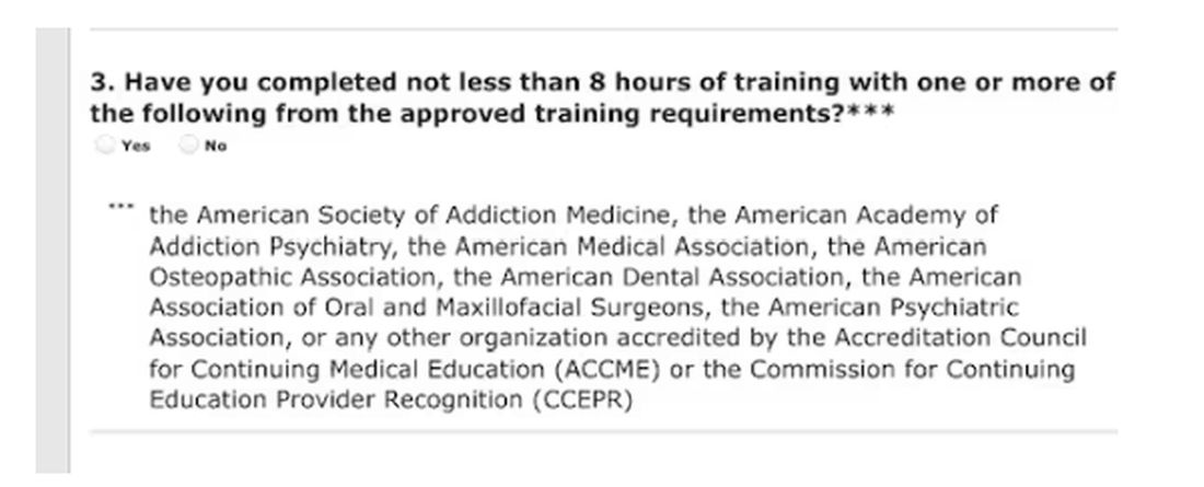
Of course, I would always be truthful when asked to check the box on the DEA renewal application attesting to my having completed the required education. On the outside chance that you plan to check the yes box without completing the relevant courses, those found guilty of such false claims could be fined up to $250,000 and subject to “not more than four years in prison,” or both. Yikes! 
Larry Houck, a former DEA investigator, explained that “[t]here are lot of people who are coming up for renewal and log on but still don’t know this is a requirement.” Neither ignorance nor complacency is an acceptable defense.
Changes Needed
The only good thing that came of those 4 long days of opioid education was a motivation to drive change in our current licensing and educational experience. Why not use this opportunity to reform the DEA-physician/prescriber relationship?
The educational requirements should be curtailed for those of us who do not provide outpatient narcotic prescriptions even if we use inpatient opioids. Meds with low abuse potential should be rescheduled to minimize who gets caught in the broad net of the education requirement.
We should reduce overregulation of the legitimate prescribers by lowering, instead of increasing, licensing fees. We should change to a single license number that covers every state. In this digital age, there is no legitimate excuse to prevent this from happening.
After all, the settlements from opioid manufacturers and distributors will in time total $50 billion. It seems that at least some of the responsibilities of the DEA could shift to states, cities, and towns.
My friend Siamak Karimian, MD, who provides locum services in multiple states, pays for seven active DEA licenses every 3 years. He pointed out the hypocrisy in the current regulatory system: “It’s funny that you can have only one DEA or state license and work for the government in all other states or territories with no limits, including the VA, Indian healthcare systems, or prison systems.”
All other prescribers require a separate DEA number for every state. Ultimately, you’d think tracking prescriptions for a single DEA number should be far simpler than tracking someone with seven.
Competent physicians not guilty of criminal overprescribing seem to be the last to be considered in nearly every healthcare endeavor these days. It would be refreshing if they would reduce our fees and prevent this waste of our time.
And while we are at it, perhaps a more fitting punishment is due for Richard Sackler and all the Purdue Pharma–affiliated family members. The Sacklers will pay out $6 billion in exchange for immunity against civil litigation. That doesn’t seem like much when they are worth $11 billion.
Perhaps they should be made to take an 8-hour course on opioid prescribing, annually and in perpetuity. Let’s see them complete a few quizlets and sit through screens of instruction on how to administer Naloxone. Of course, that would be a mild punishment for those who manufactured a drug that killed hundreds of thousands. But it would be a start.
Dr. Walton-Shirley, a clinical cardiologist in Nashville, Tennessee, has disclosed no relevant financial relationships.
A version of this article appeared on Medscape.com.
It’s time to renew two of my three narcotic prescribing licenses. For the first time in my career, I’ve waffled on whether the financial outlay to the US Drug Enforcement Agency (DEA) is worth it.
At $888 each, I’ve considered letting two licenses lapse because I only work part-time in Montana. But several friends advised me to keep a “spare” in case I transfer to a new location.
I thought about just paying the fees until I could do a little more research, but there is no mechanism for a refund unless I die within the first year of the 3-year cycle, provide incorrect credit card digits, or accidentally duplicate payments.
The renewal fee is just part of the issue.
Mandatory 8-Hour Training
I also received an alert about the requirement for more “narcotics prescribing education” thanks to the Medication Access and Training Expansion Act (MATE).
The requirement seems counterintuitive because opioid prescribing has decreased for the 10th consecutive year, according to the AMA Overdose Epidemic Report. The continuing rise in overdose deaths is largely due to illegitimate manufacturing of synthetic opioids.
I’ve written zero outpatient narcotics prescriptions in the past 6 years, and I’ve written very few in my 33 years of practice. My use is limited to intravenous morphine for flash pulmonary edema or refractory angina, but unless you graduated from a training program within 5 years of the June 2023 mandate or are boarded in addiction medicine, there is no way to escape the 8-hour education requirement.
The problem is that these courses are never just 8 hours in duration. After signing up for one such CME course that cost $150, I was still dying of boredom and at risk for DVT 4 days later. That’s how long it took to sit through.
Instead of the 30 seconds it should have taken to review the simple instructions to deliver Narcan, there were scores of screens followed by juvenile quizlets and cartoons. All but about 2 hours out of the 4 days is now relegated to that category of “hours of my life that I can never get back.” Additionally, none of that mandatory “education” will change my prescribing habits one whit.
And beware the penalty.
Of course, I would always be truthful when asked to check the box on the DEA renewal application attesting to my having completed the required education. On the outside chance that you plan to check the yes box without completing the relevant courses, those found guilty of such false claims could be fined up to $250,000 and subject to “not more than four years in prison,” or both. Yikes!
Larry Houck, a former DEA investigator, explained that “[t]here are lot of people who are coming up for renewal and log on but still don’t know this is a requirement.” Neither ignorance nor complacency is an acceptable defense.
Changes Needed
The only good thing that came of those 4 long days of opioid education was a motivation to drive change in our current licensing and educational experience. Why not use this opportunity to reform the DEA-physician/prescriber relationship?
The educational requirements should be curtailed for those of us who do not provide outpatient narcotic prescriptions even if we use inpatient opioids. Meds with low abuse potential should be rescheduled to minimize who gets caught in the broad net of the education requirement.
We should reduce overregulation of the legitimate prescribers by lowering, instead of increasing, licensing fees. We should change to a single license number that covers every state. In this digital age, there is no legitimate excuse to prevent this from happening.
After all, the settlements from opioid manufacturers and distributors will in time total $50 billion. It seems that at least some of the responsibilities of the DEA could shift to states, cities, and towns.
My friend Siamak Karimian, MD, who provides locum services in multiple states, pays for seven active DEA licenses every 3 years. He pointed out the hypocrisy in the current regulatory system: “It’s funny that you can have only one DEA or state license and work for the government in all other states or territories with no limits, including the VA, Indian healthcare systems, or prison systems.”
All other prescribers require a separate DEA number for every state. Ultimately, you’d think tracking prescriptions for a single DEA number should be far simpler than tracking someone with seven.
Competent physicians not guilty of criminal overprescribing seem to be the last to be considered in nearly every healthcare endeavor these days. It would be refreshing if they would reduce our fees and prevent this waste of our time.
And while we are at it, perhaps a more fitting punishment is due for Richard Sackler and all the Purdue Pharma–affiliated family members. The Sacklers will pay out $6 billion in exchange for immunity against civil litigation. That doesn’t seem like much when they are worth $11 billion.
Perhaps they should be made to take an 8-hour course on opioid prescribing, annually and in perpetuity. Let’s see them complete a few quizlets and sit through screens of instruction on how to administer Naloxone. Of course, that would be a mild punishment for those who manufactured a drug that killed hundreds of thousands. But it would be a start.
Dr. Walton-Shirley, a clinical cardiologist in Nashville, Tennessee, has disclosed no relevant financial relationships.
A version of this article appeared on Medscape.com.
Yoga May Augment Medical Therapy in Heart Failure
LISBON, PORTUGAL — The addition of a yearlong customized yoga therapy intervention to guideline-directed medical therapy (GDMT) appears to significantly improve heart failure measures associated with long-term prognosis, findings from an Indian study suggested.
The research, presented at the Heart Failure Association of the European Society of Cardiology (HFA-ESC) 2024 congress, involved 105 patients assigned to yoga plus GDMT or GDMT alone and demonstrated that there was a large shift in the New York Heart Association (NYHA) functional class from baseline to the 52-week follow-up.
“Yoga therapy has a beneficial impact on heart failure patients on optimal medical management,” said study presenter Ajit Singh, MD, Department of Medicine, Kasturba Medical College, Manipal, Karnataka, India, and the study “demonstrated an overall improvement in left ventricle dimensions and function.”
However, because patients were followed every day and almost a quarter had dropped out by 6 months, the study was “a challenge,” he noted. Nevertheless, the addition of yoga to GDMT could be a “game changer if we try for longer duration.”
For yoga therapy to be considered in clinical practice, a randomized study is required, said session cochair Dana Dawson, MD, PhD, professor of cardiovascular medicine and lead of the Cardiology and Cardiovascular Research Unit, University of Aberdeen, Scotland.
Patients in the current analysis, however, were not randomly allocated to treatment group, which resulted in baseline discrepancies that made the groups “incomparable,” Dr. Dawson explained.
Still, the study showed that yoga is feasible in this patient group and that, even just comparing baseline and follow-up outcomes in the yoga group, there were some significant results.
“It is effective in implementing a change,” she said, “and whether that change is clinically effective needs to be tested in a clinic in a randomized study.”
Why Yoga May Be Particularly Effective
Yoga may be different from other exercise and lifestyle interventions because it is “also about meditation and meeting with your own self,” which corresponds to a form of cognitive behavioral therapy, albeit “conducted in singular manner,” she added.
“It’s not going to be everyone’s cup of tea, and not everyone is going to be inclined to do it,” but it could be suitable in countries where yoga is more commonly practiced as a behavioral, as opposed to lifestyle, intervention, said Dr. Dawson.
Heart failure is a “complex chronic disease” that is a “prime cause of concern for healthcare sectors worldwide,” not least in India, where there is a “very high prevalence” of the disease, Dr. Singh noted.
Evidence from the literature indicates that yoga and other lifestyle modifications can improve the quality of life of patients with heart failure, alongside measures such as left ventricular ejection fraction (LVEF) and NYHA functional class, he said. However, the researchers did not find any study that looked at yoga therapy as an adjunct to standard-of-care treatment.
How Yoga Was Applied
They recruited patients aged 30-70 years with persistent heart failure symptoms, an LVEF of < 45%, and NYHA class III or lower heart failure. All participants had undergone a cardiac procedure 6-12 months previously, and all were receiving optimal GDMT.
Patients were assigned in a nonrandomized fashion to GDMT with or without a customized yoga program. Eight forms of pranayama breath work, meditation, and relaxation techniques were taught to patients in the yoga group by experienced hospital faculty.
They were supervised for 1 week and then advised to continue self-administered yoga at home once a week for 45 minutes. After each home session, an instructor followed up with each study participant to monitor progress.
All participants were assessed with echocardiography and other measures, including physical activities, to determine NYHA functional status at baseline, 6 months, and 1 year.
Of the 110 patients recruited, 25 had dropped out by 6 months. Of the remaining 85 patients included in the analysis, 40 were assigned to the yoga group. The average age was 49 years, and 70 (82%) of the participants were men. The lack of women in the study is a “major drawback,” Dr. Singh noted.
Women did not want to participate, he explained, “because they were afraid to get the follow-up,” saying, “We will not be able to follow this yoga therapy for 1 year.”
After 52 weeks, patients in the yoga group had significantly greater reductions from baseline in systolic and diastolic blood pressure, heart rate, and body mass index than those in the GDMT-alone group (P < .05 for all).
Patients in the yoga group also experienced significantly greater improvements in ejection fraction, increasing from an average of 41.5% to 44.4% over the course of the study. In contrast, ejection fraction decreased from 42.3% to 41.6% in the GDMT-alone group (P < .05).
Crucially, there was a marked improvement in the NYHA class in the yoga group.
With yoga, the proportion of patients with class I heart failure increased from 12% to 47% over the 52 weeks of the study, whereas the proportion with class II heart failure decreased from 57% to 30%, and the proportion with class III heart failure decreased from 30% to 12% (P < .001). In both the yoga and GDMT-alone groups, the proportion of patients with class IV disease increased from 0% to about 10%.
No funding was declared. No relevant financial relationships were declared.
A version of this article appeared on Medscape.com.
AMA Wrestles With AI But Acts on Prior Authorization, Other Concerns
The largest US physician organization wrestled with the professional risks and rewards of artificial intelligence (AI) at its annual meeting, delaying action even as it adopted new policies on prior authorization and other concerns for clinicians and patients.
Physicians and medical students at the annual meeting of the American Medical Association (AMA) House of Delegates in Chicago intensely debated a report and two key resolutions on AI but could not reach consensus, pushing off decision-making until a future meeting in November.
One resolution would establish “augmented intelligence” as the preferred term for AI, reflecting the desired role of these tools in supporting — not making — physicians’ decisions. The other resolution focused on insurers’ use of AI in determining medical necessity.
(See specific policies adopted at the meeting, held June 8-12, below.)
A comprehensive AMA trustees’ report on AI considered additional issues including requirements for disclosing AI use, liability for harms due to flawed application of AI, data privacy, and cybersecurity.
The AMA intends to “continue to methodically assess these issues and make informed recommendations in proposing new policy,” said Bobby Mukkamala, MD, an otolaryngologist from Flint, Michigan, who became the AMA’s new president-elect.
AMA members at the meeting largely applauded the aim of these AI proposals, but some objected to parts of the trustees’ report.
They raised questions about what, exactly, constitutes an AI-powered service and whether all AI tools need the kind of guardrails the AMA may seek. There also were concerns about calls to make AI use more transparent.
While transparency might be an admirable goal, it might prove too hard to achieve given that AI-powered tools and products are already woven into medical practice in ways that physicians may not know or understand, said Christopher Libby, MD, MPH, a clinical informaticist and emergency physician at Cedars Sinai Medical Center in Los Angeles.
“It’s hard for the practicing clinician to know how every piece of technology works in order to describe it to the patient,” Dr. Libby said at the meeting. “How many people here can identify when algorithms are used in their EHR today?”
He suggested asking for more transparency from the companies that make and sell AI-powered software and tools to insurers and healthcare systems.
Steven H. Kroft, MD, the editor of the American Journal of Clinical Pathology, raised concerns about the unintended harm that unchecked use of AI may pose to scientific research.
He asked the AMA to address “a significant omission in an otherwise comprehensive report” — the need to protect the integrity of study results that can direct patient care.
“While sham science is not a new issue, large language models make it far easier for authors to generate fake papers and far harder for editors, reviewers, and publishers to identify them,” Dr. Kroft said. “This is a rapidly growing phenomenon that is threatening the integrity of the literature. These papers become embedded in the evidence bases that drive clinical decision-making.”
AMA has been working with specialty societies and outside AI experts to refine an effective set of recommendations. The new policies, once finalized, are intended to build on steps AMA already has taken, including last year releasing principles for AI development, deployment, and use.
Congress Mulling
The AMA delegates are far from alone in facing AI policy challenges.
Leaders in Congress also are examining AI guardrails, with influential panels such as the Senate Finance and House Energy and Commerce committees holding hearings.
A key congressional AI effort to watch is the expected implementation of a bipartisan Senate “road map,” which Senate Majority Leader Chuck Schumer (D-NY) and colleagues released in May, said Miranda A. Franco, a senior policy advisor at the law firm Holland & Knight.
The product of many months of deliberation, this Senate road map identifies priorities for future legislation, including:
- Creating appropriate guardrails and safety measures to protect patients.
- Making healthcare and biomedical data available for machine learning and data science research while carefully addressing privacy issues.
- Providing transparency for clinicians and the public about the use of AI in medical products and clinical support services, including the data used to train models.
- Examining the Centers for Medicare & Medicaid Services’ reimbursement mechanisms as well as guardrails to ensure accountability, appropriate use, and broad application of AI across all populations.
Congress likely will address issues of AI in healthcare in piecemeal fashion, taking on different aspects of these challenges at different times, Ms. Franco said. The Senate road map gives the key committees directions on where to proceed in their efforts to develop new laws.
“I think this is all going to be slow and rolling, not big and sweeping,” Ms. Franco told this news organization. “I don’t think we’re going to see an encompassing AI bill.”
AMA Policies Adopted on Other Issues
At the June meeting, AMA delegates adopted the following policies aiming to:
- Increase oversight and accountability of health insurers’ use of prior authorization controls on patient access to care.
- Encourage policy changes allowing physicians to receive loan forgiveness when they practice in an Indian Health Service, Tribal, or Urban Indian Health Program, similar to physicians practicing in a Veterans Administration facility.
- Advocate for federal policy that limits a patient’s out-of-pocket cost to be the same or less than the amount that a patient with traditional Medicare plus a Medigap plan would pay.
- Oppose state or national legislation that could criminalize in vitro fertilization.
- Limit what the AMA calls the “expensive” cost for Medicare Advantage enrollees who need physician-administered drugs or biologics.
- Help physicians address the handling of de-identified patient data in a rapidly changing digital health ecosystem.
- Support efforts to decriminalize the possession of non-prescribed buprenorphine for personal use by individuals who lack access to a physician for the treatment of opioid use disorder.
- Expand access to hearing, vision, and dental care. The new AMA policy advocates working with state medical associations to support coverage of hearing exams, hearing aids, cochlear implants, and vision exams and aids. The revised AMA policy also supports working with the American Dental Association and other national organizations to improve access to dental care for people enrolled in Medicare, Medicaid, and CHIP programs.
- Increase enrollment of more women and sexual and gender minority populations in clinical trials.
A version of this article first appeared on Medscape.com.
The Tyranny of Beta-Blockers
Beta-blockers are excellent drugs. They’re cheap and effective; feature prominently in hypertension guidelines; and remain a sine qua non for coronary artery disease, myocardial infarction, and heart failure treatment. They’ve been around forever, and we know they work. Good luck finding an adult medicine patient who isn’t on one.
Beta-blockers act by slowing resting heart rate (and blunting the heart rate response to exercise. The latter is a pernicious cause of activity intolerance that often goes unchecked. Even when the adverse effects of beta-blockers are appreciated, providers are loath to alter dosing, much less stop the drug. After all, beta-blockers are an integral part of guideline-directed medical therapy (GDMT), and GDMT saves lives.
Balancing Heart Rate and Stroke Volume Effects
To augment cardiac output and optimize oxygen uptake (VO2) during exercise, we need the heart rate response. In fact, the heart rate response contributes more to cardiac output than augmenting stroke volume (SV) and more to VO2 than the increase in arteriovenous (AV) oxygen difference. An inability to increase the heart rate commensurate with physiologic work is called chronotropic incompetence (CI). That’s what beta-blockers do ─ they cause CI.
Physiology dictates that CI will cause activity intolerance. That said, it’s hard to quantify the impact from beta-blockers at the individual patient level. Data suggest the heart rate effect is profound. A study in patients without heart failure found that 22% of participants on beta-blockers had CI, and the investigators used a conservative CI definition (≤ 62% of heart rate reserve used). A recent report published in JAMA Cardiology found that stopping beta-blockers in patients with heart failure allowed for an extra 30 beats/min at max exercise.
Wasserman and Whipp’s textbook, the last word on all things exercise, presents a sample subject who undergoes two separate cardiopulmonary exercise tests (CPETs). Before the first, he’s given a placebo, and before the second, he gets an intravenous beta-blocker. He’s a 23-year-old otherwise healthy male — the perfect test case for isolating beta-blocker impact without confounding by comorbid diseases, other medications, or deconditioning. His max heart rate dropped by 30 beats/min after the beta-blocker, identical to what we saw in the JAMA Cardiology study (with the heart rate increasing by 30 beats/min following withdrawal). Case closed. Stop the beta-blockers on your patients so they can meet their exercise goals and get healthy!
Such pithy enthusiasm discounts physiology’s complexities. When blunting our patient’s heart rate response with beta-blockers, we also increase diastolic filling time, which increases SV. For the 23-year-old in Wasserman and Whipp’s physiology textbook, the beta-blocker increased O2 pulse (the product of SV and AV difference). Presumably, this is mediated by the increased SV. There was a net reduction in VO2 peak, but it was nominal, suggesting that the drop in heart rate was largely offset by the increase in O2 pulse. For the patients in the JAMA Cardiology study, the entire group had a small increase in VO2 peak with beta-blocker withdrawal, but the effect differed by left ventricular function. Across different studies, the beta-blocker effect on heart rate is consistent but the change in overall exercise capacity is not.
Patient Variability in Beta-Blocker Response
In addition to left ventricular function, there are other factors likely to drive variability at the patient level. We’ve treated the response to beta-blockers as a class effect — an obvious oversimplification. The impact on exercise and the heart will vary by dose and drug (eg, atenolol vs metoprolol vs carvedilol, and so on). Beta-blockers can also affect the lungs, and we’re still debating how cautious to be in the presence of asthma or chronic obstructive pulmonary disease.
In a world of infinite time, resources, and expertise, we’d CPET everyone before and after beta-blocker use. Our current reality requires the unthinkable: We’ll have to talk to each other and our patients. For example, heart failure guidelines recommend titrating drugs to match the dose from trials that proved efficacy. These doses are quite high. Simple discussion with the cardiologist and the patient may allow for an adjustment back down with careful monitoring and close attention to activity tolerance. With any luck, you’ll preserve the benefits from GDMT while optimizing your patient›s ability to meet their exercise goals.
Dr. Holley, professor in the department of medicine, Uniformed Services University, Bethesda, Maryland, and a pulmonary/sleep and critical care medicine physician at MedStar Washington Hospital Center, Washington, disclosed ties with Metapharm, CHEST College, and WebMD.
A version of this article appeared on Medscape.com.
Early-Life Excess Weight Tied to Subsequent Stroke Risk
, new research suggested.
An analysis of more than five decades of health data on 10,000 adults revealed that close to 5% experienced a stroke during the follow-up period, with the risk for ischemic stroke being more than twice as high in women who had obesity as teens or young adults. The risk was even higher for hemorrhagic stroke in both men and women with a history of obesity in youth.
“Our findings suggest that being overweight may have long-term health effects, even if the excess weight is temporary,” lead author Ursula Mikkola, BM, an investigator in the Research Unit of Population Health at the University of Oulu, Oulu, Finland, said in a news release.

“Health care professionals should pay attention to overweight and obesity in young people and work with them to develop healthier eating patterns and physical activity — however, conversations with teens and young adults about weight should be approached in a nonjudgmental and nonstigmatizing manner,” she added.
The study was published online in Stroke.
Gender Differences
Childhood obesity has been associated with a heightened risk for cerebrovascular disease later in life, but most studies have focused on body mass index (BMI) at a single time point without considering its fluctuations throughout life, the investigators noted.
For the study, investigators used data from the Northern Finland Birth Cohort 1966, a prospective, general population-based birth cohort that followed 10,491 individuals (5185 women) until 2020 or the first stroke, death, or moving abroad, whichever came first.
Mean (SD) follow-up for each participant was 39 years from age 14 onward and 23 years from age 31 onward. The analysis was conducted between 1980 and 2020.
BMI data were collected from participants at the age of 14 and 31 years. Age 14 covariates included smoking, parental socioeconomic status, and age at menarche (for girls). Age 31 covariates included smoking and participants’ educational level.
During the follow-up period, 4.7% of participants experienced stroke. Of these events, 31% were ischemic strokes and 40% were transient ischemic attacks. The remainder were hemorrhagic or other cerebrovascular events.
Using normal weight as a reference, researchers found that the risk for ischemic stroke was over twice as high for women who had been overweight at ages 14 (hazard ratio [HR], 2.49; 95% confidence interval [CI], 1.44-4.31) and 31 (HR, 2.13; 95% CI, 1.14-3.97) years. The risk was also considerably higher for women who had obesity at ages 14 (HR, 1.87; 95% CI, 0.76-4.58) and 31 (HR, 2.67; 95% CI, 1.26-5.65) years.
The risk for hemorrhagic stroke was even higher, both among women (HR, 3.49; 95% CI, 1.13-10.7) and men (HR, 5.75; 95% CI, 1.43-23.1) who had obesity at age 31.
No similar associations were found among men, and the findings were independent of earlier or later BMI.
The risk for any cerebrovascular disease related to overweight at age 14 was twice as high among girls vs boys (HR, 2.09; 95% CI, 1.06-4.15), and the risk for ischemic stroke related to obesity at age 31 was nearly seven times higher among women vs men (HR, 6.96; 95% CI, 1.36-35.7).
“Stroke at a young age is rare, so the difference of just a few strokes could have an outsized impact on the risk estimates,” the study authors said. “Also, BMI relies solely on a person’s height and weight; therefore, a high BMI may be a misleading way to define obesity, especially in muscular people who may carry little fat even while weighing more.”
Caveats
In an accompanying editorial, Larry Goldstein, MD, chair of the Department of Neurology, University of Kentucky, Lexington, Kentucky, and codirector of the Kentucky Neuroscience Institute, said the study “provides additional evidence of an association between overweight/obesity and stroke in young adults.”
However, Dr. Goldstein added that “while it is tempting to assume that reductions in overweight/obesity in younger populations would translate to lower stroke rates in young adults, this remains to be proven.”
Moreover, it is “always important to acknowledge that associations found in observational studies may not reflect causality.”
This study was supported by Orion Research Foundation, Päivikki and Sakari Sohlberg Foundation, and Paulo Foundation. Dr. Mikkola reported no relevant financial relationships. The other authors’ disclosures are listed on the original paper. Dr. Goldstein reported no relevant financial relationships.
A version of this article appeared on Medscape.com.
, new research suggested.
An analysis of more than five decades of health data on 10,000 adults revealed that close to 5% experienced a stroke during the follow-up period, with the risk for ischemic stroke being more than twice as high in women who had obesity as teens or young adults. The risk was even higher for hemorrhagic stroke in both men and women with a history of obesity in youth.
“Our findings suggest that being overweight may have long-term health effects, even if the excess weight is temporary,” lead author Ursula Mikkola, BM, an investigator in the Research Unit of Population Health at the University of Oulu, Oulu, Finland, said in a news release.
“Health care professionals should pay attention to overweight and obesity in young people and work with them to develop healthier eating patterns and physical activity — however, conversations with teens and young adults about weight should be approached in a nonjudgmental and nonstigmatizing manner,” she added.
The study was published online in Stroke.
Gender Differences
Childhood obesity has been associated with a heightened risk for cerebrovascular disease later in life, but most studies have focused on body mass index (BMI) at a single time point without considering its fluctuations throughout life, the investigators noted.
For the study, investigators used data from the Northern Finland Birth Cohort 1966, a prospective, general population-based birth cohort that followed 10,491 individuals (5185 women) until 2020 or the first stroke, death, or moving abroad, whichever came first.
Mean (SD) follow-up for each participant was 39 years from age 14 onward and 23 years from age 31 onward. The analysis was conducted between 1980 and 2020.
BMI data were collected from participants at the age of 14 and 31 years. Age 14 covariates included smoking, parental socioeconomic status, and age at menarche (for girls). Age 31 covariates included smoking and participants’ educational level.
During the follow-up period, 4.7% of participants experienced stroke. Of these events, 31% were ischemic strokes and 40% were transient ischemic attacks. The remainder were hemorrhagic or other cerebrovascular events.
Using normal weight as a reference, researchers found that the risk for ischemic stroke was over twice as high for women who had been overweight at ages 14 (hazard ratio [HR], 2.49; 95% confidence interval [CI], 1.44-4.31) and 31 (HR, 2.13; 95% CI, 1.14-3.97) years. The risk was also considerably higher for women who had obesity at ages 14 (HR, 1.87; 95% CI, 0.76-4.58) and 31 (HR, 2.67; 95% CI, 1.26-5.65) years.
The risk for hemorrhagic stroke was even higher, both among women (HR, 3.49; 95% CI, 1.13-10.7) and men (HR, 5.75; 95% CI, 1.43-23.1) who had obesity at age 31.
No similar associations were found among men, and the findings were independent of earlier or later BMI.
The risk for any cerebrovascular disease related to overweight at age 14 was twice as high among girls vs boys (HR, 2.09; 95% CI, 1.06-4.15), and the risk for ischemic stroke related to obesity at age 31 was nearly seven times higher among women vs men (HR, 6.96; 95% CI, 1.36-35.7).
“Stroke at a young age is rare, so the difference of just a few strokes could have an outsized impact on the risk estimates,” the study authors said. “Also, BMI relies solely on a person’s height and weight; therefore, a high BMI may be a misleading way to define obesity, especially in muscular people who may carry little fat even while weighing more.”
Caveats
In an accompanying editorial, Larry Goldstein, MD, chair of the Department of Neurology, University of Kentucky, Lexington, Kentucky, and codirector of the Kentucky Neuroscience Institute, said the study “provides additional evidence of an association between overweight/obesity and stroke in young adults.”
However, Dr. Goldstein added that “while it is tempting to assume that reductions in overweight/obesity in younger populations would translate to lower stroke rates in young adults, this remains to be proven.”
Moreover, it is “always important to acknowledge that associations found in observational studies may not reflect causality.”
This study was supported by Orion Research Foundation, Päivikki and Sakari Sohlberg Foundation, and Paulo Foundation. Dr. Mikkola reported no relevant financial relationships. The other authors’ disclosures are listed on the original paper. Dr. Goldstein reported no relevant financial relationships.
A version of this article appeared on Medscape.com.
Narcolepsy an Independent Cardiovascular Disease Risk Factor
HOUSTON — Narcolepsy is associated with a significantly increased risk for cardiovascular disease (CVD) and major adverse cardiac events (MACEs), independent of common comorbid conditions and medications commonly used to treat the chronic sleep disorder, according to two new studies.
A nationwide analysis revealed that people with narcolepsy had a 77% higher risk for CVD and an 82% higher risk for MACE than those without the disorder.
“These findings indicate that it is important for clinicians to regularly monitor patients for cardiovascular disease and take this into consideration when recommending specific treatments for narcolepsy,” study investigators Christopher Kaufmann, PhD; Munaza Riaz, PharmD, MPhil; and Rakesh Bhattacharjee, MD, told this news organization.
“Additionally, physicians should consider monitoring the presence of other health conditions as contributing factors to the risk of CVD,” they said. Dr. Kaufmann and Dr. Riaz are with the University of Florida, Gainesville, Florida, and Dr. Bhattacharjee is with the University of California, San Diego.
They presented their research at SLEEP 2024: 38th Annual Meeting of the Associated Professional Sleep Societies.
Independent Risk Factor
The National Institute of Neurological Disorders and Stroke reports an estimated 125,000 to 200,000 people in the United States live with narcolepsy. The condition often coexists with other common health conditions including obstructive sleep apnea (OSA), diabetes, and other comorbidities, which can all contribute to the risk for CVD.
This raises doubt as to whether narcolepsy itself directly leads to CVD or if it is the result of these comorbid health conditions. Additionally, some medications used to treat narcolepsy carry their own cardiovascular risks.
Using the IBM MarketScan Commercial and Medicare supplemental databases, the researchers identified 34,562 adults with a diagnosis of narcolepsy and a propensity-matched comparison cohort of 100,405 adults without narcolepsy. The patients had a mean age of 40 years, and 62% were women.
Compared with adults without narcolepsy, those with the chronic sleep disorder that causes overwhelming daytime drowsiness had a 77% increased risk for any CVD (hazard ratio [HR], 1.77) and an 82% increased risk for MACE (HR, 1.82).
They also had an increased risk for stroke (HR, 2.04), heart failure or myocardial infarction (MI; HR, 1.64), and atrial fibrillation (HR, 1.58).
The results were similar in a separate analysis of the same population that also controlled for baseline use of stimulants, oxybates, and wake-promoting agents — medications commonly used to treat excessive daytime sleepiness associated with narcolepsy.
In this analysis, narcolepsy was associated with an 89% higher risk for CVD (HR, 1.89) and a 95% increased risk for MACE (HR, 1.95). The risk for any stroke (HR, 2.06), heart failure (HR, 1.90), atrial fibrillation (HR, 1.66), and MI (HR, 1.93) was also higher in those with narcolepsy.
“Our study found that even after considering the presence of health conditions like obstructive sleep apnea, diabetes, hypertension, hyperlipidemia, and even depression, as well as medication use, there still appears to be an independent relationship between narcolepsy and CVD,” the investigators said.
They cautioned that the mechanisms explaining the link between CVD and narcolepsy are unclear and warrant further study.
“Sleep fragmentation is a hallmark of narcolepsy, and it is speculated that this fragmentation, which may trigger disturbances in autonomic function, predisposes individuals to CVD. In rodent models, a possible link has been observed between hypocretin — a central neurotransmitter that is reduced or deficient in patients with narcolepsy — and atherosclerosis.
“However, it remains uncertain whether this is the primary mechanism related to CVD,” they commented.
Compelling Evidence for Higher CVD
Commenting on the findings for this news organization, Shaheen Lakhan, MD, a neurologist and researcher based in Miami, Florida, called for narcolepsy to be recognized as a significant contributor to higher CVD risk.
“Given the compelling evidence linking narcolepsy to a higher incidence of cardiovascular disease, it is crucial that narcolepsy be included in clinical guidelines and risk assessment tools alongside other known risk factors,” said Dr. Lakhan, who was not involved in this research.
“Physicians and health care providers should proactively address the increased cardiovascular risk associated with narcolepsy by incorporating preventive strategies and interventions into the management of patients with this condition,” Dr. Lakhan suggested.
Regular CVD screening, a healthier lifestyle, and targeted therapies could all decrease cardiac risk, Dr. Lakhan added.
“Ultimately, novel disease-modifying therapies for narcolepsy should target the core mechanisms driving the increased cardiovascular risk associated with this condition. By elucidating the specific biological pathways and developing targeted therapies that address the unique challenges faced by narcolepsy patients, we can effectively mitigate the risk,” Dr. Lakhan said.
The studies were funded by the Sleep Research Society Foundation. The authors and Dr. Lakhan had no relevant disclosures.
A version of this article appeared on Medscape.com.
FROM SLEEP 2024