User login
AAA Screening to be held in DC
For the first time ever, the SVS Foundation is teaming up with the Society for Vascular Nursing and the Society for Vascular Ultrasound to provide free AAA and peripheral artery disease ultrasound screenings. The event will take place from 8am to 12pm, June 8 at the Medical Faculty Associates Department of Surgery in Washington, DC. Spread the word if you know someone in the DC area who might be interested in attending. Appointments may be scheduled by calling 888-871-3801. View the full details here.
For the first time ever, the SVS Foundation is teaming up with the Society for Vascular Nursing and the Society for Vascular Ultrasound to provide free AAA and peripheral artery disease ultrasound screenings. The event will take place from 8am to 12pm, June 8 at the Medical Faculty Associates Department of Surgery in Washington, DC. Spread the word if you know someone in the DC area who might be interested in attending. Appointments may be scheduled by calling 888-871-3801. View the full details here.
For the first time ever, the SVS Foundation is teaming up with the Society for Vascular Nursing and the Society for Vascular Ultrasound to provide free AAA and peripheral artery disease ultrasound screenings. The event will take place from 8am to 12pm, June 8 at the Medical Faculty Associates Department of Surgery in Washington, DC. Spread the word if you know someone in the DC area who might be interested in attending. Appointments may be scheduled by calling 888-871-3801. View the full details here.
Register for the UCLA-SVS Review Course
Register today for the Fourth Annual UCLA / SVS Symposium. This year’s program, “A Comprehensive Review and Update of What’s New in Vascular and Endovascular Surgery” will be held Aug. 24 to 26 at the Beverly Hilton, Beverly Hills, Calif. The course, a joint effort of the Division of Vascular and Endovascular Surgery at UCLA and the Society for Vascular Surgery, will offer an in-depth review of the specialty to those preparing to take the vascular board examinations. It will also provide basic didactic education for vascular residents and fellows in training. Read the full details and register here.
Register today for the Fourth Annual UCLA / SVS Symposium. This year’s program, “A Comprehensive Review and Update of What’s New in Vascular and Endovascular Surgery” will be held Aug. 24 to 26 at the Beverly Hilton, Beverly Hills, Calif. The course, a joint effort of the Division of Vascular and Endovascular Surgery at UCLA and the Society for Vascular Surgery, will offer an in-depth review of the specialty to those preparing to take the vascular board examinations. It will also provide basic didactic education for vascular residents and fellows in training. Read the full details and register here.
Register today for the Fourth Annual UCLA / SVS Symposium. This year’s program, “A Comprehensive Review and Update of What’s New in Vascular and Endovascular Surgery” will be held Aug. 24 to 26 at the Beverly Hilton, Beverly Hills, Calif. The course, a joint effort of the Division of Vascular and Endovascular Surgery at UCLA and the Society for Vascular Surgery, will offer an in-depth review of the specialty to those preparing to take the vascular board examinations. It will also provide basic didactic education for vascular residents and fellows in training. Read the full details and register here.
Attend PA Programming at VAM
PA programming is set to take place from 1:15 – 5:15pm Thursday, June 13. There will be a PA networking lunch before the section session from 12-1pm, and a PA networking meet-up afterwards at 5:30 in the exhibit hall. The entire Vascular Annual Meeting has been reviewed by the AAPA Review Panel and is compliant with AAPA CME Criteria. This activity is designated for 30 AAPA Category 1 CME credits. PAs should only claim credit commensurate with the extent of their participation. Register for VAM today.
PA programming is set to take place from 1:15 – 5:15pm Thursday, June 13. There will be a PA networking lunch before the section session from 12-1pm, and a PA networking meet-up afterwards at 5:30 in the exhibit hall. The entire Vascular Annual Meeting has been reviewed by the AAPA Review Panel and is compliant with AAPA CME Criteria. This activity is designated for 30 AAPA Category 1 CME credits. PAs should only claim credit commensurate with the extent of their participation. Register for VAM today.
PA programming is set to take place from 1:15 – 5:15pm Thursday, June 13. There will be a PA networking lunch before the section session from 12-1pm, and a PA networking meet-up afterwards at 5:30 in the exhibit hall. The entire Vascular Annual Meeting has been reviewed by the AAPA Review Panel and is compliant with AAPA CME Criteria. This activity is designated for 30 AAPA Category 1 CME credits. PAs should only claim credit commensurate with the extent of their participation. Register for VAM today.
Give Her a Shoulder to Cry on
ANSWER
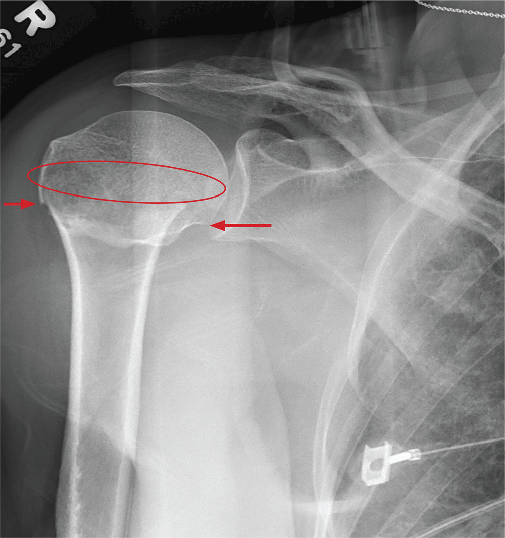
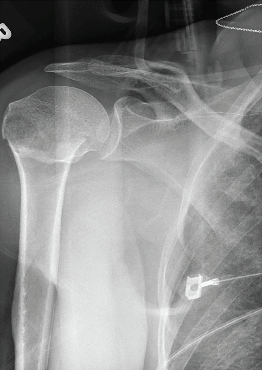
The radiograph demonstrates an acute horizontal fracture through the humeral neck. There is some slight lateral displacement of the fracture fragment.
The patient’s right arm was placed in a sling. Prompt orthopedic consultation was then obtained.
ANSWER
The radiograph demonstrates an acute horizontal fracture through the humeral neck. There is some slight lateral displacement of the fracture fragment.
The patient’s right arm was placed in a sling. Prompt orthopedic consultation was then obtained.
ANSWER
The radiograph demonstrates an acute horizontal fracture through the humeral neck. There is some slight lateral displacement of the fracture fragment.
The patient’s right arm was placed in a sling. Prompt orthopedic consultation was then obtained.
After a motor vehicle collision, a 70-year-old woman is brought to your emergency department by EMS personnel. She was a restrained driver in a vehicle crossing an intersection when she was broadsided by a tractor trailer traveling at high speed. Her airbags deployed, and she believes she briefly lost consciousness. Her biggest complaint is pain in her right shoulder.
Her medical history is significant for hypertension and hypothyroidism. On primary survey, you note an elderly woman who is in full cervical spine immobilization on a long backboard. Her Glasgow Coma Scale score is 15. She is in mild distress but has normal vital signs.
The patient has scattered abrasions and bruises on her body. Her right shoulder has mild to moderate tenderness to palpation and a decreased range of motion. Distally in that arm, she has good pulses and is neurovascularly intact.
You obtain a portable radiograph of the right shoulder (shown). What is your impression?
CardioMEMS cuts heart failure hospitalizations in post-approval study
NEW ORLEANS – Frequent, noninvasive measurement of pulmonary artery pressure in patients with advanced heart failure and an implanted CardioMEMS device that allows this measurement led to management that produced a substantial reduction in heart failure hospitalizations, compared with each patient’s history, in a real-world study.
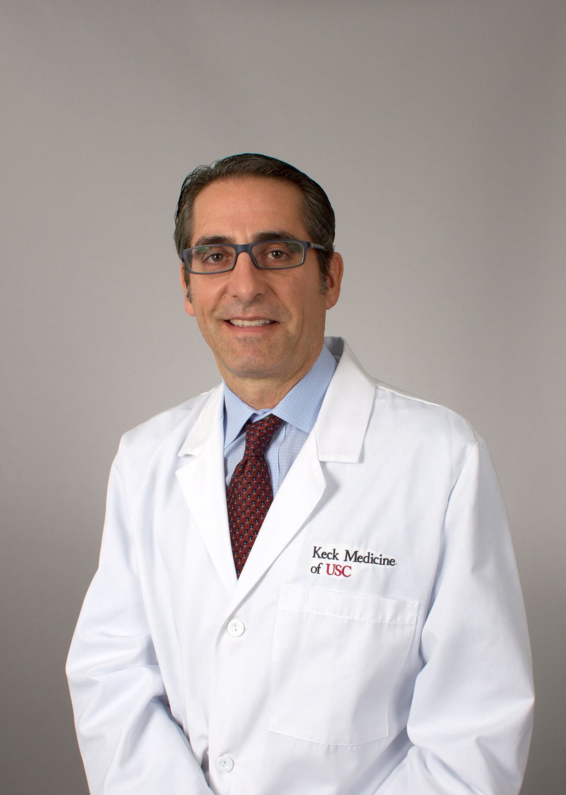
The Food and Drug Administration–mandated CardioMEMS Post-Approval Study included 1,200 patients who received CardioMEMS implants after it received U.S. marketing approval. The study showed that when clinicians and patients used the device in routine practice, presumably as part of a structured management system designed to take advantage of the pulmonary artery (PA) pressures the device provides, the result safely produced a 58% cut in heart failure hospitalizations during the year following device placement when compared to each patient’s own hospitalization history during the year before they got the CardioMEMS device, David M. Shavelle, MD, said at the at the annual meeting of the American College of Cardiology. This statistically significant result for the study’s primary endpoint showed an absolute reduction in the average rate of heart failure hospitalizations from 1.24 per patient during the year before the CardioMEMS placement to 0.52 hospitalizations per patient during the 12 months after placement, an average reduction of 0.72 hospitalizations/patient, said Dr. Shavelle, an interventional cardiologist at the University of Southern California in Los Angeles.
Another notable finding was that this benefit from CardioMEMS placement and use occurred at roughly similar rates in patients with New York Heart Association class III heart failure regardless of whether they had a reduced ejection fraction (40% or less), a mid-range ejection fraction (41%-50%), or preserved ejection fraction (greater than 50%), making CardioMEMS use one of the few treatments to produce any proven benefit in patients with heart failure with preserved ejection fraction. In that subgroup, 30% of the 1,200 enrolled patients had an average cut of 0.68 hospitalizations in the year after CardioMEMS implantation, a 61% drop, relative to the year before they received the device.
The results also fulfilled the study’s two prespecified safety measures. Among the 1,214 patients in the study assessed for safety, which included the 1,200 patients who received the device and 4 patients in whom placement failed, 4 patients had a device or system related complication during the study, a 0.3% rate, compared with a prespecified objective performance criteria of less than 20%. Among the 1,200 patients with a functioning CardioMEMS sensor, one patient (0.1%) had a device failure, compared with the study’s objective performance criteria of less than 10%.
The performance of the CardioMEMS device and the benefit it provided to patients in the post-approval study closely tracked its performance during the published pivotal trial (Lancet. 2011 Feb 19;377[9766]:658-66). On the basis of the pivotal trial results, the FDA approved CardioMEMS for U.S. marketing in 2014. Since then, the company has reported that about 10,000 U.S. heart failure patients have received these devices, Dr. Shavelle said.
“The benefit was seen across the range of ejection fractions; that’s very important,” commented Gurusher Panjrath, MD, director of advanced heart failure at George Washington University in Washington and a designated discussant for Dr. Shavelle’s report. “The safety seemed very good, and the efficacy was consistent” with prior reports. “There also was high compliance. The key to success is the structure” of patient management, Dr. Pangroth said. “The data are limited by who is monitoring patients and their data and how much of that contact influences patient outcomes.”
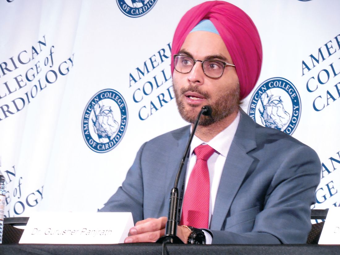
That final comment by Dr. Panjrath highlighted the biggest caveat that heart failure clinicians have raised about judging the efficacy of CardioMEMS. To achieve clinical efficacy, the implanted device requires diligent, virtually daily interrogation and data transmission by the patient, assessment of a large amount of data for each patient by the patient’s clinical team, and responsiveness by the patient to medication adjustments directed by the clinical team to deal with episodes of rising PA pressure.
“The device itself has no benefit. It’s the actions prompted by the device that have benefit,” noted Clyde W. Yancy, MD, professor of medicine and chief of cardiology at Northwestern University in Chicago and a second designated discussant for the report.

Dr. Shavelle agreed that for the CardioMEMS device to have an impact, one basic requirement is to identify patients who will cooperate with data collection and transmission and also with changes in their medications that are sent to them in response to PA pressure changes. This means selecting patients who appear to have problems with volume overload, including prior hospitalizations for decompensation, and patients who are comfortable interacting with their clinical-care providers. It also means excluding patients who are too sick to benefit from this intervention. He estimated that at his center more than 95% of class III heart failure patients who qualified for inclusion in the post-approval study by clinical criteria were also judged reasonable recipients of the device based on their willingness to cooperate with this system. He also estimated that at the University of Southern California the heart failure clinical team is now caring for about 150 patients with a CardioMEMS device implanted.
Another concern is teasing apart the specific benefit of collecting and using PA pressure data from the contact that the clinical team maintains with CardioMEMS patients.
“If nurses are contacting patients more often, is it the device or the communication? We need to look at that very carefully in a study that had no control group,” Dr. Yancy said in an interview. Contact with a nurse “is the best thing you can do for heart failure patients.”
Dr. Shavelle countered that several reports from past studies that assessed case management and regular monitoring of and contact with heart failure patients but without PA pressure data failed to showed any consistent benefit to patients.
“If you pick the right patients, CardioMEMS works. There is no question in my mind that the device works,” Dr. Shavelle said in an interview. “If you pick the wrong patient, who will not send the data or follow dose changes, then it won’t work.”
The study was sponsored by Abbott, the company that markets the CardioMEMS HF System. Dr. Shavelle has been a consultant to and speaker on behalf of Abbott Vascular and he has received research funding from Abbott Vascular, Abiomed, Biocardia, and V-Wave. Dr. Yancy had an unspecified financial relationship with Abbott Laboratories. Dr. Panjrath had no disclosures.
mzoler@mdedge.com
On Twitter @mitchelzoler
SOURCE: Shavelle DM et al. American College of Cardiology annual meeting, abstract 405-16.
NEW ORLEANS – Frequent, noninvasive measurement of pulmonary artery pressure in patients with advanced heart failure and an implanted CardioMEMS device that allows this measurement led to management that produced a substantial reduction in heart failure hospitalizations, compared with each patient’s history, in a real-world study.
The Food and Drug Administration–mandated CardioMEMS Post-Approval Study included 1,200 patients who received CardioMEMS implants after it received U.S. marketing approval. The study showed that when clinicians and patients used the device in routine practice, presumably as part of a structured management system designed to take advantage of the pulmonary artery (PA) pressures the device provides, the result safely produced a 58% cut in heart failure hospitalizations during the year following device placement when compared to each patient’s own hospitalization history during the year before they got the CardioMEMS device, David M. Shavelle, MD, said at the at the annual meeting of the American College of Cardiology. This statistically significant result for the study’s primary endpoint showed an absolute reduction in the average rate of heart failure hospitalizations from 1.24 per patient during the year before the CardioMEMS placement to 0.52 hospitalizations per patient during the 12 months after placement, an average reduction of 0.72 hospitalizations/patient, said Dr. Shavelle, an interventional cardiologist at the University of Southern California in Los Angeles.
Another notable finding was that this benefit from CardioMEMS placement and use occurred at roughly similar rates in patients with New York Heart Association class III heart failure regardless of whether they had a reduced ejection fraction (40% or less), a mid-range ejection fraction (41%-50%), or preserved ejection fraction (greater than 50%), making CardioMEMS use one of the few treatments to produce any proven benefit in patients with heart failure with preserved ejection fraction. In that subgroup, 30% of the 1,200 enrolled patients had an average cut of 0.68 hospitalizations in the year after CardioMEMS implantation, a 61% drop, relative to the year before they received the device.
The results also fulfilled the study’s two prespecified safety measures. Among the 1,214 patients in the study assessed for safety, which included the 1,200 patients who received the device and 4 patients in whom placement failed, 4 patients had a device or system related complication during the study, a 0.3% rate, compared with a prespecified objective performance criteria of less than 20%. Among the 1,200 patients with a functioning CardioMEMS sensor, one patient (0.1%) had a device failure, compared with the study’s objective performance criteria of less than 10%.
The performance of the CardioMEMS device and the benefit it provided to patients in the post-approval study closely tracked its performance during the published pivotal trial (Lancet. 2011 Feb 19;377[9766]:658-66). On the basis of the pivotal trial results, the FDA approved CardioMEMS for U.S. marketing in 2014. Since then, the company has reported that about 10,000 U.S. heart failure patients have received these devices, Dr. Shavelle said.
“The benefit was seen across the range of ejection fractions; that’s very important,” commented Gurusher Panjrath, MD, director of advanced heart failure at George Washington University in Washington and a designated discussant for Dr. Shavelle’s report. “The safety seemed very good, and the efficacy was consistent” with prior reports. “There also was high compliance. The key to success is the structure” of patient management, Dr. Pangroth said. “The data are limited by who is monitoring patients and their data and how much of that contact influences patient outcomes.”
That final comment by Dr. Panjrath highlighted the biggest caveat that heart failure clinicians have raised about judging the efficacy of CardioMEMS. To achieve clinical efficacy, the implanted device requires diligent, virtually daily interrogation and data transmission by the patient, assessment of a large amount of data for each patient by the patient’s clinical team, and responsiveness by the patient to medication adjustments directed by the clinical team to deal with episodes of rising PA pressure.
“The device itself has no benefit. It’s the actions prompted by the device that have benefit,” noted Clyde W. Yancy, MD, professor of medicine and chief of cardiology at Northwestern University in Chicago and a second designated discussant for the report.
Dr. Shavelle agreed that for the CardioMEMS device to have an impact, one basic requirement is to identify patients who will cooperate with data collection and transmission and also with changes in their medications that are sent to them in response to PA pressure changes. This means selecting patients who appear to have problems with volume overload, including prior hospitalizations for decompensation, and patients who are comfortable interacting with their clinical-care providers. It also means excluding patients who are too sick to benefit from this intervention. He estimated that at his center more than 95% of class III heart failure patients who qualified for inclusion in the post-approval study by clinical criteria were also judged reasonable recipients of the device based on their willingness to cooperate with this system. He also estimated that at the University of Southern California the heart failure clinical team is now caring for about 150 patients with a CardioMEMS device implanted.
Another concern is teasing apart the specific benefit of collecting and using PA pressure data from the contact that the clinical team maintains with CardioMEMS patients.
“If nurses are contacting patients more often, is it the device or the communication? We need to look at that very carefully in a study that had no control group,” Dr. Yancy said in an interview. Contact with a nurse “is the best thing you can do for heart failure patients.”
Dr. Shavelle countered that several reports from past studies that assessed case management and regular monitoring of and contact with heart failure patients but without PA pressure data failed to showed any consistent benefit to patients.
“If you pick the right patients, CardioMEMS works. There is no question in my mind that the device works,” Dr. Shavelle said in an interview. “If you pick the wrong patient, who will not send the data or follow dose changes, then it won’t work.”
The study was sponsored by Abbott, the company that markets the CardioMEMS HF System. Dr. Shavelle has been a consultant to and speaker on behalf of Abbott Vascular and he has received research funding from Abbott Vascular, Abiomed, Biocardia, and V-Wave. Dr. Yancy had an unspecified financial relationship with Abbott Laboratories. Dr. Panjrath had no disclosures.
mzoler@mdedge.com
On Twitter @mitchelzoler
SOURCE: Shavelle DM et al. American College of Cardiology annual meeting, abstract 405-16.
NEW ORLEANS – Frequent, noninvasive measurement of pulmonary artery pressure in patients with advanced heart failure and an implanted CardioMEMS device that allows this measurement led to management that produced a substantial reduction in heart failure hospitalizations, compared with each patient’s history, in a real-world study.
The Food and Drug Administration–mandated CardioMEMS Post-Approval Study included 1,200 patients who received CardioMEMS implants after it received U.S. marketing approval. The study showed that when clinicians and patients used the device in routine practice, presumably as part of a structured management system designed to take advantage of the pulmonary artery (PA) pressures the device provides, the result safely produced a 58% cut in heart failure hospitalizations during the year following device placement when compared to each patient’s own hospitalization history during the year before they got the CardioMEMS device, David M. Shavelle, MD, said at the at the annual meeting of the American College of Cardiology. This statistically significant result for the study’s primary endpoint showed an absolute reduction in the average rate of heart failure hospitalizations from 1.24 per patient during the year before the CardioMEMS placement to 0.52 hospitalizations per patient during the 12 months after placement, an average reduction of 0.72 hospitalizations/patient, said Dr. Shavelle, an interventional cardiologist at the University of Southern California in Los Angeles.
Another notable finding was that this benefit from CardioMEMS placement and use occurred at roughly similar rates in patients with New York Heart Association class III heart failure regardless of whether they had a reduced ejection fraction (40% or less), a mid-range ejection fraction (41%-50%), or preserved ejection fraction (greater than 50%), making CardioMEMS use one of the few treatments to produce any proven benefit in patients with heart failure with preserved ejection fraction. In that subgroup, 30% of the 1,200 enrolled patients had an average cut of 0.68 hospitalizations in the year after CardioMEMS implantation, a 61% drop, relative to the year before they received the device.
The results also fulfilled the study’s two prespecified safety measures. Among the 1,214 patients in the study assessed for safety, which included the 1,200 patients who received the device and 4 patients in whom placement failed, 4 patients had a device or system related complication during the study, a 0.3% rate, compared with a prespecified objective performance criteria of less than 20%. Among the 1,200 patients with a functioning CardioMEMS sensor, one patient (0.1%) had a device failure, compared with the study’s objective performance criteria of less than 10%.
The performance of the CardioMEMS device and the benefit it provided to patients in the post-approval study closely tracked its performance during the published pivotal trial (Lancet. 2011 Feb 19;377[9766]:658-66). On the basis of the pivotal trial results, the FDA approved CardioMEMS for U.S. marketing in 2014. Since then, the company has reported that about 10,000 U.S. heart failure patients have received these devices, Dr. Shavelle said.
“The benefit was seen across the range of ejection fractions; that’s very important,” commented Gurusher Panjrath, MD, director of advanced heart failure at George Washington University in Washington and a designated discussant for Dr. Shavelle’s report. “The safety seemed very good, and the efficacy was consistent” with prior reports. “There also was high compliance. The key to success is the structure” of patient management, Dr. Pangroth said. “The data are limited by who is monitoring patients and their data and how much of that contact influences patient outcomes.”
That final comment by Dr. Panjrath highlighted the biggest caveat that heart failure clinicians have raised about judging the efficacy of CardioMEMS. To achieve clinical efficacy, the implanted device requires diligent, virtually daily interrogation and data transmission by the patient, assessment of a large amount of data for each patient by the patient’s clinical team, and responsiveness by the patient to medication adjustments directed by the clinical team to deal with episodes of rising PA pressure.
“The device itself has no benefit. It’s the actions prompted by the device that have benefit,” noted Clyde W. Yancy, MD, professor of medicine and chief of cardiology at Northwestern University in Chicago and a second designated discussant for the report.
Dr. Shavelle agreed that for the CardioMEMS device to have an impact, one basic requirement is to identify patients who will cooperate with data collection and transmission and also with changes in their medications that are sent to them in response to PA pressure changes. This means selecting patients who appear to have problems with volume overload, including prior hospitalizations for decompensation, and patients who are comfortable interacting with their clinical-care providers. It also means excluding patients who are too sick to benefit from this intervention. He estimated that at his center more than 95% of class III heart failure patients who qualified for inclusion in the post-approval study by clinical criteria were also judged reasonable recipients of the device based on their willingness to cooperate with this system. He also estimated that at the University of Southern California the heart failure clinical team is now caring for about 150 patients with a CardioMEMS device implanted.
Another concern is teasing apart the specific benefit of collecting and using PA pressure data from the contact that the clinical team maintains with CardioMEMS patients.
“If nurses are contacting patients more often, is it the device or the communication? We need to look at that very carefully in a study that had no control group,” Dr. Yancy said in an interview. Contact with a nurse “is the best thing you can do for heart failure patients.”
Dr. Shavelle countered that several reports from past studies that assessed case management and regular monitoring of and contact with heart failure patients but without PA pressure data failed to showed any consistent benefit to patients.
“If you pick the right patients, CardioMEMS works. There is no question in my mind that the device works,” Dr. Shavelle said in an interview. “If you pick the wrong patient, who will not send the data or follow dose changes, then it won’t work.”
The study was sponsored by Abbott, the company that markets the CardioMEMS HF System. Dr. Shavelle has been a consultant to and speaker on behalf of Abbott Vascular and he has received research funding from Abbott Vascular, Abiomed, Biocardia, and V-Wave. Dr. Yancy had an unspecified financial relationship with Abbott Laboratories. Dr. Panjrath had no disclosures.
mzoler@mdedge.com
On Twitter @mitchelzoler
SOURCE: Shavelle DM et al. American College of Cardiology annual meeting, abstract 405-16.
REPORTING FROM ACC 2019
Ocrelizumab effective, safe in relapsing-remitting MS patients
PHILADELPHIA – Ocrelizumab is effective and safe in patients with relapsing-remitting multiple sclerosis who had inadequate responses to previous disease-modifying treatments, said an investigator reporting interim results of a recent nonrandomized study.
The findings of the 600+ patient CHORDS study suggest a positive risk-to-benefit ratio over nearly 1 year for this anti-CD20 monoclonal antibody, said Thomas P. Leist, MD, of Thomas Jefferson University, Philadelphia, at the annual meeting of the American Academy of Neurology (AAN).
“MRI activity has been significantly attenuated in these individuals, particularly, in the period from 24 to 48 weeks, and the overall safety that has been observed to date has been in line with what has been observed in clinical trials with this medication,” Dr. Leist said in a podium presentation.
While previous investigations demonstrated superiority to treatment with interferon in patients with relapsing multiple sclerosis, this phase IIIb study was needed to further clarify the effects of the treatment following suboptimal response to several disease modifying treatments, according to Dr. Leist and his co-investigators.
The intention-to-treat population of CHORDS consisted of 608 patients who received disease-modifying therapy for 6 or more months and discontinued it due to suboptimal response, which was defined as one or more clinically reported relapses, one or more T1 gadolinium-enhancing lesions, or two or more enlarging T2 lesions. They all received a 600 mg dose of ocrelizumab every 24 weeks for as many as 96 weeks.
With 48 weeks of follow-up, the majority of patients had no relapses, no enhancing T1 lesions, no new or enlarging T2 lesions, and no confirmed progression of disability. In all, 54.5% of the patients experienced none of those events, according to Dr. Leist.
The adjusted annualized relapse rate in this cohort was 0.065, he reported.
New MRI activity included 48 new T1 gadolinium-enhancing lesions in 1,174 MRI scans, for an adjusted rate of 0.023, he also reported, while there were 679 new or enlarging T2 lesions on 1,175 scans, for an adjusted rate of 0.581.
The safety in this population was comparable to the overall safety profile of ocrelizumab seen in other studies, according to Dr. Leist. A total of 25 patients, or 4.1%, experienced a serious adverse event, though none led to treatment withdrawal and only one led to a dose modification or interruption, the data show.
Dr. Leist reported disclosures related to Alkermes, Bayer, Biogen, EMD Serono, Genentech, Inc., Novartis, Sanofi Genzyme, Sun Pharma, and Teva Neuroscience.
SOURCE: Leist TP, et al. Presented at the 2019 American Academy of Neurology (AAN) Annual Meeting, May 4-10, 2019. Philadelphia. Presentation S56.007.
PHILADELPHIA – Ocrelizumab is effective and safe in patients with relapsing-remitting multiple sclerosis who had inadequate responses to previous disease-modifying treatments, said an investigator reporting interim results of a recent nonrandomized study.
The findings of the 600+ patient CHORDS study suggest a positive risk-to-benefit ratio over nearly 1 year for this anti-CD20 monoclonal antibody, said Thomas P. Leist, MD, of Thomas Jefferson University, Philadelphia, at the annual meeting of the American Academy of Neurology (AAN).
“MRI activity has been significantly attenuated in these individuals, particularly, in the period from 24 to 48 weeks, and the overall safety that has been observed to date has been in line with what has been observed in clinical trials with this medication,” Dr. Leist said in a podium presentation.
While previous investigations demonstrated superiority to treatment with interferon in patients with relapsing multiple sclerosis, this phase IIIb study was needed to further clarify the effects of the treatment following suboptimal response to several disease modifying treatments, according to Dr. Leist and his co-investigators.
The intention-to-treat population of CHORDS consisted of 608 patients who received disease-modifying therapy for 6 or more months and discontinued it due to suboptimal response, which was defined as one or more clinically reported relapses, one or more T1 gadolinium-enhancing lesions, or two or more enlarging T2 lesions. They all received a 600 mg dose of ocrelizumab every 24 weeks for as many as 96 weeks.
With 48 weeks of follow-up, the majority of patients had no relapses, no enhancing T1 lesions, no new or enlarging T2 lesions, and no confirmed progression of disability. In all, 54.5% of the patients experienced none of those events, according to Dr. Leist.
The adjusted annualized relapse rate in this cohort was 0.065, he reported.
New MRI activity included 48 new T1 gadolinium-enhancing lesions in 1,174 MRI scans, for an adjusted rate of 0.023, he also reported, while there were 679 new or enlarging T2 lesions on 1,175 scans, for an adjusted rate of 0.581.
The safety in this population was comparable to the overall safety profile of ocrelizumab seen in other studies, according to Dr. Leist. A total of 25 patients, or 4.1%, experienced a serious adverse event, though none led to treatment withdrawal and only one led to a dose modification or interruption, the data show.
Dr. Leist reported disclosures related to Alkermes, Bayer, Biogen, EMD Serono, Genentech, Inc., Novartis, Sanofi Genzyme, Sun Pharma, and Teva Neuroscience.
SOURCE: Leist TP, et al. Presented at the 2019 American Academy of Neurology (AAN) Annual Meeting, May 4-10, 2019. Philadelphia. Presentation S56.007.
PHILADELPHIA – Ocrelizumab is effective and safe in patients with relapsing-remitting multiple sclerosis who had inadequate responses to previous disease-modifying treatments, said an investigator reporting interim results of a recent nonrandomized study.
The findings of the 600+ patient CHORDS study suggest a positive risk-to-benefit ratio over nearly 1 year for this anti-CD20 monoclonal antibody, said Thomas P. Leist, MD, of Thomas Jefferson University, Philadelphia, at the annual meeting of the American Academy of Neurology (AAN).
“MRI activity has been significantly attenuated in these individuals, particularly, in the period from 24 to 48 weeks, and the overall safety that has been observed to date has been in line with what has been observed in clinical trials with this medication,” Dr. Leist said in a podium presentation.
While previous investigations demonstrated superiority to treatment with interferon in patients with relapsing multiple sclerosis, this phase IIIb study was needed to further clarify the effects of the treatment following suboptimal response to several disease modifying treatments, according to Dr. Leist and his co-investigators.
The intention-to-treat population of CHORDS consisted of 608 patients who received disease-modifying therapy for 6 or more months and discontinued it due to suboptimal response, which was defined as one or more clinically reported relapses, one or more T1 gadolinium-enhancing lesions, or two or more enlarging T2 lesions. They all received a 600 mg dose of ocrelizumab every 24 weeks for as many as 96 weeks.
With 48 weeks of follow-up, the majority of patients had no relapses, no enhancing T1 lesions, no new or enlarging T2 lesions, and no confirmed progression of disability. In all, 54.5% of the patients experienced none of those events, according to Dr. Leist.
The adjusted annualized relapse rate in this cohort was 0.065, he reported.
New MRI activity included 48 new T1 gadolinium-enhancing lesions in 1,174 MRI scans, for an adjusted rate of 0.023, he also reported, while there were 679 new or enlarging T2 lesions on 1,175 scans, for an adjusted rate of 0.581.
The safety in this population was comparable to the overall safety profile of ocrelizumab seen in other studies, according to Dr. Leist. A total of 25 patients, or 4.1%, experienced a serious adverse event, though none led to treatment withdrawal and only one led to a dose modification or interruption, the data show.
Dr. Leist reported disclosures related to Alkermes, Bayer, Biogen, EMD Serono, Genentech, Inc., Novartis, Sanofi Genzyme, Sun Pharma, and Teva Neuroscience.
SOURCE: Leist TP, et al. Presented at the 2019 American Academy of Neurology (AAN) Annual Meeting, May 4-10, 2019. Philadelphia. Presentation S56.007.
FROM AAN 2019
Gentamicin restores wound healing in hereditary epidermolysis bullosa
Rare progress seen in challenging disease
CHICAGO – Topical gentamicin counters the nonsense mutations that inhibit production of laminin 332 in infants with Herlitz junctional epidermolysis bullosa (H-JEB) to allow lesion healing, according to results of a small clinical study presented at the annual meeting of the Society for Investigative Dermatology.
“All of the children treated so far have responded,” reported Andrew Kwong, who will soon graduate from the Keck School of Medicine at the University of Southern California, Los Angeles.
H-JEB is an inherited blistering skin disease associated with nonsense mutations in the LAMA3, LAMB3, or LAMC2 genes that result in impaired production of functional laminin 332, an essential protein for epidermal-dermal adherence. At this time there are no effective therapies, and the disease is fatal.
The small clinical study was initiated after in vitro studies demonstrated that gentamicin restored functional laminin 332 in cultured keratinocytes from infants with H-JEB. The dose-dependent effect was credited to the ability of gentamicin to induce readthrough of premature stop codons that block production of laminin 332.
Data were presented on the first three infants with H-JEB treated with oral gentamicin. In each child, lesions were treated with topical 0.5% gentamicin twice daily for two weeks. Biopsies were taken prior to the initiation of treatment and at one and three months after treatment. The primary outcome was change in laminin 332, but clinical improvement was also monitored.
Although none of the infants had measurable laminin 332 prior to treatment, all lesions treated with topical gentamicin developed localized laminin 332 at the dermal-epidermal junction of the skin, Mr. Kwong reported. This expression, which was about 40% to 60% of that seen in normal skin, still persisted when evaluated three months after treatment.
The expression was associated with resolution of existing lesions and a reduced risk of developing new lesions, according to Mr. Kwong. In lesions that went untreated, there was no change.
Other molecular changes in the skin, such as increased expression and polarization of beta-4 integrin, were consistent with the ability of gentamicin to address the underlying pathophysiology of H-JEB. There were no adverse events observed.
By restoring functional laminin 332 in the skin, topical gentamicin appears to address the underlying cause of the bullae associated with H-JEB, but Mr. Kwong said that the next step is to determine whether intravenous gentamicin can address the systemic effects. If so, this treatment has the potential to improve survival. He reported that an infant with H-JEB was recently started on intravenous treatment, and initial results were encouraging.
Asked whether he would recommend topical gentamicin on the basis of these findings, Mr. Kwong cautioned that the case series remains very small, but he noted that the uniformity of the positive response is encouraging. He expects that off-label use of this novel and low-cost approach might be warranted in a population that has very limited therapeutic options.
SOURCE: Kwong A. SID 2019;S102, Abstract 594. Annual meeting of the Society for Investigative Dermatology.
Rare progress seen in challenging disease
Rare progress seen in challenging disease
CHICAGO – Topical gentamicin counters the nonsense mutations that inhibit production of laminin 332 in infants with Herlitz junctional epidermolysis bullosa (H-JEB) to allow lesion healing, according to results of a small clinical study presented at the annual meeting of the Society for Investigative Dermatology.
“All of the children treated so far have responded,” reported Andrew Kwong, who will soon graduate from the Keck School of Medicine at the University of Southern California, Los Angeles.
H-JEB is an inherited blistering skin disease associated with nonsense mutations in the LAMA3, LAMB3, or LAMC2 genes that result in impaired production of functional laminin 332, an essential protein for epidermal-dermal adherence. At this time there are no effective therapies, and the disease is fatal.
The small clinical study was initiated after in vitro studies demonstrated that gentamicin restored functional laminin 332 in cultured keratinocytes from infants with H-JEB. The dose-dependent effect was credited to the ability of gentamicin to induce readthrough of premature stop codons that block production of laminin 332.
Data were presented on the first three infants with H-JEB treated with oral gentamicin. In each child, lesions were treated with topical 0.5% gentamicin twice daily for two weeks. Biopsies were taken prior to the initiation of treatment and at one and three months after treatment. The primary outcome was change in laminin 332, but clinical improvement was also monitored.
Although none of the infants had measurable laminin 332 prior to treatment, all lesions treated with topical gentamicin developed localized laminin 332 at the dermal-epidermal junction of the skin, Mr. Kwong reported. This expression, which was about 40% to 60% of that seen in normal skin, still persisted when evaluated three months after treatment.
The expression was associated with resolution of existing lesions and a reduced risk of developing new lesions, according to Mr. Kwong. In lesions that went untreated, there was no change.
Other molecular changes in the skin, such as increased expression and polarization of beta-4 integrin, were consistent with the ability of gentamicin to address the underlying pathophysiology of H-JEB. There were no adverse events observed.
By restoring functional laminin 332 in the skin, topical gentamicin appears to address the underlying cause of the bullae associated with H-JEB, but Mr. Kwong said that the next step is to determine whether intravenous gentamicin can address the systemic effects. If so, this treatment has the potential to improve survival. He reported that an infant with H-JEB was recently started on intravenous treatment, and initial results were encouraging.
Asked whether he would recommend topical gentamicin on the basis of these findings, Mr. Kwong cautioned that the case series remains very small, but he noted that the uniformity of the positive response is encouraging. He expects that off-label use of this novel and low-cost approach might be warranted in a population that has very limited therapeutic options.
SOURCE: Kwong A. SID 2019;S102, Abstract 594. Annual meeting of the Society for Investigative Dermatology.
CHICAGO – Topical gentamicin counters the nonsense mutations that inhibit production of laminin 332 in infants with Herlitz junctional epidermolysis bullosa (H-JEB) to allow lesion healing, according to results of a small clinical study presented at the annual meeting of the Society for Investigative Dermatology.
“All of the children treated so far have responded,” reported Andrew Kwong, who will soon graduate from the Keck School of Medicine at the University of Southern California, Los Angeles.
H-JEB is an inherited blistering skin disease associated with nonsense mutations in the LAMA3, LAMB3, or LAMC2 genes that result in impaired production of functional laminin 332, an essential protein for epidermal-dermal adherence. At this time there are no effective therapies, and the disease is fatal.
The small clinical study was initiated after in vitro studies demonstrated that gentamicin restored functional laminin 332 in cultured keratinocytes from infants with H-JEB. The dose-dependent effect was credited to the ability of gentamicin to induce readthrough of premature stop codons that block production of laminin 332.
Data were presented on the first three infants with H-JEB treated with oral gentamicin. In each child, lesions were treated with topical 0.5% gentamicin twice daily for two weeks. Biopsies were taken prior to the initiation of treatment and at one and three months after treatment. The primary outcome was change in laminin 332, but clinical improvement was also monitored.
Although none of the infants had measurable laminin 332 prior to treatment, all lesions treated with topical gentamicin developed localized laminin 332 at the dermal-epidermal junction of the skin, Mr. Kwong reported. This expression, which was about 40% to 60% of that seen in normal skin, still persisted when evaluated three months after treatment.
The expression was associated with resolution of existing lesions and a reduced risk of developing new lesions, according to Mr. Kwong. In lesions that went untreated, there was no change.
Other molecular changes in the skin, such as increased expression and polarization of beta-4 integrin, were consistent with the ability of gentamicin to address the underlying pathophysiology of H-JEB. There were no adverse events observed.
By restoring functional laminin 332 in the skin, topical gentamicin appears to address the underlying cause of the bullae associated with H-JEB, but Mr. Kwong said that the next step is to determine whether intravenous gentamicin can address the systemic effects. If so, this treatment has the potential to improve survival. He reported that an infant with H-JEB was recently started on intravenous treatment, and initial results were encouraging.
Asked whether he would recommend topical gentamicin on the basis of these findings, Mr. Kwong cautioned that the case series remains very small, but he noted that the uniformity of the positive response is encouraging. He expects that off-label use of this novel and low-cost approach might be warranted in a population that has very limited therapeutic options.
SOURCE: Kwong A. SID 2019;S102, Abstract 594. Annual meeting of the Society for Investigative Dermatology.
REPORTING FROM SID 2019
Magnetic beads functionalized with VEGF could treat preeclampsia
A method of apheresis using vascular endothelial growth factor functionalized magnetic beads reduced levels of the soluble form of the vascular endothelial growth factor 1 in blood from women with preeclampsia, according to recent research published in the journal Hypertension.
The approach both reduces levels of the soluble form of the vascular endothelial growth factor 1 (sFlt-1) and releases placental growth factor (PlGF), which could help restore endothelial function in women with preeclampsia. The researchers said they chose sFlt-1 as a target because of “mounting evidence of its involvement in the pathogenesis of preeclampsia.” sFlt-1 has been suspected of inhibiting angiogenic signaling through “direct sequestration of angiogenic ligands” vascular endothelial growth factor (VEGF) and PlGF as well as “dominant-negative heterodimerization with surface VEGFRs.”
“During normal pregnancy, massive amounts of PlGF are produced by the placenta, reaching concentrations of free PlGF around 400 pg/mL, whereas during preeclampsia, free PlGF is extremely low due to the release of sFlt-1 into the maternal circulation,” the researchers said.
Using VEGF-functionalized magnetic beads, the researchers performed static and dynamic experiments using phosphate buffered saline (PBS), conditioned media, and plasma from women with preeclampsia. Under static conditions, there was a decrease of 33% for sFlt-1 and an increase of 27% for PlGF, while in dynamic conditions, there was a 40% decrease in sFlt-1 and a twofold increase in freed PlGF. When tested with plasma from women with preeclampsia, the ratio of sFlt-1/PlGF decreased by 63%, and VEGF release was associated with apheresis.
“This was a proof of concept study and our approach aims to restore physiologic levels of angiogenic factors,” Vassilis Tsatsaris, MD, PhD, of Cochin Hospital, Paris, said in a press release. “The reduction of sFlt-1 and the release of angiogenic factors is very significant and promising.”
Dr. Tsatsaris and his colleagues noted their next steps are to optimize the process of reducing sFlt-1 and examining how the approach works in an animal model.
“During normal pregnancy, circulating free VEGF levels are very low, almost undetectable with noncompetitive [enzyme-linked immunosorbent assay] ELISA. Whether these extremely low levels of VEGF have a physiological role during pregnancy is not known,” they wrote.
This study was funded by Agence Nationale pour la recherche, Institut Pierre Gilles de Gennes and the PremUP Foundation. One author reported receiving a grant from the Ecole Normale Supérieure and a second author reported receiving a grant from the Fondation pour la Recherche Médicale. The other authors report no relevant conflicts of interest.
SOURCE: Trapiella-Alfonso L et al. Hypertension. 2019. doi: 10.1161/HYPERTENSIONAHA.118.12380.
A method of apheresis using vascular endothelial growth factor functionalized magnetic beads reduced levels of the soluble form of the vascular endothelial growth factor 1 in blood from women with preeclampsia, according to recent research published in the journal Hypertension.
The approach both reduces levels of the soluble form of the vascular endothelial growth factor 1 (sFlt-1) and releases placental growth factor (PlGF), which could help restore endothelial function in women with preeclampsia. The researchers said they chose sFlt-1 as a target because of “mounting evidence of its involvement in the pathogenesis of preeclampsia.” sFlt-1 has been suspected of inhibiting angiogenic signaling through “direct sequestration of angiogenic ligands” vascular endothelial growth factor (VEGF) and PlGF as well as “dominant-negative heterodimerization with surface VEGFRs.”
“During normal pregnancy, massive amounts of PlGF are produced by the placenta, reaching concentrations of free PlGF around 400 pg/mL, whereas during preeclampsia, free PlGF is extremely low due to the release of sFlt-1 into the maternal circulation,” the researchers said.
Using VEGF-functionalized magnetic beads, the researchers performed static and dynamic experiments using phosphate buffered saline (PBS), conditioned media, and plasma from women with preeclampsia. Under static conditions, there was a decrease of 33% for sFlt-1 and an increase of 27% for PlGF, while in dynamic conditions, there was a 40% decrease in sFlt-1 and a twofold increase in freed PlGF. When tested with plasma from women with preeclampsia, the ratio of sFlt-1/PlGF decreased by 63%, and VEGF release was associated with apheresis.
“This was a proof of concept study and our approach aims to restore physiologic levels of angiogenic factors,” Vassilis Tsatsaris, MD, PhD, of Cochin Hospital, Paris, said in a press release. “The reduction of sFlt-1 and the release of angiogenic factors is very significant and promising.”
Dr. Tsatsaris and his colleagues noted their next steps are to optimize the process of reducing sFlt-1 and examining how the approach works in an animal model.
“During normal pregnancy, circulating free VEGF levels are very low, almost undetectable with noncompetitive [enzyme-linked immunosorbent assay] ELISA. Whether these extremely low levels of VEGF have a physiological role during pregnancy is not known,” they wrote.
This study was funded by Agence Nationale pour la recherche, Institut Pierre Gilles de Gennes and the PremUP Foundation. One author reported receiving a grant from the Ecole Normale Supérieure and a second author reported receiving a grant from the Fondation pour la Recherche Médicale. The other authors report no relevant conflicts of interest.
SOURCE: Trapiella-Alfonso L et al. Hypertension. 2019. doi: 10.1161/HYPERTENSIONAHA.118.12380.
A method of apheresis using vascular endothelial growth factor functionalized magnetic beads reduced levels of the soluble form of the vascular endothelial growth factor 1 in blood from women with preeclampsia, according to recent research published in the journal Hypertension.
The approach both reduces levels of the soluble form of the vascular endothelial growth factor 1 (sFlt-1) and releases placental growth factor (PlGF), which could help restore endothelial function in women with preeclampsia. The researchers said they chose sFlt-1 as a target because of “mounting evidence of its involvement in the pathogenesis of preeclampsia.” sFlt-1 has been suspected of inhibiting angiogenic signaling through “direct sequestration of angiogenic ligands” vascular endothelial growth factor (VEGF) and PlGF as well as “dominant-negative heterodimerization with surface VEGFRs.”
“During normal pregnancy, massive amounts of PlGF are produced by the placenta, reaching concentrations of free PlGF around 400 pg/mL, whereas during preeclampsia, free PlGF is extremely low due to the release of sFlt-1 into the maternal circulation,” the researchers said.
Using VEGF-functionalized magnetic beads, the researchers performed static and dynamic experiments using phosphate buffered saline (PBS), conditioned media, and plasma from women with preeclampsia. Under static conditions, there was a decrease of 33% for sFlt-1 and an increase of 27% for PlGF, while in dynamic conditions, there was a 40% decrease in sFlt-1 and a twofold increase in freed PlGF. When tested with plasma from women with preeclampsia, the ratio of sFlt-1/PlGF decreased by 63%, and VEGF release was associated with apheresis.
“This was a proof of concept study and our approach aims to restore physiologic levels of angiogenic factors,” Vassilis Tsatsaris, MD, PhD, of Cochin Hospital, Paris, said in a press release. “The reduction of sFlt-1 and the release of angiogenic factors is very significant and promising.”
Dr. Tsatsaris and his colleagues noted their next steps are to optimize the process of reducing sFlt-1 and examining how the approach works in an animal model.
“During normal pregnancy, circulating free VEGF levels are very low, almost undetectable with noncompetitive [enzyme-linked immunosorbent assay] ELISA. Whether these extremely low levels of VEGF have a physiological role during pregnancy is not known,” they wrote.
This study was funded by Agence Nationale pour la recherche, Institut Pierre Gilles de Gennes and the PremUP Foundation. One author reported receiving a grant from the Ecole Normale Supérieure and a second author reported receiving a grant from the Fondation pour la Recherche Médicale. The other authors report no relevant conflicts of interest.
SOURCE: Trapiella-Alfonso L et al. Hypertension. 2019. doi: 10.1161/HYPERTENSIONAHA.118.12380.
FROM HYPERTENSION
Key clinical point: Use of magnetic beads functionalized with vascular endothelial growth factor (VEGF) reduced the soluble form of endothelial growth factor 1 (sFlt-1) in the blood of women with preeclampsia.
Major finding: sFlt-1 was reduced by 40% under dynamic conditions, and there was a twofold increase in the amount of freed placental growth factor.
Study details: A proof-of-concept study using VEGF-functionalized magnetic beads and phosphate buffered saline (PBS), conditioned media, and plasma from women with preeclampsia.
Disclosures: This study was funded by Agence Nationale pour la recherche, Institut Pierre Gilles de Gennes, and the PremUP Foundation. One author reported receiving a grant from the Ecole Normale Supérieure and a second author reported receiving a grant from the Fondation pour la Recherche Médicale. The other authors reported no relevant conflicts of interest.
Source: Trapiella-Alfonso L et al. Hypertension. 2019. doi: 10.1161/HYPERTENSIONAHA.118.12380.
Post-treatment persistence of oral HPV in HNSCC predicts recurrence, death
Persistence of oral human papillomavirus (HPV) DNA after primary treatment of HPV-positive oral cavity or oropharyngeal head and neck squamous cell carcinoma (HNSCC) is a strong risk factor for poor outcomes, finds a prospective cohort study.

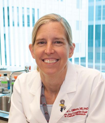
Investigators working under senior author Maura L. Gillison, MD, PhD, of the department of thoracic head and neck medical oncology at the University of Texas MD Anderson Cancer Center, Houston, collected serial oral rinses from 396 patients with HNSCC (217 with oropharyngeal cancer, 170 with oral cavity cancer, 9 with unknown primary cancer) treated at two institutions. Overall, 51% had HPV-positive tumors.
Patients with HPV-positive tumors were much more likely to have detectable oral HPV at diagnosis than their counterparts with HPV-negative tumors (84.2% vs 12.4%; P less than .001). Detection of oral HPV-16 DNA had good sensitivity (81%) and excellent specificity (100%) for HPV-16-positive tumors, Dr. Gillison and colleagues reported in JAMA Oncology.
Patients’ odds of having detectable tumor-type HPV fell during primary therapy (odds ratio per each postdiagnosis month, 0.41; P less than .001); in contrast, their odds of having of having detectable nontumor types did not. Current smokers were about half as likely to achieve clearing of tumor-type HPV DNA (hazard ratio, 0.49; P = .01).
Compared with counterparts who no longer had detectable tumor-type DNA after therapy, HPV-positive patients who did had dramatically poorer recurrence-free survival (55% vs. 88%; adjusted hazard ratio, 3.72; P less than .001) and overall survival (68% vs. 95%; adjusted hazard ratio, 6.61; P = .003).
In contrast, persistence of nontumor-type HPV DNA did not predict these outcomes among either patients with HPV-positive tumors or patients with HPV-negative tumors.
“Analysis of tumor type HPV DNA has considerable promise as a biomarker for treatment response and risk of progression,” Dr. Gillison and coinvestigators maintain.
“Our data suggest that a subset of patients with HPV-positive HNSCC at high-risk for locoregional recurrence can be identified by detection of persistent, oral HPV after treatment. However, the clinical utility may be constrained by a need to identify the tumor-type infection, a low-moderate positive predictive value for recurrence, and weak associations with risk of distant metastases,” they conclude. “Ongoing studies will evaluate whether multiplexed detection of plasma HPV DNA can improve these limitations.”
Dr. Gillison disclosed consulting for Roche Holding AG, Bristol-Myers Squibb, Merck & Co Inc., Celgene Corporation, Amgen, AstraZeneca, Rakuten Aspyrian Inc. (now known as Rakuten Medical), EMD Serono Inc., NewLink Genetics Corporation, and Genocea Biosciences. The study was supported by the Oral Cancer Foundation and The Ohio State University Comprehensive Cancer Center. Dr. Gillison is a Cancer Prevention and Research Institute of Texas Scholar.
SOURCE: Fakhry C et al. JAMA Oncol. 2019 May 2. doi: 10.1001/jamaoncol.2019.0439.
Persistence of oral human papillomavirus (HPV) DNA after primary treatment of HPV-positive oral cavity or oropharyngeal head and neck squamous cell carcinoma (HNSCC) is a strong risk factor for poor outcomes, finds a prospective cohort study.
Investigators working under senior author Maura L. Gillison, MD, PhD, of the department of thoracic head and neck medical oncology at the University of Texas MD Anderson Cancer Center, Houston, collected serial oral rinses from 396 patients with HNSCC (217 with oropharyngeal cancer, 170 with oral cavity cancer, 9 with unknown primary cancer) treated at two institutions. Overall, 51% had HPV-positive tumors.
Patients with HPV-positive tumors were much more likely to have detectable oral HPV at diagnosis than their counterparts with HPV-negative tumors (84.2% vs 12.4%; P less than .001). Detection of oral HPV-16 DNA had good sensitivity (81%) and excellent specificity (100%) for HPV-16-positive tumors, Dr. Gillison and colleagues reported in JAMA Oncology.
Patients’ odds of having detectable tumor-type HPV fell during primary therapy (odds ratio per each postdiagnosis month, 0.41; P less than .001); in contrast, their odds of having of having detectable nontumor types did not. Current smokers were about half as likely to achieve clearing of tumor-type HPV DNA (hazard ratio, 0.49; P = .01).
Compared with counterparts who no longer had detectable tumor-type DNA after therapy, HPV-positive patients who did had dramatically poorer recurrence-free survival (55% vs. 88%; adjusted hazard ratio, 3.72; P less than .001) and overall survival (68% vs. 95%; adjusted hazard ratio, 6.61; P = .003).
In contrast, persistence of nontumor-type HPV DNA did not predict these outcomes among either patients with HPV-positive tumors or patients with HPV-negative tumors.
“Analysis of tumor type HPV DNA has considerable promise as a biomarker for treatment response and risk of progression,” Dr. Gillison and coinvestigators maintain.
“Our data suggest that a subset of patients with HPV-positive HNSCC at high-risk for locoregional recurrence can be identified by detection of persistent, oral HPV after treatment. However, the clinical utility may be constrained by a need to identify the tumor-type infection, a low-moderate positive predictive value for recurrence, and weak associations with risk of distant metastases,” they conclude. “Ongoing studies will evaluate whether multiplexed detection of plasma HPV DNA can improve these limitations.”
Dr. Gillison disclosed consulting for Roche Holding AG, Bristol-Myers Squibb, Merck & Co Inc., Celgene Corporation, Amgen, AstraZeneca, Rakuten Aspyrian Inc. (now known as Rakuten Medical), EMD Serono Inc., NewLink Genetics Corporation, and Genocea Biosciences. The study was supported by the Oral Cancer Foundation and The Ohio State University Comprehensive Cancer Center. Dr. Gillison is a Cancer Prevention and Research Institute of Texas Scholar.
SOURCE: Fakhry C et al. JAMA Oncol. 2019 May 2. doi: 10.1001/jamaoncol.2019.0439.
Persistence of oral human papillomavirus (HPV) DNA after primary treatment of HPV-positive oral cavity or oropharyngeal head and neck squamous cell carcinoma (HNSCC) is a strong risk factor for poor outcomes, finds a prospective cohort study.
Investigators working under senior author Maura L. Gillison, MD, PhD, of the department of thoracic head and neck medical oncology at the University of Texas MD Anderson Cancer Center, Houston, collected serial oral rinses from 396 patients with HNSCC (217 with oropharyngeal cancer, 170 with oral cavity cancer, 9 with unknown primary cancer) treated at two institutions. Overall, 51% had HPV-positive tumors.
Patients with HPV-positive tumors were much more likely to have detectable oral HPV at diagnosis than their counterparts with HPV-negative tumors (84.2% vs 12.4%; P less than .001). Detection of oral HPV-16 DNA had good sensitivity (81%) and excellent specificity (100%) for HPV-16-positive tumors, Dr. Gillison and colleagues reported in JAMA Oncology.
Patients’ odds of having detectable tumor-type HPV fell during primary therapy (odds ratio per each postdiagnosis month, 0.41; P less than .001); in contrast, their odds of having of having detectable nontumor types did not. Current smokers were about half as likely to achieve clearing of tumor-type HPV DNA (hazard ratio, 0.49; P = .01).
Compared with counterparts who no longer had detectable tumor-type DNA after therapy, HPV-positive patients who did had dramatically poorer recurrence-free survival (55% vs. 88%; adjusted hazard ratio, 3.72; P less than .001) and overall survival (68% vs. 95%; adjusted hazard ratio, 6.61; P = .003).
In contrast, persistence of nontumor-type HPV DNA did not predict these outcomes among either patients with HPV-positive tumors or patients with HPV-negative tumors.
“Analysis of tumor type HPV DNA has considerable promise as a biomarker for treatment response and risk of progression,” Dr. Gillison and coinvestigators maintain.
“Our data suggest that a subset of patients with HPV-positive HNSCC at high-risk for locoregional recurrence can be identified by detection of persistent, oral HPV after treatment. However, the clinical utility may be constrained by a need to identify the tumor-type infection, a low-moderate positive predictive value for recurrence, and weak associations with risk of distant metastases,” they conclude. “Ongoing studies will evaluate whether multiplexed detection of plasma HPV DNA can improve these limitations.”
Dr. Gillison disclosed consulting for Roche Holding AG, Bristol-Myers Squibb, Merck & Co Inc., Celgene Corporation, Amgen, AstraZeneca, Rakuten Aspyrian Inc. (now known as Rakuten Medical), EMD Serono Inc., NewLink Genetics Corporation, and Genocea Biosciences. The study was supported by the Oral Cancer Foundation and The Ohio State University Comprehensive Cancer Center. Dr. Gillison is a Cancer Prevention and Research Institute of Texas Scholar.
SOURCE: Fakhry C et al. JAMA Oncol. 2019 May 2. doi: 10.1001/jamaoncol.2019.0439.
FROM JAMA ONCOLOGY
Key clinical point: Persistence of detectable tumor-type HPV DNA in oral rinses after primary treatment of HPV-positive HNSCC identifies patients at high risk for poor outcomes.
Major finding: HPV-positive patients with persistent oral tumor-type HPV DNA had sharply higher risks of 2-year recurrence-free survival events (hazard ratio, 3.72; P less than .001) and death (hazard ratio, 6.61; P = .003).
Study details: Prospective cohort study of 396 patients with newly diagnosed oral cavity or oropharyngeal HNSCC.
Disclosures: Dr. Gillison disclosed consulting for Roche Holding AG, Bristol-Myers Squibb, Merck & Co Inc., Celgene Corporation, Amgen, AstraZeneca, Rakuten Aspyrian Inc. (now known as Rakuten Medical), EMD Serono Inc., NewLink Genetics Corporation, and Genocea Biosciences. The study was supported by the Oral Cancer Foundation and The Ohio State University Comprehensive Cancer Center. Dr. Gillison is a Cancer Prevention and Research Institute of Texas Scholar.
Source: Gillison ML et al. JAMA Oncol. 2019 May 2. doi: 10.1001/jamaoncol.2019.0439.
FMT explored as a potential weight loss treatment
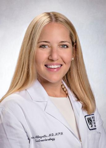
“Currently, fecal microbiota transplantation [FMT] can only be performed clinically in the US to treat recurrent Clostridium difficile infection,” lead study author Jessica R. Allegretti, MD, MPH, said during a media briefing in advance of the annual Digestive Disease Week.
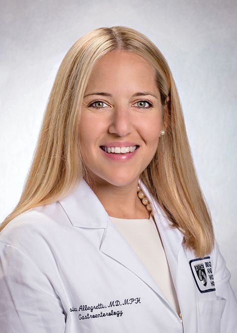
“However, there is ongoing research to find out whether FMT works for other health conditions such as obesity, a condition which affects millions of people worldwide. [It’s] a condition that also increases the risk of developing many other illnesses, including diabetes, heart disease, and certain cancers. In my clinical practice, we regularly see patients who have not yet developed some of these other conditions related to obesity, but really do have difficulty losing weight. Through our research we wanted to focus on a population we call the medically uncompromised obese, and understand if FMT might be a viable treatment option for them,” she said.
Dr. Allegretti, director of the fecal microbiota transplant program at Brigham and Women’s Hospital, Boston, conducted a parallel study of 22 patients with a body mass index of 35 kg/m2 or higher who were metabolically healthy – defined as having no type 2 diabetes, nonalcoholic steatohepatitis (NASH), or metabolic syndrome. They randomized study participants 1:1 to receive 30 FMT capsules followed by two doses of 12 capsules over a 12-week period, or identical placebo capsules. A single healthy lean donor with a BMI of 17 kg/m2 was used.
The researchers assessed patients with a mixed meal tolerance test at baseline, week 6, and week 12 post-FMT, at which biomarkers GLP-1 and leptin were measured. Stool was collected at baseline and at one, four, six, eight, and 12 weeks post-FMT. The primary outcomes were safety and change in the area under the curve for GLP-1 at 12 weeks compared to baseline. Secondary endpoints include gut microbiome profiles and diversity as well as bile acid profiles at 12 weeks post FMT. Additional endpoints include a decrease in BMI and waist circumference at week 12. Standard stool microbiome and bile acid analysis was performed.
The mean age of the patients at baseline was 43 years, and their mean BMI was 41 kg/m2. Between baseline and week 12, the research observed no increase of GLP-1 in either group, while the change in leptin revealed an increase in the placebo group only (P less than .001). At week 12, no early changes in BMI were noted in either group (P = .51). No serious adverse events occurred in either arm.
Dr. Allegretti and her colleagues observed global signals of donor community engraftment following FMT, including an increase in alpha diversity and increased similarity to stool samples from the FMT donor – trends that were not observed in the placebo arm. In addition, bile acid analysis suggested a sustained decrease in taurocholic acid in the FMT arm, comparable with the donor – an effect that was not seen in the placebo arm. “We know that what leads to the germination of C. diff. spores is brought out by bile acids,” she said. “That’s one of the critical components of pathogenesis in that disease. My group has been able to show that after FMT, you regain bile acid homeostasis.”
Dr. Allegretti concluded her remarks by noting that the current study “adds an encouraging first step in trying to understand the role that the gut microbiome is playing in the pathogenesis of medically uncompromised obese patients. As a next step, we plan to seek more sensitive measures of GLP-1, as well as conduct additional research into varied doses of FMT capsules, as well as potentially investigating other microbial pathways to better understand the role the microbiome is playing in obesity.”
Somerville, Mass.–based Finch Therapeutics provided funding for the research. Dr. Allegretti reported having no financial disclosures.
SOURCE: Allegretti J et al. DDW 2019. Abstract 621.
“Currently, fecal microbiota transplantation [FMT] can only be performed clinically in the US to treat recurrent Clostridium difficile infection,” lead study author Jessica R. Allegretti, MD, MPH, said during a media briefing in advance of the annual Digestive Disease Week.
“However, there is ongoing research to find out whether FMT works for other health conditions such as obesity, a condition which affects millions of people worldwide. [It’s] a condition that also increases the risk of developing many other illnesses, including diabetes, heart disease, and certain cancers. In my clinical practice, we regularly see patients who have not yet developed some of these other conditions related to obesity, but really do have difficulty losing weight. Through our research we wanted to focus on a population we call the medically uncompromised obese, and understand if FMT might be a viable treatment option for them,” she said.
Dr. Allegretti, director of the fecal microbiota transplant program at Brigham and Women’s Hospital, Boston, conducted a parallel study of 22 patients with a body mass index of 35 kg/m2 or higher who were metabolically healthy – defined as having no type 2 diabetes, nonalcoholic steatohepatitis (NASH), or metabolic syndrome. They randomized study participants 1:1 to receive 30 FMT capsules followed by two doses of 12 capsules over a 12-week period, or identical placebo capsules. A single healthy lean donor with a BMI of 17 kg/m2 was used.
The researchers assessed patients with a mixed meal tolerance test at baseline, week 6, and week 12 post-FMT, at which biomarkers GLP-1 and leptin were measured. Stool was collected at baseline and at one, four, six, eight, and 12 weeks post-FMT. The primary outcomes were safety and change in the area under the curve for GLP-1 at 12 weeks compared to baseline. Secondary endpoints include gut microbiome profiles and diversity as well as bile acid profiles at 12 weeks post FMT. Additional endpoints include a decrease in BMI and waist circumference at week 12. Standard stool microbiome and bile acid analysis was performed.
The mean age of the patients at baseline was 43 years, and their mean BMI was 41 kg/m2. Between baseline and week 12, the research observed no increase of GLP-1 in either group, while the change in leptin revealed an increase in the placebo group only (P less than .001). At week 12, no early changes in BMI were noted in either group (P = .51). No serious adverse events occurred in either arm.
Dr. Allegretti and her colleagues observed global signals of donor community engraftment following FMT, including an increase in alpha diversity and increased similarity to stool samples from the FMT donor – trends that were not observed in the placebo arm. In addition, bile acid analysis suggested a sustained decrease in taurocholic acid in the FMT arm, comparable with the donor – an effect that was not seen in the placebo arm. “We know that what leads to the germination of C. diff. spores is brought out by bile acids,” she said. “That’s one of the critical components of pathogenesis in that disease. My group has been able to show that after FMT, you regain bile acid homeostasis.”
Dr. Allegretti concluded her remarks by noting that the current study “adds an encouraging first step in trying to understand the role that the gut microbiome is playing in the pathogenesis of medically uncompromised obese patients. As a next step, we plan to seek more sensitive measures of GLP-1, as well as conduct additional research into varied doses of FMT capsules, as well as potentially investigating other microbial pathways to better understand the role the microbiome is playing in obesity.”
Somerville, Mass.–based Finch Therapeutics provided funding for the research. Dr. Allegretti reported having no financial disclosures.
SOURCE: Allegretti J et al. DDW 2019. Abstract 621.
“Currently, fecal microbiota transplantation [FMT] can only be performed clinically in the US to treat recurrent Clostridium difficile infection,” lead study author Jessica R. Allegretti, MD, MPH, said during a media briefing in advance of the annual Digestive Disease Week.
“However, there is ongoing research to find out whether FMT works for other health conditions such as obesity, a condition which affects millions of people worldwide. [It’s] a condition that also increases the risk of developing many other illnesses, including diabetes, heart disease, and certain cancers. In my clinical practice, we regularly see patients who have not yet developed some of these other conditions related to obesity, but really do have difficulty losing weight. Through our research we wanted to focus on a population we call the medically uncompromised obese, and understand if FMT might be a viable treatment option for them,” she said.
Dr. Allegretti, director of the fecal microbiota transplant program at Brigham and Women’s Hospital, Boston, conducted a parallel study of 22 patients with a body mass index of 35 kg/m2 or higher who were metabolically healthy – defined as having no type 2 diabetes, nonalcoholic steatohepatitis (NASH), or metabolic syndrome. They randomized study participants 1:1 to receive 30 FMT capsules followed by two doses of 12 capsules over a 12-week period, or identical placebo capsules. A single healthy lean donor with a BMI of 17 kg/m2 was used.
The researchers assessed patients with a mixed meal tolerance test at baseline, week 6, and week 12 post-FMT, at which biomarkers GLP-1 and leptin were measured. Stool was collected at baseline and at one, four, six, eight, and 12 weeks post-FMT. The primary outcomes were safety and change in the area under the curve for GLP-1 at 12 weeks compared to baseline. Secondary endpoints include gut microbiome profiles and diversity as well as bile acid profiles at 12 weeks post FMT. Additional endpoints include a decrease in BMI and waist circumference at week 12. Standard stool microbiome and bile acid analysis was performed.
The mean age of the patients at baseline was 43 years, and their mean BMI was 41 kg/m2. Between baseline and week 12, the research observed no increase of GLP-1 in either group, while the change in leptin revealed an increase in the placebo group only (P less than .001). At week 12, no early changes in BMI were noted in either group (P = .51). No serious adverse events occurred in either arm.
Dr. Allegretti and her colleagues observed global signals of donor community engraftment following FMT, including an increase in alpha diversity and increased similarity to stool samples from the FMT donor – trends that were not observed in the placebo arm. In addition, bile acid analysis suggested a sustained decrease in taurocholic acid in the FMT arm, comparable with the donor – an effect that was not seen in the placebo arm. “We know that what leads to the germination of C. diff. spores is brought out by bile acids,” she said. “That’s one of the critical components of pathogenesis in that disease. My group has been able to show that after FMT, you regain bile acid homeostasis.”
Dr. Allegretti concluded her remarks by noting that the current study “adds an encouraging first step in trying to understand the role that the gut microbiome is playing in the pathogenesis of medically uncompromised obese patients. As a next step, we plan to seek more sensitive measures of GLP-1, as well as conduct additional research into varied doses of FMT capsules, as well as potentially investigating other microbial pathways to better understand the role the microbiome is playing in obesity.”
Somerville, Mass.–based Finch Therapeutics provided funding for the research. Dr. Allegretti reported having no financial disclosures.
SOURCE: Allegretti J et al. DDW 2019. Abstract 621.
REPORTING FROM DDW 2019
Key clinical point: Using capsules filled with fecal matter from a lean donor, researchers successfully changed some of the composition of the gut microbiota of patients with obesity.
Major finding: Following fecal microbiota transplantation, researchers detected a decrease in a specific bile acid and alterations in stool samples that showed increased similarity to those of the lean donor.
Study details: A randomized, placebo-controlled trial of 22 healthy obese patients.
Disclosures: Somerville, Mass.–based Finch Therapeutics provided funding for the research. Dr. Allegretti reported having no financial disclosures.
Source: Allegretti J et al. DDW 2019. Abstract 621.