User login
FDA expands approval of fulvestrant for HR+/HER2– advanced breast cancer
The Food and Drug Administration has approved fulvestrant (Faslodex) as monotherapy for women with hormone receptor–positive, human epidermal growth factor receptor 2–negative (HR+/HER2–) advanced breast cancer who are postmenopausal and previously untreated.
Approval was based on improved progression-free survival (PFS) in the phase 3 FALCON trial of 462 postmenopausal women with HR+/HER2– metastatic or locally advanced breast cancer who had not previously received hormonal therapy, drug maker AstraZeneca said in a press release.
The most common adverse events were arthralgia (16.7% of patients on fulvestrant vs. 10.3% on anastrozole), and hot flushes/flashes (11.4% vs. 10.3%).
Grade 3 or greater adverse events occurred in 22.4% vs. 17.7%. Deaths from adverse events occurred in six patients on fulvestrant vs. seven on anastrozole, according to results published in The Lancet.
The Food and Drug Administration has approved fulvestrant (Faslodex) as monotherapy for women with hormone receptor–positive, human epidermal growth factor receptor 2–negative (HR+/HER2–) advanced breast cancer who are postmenopausal and previously untreated.
Approval was based on improved progression-free survival (PFS) in the phase 3 FALCON trial of 462 postmenopausal women with HR+/HER2– metastatic or locally advanced breast cancer who had not previously received hormonal therapy, drug maker AstraZeneca said in a press release.
The most common adverse events were arthralgia (16.7% of patients on fulvestrant vs. 10.3% on anastrozole), and hot flushes/flashes (11.4% vs. 10.3%).
Grade 3 or greater adverse events occurred in 22.4% vs. 17.7%. Deaths from adverse events occurred in six patients on fulvestrant vs. seven on anastrozole, according to results published in The Lancet.
The Food and Drug Administration has approved fulvestrant (Faslodex) as monotherapy for women with hormone receptor–positive, human epidermal growth factor receptor 2–negative (HR+/HER2–) advanced breast cancer who are postmenopausal and previously untreated.
Approval was based on improved progression-free survival (PFS) in the phase 3 FALCON trial of 462 postmenopausal women with HR+/HER2– metastatic or locally advanced breast cancer who had not previously received hormonal therapy, drug maker AstraZeneca said in a press release.
The most common adverse events were arthralgia (16.7% of patients on fulvestrant vs. 10.3% on anastrozole), and hot flushes/flashes (11.4% vs. 10.3%).
Grade 3 or greater adverse events occurred in 22.4% vs. 17.7%. Deaths from adverse events occurred in six patients on fulvestrant vs. seven on anastrozole, according to results published in The Lancet.

Office-based tests can have questionable value
Office based-testing is convenient and often useful.
Often, but not always.
Sadly, a number of practices are increasingly turning to tests of questionable value as a way to make up for decreasing reimbursements. While I have nothing against making money, I have to question where this trend is going. I see many of them even being done by physicians outside of their fields.
I get ads all the time from companies selling gadgets to test balance (“Be the first in your area! Fully reimbursed!”). I see reports from family doctors with the results of a pseudo-EMG/NCV surface test done there by an assistant saying a patient with diabetes has a neuropathy (shocker!).
I have nothing against tests. Lord knows I order plenty of them. But I always try to ask myself if the results will change my management plan or answer another crucial question. If the answer is “no” to both, why bother?
Test are shiny. They impress patients and their families. They represent technological progress in medicine to many. But a lot of time we forget that clinical skills are pretty useful, too. If a diabetic patient comes in with numb feet and an exam that shows decreased distal sensation, do we really need a pseudo-surface EMG/NCV (especially when done by someone who isn’t a neurologist or physiatrist) or skin punch biopsy to tell us they have a neuropathy?
If the patient is stumbling all over and is clearly ataxic, do we need a machine to say, “Hey, you’re off balance. You could fall.”
An old mentor always told me “clinical correlation is advised.” (Al, I hated you then and miss you now.)
And convenience doesn’t always mean something is good. Remember Theranos?
Like all doctors, I worry about my bottom line and keeping my practice afloat. These tests are alluring in that they promise to increase practice revenue without much change in your overhead (if you believe that). But they can also be a siren song that lures you to put money ahead of what’s best for a patient. That’s never the right course.
Dr. Block has a solo neurology practice in Scottsdale, Ariz.
Office based-testing is convenient and often useful.
Often, but not always.
Sadly, a number of practices are increasingly turning to tests of questionable value as a way to make up for decreasing reimbursements. While I have nothing against making money, I have to question where this trend is going. I see many of them even being done by physicians outside of their fields.
I get ads all the time from companies selling gadgets to test balance (“Be the first in your area! Fully reimbursed!”). I see reports from family doctors with the results of a pseudo-EMG/NCV surface test done there by an assistant saying a patient with diabetes has a neuropathy (shocker!).
I have nothing against tests. Lord knows I order plenty of them. But I always try to ask myself if the results will change my management plan or answer another crucial question. If the answer is “no” to both, why bother?
Test are shiny. They impress patients and their families. They represent technological progress in medicine to many. But a lot of time we forget that clinical skills are pretty useful, too. If a diabetic patient comes in with numb feet and an exam that shows decreased distal sensation, do we really need a pseudo-surface EMG/NCV (especially when done by someone who isn’t a neurologist or physiatrist) or skin punch biopsy to tell us they have a neuropathy?
If the patient is stumbling all over and is clearly ataxic, do we need a machine to say, “Hey, you’re off balance. You could fall.”
An old mentor always told me “clinical correlation is advised.” (Al, I hated you then and miss you now.)
And convenience doesn’t always mean something is good. Remember Theranos?
Like all doctors, I worry about my bottom line and keeping my practice afloat. These tests are alluring in that they promise to increase practice revenue without much change in your overhead (if you believe that). But they can also be a siren song that lures you to put money ahead of what’s best for a patient. That’s never the right course.
Dr. Block has a solo neurology practice in Scottsdale, Ariz.
Office based-testing is convenient and often useful.
Often, but not always.
Sadly, a number of practices are increasingly turning to tests of questionable value as a way to make up for decreasing reimbursements. While I have nothing against making money, I have to question where this trend is going. I see many of them even being done by physicians outside of their fields.
I get ads all the time from companies selling gadgets to test balance (“Be the first in your area! Fully reimbursed!”). I see reports from family doctors with the results of a pseudo-EMG/NCV surface test done there by an assistant saying a patient with diabetes has a neuropathy (shocker!).
I have nothing against tests. Lord knows I order plenty of them. But I always try to ask myself if the results will change my management plan or answer another crucial question. If the answer is “no” to both, why bother?
Test are shiny. They impress patients and their families. They represent technological progress in medicine to many. But a lot of time we forget that clinical skills are pretty useful, too. If a diabetic patient comes in with numb feet and an exam that shows decreased distal sensation, do we really need a pseudo-surface EMG/NCV (especially when done by someone who isn’t a neurologist or physiatrist) or skin punch biopsy to tell us they have a neuropathy?
If the patient is stumbling all over and is clearly ataxic, do we need a machine to say, “Hey, you’re off balance. You could fall.”
An old mentor always told me “clinical correlation is advised.” (Al, I hated you then and miss you now.)
And convenience doesn’t always mean something is good. Remember Theranos?
Like all doctors, I worry about my bottom line and keeping my practice afloat. These tests are alluring in that they promise to increase practice revenue without much change in your overhead (if you believe that). But they can also be a siren song that lures you to put money ahead of what’s best for a patient. That’s never the right course.
Dr. Block has a solo neurology practice in Scottsdale, Ariz.

FDA approves first gene therapy – tisagenlecleucel for ALL
The U.S. Food and Drug Administration has approved tisagenlecleucel (Kymriah), a first-of-its-kind chimeric antigen receptor T-cell (CAR T) therapy, for the treatment of children and young adults up to age 25 years with B-cell precursor acute lymphoblastic leukemia (ALL) that is refractory or in second or later relapse.

Tisagenlecleucel will carry a boxed warning regarding the CRS risk. Additionally, due to the CRS risk and risk of neurological events, the approval requires a risk evaluation and mitigation strategy (REMS), which includes elements to assure safe use, according to an FDA press release.
Special certification will be required for hospitals and associated clinics that dispense tisagenlecleucel. As part of certification, staff will be trained in the prescribing, dispensing, or administering of the therapy, and to recognize and manage CRS and neurological events.
Novartis, the maker of tisagenlecleucel, will be required to conduct postmarketing observational study.

Indeed, FDA commissioner Scott Gottlieb, MD, said the approval marks the entry to a “new frontier in medical innovation.”
“New technologies such as gene and cell therapies hold out the potential to transform medicine and create an inflection point in our ability to treat and even cure many intractable illnesses,” he said in the press statement.
Tisagenlecleucel is a genetically modified autologous T-cell immunotherapy involving customized treatment created using a patient’s own T cells. The T cells are genetically modified to include a chimeric antigen receptor that directs the T cells to target and kill leukemia cells with CD19 surface antigen, and are then infused back into the patient.
In a phase 2 clinical trial, the overall remission rate with tisagenlecleucel therapy was 83% in 63 children and young adults with relapsed/refractory B-cell precursor ALL for whom at least two prior lines of therapy had failed; the therapy was granted Fast Track, Priority Review, and Breakthrough Therapy designations.
“Kymriah is a first-of-its-kind treatment approach that fills an important unmet need for children and young adults with this serious disease,” Peter Marks, MD, director of the FDA’s Center for Biologics Evaluation and Research said in the press statement.
“Not only does Kymriah provide these patients with a new treatment option where very limited options existed, but a treatment option that shows promising remission and survival rates in clinical trials.”
At its meeting in July, the FDA ODAC agreed nearly unanimously that the risk mitigation plan put forward by Novartis, including planned 15-year follow-up and other mitigation measures, would be adequate for detecting serious consequences of CAR T-cell therapy.
sworcester@frontlinemedcom.com
This article was updated August 30, 2017.
The U.S. Food and Drug Administration has approved tisagenlecleucel (Kymriah), a first-of-its-kind chimeric antigen receptor T-cell (CAR T) therapy, for the treatment of children and young adults up to age 25 years with B-cell precursor acute lymphoblastic leukemia (ALL) that is refractory or in second or later relapse.
Tisagenlecleucel will carry a boxed warning regarding the CRS risk. Additionally, due to the CRS risk and risk of neurological events, the approval requires a risk evaluation and mitigation strategy (REMS), which includes elements to assure safe use, according to an FDA press release.
Special certification will be required for hospitals and associated clinics that dispense tisagenlecleucel. As part of certification, staff will be trained in the prescribing, dispensing, or administering of the therapy, and to recognize and manage CRS and neurological events.
Novartis, the maker of tisagenlecleucel, will be required to conduct postmarketing observational study.
Indeed, FDA commissioner Scott Gottlieb, MD, said the approval marks the entry to a “new frontier in medical innovation.”
“New technologies such as gene and cell therapies hold out the potential to transform medicine and create an inflection point in our ability to treat and even cure many intractable illnesses,” he said in the press statement.
Tisagenlecleucel is a genetically modified autologous T-cell immunotherapy involving customized treatment created using a patient’s own T cells. The T cells are genetically modified to include a chimeric antigen receptor that directs the T cells to target and kill leukemia cells with CD19 surface antigen, and are then infused back into the patient.
In a phase 2 clinical trial, the overall remission rate with tisagenlecleucel therapy was 83% in 63 children and young adults with relapsed/refractory B-cell precursor ALL for whom at least two prior lines of therapy had failed; the therapy was granted Fast Track, Priority Review, and Breakthrough Therapy designations.
“Kymriah is a first-of-its-kind treatment approach that fills an important unmet need for children and young adults with this serious disease,” Peter Marks, MD, director of the FDA’s Center for Biologics Evaluation and Research said in the press statement.
“Not only does Kymriah provide these patients with a new treatment option where very limited options existed, but a treatment option that shows promising remission and survival rates in clinical trials.”
At its meeting in July, the FDA ODAC agreed nearly unanimously that the risk mitigation plan put forward by Novartis, including planned 15-year follow-up and other mitigation measures, would be adequate for detecting serious consequences of CAR T-cell therapy.
sworcester@frontlinemedcom.com
This article was updated August 30, 2017.
The U.S. Food and Drug Administration has approved tisagenlecleucel (Kymriah), a first-of-its-kind chimeric antigen receptor T-cell (CAR T) therapy, for the treatment of children and young adults up to age 25 years with B-cell precursor acute lymphoblastic leukemia (ALL) that is refractory or in second or later relapse.
Tisagenlecleucel will carry a boxed warning regarding the CRS risk. Additionally, due to the CRS risk and risk of neurological events, the approval requires a risk evaluation and mitigation strategy (REMS), which includes elements to assure safe use, according to an FDA press release.
Special certification will be required for hospitals and associated clinics that dispense tisagenlecleucel. As part of certification, staff will be trained in the prescribing, dispensing, or administering of the therapy, and to recognize and manage CRS and neurological events.
Novartis, the maker of tisagenlecleucel, will be required to conduct postmarketing observational study.
Indeed, FDA commissioner Scott Gottlieb, MD, said the approval marks the entry to a “new frontier in medical innovation.”
“New technologies such as gene and cell therapies hold out the potential to transform medicine and create an inflection point in our ability to treat and even cure many intractable illnesses,” he said in the press statement.
Tisagenlecleucel is a genetically modified autologous T-cell immunotherapy involving customized treatment created using a patient’s own T cells. The T cells are genetically modified to include a chimeric antigen receptor that directs the T cells to target and kill leukemia cells with CD19 surface antigen, and are then infused back into the patient.
In a phase 2 clinical trial, the overall remission rate with tisagenlecleucel therapy was 83% in 63 children and young adults with relapsed/refractory B-cell precursor ALL for whom at least two prior lines of therapy had failed; the therapy was granted Fast Track, Priority Review, and Breakthrough Therapy designations.
“Kymriah is a first-of-its-kind treatment approach that fills an important unmet need for children and young adults with this serious disease,” Peter Marks, MD, director of the FDA’s Center for Biologics Evaluation and Research said in the press statement.
“Not only does Kymriah provide these patients with a new treatment option where very limited options existed, but a treatment option that shows promising remission and survival rates in clinical trials.”
At its meeting in July, the FDA ODAC agreed nearly unanimously that the risk mitigation plan put forward by Novartis, including planned 15-year follow-up and other mitigation measures, would be adequate for detecting serious consequences of CAR T-cell therapy.
sworcester@frontlinemedcom.com
This article was updated August 30, 2017.

VIDEO: Clopidogrel bests ticagrelor in PCI for ACS in real-world study
BARCELONA – Patients who underwent percutaneous coronary intervention for acute coronary syndrome using newer-generation drug-eluting stents backed by ticagrelor-based dual-antiplatelet therapy had significantly higher net adverse event rates at 1 year than did those with clopidogrel-based DAPT in the CHANGE DAPT study, Clemens von Birgelen, MD, reported at the annual congress of the European Society of Cardiology.
Based upon the CHANGE DAPT findings and those from other recent studies, it would be appropriate to revise ESC and American College of Cardiology/American Heart Association guidelines, which now give the newer, more potent platelet inhibitors ticagrelor (Brilinta) or prasugrel (Effient) preferential status as the P2Y12 inhibitor of choice over clopidogrel, added Dr. von Birgelen, professor of cardiology at the University of Twente in Enschede, the Netherlands, and codirector of the department of cardiology at Thoraxcentrum Twente.
The video associated with this article is no longer available on this site. Please view all of our videos on the MDedge YouTube channel
“With the newer drug-eluting stents we see lower ischemic event rates, so the DAPT side effects due to bleeding become more important at this time. It could be that patients with ACS who are undergoing PCI may no longer need the most potent DAPT. Perhaps less potent DAPT with clopidogrel may be sufficient when using these more modern devices,” Dr. von Birgelen said in an interview.
CHANGE DAPT was a prospective, observational registry study that compared 1-year clinical outcomes in 2,062 consecutive ACS patients treated by PCI at Thoraxcentrum Twente, a high-volume PCI center. Half of the patients were treated before the primary DAPT regimen in the region changed from clopidogrel-based to ticagrelor-based DAPT on May 1, 2014, while the other half underwent PCI after the switch. This unique registry study design avoids selection bias, whereby cardiologists might preferentially use clopidogrel – the less potent P2Y12 inhibitor – in frailer patients.
The primary endpoint was the 1-year composite of all-cause mortality, any MI, stroke, or major bleeding. The rate was 7.8% in the ticagrelor period and significantly lower at 5.1% in the clopidogrel period. This difference was driven by the significantly lower major bleeding rate in the clopidogrel group: 1.2% versus 2.7% with ticagrelor-based DAPT.
The increased risk of bleeding associated with ticagrelor wasn’t offset by any advantage in term of ischemic events; indeed, the rate of such events was actually numerically lower with clopidogrel-based DAPT, albeit not statistically significantly so. Definite or probable stent thrombosis occurred in 0.6% of the clopidogrel group and 0.8% of the ticagrelor group, while the composite of cardiac death, MI, or stroke occurred in 3.7% of patients on clopidogrel-based DAPT compared with 4.7% on ticagrelor.
The two patient groups were closely similar at baseline in most respects, although the ticagrelor group was, on average, 1 year older, reflecting the more recent increased willingness of interventional cardiologists to utilize PCI in patients of advanced age. In terms of procedural differences, the ticagrelor group was more likely to undergo a transradial rather than transfemoral approach, less likely to receive a glycoprotein IIb/IIIa inhibitor, and more likely to get a proton pump inhibitor.
“All three of those factors should have reduced the bleeding risk during that second period,” Dr. von Birgelen observed.
In a propensity score–adjusted analysis taking account of the few between-group differences, ticagrelor-based DAPT was associated with a 1.75-fold increased risk of the primary endpoint and a 2.75-fold increased risk of major bleeding.
He noted that the CHANGE DAPT results are consistent with those of TOPIC, a 646-patient, single-center randomized trial conducted in Marseille. In TOPIC, after 1 month on ticagrelor- or prasugrel-backed DAPT, half of patients were switched to vastly less expensive clopidogrel for the remaining 11 months of DAPT. The result in the switched group was a marked decrease in bleeding with no increased risk of ischemic events.
“I see our study as a piece in a mosaic of studies and real-world registries with a similar message that have recently been reported,” the cardiologist said. “I hope the ESC looks carefully at these data.”
Session cochair Laura Mauri, MD, said that while it’s important to look at real-world observational data such as CHANGE DAPT to see if the results of randomized trials are generalizable, she’s not surprised by the evidence of increased risk of bleeding with a more potent agent such as ticagrelor.
“Why there’s a lack of benefit demonstrated, I think, is the bigger question,” said Dr. Mauri, professor of medicine at Harvard Medical School, Boston. “It could be related to changes in procedures over time, with procedures being conducted in a more complex manner, or some other residual confounding. I think whenever we see an observational study that changes the nature of the benefit that we see, it needs to be investigated more deeply. I don’t think it’s time to dismiss the results of very large randomized trials that show a meaningful benefit for the potent agents in the setting of ACS.”
CHANGE DAPT was an investigator-initiated study conducted without external funding. Dr. von Birgelen reported having no financial conflicts of interest.
BARCELONA – Patients who underwent percutaneous coronary intervention for acute coronary syndrome using newer-generation drug-eluting stents backed by ticagrelor-based dual-antiplatelet therapy had significantly higher net adverse event rates at 1 year than did those with clopidogrel-based DAPT in the CHANGE DAPT study, Clemens von Birgelen, MD, reported at the annual congress of the European Society of Cardiology.
Based upon the CHANGE DAPT findings and those from other recent studies, it would be appropriate to revise ESC and American College of Cardiology/American Heart Association guidelines, which now give the newer, more potent platelet inhibitors ticagrelor (Brilinta) or prasugrel (Effient) preferential status as the P2Y12 inhibitor of choice over clopidogrel, added Dr. von Birgelen, professor of cardiology at the University of Twente in Enschede, the Netherlands, and codirector of the department of cardiology at Thoraxcentrum Twente.
The video associated with this article is no longer available on this site. Please view all of our videos on the MDedge YouTube channel
“With the newer drug-eluting stents we see lower ischemic event rates, so the DAPT side effects due to bleeding become more important at this time. It could be that patients with ACS who are undergoing PCI may no longer need the most potent DAPT. Perhaps less potent DAPT with clopidogrel may be sufficient when using these more modern devices,” Dr. von Birgelen said in an interview.
CHANGE DAPT was a prospective, observational registry study that compared 1-year clinical outcomes in 2,062 consecutive ACS patients treated by PCI at Thoraxcentrum Twente, a high-volume PCI center. Half of the patients were treated before the primary DAPT regimen in the region changed from clopidogrel-based to ticagrelor-based DAPT on May 1, 2014, while the other half underwent PCI after the switch. This unique registry study design avoids selection bias, whereby cardiologists might preferentially use clopidogrel – the less potent P2Y12 inhibitor – in frailer patients.
The primary endpoint was the 1-year composite of all-cause mortality, any MI, stroke, or major bleeding. The rate was 7.8% in the ticagrelor period and significantly lower at 5.1% in the clopidogrel period. This difference was driven by the significantly lower major bleeding rate in the clopidogrel group: 1.2% versus 2.7% with ticagrelor-based DAPT.
The increased risk of bleeding associated with ticagrelor wasn’t offset by any advantage in term of ischemic events; indeed, the rate of such events was actually numerically lower with clopidogrel-based DAPT, albeit not statistically significantly so. Definite or probable stent thrombosis occurred in 0.6% of the clopidogrel group and 0.8% of the ticagrelor group, while the composite of cardiac death, MI, or stroke occurred in 3.7% of patients on clopidogrel-based DAPT compared with 4.7% on ticagrelor.
The two patient groups were closely similar at baseline in most respects, although the ticagrelor group was, on average, 1 year older, reflecting the more recent increased willingness of interventional cardiologists to utilize PCI in patients of advanced age. In terms of procedural differences, the ticagrelor group was more likely to undergo a transradial rather than transfemoral approach, less likely to receive a glycoprotein IIb/IIIa inhibitor, and more likely to get a proton pump inhibitor.
“All three of those factors should have reduced the bleeding risk during that second period,” Dr. von Birgelen observed.
In a propensity score–adjusted analysis taking account of the few between-group differences, ticagrelor-based DAPT was associated with a 1.75-fold increased risk of the primary endpoint and a 2.75-fold increased risk of major bleeding.
He noted that the CHANGE DAPT results are consistent with those of TOPIC, a 646-patient, single-center randomized trial conducted in Marseille. In TOPIC, after 1 month on ticagrelor- or prasugrel-backed DAPT, half of patients were switched to vastly less expensive clopidogrel for the remaining 11 months of DAPT. The result in the switched group was a marked decrease in bleeding with no increased risk of ischemic events.
“I see our study as a piece in a mosaic of studies and real-world registries with a similar message that have recently been reported,” the cardiologist said. “I hope the ESC looks carefully at these data.”
Session cochair Laura Mauri, MD, said that while it’s important to look at real-world observational data such as CHANGE DAPT to see if the results of randomized trials are generalizable, she’s not surprised by the evidence of increased risk of bleeding with a more potent agent such as ticagrelor.
“Why there’s a lack of benefit demonstrated, I think, is the bigger question,” said Dr. Mauri, professor of medicine at Harvard Medical School, Boston. “It could be related to changes in procedures over time, with procedures being conducted in a more complex manner, or some other residual confounding. I think whenever we see an observational study that changes the nature of the benefit that we see, it needs to be investigated more deeply. I don’t think it’s time to dismiss the results of very large randomized trials that show a meaningful benefit for the potent agents in the setting of ACS.”
CHANGE DAPT was an investigator-initiated study conducted without external funding. Dr. von Birgelen reported having no financial conflicts of interest.
BARCELONA – Patients who underwent percutaneous coronary intervention for acute coronary syndrome using newer-generation drug-eluting stents backed by ticagrelor-based dual-antiplatelet therapy had significantly higher net adverse event rates at 1 year than did those with clopidogrel-based DAPT in the CHANGE DAPT study, Clemens von Birgelen, MD, reported at the annual congress of the European Society of Cardiology.
Based upon the CHANGE DAPT findings and those from other recent studies, it would be appropriate to revise ESC and American College of Cardiology/American Heart Association guidelines, which now give the newer, more potent platelet inhibitors ticagrelor (Brilinta) or prasugrel (Effient) preferential status as the P2Y12 inhibitor of choice over clopidogrel, added Dr. von Birgelen, professor of cardiology at the University of Twente in Enschede, the Netherlands, and codirector of the department of cardiology at Thoraxcentrum Twente.
The video associated with this article is no longer available on this site. Please view all of our videos on the MDedge YouTube channel
“With the newer drug-eluting stents we see lower ischemic event rates, so the DAPT side effects due to bleeding become more important at this time. It could be that patients with ACS who are undergoing PCI may no longer need the most potent DAPT. Perhaps less potent DAPT with clopidogrel may be sufficient when using these more modern devices,” Dr. von Birgelen said in an interview.
CHANGE DAPT was a prospective, observational registry study that compared 1-year clinical outcomes in 2,062 consecutive ACS patients treated by PCI at Thoraxcentrum Twente, a high-volume PCI center. Half of the patients were treated before the primary DAPT regimen in the region changed from clopidogrel-based to ticagrelor-based DAPT on May 1, 2014, while the other half underwent PCI after the switch. This unique registry study design avoids selection bias, whereby cardiologists might preferentially use clopidogrel – the less potent P2Y12 inhibitor – in frailer patients.
The primary endpoint was the 1-year composite of all-cause mortality, any MI, stroke, or major bleeding. The rate was 7.8% in the ticagrelor period and significantly lower at 5.1% in the clopidogrel period. This difference was driven by the significantly lower major bleeding rate in the clopidogrel group: 1.2% versus 2.7% with ticagrelor-based DAPT.
The increased risk of bleeding associated with ticagrelor wasn’t offset by any advantage in term of ischemic events; indeed, the rate of such events was actually numerically lower with clopidogrel-based DAPT, albeit not statistically significantly so. Definite or probable stent thrombosis occurred in 0.6% of the clopidogrel group and 0.8% of the ticagrelor group, while the composite of cardiac death, MI, or stroke occurred in 3.7% of patients on clopidogrel-based DAPT compared with 4.7% on ticagrelor.
The two patient groups were closely similar at baseline in most respects, although the ticagrelor group was, on average, 1 year older, reflecting the more recent increased willingness of interventional cardiologists to utilize PCI in patients of advanced age. In terms of procedural differences, the ticagrelor group was more likely to undergo a transradial rather than transfemoral approach, less likely to receive a glycoprotein IIb/IIIa inhibitor, and more likely to get a proton pump inhibitor.
“All three of those factors should have reduced the bleeding risk during that second period,” Dr. von Birgelen observed.
In a propensity score–adjusted analysis taking account of the few between-group differences, ticagrelor-based DAPT was associated with a 1.75-fold increased risk of the primary endpoint and a 2.75-fold increased risk of major bleeding.
He noted that the CHANGE DAPT results are consistent with those of TOPIC, a 646-patient, single-center randomized trial conducted in Marseille. In TOPIC, after 1 month on ticagrelor- or prasugrel-backed DAPT, half of patients were switched to vastly less expensive clopidogrel for the remaining 11 months of DAPT. The result in the switched group was a marked decrease in bleeding with no increased risk of ischemic events.
“I see our study as a piece in a mosaic of studies and real-world registries with a similar message that have recently been reported,” the cardiologist said. “I hope the ESC looks carefully at these data.”
Session cochair Laura Mauri, MD, said that while it’s important to look at real-world observational data such as CHANGE DAPT to see if the results of randomized trials are generalizable, she’s not surprised by the evidence of increased risk of bleeding with a more potent agent such as ticagrelor.
“Why there’s a lack of benefit demonstrated, I think, is the bigger question,” said Dr. Mauri, professor of medicine at Harvard Medical School, Boston. “It could be related to changes in procedures over time, with procedures being conducted in a more complex manner, or some other residual confounding. I think whenever we see an observational study that changes the nature of the benefit that we see, it needs to be investigated more deeply. I don’t think it’s time to dismiss the results of very large randomized trials that show a meaningful benefit for the potent agents in the setting of ACS.”
CHANGE DAPT was an investigator-initiated study conducted without external funding. Dr. von Birgelen reported having no financial conflicts of interest.
AT THE ESC CONGRESS 2017
Key clinical point:
Major finding: The 1-year composite endpoint of all-cause mortality, MI, stroke, or major bleeding occurred in 5.1% of ACS patients who underwent PCI using newer-generation drug-eluting stents followed by clopidogrel-based DAPT, compared with 7.8% who received ticagrelor-based DAPT.
Data source: This unique design for a prospective observational registry study compared 1-year outcomes in 2,062 consecutive ACS patients who underwent PCI at a single high-volume center, half before a regional switch from clopidogrel- to ticagrelor-based DAPT and half afterward.
Disclosures: CHANGE DAPT was an investigator-initiated study conducted without external funding. The presenter reported having no financial conflicts of interest.

Low malignant potential tumors of the ovary: A review
Low malignant potential tumors of the ovary – otherwise known as borderline tumors – include ovarian tumors with atypical cellularity, which lack stromal invasion that differentiates them from low grade and high grade invasive carcinomas. They can coexist with extraovarian disease, however, in the setting of borderline tumors these foci are referred to as “implants” rather than metastases. As discussed below, these implants can exhibit the presence of invasion or not.
Classification
The two most common histologic categories of borderline tumors are serous and mucinous cell types. Rarer histologic types such as endometrioid, clear cell, and Brenner also exist. However, these are so infrequent that they will not be covered in this discussion as there are very limited data to make generalizations about these histologies.
Serous borderline tumors contain cellularity similar to that of fallopian tubal epithelium. Approximately 25% of all serous ovarian tumors exhibit borderline features. Compared with mucinous tumors, they are more commonly bilateral and smaller in size (mean size of 12 cm) at the time of diagnosis and they are more likely to be associated with extraovarian implants (typically peritoneal). In fact, up to 25% of serous borderline tumors have concomitant extraovarian implants. Cancer antigen (CA) 125 is commonly a tumor marker for these tumors (elevated in 45% of early stage disease and 80% of advanced stage disease).1

Incidence
The incidence of borderline ovarian tumors is 2.5 per 100,000 woman years in the United States. About 70% are diagnosed at stage I.3 They arise in a younger population compared with invasive ovarian carcinomas. Risk factors for development of borderline tumors are similar to those of invasive ovarian carcinomas (such as nulliparity) but there may be a stronger association between the development of borderline ovarian tumors and infertility, as well as prior use of infertility treatment.4
Diagnosis
The diagnosis of borderline tumors of the ovaries occurs almost exclusively at the time of surgical pathology (either frozen section or definitive pathology).
Preoperative assessments with imaging and tumor markers – usually CA 125 and carcinoembryonic antigen (CEA) – are nonspecific for this tumor type. Preoperative imaging will typically reveal complex ovarian cysts with papillations and vascularity. However, in the case of mucinous borderline tumors, unilocular cysts are common.1 The presence of ascites and peritoneal implants can be observed on preoperative imaging of serous borderline tumors with extraovarian disease. However, it is not possible for this imaging to accurately differentiate borderline tumors with implants from low grade and high grade carcinomas with metastases.
Surgical management
Borderline tumors are commonly diagnosed in women of reproductive age and decisions need to be made regarding fertility sparing surgery, ovarian sparing surgery, and whether staging is performed. The recommended surgery for women who have completed child bearing is complete hysterectomy with bilateral salpingo-oophorectomy. However, cystectomy or unilateral salpingo-oophorectomy can be considered for women who desire fertility preservation. Conservative fertility preserving surgery is associated with an increased risk of recurrence, but with no negative impact on survival.1
Staging – with at least omentectomy and comprehensive evaluation of the peritoneal cavity, with or without peritoneal biopsies – can be considered, though staging is not associated with improved survival. Lymphadenectomy is also not associated with improved oncologic outcomes and routine lymphadenectomy is not recommended for borderline tumors.1 However, about a quarter of patients with gross evidence of extraovarian disease have implants within lymph nodes. Bulky lymph nodes should be removed, particularly in this group of patients.
Complete removal of extraovarian implants is the surgical intervention that is most important for survival and recurrence.1 This requires that surgeons thoroughly evaluate the peritoneal cavity and retroperitoneum, and possess the capability to completely resect all sites of disease.
Historically appendectomy was part of surgical staging of mucinous borderline tumors in order to identify a primary appendiceal lesion, but only 1% of patients with a grossly normal appearing appendix have significant pathology identified. This is no longer recommended.2
Treatment
The primary treatment for borderline tumors of the ovary is surgery. A minimally invasive approach is appropriate when feasible, though it may be associated with an increased risk of cyst rupture, particularly if cystectomy is attempted. Outcomes are best when extraovarian implants are completely resected. Adjuvant chemotherapy is not associated with improved survival and is not routinely recommended, though the guidelines from the National Comprehensive Cancer Network include this as an option for patients with advanced stage disease that is either completely or incompletely resected.5
Prognosis
In general, prognosis is excellent for borderline tumors with 5- and 10-year survival of 99% and 97%, 98% and 90%, and 96% and 88% for stages I, II and III tumors, respectively.1 However, several pathologic, molecular, and anatomic features are important in predicting who is at highest risk for recurrence.
Serous borderline tumors with invasive implants (as opposed to desmoplastic implants) and incompletely resected extraovarian implants are associated with increased recurrence and poor prognosis.Micropapillary features and stromal invasion are histologic features that have historically been associated with worse prognosis, but it is unclear if these are independent risk factors, or instead associated with invasive implants. For mucinous borderline tumors, intraepithelial carcinoma has been inconclusively associated with poor prognosis.1,6
Surveillance
Recurrences do occur in patients with a history of borderline tumors of the ovary, however these typically occur late. For this reason, surveillance is important and should continue for many years after diagnosis. Most recurrences are within the peritoneal cavity and are treated with surgical excision and patients should be counseled regarding symptoms of recurrence that include gastrointestinal symptoms, bloating, and pain.
In accordance with guidelines from the Society of Gynecologic Oncology, surveillance examinations can take place annually as there is no evidence that more frequent evaluations improve outcomes. These visits should include physical examinations (with pelvic examinations), symptom assessment, and, if elevated preoperatively, assessment of relevant tumor markers (typically CA 125 and/or CEA).7 Surveillance should continue for at least 10 years postoperatively.
Routine imaging is not recommended for all patients in surveillance. However, for patients who have had fertility-sparing surgery, imaging with pelvic ultrasound is recommended, particularly for women with a history of cystectomy or serous borderline tumor (who are at increased risk for bilateral tumors).

Prognosis is most closely associated with the presence of invasive implants and residual disease following surgery. Surgeons who manage these tumors can safely consider fertility-sparing procedures but should be equipped to completely resect all gross disease.
Dr. Rossi is an assistant professor in the division of gynecologic oncology at the University of North Carolina at Chapel Hill. She reported having no relevant financial disclosures.
References
1. Lancet Oncol. 2012 Mar;13(3):e103-15.
2. Arch Gynecol Obstet. 2016 Nov;294(6):1283-9.
3. Cancer. 2002 Dec 1;95(11):2380-9.
4. Am J Epidemiol. 2002 Feb 1;155(3):217-24.
5. J Natl Compr Canc Netw. 2016 Sep;14(9):1134-63.
6. BJOG. 2016 Mar;123(4):498-508.
7. Gynecol Oncol. 2017 Jul;146(1):3-10.
Low malignant potential tumors of the ovary – otherwise known as borderline tumors – include ovarian tumors with atypical cellularity, which lack stromal invasion that differentiates them from low grade and high grade invasive carcinomas. They can coexist with extraovarian disease, however, in the setting of borderline tumors these foci are referred to as “implants” rather than metastases. As discussed below, these implants can exhibit the presence of invasion or not.
Classification
The two most common histologic categories of borderline tumors are serous and mucinous cell types. Rarer histologic types such as endometrioid, clear cell, and Brenner also exist. However, these are so infrequent that they will not be covered in this discussion as there are very limited data to make generalizations about these histologies.
Serous borderline tumors contain cellularity similar to that of fallopian tubal epithelium. Approximately 25% of all serous ovarian tumors exhibit borderline features. Compared with mucinous tumors, they are more commonly bilateral and smaller in size (mean size of 12 cm) at the time of diagnosis and they are more likely to be associated with extraovarian implants (typically peritoneal). In fact, up to 25% of serous borderline tumors have concomitant extraovarian implants. Cancer antigen (CA) 125 is commonly a tumor marker for these tumors (elevated in 45% of early stage disease and 80% of advanced stage disease).1
Incidence
The incidence of borderline ovarian tumors is 2.5 per 100,000 woman years in the United States. About 70% are diagnosed at stage I.3 They arise in a younger population compared with invasive ovarian carcinomas. Risk factors for development of borderline tumors are similar to those of invasive ovarian carcinomas (such as nulliparity) but there may be a stronger association between the development of borderline ovarian tumors and infertility, as well as prior use of infertility treatment.4
Diagnosis
The diagnosis of borderline tumors of the ovaries occurs almost exclusively at the time of surgical pathology (either frozen section or definitive pathology).
Preoperative assessments with imaging and tumor markers – usually CA 125 and carcinoembryonic antigen (CEA) – are nonspecific for this tumor type. Preoperative imaging will typically reveal complex ovarian cysts with papillations and vascularity. However, in the case of mucinous borderline tumors, unilocular cysts are common.1 The presence of ascites and peritoneal implants can be observed on preoperative imaging of serous borderline tumors with extraovarian disease. However, it is not possible for this imaging to accurately differentiate borderline tumors with implants from low grade and high grade carcinomas with metastases.
Surgical management
Borderline tumors are commonly diagnosed in women of reproductive age and decisions need to be made regarding fertility sparing surgery, ovarian sparing surgery, and whether staging is performed. The recommended surgery for women who have completed child bearing is complete hysterectomy with bilateral salpingo-oophorectomy. However, cystectomy or unilateral salpingo-oophorectomy can be considered for women who desire fertility preservation. Conservative fertility preserving surgery is associated with an increased risk of recurrence, but with no negative impact on survival.1
Staging – with at least omentectomy and comprehensive evaluation of the peritoneal cavity, with or without peritoneal biopsies – can be considered, though staging is not associated with improved survival. Lymphadenectomy is also not associated with improved oncologic outcomes and routine lymphadenectomy is not recommended for borderline tumors.1 However, about a quarter of patients with gross evidence of extraovarian disease have implants within lymph nodes. Bulky lymph nodes should be removed, particularly in this group of patients.
Complete removal of extraovarian implants is the surgical intervention that is most important for survival and recurrence.1 This requires that surgeons thoroughly evaluate the peritoneal cavity and retroperitoneum, and possess the capability to completely resect all sites of disease.
Historically appendectomy was part of surgical staging of mucinous borderline tumors in order to identify a primary appendiceal lesion, but only 1% of patients with a grossly normal appearing appendix have significant pathology identified. This is no longer recommended.2
Treatment
The primary treatment for borderline tumors of the ovary is surgery. A minimally invasive approach is appropriate when feasible, though it may be associated with an increased risk of cyst rupture, particularly if cystectomy is attempted. Outcomes are best when extraovarian implants are completely resected. Adjuvant chemotherapy is not associated with improved survival and is not routinely recommended, though the guidelines from the National Comprehensive Cancer Network include this as an option for patients with advanced stage disease that is either completely or incompletely resected.5
Prognosis
In general, prognosis is excellent for borderline tumors with 5- and 10-year survival of 99% and 97%, 98% and 90%, and 96% and 88% for stages I, II and III tumors, respectively.1 However, several pathologic, molecular, and anatomic features are important in predicting who is at highest risk for recurrence.
Serous borderline tumors with invasive implants (as opposed to desmoplastic implants) and incompletely resected extraovarian implants are associated with increased recurrence and poor prognosis.Micropapillary features and stromal invasion are histologic features that have historically been associated with worse prognosis, but it is unclear if these are independent risk factors, or instead associated with invasive implants. For mucinous borderline tumors, intraepithelial carcinoma has been inconclusively associated with poor prognosis.1,6
Surveillance
Recurrences do occur in patients with a history of borderline tumors of the ovary, however these typically occur late. For this reason, surveillance is important and should continue for many years after diagnosis. Most recurrences are within the peritoneal cavity and are treated with surgical excision and patients should be counseled regarding symptoms of recurrence that include gastrointestinal symptoms, bloating, and pain.
In accordance with guidelines from the Society of Gynecologic Oncology, surveillance examinations can take place annually as there is no evidence that more frequent evaluations improve outcomes. These visits should include physical examinations (with pelvic examinations), symptom assessment, and, if elevated preoperatively, assessment of relevant tumor markers (typically CA 125 and/or CEA).7 Surveillance should continue for at least 10 years postoperatively.
Routine imaging is not recommended for all patients in surveillance. However, for patients who have had fertility-sparing surgery, imaging with pelvic ultrasound is recommended, particularly for women with a history of cystectomy or serous borderline tumor (who are at increased risk for bilateral tumors).
Prognosis is most closely associated with the presence of invasive implants and residual disease following surgery. Surgeons who manage these tumors can safely consider fertility-sparing procedures but should be equipped to completely resect all gross disease.
Dr. Rossi is an assistant professor in the division of gynecologic oncology at the University of North Carolina at Chapel Hill. She reported having no relevant financial disclosures.
References
1. Lancet Oncol. 2012 Mar;13(3):e103-15.
2. Arch Gynecol Obstet. 2016 Nov;294(6):1283-9.
3. Cancer. 2002 Dec 1;95(11):2380-9.
4. Am J Epidemiol. 2002 Feb 1;155(3):217-24.
5. J Natl Compr Canc Netw. 2016 Sep;14(9):1134-63.
6. BJOG. 2016 Mar;123(4):498-508.
7. Gynecol Oncol. 2017 Jul;146(1):3-10.
Low malignant potential tumors of the ovary – otherwise known as borderline tumors – include ovarian tumors with atypical cellularity, which lack stromal invasion that differentiates them from low grade and high grade invasive carcinomas. They can coexist with extraovarian disease, however, in the setting of borderline tumors these foci are referred to as “implants” rather than metastases. As discussed below, these implants can exhibit the presence of invasion or not.
Classification
The two most common histologic categories of borderline tumors are serous and mucinous cell types. Rarer histologic types such as endometrioid, clear cell, and Brenner also exist. However, these are so infrequent that they will not be covered in this discussion as there are very limited data to make generalizations about these histologies.
Serous borderline tumors contain cellularity similar to that of fallopian tubal epithelium. Approximately 25% of all serous ovarian tumors exhibit borderline features. Compared with mucinous tumors, they are more commonly bilateral and smaller in size (mean size of 12 cm) at the time of diagnosis and they are more likely to be associated with extraovarian implants (typically peritoneal). In fact, up to 25% of serous borderline tumors have concomitant extraovarian implants. Cancer antigen (CA) 125 is commonly a tumor marker for these tumors (elevated in 45% of early stage disease and 80% of advanced stage disease).1
Incidence
The incidence of borderline ovarian tumors is 2.5 per 100,000 woman years in the United States. About 70% are diagnosed at stage I.3 They arise in a younger population compared with invasive ovarian carcinomas. Risk factors for development of borderline tumors are similar to those of invasive ovarian carcinomas (such as nulliparity) but there may be a stronger association between the development of borderline ovarian tumors and infertility, as well as prior use of infertility treatment.4
Diagnosis
The diagnosis of borderline tumors of the ovaries occurs almost exclusively at the time of surgical pathology (either frozen section or definitive pathology).
Preoperative assessments with imaging and tumor markers – usually CA 125 and carcinoembryonic antigen (CEA) – are nonspecific for this tumor type. Preoperative imaging will typically reveal complex ovarian cysts with papillations and vascularity. However, in the case of mucinous borderline tumors, unilocular cysts are common.1 The presence of ascites and peritoneal implants can be observed on preoperative imaging of serous borderline tumors with extraovarian disease. However, it is not possible for this imaging to accurately differentiate borderline tumors with implants from low grade and high grade carcinomas with metastases.
Surgical management
Borderline tumors are commonly diagnosed in women of reproductive age and decisions need to be made regarding fertility sparing surgery, ovarian sparing surgery, and whether staging is performed. The recommended surgery for women who have completed child bearing is complete hysterectomy with bilateral salpingo-oophorectomy. However, cystectomy or unilateral salpingo-oophorectomy can be considered for women who desire fertility preservation. Conservative fertility preserving surgery is associated with an increased risk of recurrence, but with no negative impact on survival.1
Staging – with at least omentectomy and comprehensive evaluation of the peritoneal cavity, with or without peritoneal biopsies – can be considered, though staging is not associated with improved survival. Lymphadenectomy is also not associated with improved oncologic outcomes and routine lymphadenectomy is not recommended for borderline tumors.1 However, about a quarter of patients with gross evidence of extraovarian disease have implants within lymph nodes. Bulky lymph nodes should be removed, particularly in this group of patients.
Complete removal of extraovarian implants is the surgical intervention that is most important for survival and recurrence.1 This requires that surgeons thoroughly evaluate the peritoneal cavity and retroperitoneum, and possess the capability to completely resect all sites of disease.
Historically appendectomy was part of surgical staging of mucinous borderline tumors in order to identify a primary appendiceal lesion, but only 1% of patients with a grossly normal appearing appendix have significant pathology identified. This is no longer recommended.2
Treatment
The primary treatment for borderline tumors of the ovary is surgery. A minimally invasive approach is appropriate when feasible, though it may be associated with an increased risk of cyst rupture, particularly if cystectomy is attempted. Outcomes are best when extraovarian implants are completely resected. Adjuvant chemotherapy is not associated with improved survival and is not routinely recommended, though the guidelines from the National Comprehensive Cancer Network include this as an option for patients with advanced stage disease that is either completely or incompletely resected.5
Prognosis
In general, prognosis is excellent for borderline tumors with 5- and 10-year survival of 99% and 97%, 98% and 90%, and 96% and 88% for stages I, II and III tumors, respectively.1 However, several pathologic, molecular, and anatomic features are important in predicting who is at highest risk for recurrence.
Serous borderline tumors with invasive implants (as opposed to desmoplastic implants) and incompletely resected extraovarian implants are associated with increased recurrence and poor prognosis.Micropapillary features and stromal invasion are histologic features that have historically been associated with worse prognosis, but it is unclear if these are independent risk factors, or instead associated with invasive implants. For mucinous borderline tumors, intraepithelial carcinoma has been inconclusively associated with poor prognosis.1,6
Surveillance
Recurrences do occur in patients with a history of borderline tumors of the ovary, however these typically occur late. For this reason, surveillance is important and should continue for many years after diagnosis. Most recurrences are within the peritoneal cavity and are treated with surgical excision and patients should be counseled regarding symptoms of recurrence that include gastrointestinal symptoms, bloating, and pain.
In accordance with guidelines from the Society of Gynecologic Oncology, surveillance examinations can take place annually as there is no evidence that more frequent evaluations improve outcomes. These visits should include physical examinations (with pelvic examinations), symptom assessment, and, if elevated preoperatively, assessment of relevant tumor markers (typically CA 125 and/or CEA).7 Surveillance should continue for at least 10 years postoperatively.
Routine imaging is not recommended for all patients in surveillance. However, for patients who have had fertility-sparing surgery, imaging with pelvic ultrasound is recommended, particularly for women with a history of cystectomy or serous borderline tumor (who are at increased risk for bilateral tumors).
Prognosis is most closely associated with the presence of invasive implants and residual disease following surgery. Surgeons who manage these tumors can safely consider fertility-sparing procedures but should be equipped to completely resect all gross disease.
Dr. Rossi is an assistant professor in the division of gynecologic oncology at the University of North Carolina at Chapel Hill. She reported having no relevant financial disclosures.
References
1. Lancet Oncol. 2012 Mar;13(3):e103-15.
2. Arch Gynecol Obstet. 2016 Nov;294(6):1283-9.
3. Cancer. 2002 Dec 1;95(11):2380-9.
4. Am J Epidemiol. 2002 Feb 1;155(3):217-24.
5. J Natl Compr Canc Netw. 2016 Sep;14(9):1134-63.
6. BJOG. 2016 Mar;123(4):498-508.
7. Gynecol Oncol. 2017 Jul;146(1):3-10.

Opioid antagonists in pregnancy: Naltrexone or not?
With the increasing concern about rising rates of opioid abuse in the general population, including women of reproductive age and pregnant and breastfeeding women, clear guidelines regarding treatment in pregnancy and lactation are needed.
The Committee on Obstetric Practice of the American College of Obstetricians and Gynecologists and the American Society of Addiction Medicine addressed this issue comprehensively in the ACOG Committee Opinion issued in August 2017.1 In this document, universal screening and medication-assisted treatment for opioid use disorder were recommended. Opioid agonists including methadone and buprenorphine were considered the treatments with the most evidence of benefit, and limited concern about adverse fetal effects, other than predictable and treatable neonatal abstinence syndrome.
Two types of scenarios make this topic relevant. In the first, a woman who has been successful in avoiding relapse by naltrexone treatment, although advised not to become pregnant, could inadvertently conceive. She would then be at risk of relapse if treatment were discontinued. In the second, a woman who overdoses with an opioid in pregnancy might require rapid detoxification with naltrexone in order to survive. In either case, there are quite limited data on potential fetal consequences.
In a 2001 report, Hulse et al. described a series of fetal outcomes following prenatal naltrexone exposure. In one set of cases accumulated from three countries, rapid opiate detoxification with naltrexone was performed for 18 pregnant women. One woman received two detoxification treatments. Two treatments occurred in the first trimester, 11 in the second, and 6 in the third. Maternal and fetal outcomes were said to be unremarkable, except for two cases of low birth weight infants (less than 2,500 g). In another set of cases, seven opioid-dependent women in Australia who had been maintained on 50 mg naltrexone per day became pregnant. In six of the seven cases, naltrexone was discontinued at 7 weeks’ gestation because of the unknown risks of teratogenicity. Of these, three restarted naltrexone maintenance therapy in the second trimester. One mother continued naltrexone throughout pregnancy. One of the seven women delivered at 36 weeks by induction for high blood pressure, and the infant was less than 2,500 g. One other term infant was small at 2,625 g. Otherwise, outcomes were considered normal.3
In two subsequent reports by some of the same authors, pregnancy outcomes in 9 and 17 heroin users with naltrexone implants were unremarkable and comparable to those of women on methadone maintenance therapy.4,5

While the very limited data on naltrexone safety in pregnancy have not suggested substantial increased risks, the numbers are too small to provide strong reassurance, and the animal data remain concerning. Long-term behavioral outcome studies are also lacking. More research in this area is needed to weigh the safety of naltrexone for the fetus against the risk of relapse with discontinuation of this drug.
Dr. Chambers is a professor of pediatrics and director of clinical research at Rady Children’s Hospital and associate director of the Clinical and Translational Research Institute at the University of California, San Diego. She is also director of MotherToBaby California, a past president of the Organization of Teratology Information Specialists, and past president of the Teratology Society. She has no relevant financial disclosures.
References
1. Obstet Gynecol. 2017 Aug;130(2):e81-e94.
2. Curr Neuropharmacol. 2008 Jun;6(2):125–50.
3. Aust N Z J Obstet Gynaecol. 2001 Nov;41(4):424-8.
4. Aust N Z J Obstet Gynaecol. 2002 Feb;42(1):104-5.
5. Int J Gynaecol Obstet. 2004 May;85(2):170-1.
6. Drugs. 2017 Jul;77(11):1211-9.
With the increasing concern about rising rates of opioid abuse in the general population, including women of reproductive age and pregnant and breastfeeding women, clear guidelines regarding treatment in pregnancy and lactation are needed.
The Committee on Obstetric Practice of the American College of Obstetricians and Gynecologists and the American Society of Addiction Medicine addressed this issue comprehensively in the ACOG Committee Opinion issued in August 2017.1 In this document, universal screening and medication-assisted treatment for opioid use disorder were recommended. Opioid agonists including methadone and buprenorphine were considered the treatments with the most evidence of benefit, and limited concern about adverse fetal effects, other than predictable and treatable neonatal abstinence syndrome.
Two types of scenarios make this topic relevant. In the first, a woman who has been successful in avoiding relapse by naltrexone treatment, although advised not to become pregnant, could inadvertently conceive. She would then be at risk of relapse if treatment were discontinued. In the second, a woman who overdoses with an opioid in pregnancy might require rapid detoxification with naltrexone in order to survive. In either case, there are quite limited data on potential fetal consequences.
In a 2001 report, Hulse et al. described a series of fetal outcomes following prenatal naltrexone exposure. In one set of cases accumulated from three countries, rapid opiate detoxification with naltrexone was performed for 18 pregnant women. One woman received two detoxification treatments. Two treatments occurred in the first trimester, 11 in the second, and 6 in the third. Maternal and fetal outcomes were said to be unremarkable, except for two cases of low birth weight infants (less than 2,500 g). In another set of cases, seven opioid-dependent women in Australia who had been maintained on 50 mg naltrexone per day became pregnant. In six of the seven cases, naltrexone was discontinued at 7 weeks’ gestation because of the unknown risks of teratogenicity. Of these, three restarted naltrexone maintenance therapy in the second trimester. One mother continued naltrexone throughout pregnancy. One of the seven women delivered at 36 weeks by induction for high blood pressure, and the infant was less than 2,500 g. One other term infant was small at 2,625 g. Otherwise, outcomes were considered normal.3
In two subsequent reports by some of the same authors, pregnancy outcomes in 9 and 17 heroin users with naltrexone implants were unremarkable and comparable to those of women on methadone maintenance therapy.4,5
While the very limited data on naltrexone safety in pregnancy have not suggested substantial increased risks, the numbers are too small to provide strong reassurance, and the animal data remain concerning. Long-term behavioral outcome studies are also lacking. More research in this area is needed to weigh the safety of naltrexone for the fetus against the risk of relapse with discontinuation of this drug.
Dr. Chambers is a professor of pediatrics and director of clinical research at Rady Children’s Hospital and associate director of the Clinical and Translational Research Institute at the University of California, San Diego. She is also director of MotherToBaby California, a past president of the Organization of Teratology Information Specialists, and past president of the Teratology Society. She has no relevant financial disclosures.
References
1. Obstet Gynecol. 2017 Aug;130(2):e81-e94.
2. Curr Neuropharmacol. 2008 Jun;6(2):125–50.
3. Aust N Z J Obstet Gynaecol. 2001 Nov;41(4):424-8.
4. Aust N Z J Obstet Gynaecol. 2002 Feb;42(1):104-5.
5. Int J Gynaecol Obstet. 2004 May;85(2):170-1.
6. Drugs. 2017 Jul;77(11):1211-9.
With the increasing concern about rising rates of opioid abuse in the general population, including women of reproductive age and pregnant and breastfeeding women, clear guidelines regarding treatment in pregnancy and lactation are needed.
The Committee on Obstetric Practice of the American College of Obstetricians and Gynecologists and the American Society of Addiction Medicine addressed this issue comprehensively in the ACOG Committee Opinion issued in August 2017.1 In this document, universal screening and medication-assisted treatment for opioid use disorder were recommended. Opioid agonists including methadone and buprenorphine were considered the treatments with the most evidence of benefit, and limited concern about adverse fetal effects, other than predictable and treatable neonatal abstinence syndrome.
Two types of scenarios make this topic relevant. In the first, a woman who has been successful in avoiding relapse by naltrexone treatment, although advised not to become pregnant, could inadvertently conceive. She would then be at risk of relapse if treatment were discontinued. In the second, a woman who overdoses with an opioid in pregnancy might require rapid detoxification with naltrexone in order to survive. In either case, there are quite limited data on potential fetal consequences.
In a 2001 report, Hulse et al. described a series of fetal outcomes following prenatal naltrexone exposure. In one set of cases accumulated from three countries, rapid opiate detoxification with naltrexone was performed for 18 pregnant women. One woman received two detoxification treatments. Two treatments occurred in the first trimester, 11 in the second, and 6 in the third. Maternal and fetal outcomes were said to be unremarkable, except for two cases of low birth weight infants (less than 2,500 g). In another set of cases, seven opioid-dependent women in Australia who had been maintained on 50 mg naltrexone per day became pregnant. In six of the seven cases, naltrexone was discontinued at 7 weeks’ gestation because of the unknown risks of teratogenicity. Of these, three restarted naltrexone maintenance therapy in the second trimester. One mother continued naltrexone throughout pregnancy. One of the seven women delivered at 36 weeks by induction for high blood pressure, and the infant was less than 2,500 g. One other term infant was small at 2,625 g. Otherwise, outcomes were considered normal.3
In two subsequent reports by some of the same authors, pregnancy outcomes in 9 and 17 heroin users with naltrexone implants were unremarkable and comparable to those of women on methadone maintenance therapy.4,5
While the very limited data on naltrexone safety in pregnancy have not suggested substantial increased risks, the numbers are too small to provide strong reassurance, and the animal data remain concerning. Long-term behavioral outcome studies are also lacking. More research in this area is needed to weigh the safety of naltrexone for the fetus against the risk of relapse with discontinuation of this drug.
Dr. Chambers is a professor of pediatrics and director of clinical research at Rady Children’s Hospital and associate director of the Clinical and Translational Research Institute at the University of California, San Diego. She is also director of MotherToBaby California, a past president of the Organization of Teratology Information Specialists, and past president of the Teratology Society. She has no relevant financial disclosures.
References
1. Obstet Gynecol. 2017 Aug;130(2):e81-e94.
2. Curr Neuropharmacol. 2008 Jun;6(2):125–50.
3. Aust N Z J Obstet Gynaecol. 2001 Nov;41(4):424-8.
4. Aust N Z J Obstet Gynaecol. 2002 Feb;42(1):104-5.
5. Int J Gynaecol Obstet. 2004 May;85(2):170-1.
6. Drugs. 2017 Jul;77(11):1211-9.

Study: Two antithrombotic drugs better than 3

BARCELONA—A new study suggests a combination of 2 antithrombotic drugs is as effective as, but safer than, a 3-drug combination for patients with atrial fibrillation who have undergone percutaneous coronary intervention (PCI).
The combinations were similarly effective in preventing thromboembolic events, unplanned revascularization, or death.
But the 2-drug combination—dabigatran plus clopidogrel/ticagrelor—reduced the risk of major or clinically relevant non-major bleeding when compared to the 3-drug combination—warfarin plus aspirin and clopidogrel/ticagrelor.
These results were presented at ESC Congress 2017 (abstract 1920) and published in NEJM. The trial, known as RE-DUAL PCI, was sponsored by Boehringer Ingelheim, makers of dabigatran.
“When we treat patients who have atrial fibrillation and need a stent, we need to strike a difficult balance between risk of clotting and risk of bleeding,” said study author Christopher Cannon, MD, of Brigham and Women’s Hospital in Boston, Massachusetts.
“Our study finds that patients who received 2 anticlotting medications—including one of a newer class of drug—had fewer bleeding events without being more at risk for a stroke or other cardiac events.”
Patients and treatment
The RE-DUAL PCI trial included 2725 patients with atrial fibrillation who had undergone PCI. Patients were randomized to receive the 3-drug combination or the 2-drug combination. The 2-drug combination included 2 different doses of dabigatran—110 mg or 150 mg twice daily.
The researchers compared the 100-mg dual-therapy group (n=981) to the entire triple-therapy group (n=981), and they compared the 150-mg dual-therapy group (n=763) to a corresponding triple-therapy group (n=764).
The corresponding triple-therapy group only included patients who had been eligible for the 150-mg dual-therapy group, meaning this group did not include elderly patients outside the US.
Results
The primary endpoint was the first major or clinically relevant non-major bleeding event.
The incidence of this endpoint was 15.4% in the 110-mg dual-therapy group and 26.9% in the triple-therapy group (hazard ratio [HR]=0.52, P<0.001 for non-inferiority, P<0.001 for superiority).
The incidence was 20.2% in the 150-mg dual-therapy group and 25.7% in the corresponding triple-therapy group (HR=0.72, P<0.001 for non-inferiority).
A main secondary endpoint was a composite efficacy endpoint of thromboembolic events (myocardial infarction, stroke, or systemic embolism), unplanned revascularization (PCI or coronary-artery bypass grafting), or death.
The incidence of this endpoint was 15.2% in the 110-mg dual-therapy group and 13.4% in the triple-therapy group (HR=1.13, P=0.30). And it was 11.8% in the 150-mg dual-therapy group and 12.8% in the corresponding triple-therapy group (HR=0.89, P=0.44).
The incidence was 13.7% in the 2 dual-therapy groups combined and 13.4% in the triple-therapy group (HR=1.04. P=0.005 for non-inferiority).
Serious adverse events during treatment occurred in 42.7% of the patients in the 110-mg dual-therapy group, 39.6% in the 150-mg dual-therapy group, and 41.8% in the triple-therapy group.
Fatal serious adverse events occurred during treatment in 3.9%, 3.2%, and 4.3%, respectively.
“These data are very reassuring,” Dr Cannon said. “We now have new information to help select the right treatment for individual patients, which has been hard to date, and this study can help.” 
BARCELONA—A new study suggests a combination of 2 antithrombotic drugs is as effective as, but safer than, a 3-drug combination for patients with atrial fibrillation who have undergone percutaneous coronary intervention (PCI).
The combinations were similarly effective in preventing thromboembolic events, unplanned revascularization, or death.
But the 2-drug combination—dabigatran plus clopidogrel/ticagrelor—reduced the risk of major or clinically relevant non-major bleeding when compared to the 3-drug combination—warfarin plus aspirin and clopidogrel/ticagrelor.
These results were presented at ESC Congress 2017 (abstract 1920) and published in NEJM. The trial, known as RE-DUAL PCI, was sponsored by Boehringer Ingelheim, makers of dabigatran.
“When we treat patients who have atrial fibrillation and need a stent, we need to strike a difficult balance between risk of clotting and risk of bleeding,” said study author Christopher Cannon, MD, of Brigham and Women’s Hospital in Boston, Massachusetts.
“Our study finds that patients who received 2 anticlotting medications—including one of a newer class of drug—had fewer bleeding events without being more at risk for a stroke or other cardiac events.”
Patients and treatment
The RE-DUAL PCI trial included 2725 patients with atrial fibrillation who had undergone PCI. Patients were randomized to receive the 3-drug combination or the 2-drug combination. The 2-drug combination included 2 different doses of dabigatran—110 mg or 150 mg twice daily.
The researchers compared the 100-mg dual-therapy group (n=981) to the entire triple-therapy group (n=981), and they compared the 150-mg dual-therapy group (n=763) to a corresponding triple-therapy group (n=764).
The corresponding triple-therapy group only included patients who had been eligible for the 150-mg dual-therapy group, meaning this group did not include elderly patients outside the US.
Results
The primary endpoint was the first major or clinically relevant non-major bleeding event.
The incidence of this endpoint was 15.4% in the 110-mg dual-therapy group and 26.9% in the triple-therapy group (hazard ratio [HR]=0.52, P<0.001 for non-inferiority, P<0.001 for superiority).
The incidence was 20.2% in the 150-mg dual-therapy group and 25.7% in the corresponding triple-therapy group (HR=0.72, P<0.001 for non-inferiority).
A main secondary endpoint was a composite efficacy endpoint of thromboembolic events (myocardial infarction, stroke, or systemic embolism), unplanned revascularization (PCI or coronary-artery bypass grafting), or death.
The incidence of this endpoint was 15.2% in the 110-mg dual-therapy group and 13.4% in the triple-therapy group (HR=1.13, P=0.30). And it was 11.8% in the 150-mg dual-therapy group and 12.8% in the corresponding triple-therapy group (HR=0.89, P=0.44).
The incidence was 13.7% in the 2 dual-therapy groups combined and 13.4% in the triple-therapy group (HR=1.04. P=0.005 for non-inferiority).
Serious adverse events during treatment occurred in 42.7% of the patients in the 110-mg dual-therapy group, 39.6% in the 150-mg dual-therapy group, and 41.8% in the triple-therapy group.
Fatal serious adverse events occurred during treatment in 3.9%, 3.2%, and 4.3%, respectively.
“These data are very reassuring,” Dr Cannon said. “We now have new information to help select the right treatment for individual patients, which has been hard to date, and this study can help.”
BARCELONA—A new study suggests a combination of 2 antithrombotic drugs is as effective as, but safer than, a 3-drug combination for patients with atrial fibrillation who have undergone percutaneous coronary intervention (PCI).
The combinations were similarly effective in preventing thromboembolic events, unplanned revascularization, or death.
But the 2-drug combination—dabigatran plus clopidogrel/ticagrelor—reduced the risk of major or clinically relevant non-major bleeding when compared to the 3-drug combination—warfarin plus aspirin and clopidogrel/ticagrelor.
These results were presented at ESC Congress 2017 (abstract 1920) and published in NEJM. The trial, known as RE-DUAL PCI, was sponsored by Boehringer Ingelheim, makers of dabigatran.
“When we treat patients who have atrial fibrillation and need a stent, we need to strike a difficult balance between risk of clotting and risk of bleeding,” said study author Christopher Cannon, MD, of Brigham and Women’s Hospital in Boston, Massachusetts.
“Our study finds that patients who received 2 anticlotting medications—including one of a newer class of drug—had fewer bleeding events without being more at risk for a stroke or other cardiac events.”
Patients and treatment
The RE-DUAL PCI trial included 2725 patients with atrial fibrillation who had undergone PCI. Patients were randomized to receive the 3-drug combination or the 2-drug combination. The 2-drug combination included 2 different doses of dabigatran—110 mg or 150 mg twice daily.
The researchers compared the 100-mg dual-therapy group (n=981) to the entire triple-therapy group (n=981), and they compared the 150-mg dual-therapy group (n=763) to a corresponding triple-therapy group (n=764).
The corresponding triple-therapy group only included patients who had been eligible for the 150-mg dual-therapy group, meaning this group did not include elderly patients outside the US.
Results
The primary endpoint was the first major or clinically relevant non-major bleeding event.
The incidence of this endpoint was 15.4% in the 110-mg dual-therapy group and 26.9% in the triple-therapy group (hazard ratio [HR]=0.52, P<0.001 for non-inferiority, P<0.001 for superiority).
The incidence was 20.2% in the 150-mg dual-therapy group and 25.7% in the corresponding triple-therapy group (HR=0.72, P<0.001 for non-inferiority).
A main secondary endpoint was a composite efficacy endpoint of thromboembolic events (myocardial infarction, stroke, or systemic embolism), unplanned revascularization (PCI or coronary-artery bypass grafting), or death.
The incidence of this endpoint was 15.2% in the 110-mg dual-therapy group and 13.4% in the triple-therapy group (HR=1.13, P=0.30). And it was 11.8% in the 150-mg dual-therapy group and 12.8% in the corresponding triple-therapy group (HR=0.89, P=0.44).
The incidence was 13.7% in the 2 dual-therapy groups combined and 13.4% in the triple-therapy group (HR=1.04. P=0.005 for non-inferiority).
Serious adverse events during treatment occurred in 42.7% of the patients in the 110-mg dual-therapy group, 39.6% in the 150-mg dual-therapy group, and 41.8% in the triple-therapy group.
Fatal serious adverse events occurred during treatment in 3.9%, 3.2%, and 4.3%, respectively.
“These data are very reassuring,” Dr Cannon said. “We now have new information to help select the right treatment for individual patients, which has been hard to date, and this study can help.”

CNS lymphoma responds to CAR T-cell therapy
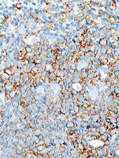
Researchers have reported the first known case of central nervous system (CNS) lymphoma responding to chimeric antigen receptor (CAR) T-cell therapy.
The investigational CAR T-cell therapy JCAR017 induced complete remission of brain metastasis in a patient with refractory diffuse large B-cell lymphoma (DLBCL).
When a subcutaneous tumor began to recur 2 months after the patient received JCAR017 and a surgical biopsy was performed, the CAR T cells spontaneously re-expanded and the tumor again went into remission.
While the patient eventually relapsed and died more than a year after receiving JCAR017, the brain tumor never recurred.
“Brain involvement in DLBCL carries a grave prognosis, and the ability to induce a complete and durable response with conventional therapies is rare,” said Jeremy Abramson, MD, of Massachusetts General Hospital in Boston.
“In addition, all available CAR T-cell trials have excluded patients with central nervous system involvement. This result has implications not only for secondary DLBCL like this case but also for primary central nervous system lymphoma, for which treatment options are similarly limited after relapse and few patents are cured.”
Dr Abramson and his colleagues described this case in a letter to NEJM. The patient was involved in a trial of JCAR017, which was sponsored by Juno Therapeutics.
The patient was a 68-year-old woman with germinal center B-cell-like DLBCL with a BCL2 rearrangement and multiple copies of MYC and BCL6.
The patients’ disease was refractory to conventional chemotherapy and an 8/8 HLA-matched stem cell transplant. After she enrolled in a phase 1 trial of JCAR017, the patient was found to have a new lesion in the right temporal lobe of her brain.
One month after the patient received JCAR017—given after lymphodepletion with fludarabine and cyclophosphamide—imaging showed complete remission of the brain lesion.
The subcutaneous lesion that recurred 2 months later disappeared after the biopsy with no further treatment. Blood testing showed an expansion of CAR T cells that coincided with the tumor’s regression.
While re-expansion of CAR T cells has been reported in response to other immunotherapy drugs, this is the first report of such a response to a biopsy.
“Typically, the drugs we use to fight cancer and other diseases wear off over time,” Dr Abramson said. “This spontaneous re-expansion after biopsy highlights this therapy as something entirely different, a ‘living drug’ that can re-expand and proliferate in response to biologic stimuli.”
Researchers have reported the first known case of central nervous system (CNS) lymphoma responding to chimeric antigen receptor (CAR) T-cell therapy.
The investigational CAR T-cell therapy JCAR017 induced complete remission of brain metastasis in a patient with refractory diffuse large B-cell lymphoma (DLBCL).
When a subcutaneous tumor began to recur 2 months after the patient received JCAR017 and a surgical biopsy was performed, the CAR T cells spontaneously re-expanded and the tumor again went into remission.
While the patient eventually relapsed and died more than a year after receiving JCAR017, the brain tumor never recurred.
“Brain involvement in DLBCL carries a grave prognosis, and the ability to induce a complete and durable response with conventional therapies is rare,” said Jeremy Abramson, MD, of Massachusetts General Hospital in Boston.
“In addition, all available CAR T-cell trials have excluded patients with central nervous system involvement. This result has implications not only for secondary DLBCL like this case but also for primary central nervous system lymphoma, for which treatment options are similarly limited after relapse and few patents are cured.”
Dr Abramson and his colleagues described this case in a letter to NEJM. The patient was involved in a trial of JCAR017, which was sponsored by Juno Therapeutics.
The patient was a 68-year-old woman with germinal center B-cell-like DLBCL with a BCL2 rearrangement and multiple copies of MYC and BCL6.
The patients’ disease was refractory to conventional chemotherapy and an 8/8 HLA-matched stem cell transplant. After she enrolled in a phase 1 trial of JCAR017, the patient was found to have a new lesion in the right temporal lobe of her brain.
One month after the patient received JCAR017—given after lymphodepletion with fludarabine and cyclophosphamide—imaging showed complete remission of the brain lesion.
The subcutaneous lesion that recurred 2 months later disappeared after the biopsy with no further treatment. Blood testing showed an expansion of CAR T cells that coincided with the tumor’s regression.
While re-expansion of CAR T cells has been reported in response to other immunotherapy drugs, this is the first report of such a response to a biopsy.
“Typically, the drugs we use to fight cancer and other diseases wear off over time,” Dr Abramson said. “This spontaneous re-expansion after biopsy highlights this therapy as something entirely different, a ‘living drug’ that can re-expand and proliferate in response to biologic stimuli.”
Researchers have reported the first known case of central nervous system (CNS) lymphoma responding to chimeric antigen receptor (CAR) T-cell therapy.
The investigational CAR T-cell therapy JCAR017 induced complete remission of brain metastasis in a patient with refractory diffuse large B-cell lymphoma (DLBCL).
When a subcutaneous tumor began to recur 2 months after the patient received JCAR017 and a surgical biopsy was performed, the CAR T cells spontaneously re-expanded and the tumor again went into remission.
While the patient eventually relapsed and died more than a year after receiving JCAR017, the brain tumor never recurred.
“Brain involvement in DLBCL carries a grave prognosis, and the ability to induce a complete and durable response with conventional therapies is rare,” said Jeremy Abramson, MD, of Massachusetts General Hospital in Boston.
“In addition, all available CAR T-cell trials have excluded patients with central nervous system involvement. This result has implications not only for secondary DLBCL like this case but also for primary central nervous system lymphoma, for which treatment options are similarly limited after relapse and few patents are cured.”
Dr Abramson and his colleagues described this case in a letter to NEJM. The patient was involved in a trial of JCAR017, which was sponsored by Juno Therapeutics.
The patient was a 68-year-old woman with germinal center B-cell-like DLBCL with a BCL2 rearrangement and multiple copies of MYC and BCL6.
The patients’ disease was refractory to conventional chemotherapy and an 8/8 HLA-matched stem cell transplant. After she enrolled in a phase 1 trial of JCAR017, the patient was found to have a new lesion in the right temporal lobe of her brain.
One month after the patient received JCAR017—given after lymphodepletion with fludarabine and cyclophosphamide—imaging showed complete remission of the brain lesion.
The subcutaneous lesion that recurred 2 months later disappeared after the biopsy with no further treatment. Blood testing showed an expansion of CAR T cells that coincided with the tumor’s regression.
While re-expansion of CAR T cells has been reported in response to other immunotherapy drugs, this is the first report of such a response to a biopsy.
“Typically, the drugs we use to fight cancer and other diseases wear off over time,” Dr Abramson said. “This spontaneous re-expansion after biopsy highlights this therapy as something entirely different, a ‘living drug’ that can re-expand and proliferate in response to biologic stimuli.”

TKI granted priority review for newly diagnosed CML

The US Food and Drug Administration (FDA) has granted priority review to a supplemental new drug application (sNDA) for the tyrosine kinase inhibitor (TKI) bosutinib (Bosulif®).
If approved, the sNDA would expand the use of bosutinib to include patients with newly diagnosed, chronic phase, Philadelphia chromosome-positive (Ph+) chronic myeloid leukemia (CML).
Bosutinib is currently FDA-approved to treat adults with chronic, accelerated, or blast phase Ph+ CML with resistance or intolerance to prior therapy.
The FDA grants priority review to applications for products that may provide significant improvements in the treatment, diagnosis, or prevention of serious conditions.
The agency’s goal is to take action on a priority review application within 6 months of receiving it, rather than the standard 10 months.
The FDA plans to make a decision on the sNDA for bosutinib by the end of this year.
Meanwhile, the European Medicines Agency (EMA) has validated for review a type II variation application for bosutinib in patients with newly diagnosed, chronic phase Ph+ CML.
Bosutinib already has conditional marketing authorization in the European Economic Area for the treatment of adults with Ph+ CML who previously received at least 1 TKI and adults with Ph+ CML for whom imatinib, nilotinib, and dasatinib are not considered appropriate.
Phase 3 trial
The applications submitted to the EMA and FDA are both supported by early results from the phase 3 BFORE trial. Results from this trial were presented at the ASCO Annual Meeting in May.
In this ongoing study, researchers are comparing bosutinib and imatinib as first-line treatment of chronic phase CML.
As of the ASCO presentation, the trial had enrolled 536 patients who were randomized 1:1 to receive bosutinib (n=268) or imatinib (n=268).
The presentation included results in a modified intent-to-treat population of Ph+ patients with e13a2/e14a2 transcripts who had at least 12 months of follow-up. In this group, there were 246 patients in the bosutinib arm and 241 in the imatinib arm.
Most of the patients were still on therapy at the 12-month mark or beyond—78% in the bosutinib arm and 73.2% in the imatinib arm. The median treatment duration was 14.1 months and 13.8 months, respectively.
At 12 months, the rate of major molecular response was 47.2% in the bosutinib arm and 36.9% in the imatinib arm (P= 0.02). The rate of complete cytogenetic response was 77.2% and 66.4%, respectively (P<0.008).
One patient in the bosutinib arm and 4 in the imatinib arm discontinued treatment due to disease progression, while 12.7% and 8.7%, respectively, discontinued treatment due to drug-related toxicity.
Adverse events that were more common in the bosutinib arm than the imatinib arm included grade 3 or higher diarrhea (7.8% vs 0.8%), increased alanine levels (19% vs 1.5%), increased aspartate levels (9.7% vs 1.9%), cardiovascular events (3% vs 0.4%), and peripheral vascular events (1.5% vs 1.1%). Cerebrovascular events were more common with imatinib than bosutinib (0.4% and 0%, respectively).
The US Food and Drug Administration (FDA) has granted priority review to a supplemental new drug application (sNDA) for the tyrosine kinase inhibitor (TKI) bosutinib (Bosulif®).
If approved, the sNDA would expand the use of bosutinib to include patients with newly diagnosed, chronic phase, Philadelphia chromosome-positive (Ph+) chronic myeloid leukemia (CML).
Bosutinib is currently FDA-approved to treat adults with chronic, accelerated, or blast phase Ph+ CML with resistance or intolerance to prior therapy.
The FDA grants priority review to applications for products that may provide significant improvements in the treatment, diagnosis, or prevention of serious conditions.
The agency’s goal is to take action on a priority review application within 6 months of receiving it, rather than the standard 10 months.
The FDA plans to make a decision on the sNDA for bosutinib by the end of this year.
Meanwhile, the European Medicines Agency (EMA) has validated for review a type II variation application for bosutinib in patients with newly diagnosed, chronic phase Ph+ CML.
Bosutinib already has conditional marketing authorization in the European Economic Area for the treatment of adults with Ph+ CML who previously received at least 1 TKI and adults with Ph+ CML for whom imatinib, nilotinib, and dasatinib are not considered appropriate.
Phase 3 trial
The applications submitted to the EMA and FDA are both supported by early results from the phase 3 BFORE trial. Results from this trial were presented at the ASCO Annual Meeting in May.
In this ongoing study, researchers are comparing bosutinib and imatinib as first-line treatment of chronic phase CML.
As of the ASCO presentation, the trial had enrolled 536 patients who were randomized 1:1 to receive bosutinib (n=268) or imatinib (n=268).
The presentation included results in a modified intent-to-treat population of Ph+ patients with e13a2/e14a2 transcripts who had at least 12 months of follow-up. In this group, there were 246 patients in the bosutinib arm and 241 in the imatinib arm.
Most of the patients were still on therapy at the 12-month mark or beyond—78% in the bosutinib arm and 73.2% in the imatinib arm. The median treatment duration was 14.1 months and 13.8 months, respectively.
At 12 months, the rate of major molecular response was 47.2% in the bosutinib arm and 36.9% in the imatinib arm (P= 0.02). The rate of complete cytogenetic response was 77.2% and 66.4%, respectively (P<0.008).
One patient in the bosutinib arm and 4 in the imatinib arm discontinued treatment due to disease progression, while 12.7% and 8.7%, respectively, discontinued treatment due to drug-related toxicity.
Adverse events that were more common in the bosutinib arm than the imatinib arm included grade 3 or higher diarrhea (7.8% vs 0.8%), increased alanine levels (19% vs 1.5%), increased aspartate levels (9.7% vs 1.9%), cardiovascular events (3% vs 0.4%), and peripheral vascular events (1.5% vs 1.1%). Cerebrovascular events were more common with imatinib than bosutinib (0.4% and 0%, respectively).
The US Food and Drug Administration (FDA) has granted priority review to a supplemental new drug application (sNDA) for the tyrosine kinase inhibitor (TKI) bosutinib (Bosulif®).
If approved, the sNDA would expand the use of bosutinib to include patients with newly diagnosed, chronic phase, Philadelphia chromosome-positive (Ph+) chronic myeloid leukemia (CML).
Bosutinib is currently FDA-approved to treat adults with chronic, accelerated, or blast phase Ph+ CML with resistance or intolerance to prior therapy.
The FDA grants priority review to applications for products that may provide significant improvements in the treatment, diagnosis, or prevention of serious conditions.
The agency’s goal is to take action on a priority review application within 6 months of receiving it, rather than the standard 10 months.
The FDA plans to make a decision on the sNDA for bosutinib by the end of this year.
Meanwhile, the European Medicines Agency (EMA) has validated for review a type II variation application for bosutinib in patients with newly diagnosed, chronic phase Ph+ CML.
Bosutinib already has conditional marketing authorization in the European Economic Area for the treatment of adults with Ph+ CML who previously received at least 1 TKI and adults with Ph+ CML for whom imatinib, nilotinib, and dasatinib are not considered appropriate.
Phase 3 trial
The applications submitted to the EMA and FDA are both supported by early results from the phase 3 BFORE trial. Results from this trial were presented at the ASCO Annual Meeting in May.
In this ongoing study, researchers are comparing bosutinib and imatinib as first-line treatment of chronic phase CML.
As of the ASCO presentation, the trial had enrolled 536 patients who were randomized 1:1 to receive bosutinib (n=268) or imatinib (n=268).
The presentation included results in a modified intent-to-treat population of Ph+ patients with e13a2/e14a2 transcripts who had at least 12 months of follow-up. In this group, there were 246 patients in the bosutinib arm and 241 in the imatinib arm.
Most of the patients were still on therapy at the 12-month mark or beyond—78% in the bosutinib arm and 73.2% in the imatinib arm. The median treatment duration was 14.1 months and 13.8 months, respectively.
At 12 months, the rate of major molecular response was 47.2% in the bosutinib arm and 36.9% in the imatinib arm (P= 0.02). The rate of complete cytogenetic response was 77.2% and 66.4%, respectively (P<0.008).
One patient in the bosutinib arm and 4 in the imatinib arm discontinued treatment due to disease progression, while 12.7% and 8.7%, respectively, discontinued treatment due to drug-related toxicity.
Adverse events that were more common in the bosutinib arm than the imatinib arm included grade 3 or higher diarrhea (7.8% vs 0.8%), increased alanine levels (19% vs 1.5%), increased aspartate levels (9.7% vs 1.9%), cardiovascular events (3% vs 0.4%), and peripheral vascular events (1.5% vs 1.1%). Cerebrovascular events were more common with imatinib than bosutinib (0.4% and 0%, respectively).

HERDOO2 may guide duration of treatment for unprovoked VTE
Clinical Question: Can HERDOO2 guide anticoagulation cessation in women with unprovoked venous thromboembolism (VTE)?
Background: Patients with unprovoked VTE have increased recurrence rates after stopping anticoagulation, but no tools have been validated to identify low risk patients.
Setting: Forty-four referral centers in seven countries.
Synopsis: Of patients with unprovoked, symptomatic VTE, 2,747 were evaluated after receiving anticoagulation for 5-12 months. HERDOO2 was used to classify women as low (0-1 points) or high (equal to or greater than 2 points) risk categories. Men were considered high risk. Anticoagulation was stopped for low risk patients. Treatment of high risk patients was left to physician choice.
Overall, high risk patients who continued anticoagulation had a 1.6% recurrence rate. Low risk women who stopped anticoagulation had a 3% recurrence rate per patient year, but postmenopausal women aged 50 years or older had a rate of 5.7%. High risk patients who stopped anticoagulation had a 7.4% recurrence rate. This study included multiple sites, but only 44% of participants were women. HERDOO2 should be used cautiously in postmenopausal women aged 50 years or older and in nonwhite women.
Bottom Line: HERDOO2 may help guide the decision to stop anticoagulation in select low-risk women with unprovoked VTE.
Citation: Rodger MA, Gregoire LG, Anderson DR, et al. Validating the HERDOO2 rule to guide treatment duration for women with unprovoked venous thrombosis: Multinational prospective cohort management study. BMJ. 2017 March;356:j1065.
Dr. Helfrich is an assistant professor in the University of Kentucky division of hospital medicine.
Clinical Question: Can HERDOO2 guide anticoagulation cessation in women with unprovoked venous thromboembolism (VTE)?
Background: Patients with unprovoked VTE have increased recurrence rates after stopping anticoagulation, but no tools have been validated to identify low risk patients.
Setting: Forty-four referral centers in seven countries.
Synopsis: Of patients with unprovoked, symptomatic VTE, 2,747 were evaluated after receiving anticoagulation for 5-12 months. HERDOO2 was used to classify women as low (0-1 points) or high (equal to or greater than 2 points) risk categories. Men were considered high risk. Anticoagulation was stopped for low risk patients. Treatment of high risk patients was left to physician choice.
Overall, high risk patients who continued anticoagulation had a 1.6% recurrence rate. Low risk women who stopped anticoagulation had a 3% recurrence rate per patient year, but postmenopausal women aged 50 years or older had a rate of 5.7%. High risk patients who stopped anticoagulation had a 7.4% recurrence rate. This study included multiple sites, but only 44% of participants were women. HERDOO2 should be used cautiously in postmenopausal women aged 50 years or older and in nonwhite women.
Bottom Line: HERDOO2 may help guide the decision to stop anticoagulation in select low-risk women with unprovoked VTE.
Citation: Rodger MA, Gregoire LG, Anderson DR, et al. Validating the HERDOO2 rule to guide treatment duration for women with unprovoked venous thrombosis: Multinational prospective cohort management study. BMJ. 2017 March;356:j1065.
Dr. Helfrich is an assistant professor in the University of Kentucky division of hospital medicine.
Clinical Question: Can HERDOO2 guide anticoagulation cessation in women with unprovoked venous thromboembolism (VTE)?
Background: Patients with unprovoked VTE have increased recurrence rates after stopping anticoagulation, but no tools have been validated to identify low risk patients.
Setting: Forty-four referral centers in seven countries.
Synopsis: Of patients with unprovoked, symptomatic VTE, 2,747 were evaluated after receiving anticoagulation for 5-12 months. HERDOO2 was used to classify women as low (0-1 points) or high (equal to or greater than 2 points) risk categories. Men were considered high risk. Anticoagulation was stopped for low risk patients. Treatment of high risk patients was left to physician choice.
Overall, high risk patients who continued anticoagulation had a 1.6% recurrence rate. Low risk women who stopped anticoagulation had a 3% recurrence rate per patient year, but postmenopausal women aged 50 years or older had a rate of 5.7%. High risk patients who stopped anticoagulation had a 7.4% recurrence rate. This study included multiple sites, but only 44% of participants were women. HERDOO2 should be used cautiously in postmenopausal women aged 50 years or older and in nonwhite women.
Bottom Line: HERDOO2 may help guide the decision to stop anticoagulation in select low-risk women with unprovoked VTE.
Citation: Rodger MA, Gregoire LG, Anderson DR, et al. Validating the HERDOO2 rule to guide treatment duration for women with unprovoked venous thrombosis: Multinational prospective cohort management study. BMJ. 2017 March;356:j1065.
Dr. Helfrich is an assistant professor in the University of Kentucky division of hospital medicine.