User login
Blinatumomab bests chemo in rel/ref B-ALL

Blinatumomab proved more effective than chemotherapy in a phase 3 trial of adults with Ph-negative, relapsed/refractory B-cell precursor acute lymphoblastic leukemia (B-ALL).
Blinatumomab produced higher remission rates and nearly doubled overall survival (OS) when compared to standard care, which encompassed 4 different chemotherapy regimens (investigator’s choice).
The incidence of grade 3 or higher adverse events (AEs) was higher for patients who received chemotherapy, but the incidence of serious AEs was higher in the blinatumomab arm.
These results were published in NEJM. The trial, known as TOWER, was sponsored by Amgen, the company developing blinatumomab.
“Results from the TOWER study reinforce the potential of this single-agent, bispecific T-cell engager immunotherapy, which helped a higher percentage of patients achieve minimal residual disease response versus standard of care chemotherapy, highlighting the depth and quality of remissions achieved,” said study author Hagop M. Kantarjian, MD, of The University of Texas MD Anderson Cancer Center in Houston.
Treatment
TOWER enrolled 405 patients with Ph-negative, relapsed/refractory B-ALL, 376 of whom ultimately received treatment.
The patients received blinatumomab (n=267) or investigator’s choice of 1 of 4 protocol-defined standard chemotherapy regimens (n=109):
- FLAG (fludarabine, high-dose cytarabine arabinoside, and granulocyte-colony stimulating factor), with or without an anthracycline (n=49, 45%)
- A high-dose cytarabine arabinoside-based regimen (n=19, 17%)
- A high-dose methotrexate-based regimen (n=22, 20%)
- A clofarabine-based regimen (n=19, 17%).
Patients who received blinatumomab received it as a continuous infusion, 4 weeks on and 2 weeks off, at 9 µg/day for 7 days, then 28 µg/day on weeks 2-4. They received 2 cycles of induction, which was followed by 3 cycles of consolidation if they had ≤5% blasts.
If patients still had ≤5% blasts after consolidation, they received up to 12 months of blinatumomab maintenance. Maintenance was a continuous infusion, 4 weeks on and 8 weeks off, at 28 µg/day.
Patients in the blinatumomab arm received a median of 2 cycles of therapy (range, 1-9). And patients in the chemotherapy arm received a median of 1 cycle (range, 1 to 4).
Thirty-two percent of patients in the blinatumomab arm received consolidation, as did 3% of patients in the chemotherapy arm.
Patients
Patient characteristics were similar between the treatment arms. The mean age was 41 in both arms (range, 18-80). Nearly 60% of patients in both arms were male.
About 40% of patients in the blinatumomab arm and 50% in the chemotherapy arm had not received any prior salvage regimens.
Thirty-five percent of patients in the blinatumomab arm and 34% in the chemotherapy arm had an allogeneic hematopoietic stem cell transplant (allo-HSCT) prior to study enrollment. Seventeen percent and 20%, respectively, relapsed after HSCT.
Forty-two percent and 40%, respectively, were refractory to primary or salvage therapy. Twenty-eight percent of patients in both arms were in their first relapse, and their first remission had lasted less than 12 months. Twelve percent of patients in both arms had an untreated second or greater relapse.
Remission
Within 12 weeks of treatment initiation, complete remission (CR) rates were significantly higher in the blinatumomab arm than the chemotherapy arm. (This was in the intent-to-treat population, which included 271 patients in the blinatumomab arm and 134 patients in the chemotherapy arm.)
The rate of CR with full hematologic recovery was 34% in the blinatumomab arm and 16% in the chemotherapy arm (P<0.001). The rate of CR with full, partial, or incomplete hematologic recovery was 44% and 25%, respectively (P<0.001).
Among patients who achieved a CR with full, partial, or incomplete hematologic recovery, 76% of those in the blinatumomab arm and 48% of those in the chemotherapy arm were negative for minimal residual disease.
Survival
At a median follow-up of 11.7 months for the blinatumomab arm and 11.8 months for the chemotherapy arm, the OS was significantly longer in the blinatumomab arm.
The median OS was 7.7 months and 4.0 months, respectively (hazard ratio for death=0.71, P=0.01).
The improvement in OS with blinatumomab was consistent across subgroups, regardless of age, prior salvage therapy, or prior allo-HSCT.
The investigators also considered the effect that post-treatment allo-HSCT might have on OS. Sixty-five patients in the blinatumomab arm and 32 in the chemotherapy arm went on to receive an allo-HSCT (24% of patients in both arms).
When the investigators censored for post-treatment allo-HSCT, the median OS was 6.9 months in the blinatumomab arm and 3.9 months in the chemotherapy arm (hazard ratio=0.66, P=0.004).
Safety
Nearly all patients in both arms (99%) experienced AEs. Grade 3 or higher AEs occurred in 87% of patients in the blinatumomab arm and 92% of those in the chemotherapy arm. Serious AEs occurred in 62% and 45%, respectively.
Grade 3 or higher AEs of interest, according to the researchers, were infection (34% with blinatumomab and 52% with chemotherapy), neutropenia (38% and 58%, respectively), elevated liver enzymes (13% and 15%, respectively), neurologic events (9% and 8%, respectively), cytokine release syndrome (5% and 0%, respectively), infusion reactions (3% and 1%, respectively), and lymphopenia (2% and 4%, respectively).
Fatal AEs occurred in 19% of patients in the blinatumomab arm and 17% of those in the chemotherapy arm.
Fatal AEs that occurred in at least 1% of patients in either arm (blinatumomab and chemotherapy, respectively) were sepsis (3% and 4%), septic shock (2% and 0%), multiorgan failure (1% and 0%), respiratory failure (<1% and 2%), and bacteremia (0% and 2%).
Arteriovenous coupler for structural hypertension poised for sham-controlled study
WASHINGTON – A new device may be the best opportunity yet to achieve sustained blood pressure control in individuals aged 60 years and older, according to Paul Sobotka, MD, Chief Scientific Officer at ROX Medical, the maker of the device.
The experimental device is an arteriovenous coupler that provides an anastomosis between artery and vein to lower arterial volume and reduce blood pressure due to a structural cause. The device has already performed well in initial clinical studies, including a controlled, open-label trial, he reported at CRT 2017 sponsored by the Cardiovascular Research Institute at Washington Hospital Center.
While neurohormonally driven elevations drive increases in diastolic and systolic blood pressures in younger patients, aortic stiffness and loss of vascular elasticity characterize the structurally driven hypertension of older patients, he said. While diuretics can lower arterial volume to achieve reductions in structurally related systolic hypertension, large doses are typically required to have meaningful results and are likely to be accompanied by unacceptable side effects in a large proportion of patients, he said.
“Somewhere between the age of 50 and 60 years, the majority of hypertensive patients will no longer be principally responsive to drugs acting on neurohormonal pathways or to renal denervation strategies.” In the elderly, cardiovascular risk is largely driven by hypertension principally related to the loss of aortic elasticity, which does not respond to most antihypertensive drugs.
The investigational arteriovenous coupler being developed by ROX Medical is intended to lower systolic blood pressure by reducing vascular resistance and therefore arterial volume. The coupler can be placed during cardiac catheterization, does not require sedation, takes about 1 hour to insert, and can be removed if necessary.
In a randomized, open-label study (Lancet. 2015 Apr;385:1634-41), mean systolic blood pressure was reduced by 26.9 mmHg on average in the 44 patients who received the arteriovenous coupler (P less than .001 vs. baseline) and by 3.7 mmHg (P = .31 vs. baseline) in the 39 patients who were maintained on antihypertensive medication, said Dr. Sobotka, who was a coauthor on this and several other clinical studies testing the efficacy and safety of the coupler. Systolic blood pressure falls almost immediately after the device is positioned, and blood pressure control has been sustained for up to 2 years of follow-up so far.
The procedure has been effective in patients resistant to antihypertensive medications and in those who have failed renal denervation, he said.
“The most significant adverse event observed has been venous stenosis related to turbulence, which occurs within in the first 12 months” after device placement, Dr. Sobotka reported. He said that venous stenting has resolved the problem in all affected patients. “Adverse events beyond that have been trivial.”
EXPERT ANALYSIS AT CRT 2017
Chronic rhinosinusitis exacerbation factors identified
ATLANTA – Clinical factors associated with acute exacerbations of chronic rhinosinusitis include female sex, nonwhite race, and a higher body mass index, results from a retrospective study suggest.
“Previous research has shown associations between various risk factors and the development of chronic rhinosinusitis,” study author Jason H. Kwah, MD, said in an interview in advance of the annual meeting of the American Academy of Allergy, Asthma, and Immunology. “However, there is minimal data regarding risk factor associations with frequent acute exacerbations of chronic rhinosinusitis. This is an important question as much of the morbidity and financial burden of this disease stems from costs and symptoms related to the management of acute exacerbations with antibiotics and steroids. If associations can be identified, the hope would be that modification of some of these risk factors could lead to improved control of the condition.”
Of the 3,109 CRS patients, 936 were frequent exacerbators while 2,173 were infrequent exacerbators. Univariate analysis revealed that the following factors were associated with frequent exacerbations, compared with infrequent exacerbations: female sex, nonwhite race, higher body mass index, having any drug allergy, allergic rhinitis, peripheral eosinophilia, asthma, lower forced expiratory volume in 1 second, previous sinus surgery, severe sinusitis on CT, one or more steroid prescriptions, and the presence of autoimmune disease. Multivariate analysis adjusted for race and sex revealed that allergic rhinitis (odds ratio, 1.48), peripheral eosinophilia (OR, 1.30), asthma (OR, 1.90), and autoimmune disease (OR, 1.61) remained significantly associated with frequent CRS exacerbations.
“We found it surprising that there were so many risk factors associated with frequent acute exacerbations of CRS, as well as the strengths of these associations,” Dr. Kwah said. “Acute exacerbations of CRS cause significant morbidity and financial burden, and based on the results of our study, are likely associated with many clinically relevant risk factors. Further study is needed to confirm the presence of these associations, study the mechanism of disease, and ultimately establish treatment protocols that can improve the control of frequent acute exacerbations of CRS.”
He acknowledged certain limitations of the study, including the challenges of establishing an accurate definition for acute exacerbations of CRS in a retrospective study. “While we felt our protocol for retrospective analysis was sufficient given our inclusion criteria, a prospective trial would be helpful in further delineating the presence of acute exacerbations of CRS and how they are associated with clinical risk factors,” he said.
The study was supported in part by the NIH National Center for Advancing Translational Sciences, the Chronic Rhinosinusitis Integrative Studies Program, the Ernest Bazley Foundation, and the Division of Allergy and Immunology at the Northwestern Feinberg School of Medicine. Dr. Kwah reported having no financial disclosures.
dbrunk@frontlinemedcom.com
AT THE 2017 AAAAI ANNUAL MEETING
Key clinical point:
Major finding: On multivariate analysis, allergic rhinitis (odds ratio, 1.48), peripheral eosinophilia (OR, 1.30), asthma (OR, 1.90), and autoimmune disease (OR, 1.61) remained significantly associated with frequent CRS exacerbations.
Data source: A retrospective review of 3,109 patients who were treated for CRS from January 2014 to December 2015.
Disclosures: The study was supported in part by the NIH National Center for Advancing Translational Sciences, the Chronic Rhinosinusitis Integrative Studies Program, the Ernest Bazley Foundation, and the Division of Allergy and Immunology at the Northwestern Feinberg School of Medicine. Dr. Kwah reported having no financial disclosures.
When STEMI door-to-balloon times deteriorate
WASHINGTON – The interventional cardiology team at Geisinger Medical Center, Danville, Pa., reorganized their care for ST segment elevation MI (STEMI) in 2004 to improve their door-to-balloon angioplasty times. Their highly successful efforts soon streamlined the process, placing Geisinger in the top 10% nationally for low average door-to-balloon (DTB) times.
Then, Geisinger began to see their DTB times creep up.
After witnessing a 5- to 10-minute increase in DTB times for STEMI patients – whether picked up by ambulance, appearing in the emergency department (ED), or referred from neighboring hospitals – James Blankenship, MD, director of the division of cardiology, and fellow Geisinger researchers took on an institutional analysis, which was presented at CRT 2017 sponsored by the Cardiovascular Research Institute at Washington Hospital Center.
Their analysis failed to identify a single explanation, but it did reinforce the importance of constantly reinvigorating processes, Dr. Blankenship said. And their analysis indicated that delays don’t necessarily translate into poorer outcomes.
One proposed explanation was that delays had been caused by an increased reliance on radial rather than femoral catheter access. Dr. Blankenship cited studies suggesting that radial access increases DTB times by 1 to 12 minutes. The proportion of percutaneous coronary interventions performed radially at Geisinger had risen from 2% to 85% over the time period that DTB times had risen.
“There is evidence of benefit from radial access, so even if it slows you down a few minutes, it may be worth doing,” Dr. Blankenship noted. However, when this variable was evaluated, the DTB times were, if anything, slightly faster with radial relative to femoral access.
Another theory was that the decision to provide fellows with a greater role in STEMI management had produced treatment delays. In the cath lab at Geisinger, the increased fellow participation “correlated perfectly” with the decline in DTB times, according to Dr. Blankenship. However, a close look at this variable failed to show any meaningful impact on DTB times.
Changes in process were also examined. For one example, a form must now be completed documenting airway assessment. However, Dr. Blankenship found that filling out this form only takes about a minute and could account for only a small part of the observed loss.
The most significant cause for the increased DTB times among STEMI patients presenting in the ED may well have been a 2012 change in the configuration of the hospital. Prior to 2012, the distance from the ED to the cath lab was less than 100 yards and a 1-minute walk. After the change in the configuration, the cath lab was approximately 7 minutes away, a change that “correlated somewhat” to a prolongation in DTB times.
Similarly, regional hospital STEMI referral patterns changed when a hospital in relatively close proximity opened a cath lab. Up until that time, most referrals had been a 5-minute helicopter transfer, according to Dr. Blankenship. Afterwards, some helicopter transfer times rose to 25 minutes.
Yet, no explanation seemed to be more important than simply ensuring that new staff understand and adhere to the processes. Recounting his experience with a “secret shopper” approach in which he called Geisinger posing as a referring physician to report a potential STEMI, he was disappointed to reach a staff member uncertain of the meaning of the term STEMI.
“STEMI systems need a lot of maintenance,” Dr. Blankenship said. He cautioned that staff changes are common and frequent, making it important to continually assess whether all the participants in delivering STEMI care understand their role.
Some published studies challenge the importance of small changes in DTB times as a predictor of STEMI survival, according to Dr. Blankenship, citing one national survey unable to link a reduction in DTB times with a change in in-hospital mortality (N Engl J Med. 2013;369:901-09). However, he has been more convinced by those studies that do show a relationship. He cited one large study that found an 8% loss in the mortality benefit for each 10-minute delay in DTB (Lancet. 2015;385:1114-22).
Rapid DTB times cannot be divorced from quality of care, Dr. Blankenship said. He cited an experience at one center in which an aggressive program for reducing DTB times led to an increase in mortality among false-positive patients. “It is okay to sacrifice time for quality.”
It was at the 2016 CRT meeting that Dr. Blankenship first provided data showing the decline in DTB times at his institution. At that time and prior to the more comprehensive evaluation of the reasons for the delay presented at this year’s meeting, he speculated that it may be just a question of fatigue from sustaining a rapid response posture over several years.
“My original observation, that after a while you just get tired, is probably still true,” he said.
AT CRT 2017
Key clinical point: With small deviations in process over time, delays build in the emergency treatment of ST segment elevation MI (STEMI).
Major finding: Processes must be regularly reinvigorated, a systematic evaluation of deteriorating door-to-balloon times for STEMI care suggests.
Data source: Nonrandomized retrospective analysis.
Disclosures: Dr. Blankenship reported no financial relationships to disclose.
Psoriasis and depression may pack a PsA punch
Patients with both psoriasis and major depressive disorder face an adjusted 37% greater risk of developing psoriatic arthritis (PsA) over a median follow-up of 5.1 years, a new study finds.
The findings don’t definitively prove that depression plays a role in PsA. Still, “,” study coauthor Cheryl Barnabe, MD, MSc, said in an interview.
For the new study published by the Journal of Investigative Dermatology, researchers tracked 73,447 people in the United Kingdom with psoriasis through a primary care medical records database for up to 25 years. The study statistics come from the years 1987-2012, reported Ryan T. Lewinson of the Cumming School of Medicine, in Calgary, Alta., and his associates (J Invest Dermatol. 2017. doi: 10.1016/j.jid.2016.11.032).
The median age at psoriasis diagnosis was 49.5 years (range, 20-90 years) and the median follow-up time was 5.1 years; 2% of the patients developed PsA and 7% developed major depression.
Via an unadjusted model, those with signs of depression were 1.56 times more likely to develop PsA (hazard ratio; 95% CI, 1.28-1.90; P less than .0001). In a model adjusted for factors such as age, gender, and obesity status, the extra risk of PsA was 1.37 (HR; 95% CI, 1.05-1.80; P = .021).
“The study draws into question the biological mechanisms by which depression increases the risk for developing psoriatic arthritis,” said Dr. Barnabe, an associate professor with the departments of medicine and community health sciences at the University of Calgary and a rheumatologist with Alberta Health Services.
The study notes that depression is linked to poor diet and lack of exercise, factors that could contribute to PsA. The authors also point out that researchers have linked depression to inflammation, a crucial component of both psoriasis and PsA, although they note that the study doesn’t examine systemic inflammation.
What’s next? “Mental health in chronic inflammatory diseases is not well addressed at the present time, in our system anyway, and should be a prime area of focus,” Dr. Barnabe noted. “Depression occurs at elevated rates in both psoriasis and psoriatic arthritis, and there is certainly a role for treatment to assist with disease management.”
The study authors reported no specific study funding and no relevant financial disclosures.
FROM THE JOURNAL OF INVESTIGATIVE DERMATOLOGY
Key clinical point: Depression is especially common in patients with psoriasis, and those with both conditions appear to face a higher risk of psoriatic arthritis.
Major finding: In an adjusted model, patients with psoriasis and signs of major depression were 37% more likely to develop PsA.
Data source: Retrospective study of 73,447 patients with psoriasis from the U.K. tracked for up to 25 years (median follow-up, 5.1 years).
Disclosures: The authors reported no specific study funding and no relevant financial disclosures.
Protocol speeds thrombectomy stroke patients from primary centers
HOUSTON – A novel protocol designed to speed patients with large-vessel occlusion strokes in and out of primary stroke centers and on to centers where they can undergo definitive thrombectomy treatment produced significant improvements in treatment speed and outcomes among 22 Rhode Island patients managed with the full protocol.
Streamlining the path in and out of a primary stroke center is key for delivering mechanical thrombectomy as quickly as possible to patients with an emergent large vessel occlusion, said Ryan A. McTaggart, MD, at the International Stroke Conference sponsored by the American Heart Association. “Door-in door-out time should be the standard metric for all partnerships between primary and comprehensive stroke centers,” said Dr. McTaggart, a neuroradiologist at Rhode Island Hospital in Providence, the state’s only comprehensive stroke center.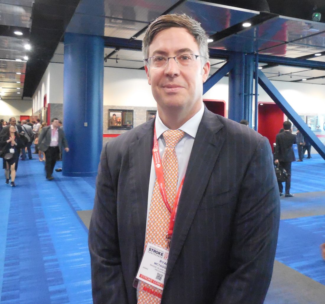
• When a patient with a suspected large vessel occlusion with a Los Angeles Motor Score of 4 or 5 arrives soon after onset at the primary stroke center, a call immediately goes out to the EMS transfer center of Rhode Island Hospital to coordinate the transport that will move the patient from the primary center to the comprehensive when needed.
• The initial CT scan at the primary center is run as the definitive scan, including a conventional CT scan to rule out hemorrhage and allow intravenous thrombolytic therapy with tissue plasminogen activator (TPA) and CT angiography to locate the occluding clot.
• The CT images are immediately uploaded to a cloud-based library so that neurologists at Rhode Island Hospital can read the images on their phones and plan the management strategy.
During the 11 months following the start of the protocol, the Rhode Island network identified 70 patients as candidates for thrombectomy, including 22 managed using the complete protocol and 48 managed using only parts of the new protocol.
The median time from onset of stroke symptoms to revascularization with thrombectomy was 184 minutes in the 22 patients managed under the full protocol and 233 minutes among 48 similar patients who were not fully managed with the protocol, Dr. McTaggart reported. This “dramatic” difference in median times was entirely driven by a difference in the door-in door-out time at the primary stroke center, which was a median of 64 minutes for the 22 patients managed with the full protocol and a median of 104 minutes without the full protocol, a 38% relative decrease that was statistically significant.
Time to initiation of intravenous TPA at the primary stroke center also improved, from a median of 65 minutes without the full protocol to a median of 40 minutes with it, a statistically significant difference. “The primary stroke center physicians tell us they have greater confidence to start TPA when they have a consult that can identify the patient’s clot,” he said.
Consistent with the shorter time to revascularization, the prevalence after 90 days of a functionally good outcome – a modified Rankin scale score of 0-2 – occurred in 50% of patients managed with the full protocol and 25% of those managed with a partial protocol, a statistically significant difference.
To put the 184 minutes median time from stroke onset to reperfusion into perspective, Dr. McTaggart noted that it is comparable to the time to reperfusion documented recently in a U.S. registry of thrombectomy patients who had been transported directly to the comprehensive stroke centers where their thrombectomy was done.
He also acknowledged the heavy lifting he and his associates had to do to set up this network. Getting buy-in from all the regional primary strokes centers was “a ton of work,” Dr. McTaggart said in an interview. “We told the primary stroke center staffs that thrombectomy is a powerful treatment, with a number needed to treat of three to get one improved outcome. That’s a convincing argument. The thrombectomy data [that became available in early 2015] made the argument for the protocol and network more compelling.”
Primary stroke centers keep the stroke patients who don’t have a clot occlusion suitable for thrombectomy, which means the comprehensive center thrombectomy team receives fewer false-alarm patients. Dr. McTaggart’s current goal is to have primary stroke centers get incoming patients out and on the road to a thrombectomy center within 45 minutes. In the future, primary stroke centers will perform CT imaging on all patients with suspected strokes, not just the severely affected patients with a Los Angeles Motor Score of 4 or 5. Additional useful steps toward speeding appropriate stroke patients to thrombectomy is direct ambulance transport of selected, high probability patients directly to a comprehensive stroke center and use of mobile stroke units to bring CT imaging and the start of TPA treatment out into the field.
Dr. McTaggart had no disclosures.
mzoler@frontlinemedcom.com
On Twitter @mitchelzoler
AT THE INTERNATIONAL STROKE CONFERENCE
Key clinical point:
Major finding: Median time from stroke onset to thrombectomy reperfusion was 184 minutes, including transfer between a primary and comprehensive stroke center.
Data source: Review of 70 acute ischemic stroke patients treated at Rhode Island Hospital.
Disclosures: Dr. McTaggart had no disclosures.
Teledermatology shows potential for grading patch test results
ORLANDO – Store-and-forward teledermatology may be useful for grading patch test results.
Erin Warshaw, MD, and Sara Hylwa, MD, both of the University of Minnesota, Minneapolis, sought to compare readings of patch test results both in person and via store-and-forward teledermatology. They patch tested patients at the Hennepin County (Minn.) Medical Center with the North American Contact Dermatitis Group screening series; photos were obtained at the 48-hour reading and the final reading (96-160 hours).
Almost all (101 of 107) of patients eligible for the trial were enrolled. Patients were overwhelmingly female (72%) with an average age of 50 years in this single-site study. Most screening panels were applied to the back.
Teledermatology assessment was categorized as successful if it matched the in-person assessment and as a failure if it did not; investigators labeled assessed pairs that did not fully match as indeterminate. Successful matches indicated there was no clinically significant difference between teledermatology and in-person assessment, indeterminate matches indicated that there was possible clinically significant difference, and failure to match indicated definite clinically significant difference.
All readings that were negative both in person and via teledermatology were excluded from the analysis.
At 48 hours, 47.2% of 705 reading pairs were labeled successful and 51.3% were labeled indeterminate. Failure, or complete disagreement, occurred in 1.6%, or 11 individual antigen pairs.
More successes – and failures – were seen at the final reading, with 53.8% of 420 final readings labeled successful, 39.8% labeled indeterminate, and 6.4%, or 27 individual antigen pairs, labeled as failures.
In general, teledermatology was more likely to miss or downplay the severity of reactions in the indeterminate pairs, Dr. Warshaw said. “This makes intuitive sense because when you are with a patient live, often the lighting catches an irritant wrinkle reaction or you can feel the lesion and be much more likely to call it irritant or a mild reaction than you would be from a flat photo.”
In the failure group, teledermatology generally overstated reactions, she added.
Dr. Warshaw said that logistical changes would be needed to make teledermatology more effective for reading patch test reactions in her practice. Their method of marking the patch test grid is to use a surgical marker on the corners, but a highlighter to mark the grid between the antigens. The highlighter simply did not show up well in photographs, she noted.
While not perfect, teledermatology does have promise for reading patch test reactions, she added. “I would love to save patients from having to come for their 48-hour reading... In Minnesota we have these horrible snowstorms. Last week there was a blizzard that was predicted. A third of our patients live 2 hours away from the clinic. If they could have taken photographs instead of trying to come through a blizzard for their final reading, that would be helpful.”
Dr. Warshaw noted that their study assessed only the 70 antigens of the North American Contact Dermatitis Research Group series and that it could have been strengthened by using additional series or the patients’ own products.
dfulton@frontlinemedcom.com
On Twitter @denisefulton
Key clinical point:
Major finding: Teledermatology readings failed to match in-person final readings 6% of the time.
Data source: Single-site study of 101 patients patch tested with the North American Contact Dermatitis Group series.
Disclosures: Dr. Warshaw declared no relevant conflicts of interest.
For bullous pemphigoid, doxycycline noninferior to prednisolone, but safer
ORLANDO – A 6-week course of doxycycline is an effective alternative to prednisolone for initial treatment of bullous pemphigoid in elderly patients, who may be particularly vulnerable to the potentially serious side effects of steroids.
While not as effective or quick-acting as prednisolone, doxycycline effected a cure in 74% of elderly patients who received it for 6 weeks in the Bullous Pemphigoid Steroids and Tetracyclines (BLISTER) Trial. More importantly, however, patients who took the antibiotic experienced half as many serious adverse events as those who took prednisolone (18% vs. 36%), Karen Harman, MD, said at the annual meeting of the American Academy of Dermatology.
The trial results will be published in the March 6 issue of The Lancet, she added.
BLISTER randomized 253 elderly patients with bullous pemphigoid to an initial, 6-week course of either 200 mg doxycycline/day or 0.5 mg/kg prednisolone/day. After the induction period, treatment could be adjusted as needed.
The primary efficacy outcome was blister count at 6 weeks; patients with fewer than three blisters were considered treatment successes. The primary safety outcome was the proportion who experienced an adverse event of at least grade 3 (severe, life-threatening, or fatal) related to the study medication.
The study was conducted at 54 centers in the United Kingdom and Germany, said Dr. Harman, who was also a primary investigator on the trial. It was a pragmatic noninferiority trial.
“We knew from the beginning that doxycycline would not be as effective as prednisolone. We estimated that we would see a 25% reduced efficacy, but we were prepared to accept that if we could also see an improvement in safety of at least 20%.”
Patients were a mean of age of 78 years, although about a quarter were 85 years or older. They scored a mean of 70 points on the 0-100 point Karnofsky scale of functional independence, with 100 being completely functionally independent.
A score of 70 means the patient is capable of self-care but cannot engage in normal activity or work because of disease. About 10% were unable to care for themselves and 42% unable to work.
Over the year-long trial, 33 patients dropped out. The most frequent reason for withdrawal was death – not an unexpected occurrence in such an elderly and infirm group, Dr. Harman noted. There was no significant difference in deaths between the treatment arms. Another 39 withdrew consent during the study. Adverse events caused three patients to drop out (two in the doxycycline group and one in the prednisolone group). One patient taking doxycycline was unable to tolerate the study medication and dropped out.
At 6 weeks, treatment succeeded in 74% of the doxycycline group and in 91% of the prednisolone group. This was an adjusted absolute treatment difference of 18% – well within the 25% margin of noninferiority.
Baseline disease severity didn’t interact with either of the drugs’ effectiveness. Prednisolone was more effective in every severity class. Among those with mild disease, the success rates were 76% with doxycycline and 97% with prednisolone. Among those with moderate disease, success rates were 78% and 97%. Among those with severe disease, both drugs were slightly less effective, with success rates of 65% and 75%.
Adverse events of grade 3 or greater occurred in significantly fewer patients taking doxycycline (18.5% vs. 36.6%), an adjusted difference of 19%. This was close to the 20% gain in safety the investigators hoped for.
Adverse events were common, however, with 82% of patients on doxycycline and 64% on prednisolone experiencing at least one mild-moderate event. Most of these in the doxycycline group were gastrointestinal, Dr. Harman noted.
About twice as many patients taking prednisolone had an event with a maximum of grade 3 (22% vs. 11%). The proportion with a maximum event of grade 4 was 4% in each group. Eleven in the prednisolone group and three in the doxycycline group died (9.7% vs. 2.5%).
The trial had several secondary efficacy outcomes.
Doxycycline had a slower onset of action, as evidenced by the significant difference in high blister count at week 6 (26% vs. 8.7%). At 6 weeks, 20.5% of the doxycycline group and 4% of the prednisolone group experienced treatment failure and chose to switch to the other treatment arm. Adherence was also worse in the doxycycline group, with 19% missing more than 3 consecutive days of treatment vs. 5% in the prednisolone group.
The trial also included a quality of life measure for the entire 52 weeks. Through week 26, this favored prednisolone, again reflecting the slower disease control of doxycycline. Thereafter, quality of life was similar between the two groups.
The trial was run in collaboration with the Nottingham Clinical Trials Unit, the UK Dermatology Clinical Trials Network, and the Medical Research Council Clinical Trials Unit. Dr. Harman had no financial disclosures.
msullivan@frontlinemedcom.com
On Twitter @Alz_Gal
Key clinical point:
Major finding: At 6 weeks, treatment succeeded in 74% of the doxycycline group and 91% of the prednisolone group; severe adverse events occurred in 18% and 36%, respectively.
Data source: The BLISTER trial randomized 253 patients.
Disclosures: The trial was run in collaboration with the Nottingham Clinical Trials Unit, the UK Dermatology Clinical Trials Network, and the Medical Research Council Clinical Trials Unit. Dr. Harman had no financial disclosures.
Patient transfer before thrombectomy worsens stroke outcomes
HOUSTON – Drip and ship may not be the most time-effective way to treat acute ischemic stroke patients who are candidates for endovascular thrombectomy.
Results from two separate real-world, observational studies showed that acute ischemic stroke patients with large vessel occlusions amenable to mechanical thrombectomy had significantly worse clinical outcomes when their management path included a stop at a primary stroke center followed by transfer to a comprehensive stroke center that had the capacity to perform thrombectomy, compared with going straight to the thrombectomy site.
The findings show “the system of care has room for improvement. Patients with large vessel occlusions clearly do better when we get them to mechanical thrombectomy as quickly as possible,” said Dr. Froehler, a vascular neurologist at Vanderbilt University in Nashville, Tenn. Thrombectomy “has a more powerful treatment effect than TPA [tissue plasminogen activator] and we need to adjust our standard of care to best deliver” thrombectomy, he said in an interview.
The study run by Dr. Froehler used data collected in the Systematic Evaluation of Patients Treated With Stroke Devices for Acute Ischemic Stroke (STRATIS) registry, which began in 2014 and has data for 984 acute ischemic stroke patients with large vessel occlusions treated by mechanical thrombectomy seen at any of 55 U.S. centers. The series included 445 (45%) patients first seen as a primary stroke center and then transferred to a comprehensive center and 539 (55%) who went directly to a comprehensive stroke center (direct patients). Prior to thrombectomy, 628 of all patients (64%) received TPA, with a roughly similar percentage in both the transferred and direct patients.
The data showed that the median time from symptom onset to revascularization was 202 minutes among the direct patients and 312 minutes among those first seen at a primary stroke center and then transferred, a statistically significant difference. The average time difference per patient between the two subgroups was 100 minutes, Dr. Froehler reported.
This difference in time to reperfusion led directly to significant differences in functional outcomes after 90 days measured on the modified Rankin Scale (mRS). The percentage of patients with a mRS score of 0 or 1 (an excellent functional outcome) was 38% for the patients first seen at primary stroke centers and 47% in direct patients, a 47% relative rise in excellent outcomes among the direct patients. The percentage of patients with a mRS score of 0-2, which identifies functional independence post stroke, was 52% among transferred patients and 60% in direct patients, a 38% relative improvement for this outcome among direct patients.
The second study of stroke transfer times and outcomes used data from 562 acute ischemic stroke patients with large vessel occlusions treated in the Providence Health & Services system in five western U.S. states during 2012-2016. Nearly half the patients required a transfer and the other half went directly to a center able to perform thrombectomy. The analysis used clinical outcomes scored on the mRS at the time of hospital discharge.
“Right now, the big delay at primary stroke centers is the door-in door-out time,” commented Ryan A. McTaggart, MD, an interventional neuroradiologist at Rhode Island Hospital in Providence, the only comprehensive stroke center in Rhode Island. He helped organize a partnership with 14 primary stroke centers in Rhode Island that uses a streamlined imaging, treatment (with TPA), and transfer protocol that hacked dozens of minutes off transfer times and produced a median time from onset of symptoms to revascularization by thrombectomy of 184 minutes in patients first seen at a primary stroke center. This clocking blows past the 202 minute median for stroke onset to revascularization in the direct patients from Dr. Froehler’s study.
Stroke transport and treatment networks are now undergoing refinement in Tennessee, said Dr. Froehler, based in part on the data he reported. Considerations in Tennessee include how EMS workers assess possible stroke patients, decisions by EMS on where to take patients, and how quality of care is measured at primary and comprehensive stroke centers.
The STRATIS registry is sponsored by Medtronic. Dr. Froehler is a consultant to Medtronic, Blockade, Stryker, and Control Medical. Dr. Smith, Dr. Tarpley, and Dr. McTaggart had no disclosures.
mzoler@frontlinemedcom.com
On Twitter @mitchelzoler
AT THE INTERNATIONAL STROKE CONFERENCE
Key clinical point:
Major finding: In STRATIS, excellent outcomes occurred in 47% of patients sent directly to a thrombectomy hospital and in 38% of transferred patients.
Data source: The STRATIS registry, with 984 U.S. acute ischemic stroke patients, and 562 U.S. acute ischemic stroke patients from the Providence Health & Services network.
Disclosures: The STRATIS registry is sponsored by Medtronic. Dr. Froehler is a consultant to Medtronic, Blockade, Stryker, and Control Medical. Dr. Smith, Dr. Tarpley, and Dr. McTaggart had no disclosures.
Maternal vitamin E isoform levels possible marker for infant wheezing risk
ATLANTA – Increasing maternal postpartum–plasma alpha-tocopherol isoform concentration was associated with a decreased likelihood of wheezing at age 2 years, defined as wheezing in the past 12 months, use of asthma medications in the past 12 months, or diagnosis of asthma, results from a large analysis showed.
“For now, this is an association and not causation,” study author Cosby Stone, MD, MPH, said in an interview in advance of the annual meeting of the American Academy of Allergy, Asthma, and Immunology. “We need a clinical trial to evaluate the effect of giving more alpha-tocopherol to mothers during pregnancy before anyone should jump to giving supplements.”
The median age of children at the time of maternal sample collection was 50 days. 47% were female, and 61% were white. Of the 652 children, 167 (26%) met criteria for wheezing at age 2 years. These children had mothers with significantly lower postpartum concentrations of plasma alpha-tocopherol, compared with those who did not meet criteria for wheezing at 2 years (a mean of 69 micromol/L vs. 75 micromol/L, respectively; P = .02). In multivariable regression analysis, the researchers detected a significant interaction between gamma-tocopherol and alpha-tocopherol, where the highest amounts of maternal gamma-tocopherol modified and mitigated the protective association of maternal alpha-tocopherol with risk of wheezing at 2 years (P = .05).
Dr. Stone cautioned that, what is currently labeled as vitamin E or alpha-tocopherol in foods and supplements, “could be any of eight different isoforms, and alpha-tocopherol may not actually be the dominant isoform being provided. In addition, the oils that we eat are the main sources of tocopherols in our diet, and they vary widely in terms of their tocopherol isoforms. Sunflower and safflower oil, for example, provide predominantly alpha-tocopherol as their isoform of vitamin E, while corn and soy oil provide predominantly gamma-tocopherol.”
For now, he continued, correcting maternal alpha-tocopherol deficiency, currently defined by a serum alpha-tocopherol concentration less than 11.6 micromol/L, is certainly reasonable. Down the road, modification of maternal alpha-tocopherol or gamma-tocopherol concentrations through dietary counseling may provide clinicians with a tool to prevent wheezing or asthma in affected children. “In the future there may be a role for checking tocopherol isoforms in pregnant women and then modifying dietary oil consumption to protect the health of their children,” he said.
He acknowledged certain limitations of the study, including that researchers obtained maternal vitamin E isoform measurements at enrollment, when the infants were, on average, 6 weeks post partum, and not during pregnancy. “However, the literature has shown that postpartum vitamin E levels at this time point are very similar to those during the second trimester of pregnancy,” Dr. Stone said. “The literature has also shown that plasma vitamin E isoform concentrations are tightly tied to diet and body stores and do not change very rapidly (such that a woman would not be likely to go from the highest quartile to the lowest, or vice versa). People’s diets don’t tend to change that much, in general.”
INSPIRE is funded by the National Institutes of Health. Dr. Stone is funded by an NIH training grant through Vanderbilt University. He reported having no relevant financial disclosures.
AT THE 2017 AAAAI ANNUAL MEETING
Key clinical point:
Major finding: Increasing maternal postpartum–plasma alpha-tocopherol concentration was associated with a decreased likelihood of wheezing requiring asthma medications at 2 years (P = .02).
Data source: A prospective evaluation of 652 children with maternal postpartum–plasma vitamin E isoforms measured at study enrollment.
Disclosures: INSPIRE is funded by the National Institutes of Health. Dr. Stone is funded by an NIH training grant through Vanderbilt University. He reported having no relevant financial disclosures.