User login
Buprenorphine implants rival daily sublingual buprenorphine for opioid dependence
Using an implant device to administer buprenorphine for adult patients being treated for opioid addiction is a viable alternative to the standard sublingual buprenorphine, a randomized clinical trial published online July 19 suggests.
“An implantable buprenorphine delivery system reduces adherence issues and may improve efficacy,” wrote Richard N. Rosenthal, MD, of the Icahn School of Medicine at Mount Sinai, New York, and his coinvestigators. “Furthermore, buprenorphine implants may reduce the need for sublingual buprenorphine, decreasing its availability for diversion, misuse, and harms.”
Dr. Rosenthal and his coinvestigators enrolled 177 patients from June 2014 to May 2015. The participants were aged 18-65 years, had received a primary diagnosis of opioid addiction, and had been receiving daily 8-mg doses of sublingual buprenorphine at an outpatient clinic for at least 24 weeks. The average age of patients was 39 years, and 40.9% were female. All of the participants were recruited from 21 treatment sites across the United States (JAMA. 2016;316[3]:282-90).
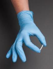
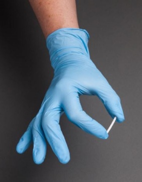
The participants were randomized into one of two cohorts: 87 subjects received buprenorphine implants, and the remaining 90 received sublingual buprenorphine. Those in the former cohort received their implants on the day of randomization, with four subdermal devices implanted along the inner upper arm. The staff involved with implanting and removing the devices did not have anything to do with study evaluation, in order to maintain blinding. Ten urine samples were collected from subjects over the course of the study period, with follow-up visits at 1 week and 2 weeks post treatment.
“The primary efficacy endpoint was the difference in proportion of responders, defined as participants with at least 4 of 6 months without evidence of illicit opioid use (based on urine test and self-report composites) by treatment group” the authors clarified.
Ultimately, 84 of those who received implants and 89 of those receiving sublingual treatment completed the trial, and were included in the primary analysis. Of those, 81 of those receiving implants (96.4%) and 78 of those receiving sublingual buprenorphine (87.6%) responded to treatment.
Based on urine tests and self-reporting, 72 of those with implants (85.7%) maintained their opioid abstinence throughout the study period, compared with 64 of those in the sublingual treatment cohort (71.9%). Additionally, Dr. Rosenthal and his coinvestigators noted that sustained abstinence in months 3-6 was more prominent in the implant cohort. Adverse events not related to the implant site occurred in 42 of the implant subjects (48.3%) and 47 of the sublingual treatment subjects (52.8%).
“To our knowledge, this was the first comparative trial to evaluate efficacy and safety of 6-month buprenorphine implants relative to sublingual buprenorphine in this understudied population of patients clinically stable taking sublingual buprenorphine,” the authors stated. They added that “because there are limited data on patients stable on sublingual buprenorphine, it is important to study maintenance and improvement of stability in patients who achieve good clinical response to initial buprenorphine treatment” with regard to further study.
Dr. Rosenthal and his coinvestigators disclosed receiving grants and nonfinancial support from Braeburn Pharmaceuticals, which was the sole provider of funding for this study. The other investigators also reported financial relationships with other companies.
“Because of the overall high prevalence and marked increases in morbidity and mortality associated with nonmedical prescription opioids and heroin, addressing opioid use disorders is a public health priority in the United States,” wrote Wilson M. Compton, MD, and Nora D. Volkow, MD, in an accompanying editorial (JAMA. 2016;316[3]:277-79).
But a fundamental challenge in addressing the treatment of chronic diseases such as opioid use disorder is nonadherence and discontinuation of care, they wrote. The use of extended-release formulations has been one approach. For example, extended-release naltrexone is administered monthly rather than the “nearly daily dosing necessary for the oral formulations of naltrexone, methadone, and buprenorphine. An additional concern for the agonist medications used for opioid use disorders is their risk of being diverted and misused.”
The results of the study by Richard N. Rosenthal, MD, and his colleagues show clearly that buprenorphine implants are not inferior to buprenorphine administered sublingually. However, as the study authors pointed out, several factors limit the generalizability of their results. For example, the patient population in their study were primarily white, employed, had at least a high school education, and were prescription opioid dependent.
“The approval of this new buprenorphine implant formulation provides a unique new tool to address the complex, often chronic and relapsing opioid use disorders,” Dr. Compton and Dr. Volkow wrote. “This novel implant system may help buttress patients’ decision-making deficits that are a core component of the addiction by making these lifesaving medication adherence decisions far more infrequent.”
Dr. Compton, deputy director of the National Institute on Drug Abuse, reported owning stock in Pfizer, General Electric, and 3M. Dr. Volkow is director of NIDA and reported no disclosures.
Using an implant device to administer buprenorphine for adult patients being treated for opioid addiction is a viable alternative to the standard sublingual buprenorphine, a randomized clinical trial published online July 19 suggests.
“An implantable buprenorphine delivery system reduces adherence issues and may improve efficacy,” wrote Richard N. Rosenthal, MD, of the Icahn School of Medicine at Mount Sinai, New York, and his coinvestigators. “Furthermore, buprenorphine implants may reduce the need for sublingual buprenorphine, decreasing its availability for diversion, misuse, and harms.”
Dr. Rosenthal and his coinvestigators enrolled 177 patients from June 2014 to May 2015. The participants were aged 18-65 years, had received a primary diagnosis of opioid addiction, and had been receiving daily 8-mg doses of sublingual buprenorphine at an outpatient clinic for at least 24 weeks. The average age of patients was 39 years, and 40.9% were female. All of the participants were recruited from 21 treatment sites across the United States (JAMA. 2016;316[3]:282-90).
The participants were randomized into one of two cohorts: 87 subjects received buprenorphine implants, and the remaining 90 received sublingual buprenorphine. Those in the former cohort received their implants on the day of randomization, with four subdermal devices implanted along the inner upper arm. The staff involved with implanting and removing the devices did not have anything to do with study evaluation, in order to maintain blinding. Ten urine samples were collected from subjects over the course of the study period, with follow-up visits at 1 week and 2 weeks post treatment.
“The primary efficacy endpoint was the difference in proportion of responders, defined as participants with at least 4 of 6 months without evidence of illicit opioid use (based on urine test and self-report composites) by treatment group” the authors clarified.
Ultimately, 84 of those who received implants and 89 of those receiving sublingual treatment completed the trial, and were included in the primary analysis. Of those, 81 of those receiving implants (96.4%) and 78 of those receiving sublingual buprenorphine (87.6%) responded to treatment.
Based on urine tests and self-reporting, 72 of those with implants (85.7%) maintained their opioid abstinence throughout the study period, compared with 64 of those in the sublingual treatment cohort (71.9%). Additionally, Dr. Rosenthal and his coinvestigators noted that sustained abstinence in months 3-6 was more prominent in the implant cohort. Adverse events not related to the implant site occurred in 42 of the implant subjects (48.3%) and 47 of the sublingual treatment subjects (52.8%).
“To our knowledge, this was the first comparative trial to evaluate efficacy and safety of 6-month buprenorphine implants relative to sublingual buprenorphine in this understudied population of patients clinically stable taking sublingual buprenorphine,” the authors stated. They added that “because there are limited data on patients stable on sublingual buprenorphine, it is important to study maintenance and improvement of stability in patients who achieve good clinical response to initial buprenorphine treatment” with regard to further study.
Dr. Rosenthal and his coinvestigators disclosed receiving grants and nonfinancial support from Braeburn Pharmaceuticals, which was the sole provider of funding for this study. The other investigators also reported financial relationships with other companies.
FROM JAMA
Key clinical point: Buprenorphine implants are no less effective than sublingual buprenorphine, the current standard of care, in managing opioid dependence.
Major finding: 96.4% of those with implants and 87.6% of those with sublingual buprenorphine responded to treatment; over 6 months, 85.7% of those with implants and 71.9% of those receiving sublingual treatment maintained opioid resistance.
Data source: An outpatient, randomized, active-controlled, 24-week, double-blind, double-dummy study of 177 buprenorphine patients at 21 U.S. sites.
Disclosures: The study funded by Braeburn Pharmaceuticals. Dr. Rosenthal and other coinvestigators disclosed receiving grants and nonfinancial support from Braeburn Pharmaceuticals while this study was being conducted. The other investigators also reported financial relationships with other companies.
BTK inhibitor may treat ibrutinib-resistant cancers

Photo by Aaron Logan
KOLOA, HAWAII—Preclinical research suggests that ARQ 531, a reversible Bruton’s tyrosine kinase (BTK) inhibitor, might prove effective against ibrutinib-resistant hematologic malignancies.
The study showed that ARQ 531 inhibits wild-type BTK and the ibrutinib-resistant BTK-C481S mutant with similar potency.
The compound also suppressed proliferation of hematologic cancer cells in vitro and inhibited tumor growth in a mouse model of B-cell lymphoma.
Researchers disclosed these results in a poster presentation at the 2016 Pan Pacific Lymphoma Conference. The research was supported by ArQule Inc., the company developing ARQ 531.
The researchers first demonstrated that ARQ 531 enacts biochemical inhibition of both wild-type and C481S-mutant BTK at sub-nanomolar levels and cellular inhibition in C481S-mutant BTK cells that are resistant to ibrutinib.
The team then tested ARQ 531 in a range of cell lines encompassing a variety of leukemias and lymphomas, as well as multiple myeloma.
They found that ARQ 531 can inhibit proliferation in many types of hematologic cancer cells, but it “potently inhibits” cell lines that are addicted to BCR, PI3K/AKT, and Notch signaling pathways.
The researchers also tested ARQ 531 in the BTK-driven TMD8 xenograft mouse model (B-cell lymphoma). They said the compound demonstrated strong target and pathway inhibition, with sustained tumor growth inhibition.
The team noted that ARQ 531 exhibits a distinct kinase selectivity profile, with strong inhibitory activity against several key oncogenic drivers from TEC, Trk, and Src family kinases. And the compound inhibits the RAF/MEK/ERK and PI3K/AKT/mTOR pathways.
The researchers said these results support further investigation of ARQ 531, particularly in the setting of ibrutinib resistance.
It is currently estimated that about 10% of patients treated with ibrutinib develop resistance, and more than 80% of these patients present with the C481S mutation.
“We are beginning to see increasing resistance to ibrutinib, which is creating the need for a BTK inhibitor, like ARQ 531, that targets the C481S mutation,” said Brian Schwartz, head of research and development and chief medical officer at ArQule.
“The preclinical profile of ARQ 531 as a potent and reversible inhibitor of wild-type and mutant BTK presents the potential for a first-in-class and best-in-class molecule. We are working toward completing GLP [good laboratory practice] toxicology studies and filing an IND [investigational new drug application] in early 2017.” ![]()
FDA rejects pegfilgrastim biosimilar
The US Food and Drug Administration (FDA) has decided not to approve Novartis’s application for a biosimilar of Amgen’s Neulasta, also known by the generic name pegfilgrastim.
The FDA issued a complete response letter for the pegfilgrastim biosimilar last month.
Novartis has not provided details about the agency’s decision or the contents of the letter, but the company said it is working with the FDA to answer its questions about the drug.
Novartis was seeking approval for its pegfilgrastim biosimilar for the same indication as Amgen’s Neulasta.
Neulasta is a leukocyte growth factor that is FDA-approved for the following indications:
- To decrease the incidence of infection, as manifested by febrile neutropenia, in patients with non-myeloid malignancies receiving myelosuppressive anticancer drugs associated with a clinically significant incidence of febrile neutropenia.
- To increase survival in patients acutely exposed to myelosuppressive doses of radiation.
Neulasta is not FDA-approved for the mobilization of peripheral blood progenitor cells for hematopoietic stem cell transplantation. ![]()
JAK3 inhibitors could treat NK/T-cell lymphoma
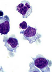
Agents that inhibit the protein kinase JAK3 could prove effective for treating natural killer/T-cell lymphoma (NKTL), according to research published in Blood.
Researchers investigated the role the cancer-promoting gene EZH2 plays in NKTL.
This revealed that EZH2 activity is regulated by JAK3, and a JAK3 inhibitor could significantly reduce the growth of NKTL cells.
Specifically, the team discovered that JAK3 activation leads to phosphorylation of EZH2.
This prompts the dissociation of EZH2 from the PRC2 complex and leads to decreased global H3K27me3 levels.
EZH2 then shifts from its normal function of suppressing gene expression to activating genes—specifically, upregulating a set of genes involved in DNA replication, cell cycle, biosynthesis, stemness, and invasiveness.
And this leads to the development of NKTL.
“As JAK3 is often mutated and activated in natural killer/T-cell lymphoma cells, this finding is particular intriguing, as it suggests a predominant non-catalytic function of EZH2 in JAK3-mutant natural killer/T-cell lymphoma,” said study author Wee-Joo Chng, MB ChB, PhD, of the National University of Singapore.
“Our study also suggests that various oncogenic mutations may modify the function of EZH2, explaining the complex roles of EZH2 in cancer.”
The researchers also found that a JAK3 inhibitor could significantly reduce the growth of NKTL cells, in an EZH2 phosphorylation-dependent manner.
However, compounds that have recently been developed to inhibit EZH2 methyltransferase activity did not have the same effect.
“Moving forward, a biomarker strategy might be needed to ensure appropriate application of EZH2 inhibitors,” Dr Chng said.
“This will help to identify tumors where EZH2 requires its catalytic activity or is actually acting through non-catalytic function. At the same time, we need to develop therapies that can target the non-catalytic function of EZH2.” ![]()
Antimalarials, antifungals may fight leukemia
A new study has shed light on the process leukemia cells use to evade apoptosis and revealed drugs that can fight this process.
Investigators found that leukemia cells expel a molecule that starts apoptosis, but antimalarial drugs and antifungal drugs can halt this process and force the leukemia cells to self-destruct.
Alexandre Chigaev, PhD, of the University of New Mexico in Albuquerque, and his colleagues described these discoveries in Oncotarget.
The investigators theorized that leukemia cells might evade death by regulating 3’-5’-cyclic adenosine monophosphate (cAMP), which is associated with pro-apoptotic signaling.
The team thought leukemic cells might possess mechanisms that efflux cAMP from the cytoplasm, thereby protecting them from apoptosis.
In studying VLA-4—an adhesion molecule that keeps each cell in its niche—the investigators discovered that cAMP reduces VLA-4’s adhesive properties, allowing cells to detach.
“That’s how we stumbled upon it,” Dr Chigaev said. “Cyclic AMP reduces cell adhesion, and maybe that’s one of the mechanisms by which [leukemic] cells leave bone marrow niches.”
The investigators then confirmed that cAMP starts apoptosis in leukemia cells. And they showed that leukemia cells could efflux cAMP, but normal blood cells could not.
The team substantiated their findings by testing several drugs that block cAMP efflux.
The drugs, which are all approved by the US Food and Drug Administration, are used to fight malaria or fungal infections. They include artesunate, dihydroartemisinin, clioquinol, cryptotanshinone, parthenolide, and patulin.
The investigators found these drugs successfully decreased cAMP efflux and induced apoptosis in a model of acute myeloid leukemia, in B-lineage acute lymphoblastic leukemia cell lines, and in samples from patients with B-lineage acute lymphoblastic leukemia.
On the other hand, the drugs did not affect peripheral blood mononuclear cells.
“This particular mechanism of action has not been reported for these drugs,” Dr Chigaev said. “And the idea that cells can have this apoptotic escape, or apoptotic evasion, through cyclic AMP pumping, that’s new. It’s never been reported previously.”
Dr Chigaev and his colleagues noted that repurposing already-approved drugs could greatly shorten the approval process to use them for leukemia.
Since the drugs were tested on cells from leukemia patients, the team hopes to continue seeing promising results. They have begun animal studies in preparation for clinical trials. ![]()
HPV vaccination rates not improved by increased awareness
Increased awareness did not increase adolescent HPV vaccination rates in a high-risk population, according to Jessica Fishman, PhD, of the University of Pennsylvania, Philadelphia, and her associates.
The study sample included 211 low-income adolescents, aged 13-18 years, and 149 parents of different adolescents, aged 9-18 years, who had not been vaccinated for HPV. In the adolescent group, 3% received an HPV vaccination after 3 months, 9% received a vaccination after 6 months, and 15% received a vaccination after 1 year. In the parent group, 5% had their daughters vaccinated after 3 months, 10% had their daughters vaccinated after 6 months, and 13% had their daughters vaccinated after 1 year.
Awareness was measured using a questionnaire asking about individual awareness of HPV, cervical cancer, HPV vaccination, and news or advertising about HPV vaccination. Both adolescents and parents were most aware of cervical cancer (73% and 94%, respectively) and least aware of news about HPV advertising (51% and 66%, respectively). A total of 14% of adolescents and 4% of parents had no awareness of any questionnaire item, while 32% of adolescents and 57% of parents had awareness of all items.
Probability of vaccination was less than 0.5 for all levels of awareness, and accuracy of HPV vaccination prediction models was poor, Dr. Fishman and her associates noted.
“For this high-risk population where vaccination is rare, evidence-based behavioral interventions are urgently needed. Ideally, interventions will target variables associated with vaccination. Interventions that do not target actual determinants can have no effect or even a ‘boomerang’ effect that increases unhealthy behavior,” the investigators concluded.
Find the full study in Pediatrics (doi: 10.1542/peds.2015-2048).
FROM PEDIATRICS
Feds plan to raise penalties for false claims
Penalties for health providers under the federal False Claims Act (FCA) are set to double under a proposed rule by the U.S. Department of Justice.
The interim final rule would increase minimum per-claim FCA fines from $5,500 to $10,781 and maximum per-claim penalties would rise from $11,000 to $21,563.
The adjusted civil penalty amounts would apply to civil penalties assessed after Aug. 1, 2016, whose associated violations occurred after Nov. 2, 2015. Violations on or before Nov. 2, 2015, and assessments made prior to Aug. 1, 2016, would continue to be subject to the lower penalties. The rise stems from the federal Civil Monetary Penalties Inflation Adjustment Act, which provides for the regular evaluation and adjustment for inflation of civil monetary penalties to ensure they maintain a deterrent effect, according to a summary of the rule.
The FCA penalizes any person who knowingly submits a false claim to the government or causes another to submit a false claim to the government or who knowingly makes a false record or statement to get a false claim paid by the government. In 2015, the DOJ recovered more than $3.5 billion in FCA settlements and judgments.
Health law experts say the higher penalties may encourage more whistleblowers to file FCA claims against doctors since the potential recoveries would be higher.
“The new maximums may make things still more enticing for relators with visions of increasingly large relators’ shares on the table,” said William W. Horton, a Birmingham, Ala.-based health law attorney and chair of the American Bar Association Health Law Section.
However, Mr. Horton does not believe the rates will have much practical effect in terms of strategy or settlement rates. Right now, the hypothetical penalties in such cases are so enormous they are almost not meaningful, he said in an interview.
“In reality, cases settle based on the amount of actual damages – overpayments, etc. – and not on the penalties, because the penalties are so high,” he said. “I don’t think making them higher is going to change that, because it doesn’t increase the amount of money available for defendants to settle with.”
The ultimate question is whether the higher penalties will help deter health fraud, adds Houston health law attorney Michael E. Clark.
“I don’t see it making a difference,” he said in an interview. “The FCA penalties are particularly ruinous in the health care field since so many claims get made and courts have accepted broad theories of liability.”
The DOJ is accepting comments on the interim final rule until Aug. 29.
On Twitter @legal_med
Pediatric Photosensitivity Disorders
Review the PDF of the fact sheet on pediatric photosensitivity disorders with board-relevant, easy-to-review material. This month's fact sheet will review important disorders in the pediatric population where photosensitivity is a major feature
Practice Questions
1. Which photosensitivity disorder is characterized by decreased immunoglobulin-mediated immunity?
a. Bloom syndrome
b. Cockayne syndrome
c. hydroa vacciniforme
d. Kindler syndrome
e. poikiloderma congenitale
2. Which of the following is an inappropriate treatment for a young Mexican girl with cheilitis and treatment-resistant chronic pruritic crusted papules and scars on both sun-exposed and nonexposed sites?
a. oral isotretinoin
b. oral prednisone
c. oral thalidomide
d. topical calcineurin inhibitors
e. topical corticosteroids
3. Which gene is mutated in a patient with a history of congenital acral blistering and then gradual onset of cutaneous atrophy and fragility, oral lesions, and photosensitivity?
a. DHCR7
b. KIND1
c. RECQL4
d. SLC6A19
e. XPD
4. Which photosensitivity disorder is associated with an increased risk for osteosarcoma?
a. actinic prurigo
b. Rothmund-Thomson syndrome
c. Smith-Lemli-Opitz syndrome
d. trichothiodystrophy
e. xeroderma pigmentosum
5. All of the following are features seen in De Sanctis-Cacchione syndrome except:
a. ataxia
b. basal ganglion calcification
c. deafness
d. hypogonadism
e. short stature
Answers to practice questions provided on next page
Practice Question Answers
1. Which photosensitivity disorder is characterized by decreased immunoglobulin-mediated immunity?
a. Bloom syndrome
b. Cockayne syndrome
c. hydroa vacciniforme
d. Kindler syndrome
e. poikiloderma congenitale
2. Which of the following is an inappropriate treatment for a young Mexican girl with cheilitis and treatment-resistant chronic pruritic crusted papules and scars on both sun-exposed and nonexposed sites?
a. oral isotretinoin
b. oral prednisone
c. oral thalidomide
d. topical calcineurin inhibitors
e. topical corticosteroids
3. Which gene is mutated in a patient with a history of congenital acral blistering and then gradual onset of cutaneous atrophy and fragility, oral lesions, and photosensitivity?
a. DHCR7
b. KIND1
c. RECQL4
d. SLC6A19
e. XPD
4. Which photosensitivity disorder is associated with an increased risk for osteosarcoma?
a. actinic prurigo
b. Rothmund-Thomson syndrome
c. Smith-Lemli-Opitz syndrome
d. trichothiodystrophy
e. xeroderma pigmentosum
5. All of the following are features seen in De Sanctis-Cacchione syndrome except:
a. ataxia
b. basal ganglion calcification
c. deafness
d. hypogonadism
e. short stature
MRI-VA improves view of anomalous coronary arteries
Failure to achieve a rounded and unobstructed ostia in children who have surgery to repair anomalous coronary arteries can put these children at continued risk for sudden death, but cardiac MRI with virtual angioscopy (VA) before and after the operation can give cardiologists a clear picture of a patient’s risk for sudden death and help direct ongoing management, according to a study in the July issue of the Journal of Thoracic and Cardiovascular Surgery (2016;152:205-10).
“Cardiac MRI with virtual angioscopy is an important tool for evaluating anomalous coronary anatomy, myocardial function, and ischemia and should be considered for initial and postoperative assessment of children with anomalous coronary arteries,” lead author Julie A. Brothers, MD, and her coauthors said in reporting their findings.
Anomalous coronary artery is a rare congenital condition in which the left coronary artery (LCA) originates from the right sinus or the right coronary artery (RCA) originates from the left coronary sinus. Dr. Brothers, a pediatric cardiologist, and her colleagues from the Children’s Hospital of Philadelphia and the University of Pennsylvania, also in Philadelphia, studied nine male patients who had operations for anomalous coronary arteries during Feb. 2009-May 2015 in what they said is the first study to document anomalous coronary artery anatomy both before and after surgery. The patients’ average age was 14.1 years; seven had right anomalous coronary arteries and two had left anomalous arteries. After the operations, MRI-VA revealed that two patients still had narrowing in the neo-orifices.
Previous reports recommend surgical repair for all patients with anomalous LCA and for symptomatic patients with anomalous RCA anatomy (Ann Thorac Surg. 2011;92:691-7; Ann Thorac Surg. 2014;98:941-5). MRI-VA allows the surgical team to survey the ostial stenosis before the operation “as if standing within the vessel itself,” Dr. Brothers and her coauthors wrote. Afterward, MRI-VA lets the surgeon and team see if the operation succeeded in repairing the orifices.
In the study population, VA before surgery confirmed elliptical, slit-like orifices in all patients. The operations involved unroofing procedures; two patients also had detachment and resuspension procedures during surgery. After surgery, VA showed that seven patients had round, patent, unobstructed repaired orifices; but two had orifices that were still narrow and somewhat stenotic, Dr. Brothers and her coauthors said. The study group had postoperative MRI-VA an average of 8.6 months after surgery.
“The significance of these findings is unknown; however, if the proposed mechanism of ischemia is due to a slit-like orifice, a continued stenotic orifice may place subjects at risk for sudden death,” the researchers said. The two study patients with the narrowed, stenotic orifices have remained symptom free, with no evidence of ischemia on exercise stress test or cardiac MRI. “These subjects will need to be followed up in the future to monitor for progression or resolution,” the study authors wrote.
Sudden cardiac death (SCD) is more common in anomalous aortic origin of the LCA than the RCA, Dr. Brothers and her colleagues said. Thus, an elliptical, slit-like neo-orifice is a concern because it can become blocked during exercise, possibly leading to lethal ventricular arrhythmia, they said. Ischemia in patients with anomalous coronary artery seems to result from a cumulative effect of exercise.
Patients who undergo the modified unroofing procedure typically have electrocardiography and echocardiography afterward and then get cleared to return to competitive sports in about 3 months if their stress test indicates it. Dr. Brothers and her colleagues said this activity recommendation may need alteration for those patients who have had a heart attack or sudden cardiac arrest, because they may remain at increased risk of SCD after surgery. “At the very least, additional imaging, such as with MRI-VA, should be used in this population,” the study authors said.
While Dr. Brothers and her colleagues acknowledged the small sample size is a limitation of the study, they also pointed out that anomalous coronary artery is a rare disease. They also noted that high-quality VA images can be difficult to obtain in noncompliant patients or those have arrhythmia or irregular breathing. “The images obtained in this study were acquired at an institution very familiar with pediatric cardiac coronary MRI and would be appropriate for assessing the coronary ostia with VA,” they said.
Dr. Brothers and her coauthors had no financial disclosures.
The MRI technique that Dr. Brothers and her colleagues reported on can provide important details of the anomalous coronary anatomy and about myocardial function, Philip S. Naimo, MD, Edward Buratto, MBBS, and Igor Konstantinov, MD, PhD, FRACS, of the Royal Children’s Hospital, University of Melbourne, wrote in their invited commentary. But, the ability to evaluate the neo-ostium after surgery had “particular value,” the commentators said (J. Thorac. Cardiovasc. Surg. 2016 Jul;152:211-12).
MRI with virtual angioscopy can fill help fill in the gaps where the significance of a narrowed neo-ostium is unknown, the commentators said. “The combination of anatomic information on the ostium size, shape, and location, as well as functional information on wall motion and myocardial perfusion, which can be provided by MRI-VA, would be particularly valuable in these patients,” they said.
They also pointed out that MRI-VA could be used in patients who have ongoing but otherwise undetected narrowing of the ostia after the unroofing procedure. At the same time, the technique will also require sufficient caseloads to maintain expertise. “It is safe to say that MRI-VA is here to stay,” Dr. Naimo, Dr. Buratto, and Dr. Konstantinov wrote. “The actual application of this virtual modality will need further refinement to be used routinely.”
The commentary authors had no financial relationships to disclose.
Failure to achieve a rounded and unobstructed ostia in children who have surgery to repair anomalous coronary arteries can put these children at continued risk for sudden death, but cardiac MRI with virtual angioscopy (VA) before and after the operation can give cardiologists a clear picture of a patient’s risk for sudden death and help direct ongoing management, according to a study in the July issue of the Journal of Thoracic and Cardiovascular Surgery (2016;152:205-10).
“Cardiac MRI with virtual angioscopy is an important tool for evaluating anomalous coronary anatomy, myocardial function, and ischemia and should be considered for initial and postoperative assessment of children with anomalous coronary arteries,” lead author Julie A. Brothers, MD, and her coauthors said in reporting their findings.
Anomalous coronary artery is a rare congenital condition in which the left coronary artery (LCA) originates from the right sinus or the right coronary artery (RCA) originates from the left coronary sinus. Dr. Brothers, a pediatric cardiologist, and her colleagues from the Children’s Hospital of Philadelphia and the University of Pennsylvania, also in Philadelphia, studied nine male patients who had operations for anomalous coronary arteries during Feb. 2009-May 2015 in what they said is the first study to document anomalous coronary artery anatomy both before and after surgery. The patients’ average age was 14.1 years; seven had right anomalous coronary arteries and two had left anomalous arteries. After the operations, MRI-VA revealed that two patients still had narrowing in the neo-orifices.
Previous reports recommend surgical repair for all patients with anomalous LCA and for symptomatic patients with anomalous RCA anatomy (Ann Thorac Surg. 2011;92:691-7; Ann Thorac Surg. 2014;98:941-5). MRI-VA allows the surgical team to survey the ostial stenosis before the operation “as if standing within the vessel itself,” Dr. Brothers and her coauthors wrote. Afterward, MRI-VA lets the surgeon and team see if the operation succeeded in repairing the orifices.
In the study population, VA before surgery confirmed elliptical, slit-like orifices in all patients. The operations involved unroofing procedures; two patients also had detachment and resuspension procedures during surgery. After surgery, VA showed that seven patients had round, patent, unobstructed repaired orifices; but two had orifices that were still narrow and somewhat stenotic, Dr. Brothers and her coauthors said. The study group had postoperative MRI-VA an average of 8.6 months after surgery.
“The significance of these findings is unknown; however, if the proposed mechanism of ischemia is due to a slit-like orifice, a continued stenotic orifice may place subjects at risk for sudden death,” the researchers said. The two study patients with the narrowed, stenotic orifices have remained symptom free, with no evidence of ischemia on exercise stress test or cardiac MRI. “These subjects will need to be followed up in the future to monitor for progression or resolution,” the study authors wrote.
Sudden cardiac death (SCD) is more common in anomalous aortic origin of the LCA than the RCA, Dr. Brothers and her colleagues said. Thus, an elliptical, slit-like neo-orifice is a concern because it can become blocked during exercise, possibly leading to lethal ventricular arrhythmia, they said. Ischemia in patients with anomalous coronary artery seems to result from a cumulative effect of exercise.
Patients who undergo the modified unroofing procedure typically have electrocardiography and echocardiography afterward and then get cleared to return to competitive sports in about 3 months if their stress test indicates it. Dr. Brothers and her colleagues said this activity recommendation may need alteration for those patients who have had a heart attack or sudden cardiac arrest, because they may remain at increased risk of SCD after surgery. “At the very least, additional imaging, such as with MRI-VA, should be used in this population,” the study authors said.
While Dr. Brothers and her colleagues acknowledged the small sample size is a limitation of the study, they also pointed out that anomalous coronary artery is a rare disease. They also noted that high-quality VA images can be difficult to obtain in noncompliant patients or those have arrhythmia or irregular breathing. “The images obtained in this study were acquired at an institution very familiar with pediatric cardiac coronary MRI and would be appropriate for assessing the coronary ostia with VA,” they said.
Dr. Brothers and her coauthors had no financial disclosures.
FROM THE JOURNAL OF THORACIC AND CARDIOVASCULAR SURGERY
Key clinical point: Cardiac MRI with virtual angioscopy (VA) can perform pre- and postoperative assessment in pediatric patients with anomalous coronary arteries.
Major finding: MRI-VA showed that neo-ostium in seven patients were round and unobstructed after surgery, but remained elliptical and somewhat stenotic in two patients.
Data source: Nine male patients aged 5-19 years who had modified unroofing procedure for anomalous coronary artery anatomy at a single institution between February 2009 and May 2015.
Disclosures: Dr. Brothers and coauthors had no financial relationships to disclose.
Treatment of posttraumatic stress disorder
Traumatic events are extremely common, with as many as 60% of children experiencing some trauma by age 18 years. About 15% of these children will develop posttraumatic stress disorder (PTSD).
Case summary
Jane is a 13-year-old girl who presented because of steadily escalating angry outbursts with her mother, irritable mood, and anxiety since her father went to jail 2 years previously. Prior to the father’s departure from the family, he drank heavily and had been physically violent to Jane’s mother through most of Jane’s life.
Since these events, Jane has been extremely angry and irritable, often fighting extensively with her younger sister. She has severe difficulty separating from her mother, often following her around or demanding to know everything that her mother is doing. Jane herself reports that she feels worried, irritable, and sad much of the time. She is especially angry when thinking about anything related to her father. Jane won’t talk about her father to anyone, except occasionally her mother and one friend. She has difficulty falling asleep and has nightmares. She never thinks about the future, and instead just lives day to day. Images from the past come vividly into her mind. She has highly negative, hopeless views of the world, and doesn’t trust people, so she is unwilling to consider any therapy. Jane’s mother also is highly irritable and snaps at Jane over small things while in the office.
Discussion
The DSM-5 diagnostic criteria for PTSD require that an individual has been exposed to a severe stressor that threatens death, serious injury, or sexual violence through direct experience, witnessing the event happening to others, or learning that the event happened to a close family member or friend. Not all people who experience such events will develop PTSD, however. Additional symptoms are grouped into four areas (rather than three as in the DSM-IV), and a diagnosis requires one or two symptoms in each area:
• Intrusive symptoms including intrusive distressing memories, recurrent dreams with content related to the event, dissociative reactions such as flashbacks, intense distress at exposure to triggers that remind individuals of the event, or marked physiologic reactions to triggers.
• Avoidance of stimuli associated with the event, either memories or thoughts or external reminders.
• Negative cognitions manifesting as changes in thoughts and mood beginning or worsening after the event. These are an inability to remember the event, persistent negative beliefs about oneself or the world, distorted thoughts about the cause or results of the event, persistent negative emotional states such as anger or guilt, decreased participation in activities, feelings of estrangement from others, or an inability to experience positive emotions.
• Changes in arousal and reactivity as shown by irritable behavior, reckless behavior, hypervigilance, an exaggerated startle response, concentration problems, or sleep disturbance.
There are several screening instruments for the presence of a history of traumatic events as well as for symptoms of PTSD. The Child PTSD Symptom Scale (CPSS) is one example of a simple, readily available screening tool. More extensive assessment is an important part of treatment by mental health clinicians.
Treatment
Psychotherapy interventions are the core of treatment for PTSD in young people. Interventions based on cognitive-behavioral therapy (CBT) are the most extensively researched, with trauma-focused CBT (TF-CBT) being the specific intervention with the most research (13 randomized controlled trials showing efficacy) for children and adolescents. There are several other approaches that have evidence of efficacy through randomized controlled trials, and have been specifically studied for different ages, cultural groups, and focus of intervention (group, family, classroom). Child-parent psychotherapy focuses on traumatized 3-to 5-year-olds and works with both parent and child. Eye movement desensitization and preprocessing therapy (EMDR), extensively studied for adults, has some randomized controlled trials in children. The National Child Traumatic Stress Network (NCTSN) has a website listing evidence-based interventions with descriptions of the extent of the evidence for these and other interventions, including the population for which the intervention was designed and information on training and dissemination.
A recent meta-analysis by Morina et al. identified 39 randomized controlled trials with psychological interventions targeting PTSD in children and youth and found a large (0.83) overall effect size vs. wait list control, and a moderate (0.41) effect size vs. an active control such as supportive therapy. There were enough randomized controlled trials to analyze the TF-CBT–based interventions as a group, and these had even larger effect sizes: 1.44 vs. wait list and 0.66 vs. active control. The non-CBT approaches did not have enough studies to be evaluated separately (Clin Psychol Rev. 2016 Jul;47:41-54).
It is important to know which available therapists are trained in specific interventions such as TF-CBT and review the evidence behind other interventions that therapists are using. Advocacy for the training of local therapists, particularly therapists who are affiliated with your practice, can increase these resources.
The evidence for pharmacologic treatment for PTSD in children and adolescents, in contrast to adults, is very thin. In adults, SSRIs have shown a significant benefit, but there have been three randomized controlled trials examining this question in young people with no significant difference shown for the SSRI. One of these compared TF-CBT alone to TF-CBT plus sertraline, with no added benefit for sertraline. A second compared sertraline to placebo and showed no difference, and the third was an extremely brief trial of 1 week of fluoxetine for children with burns, with no effect. There are open label studies of citalopram that have shown some benefit.
Prazosin is an alpha-1 antagonist that decreases the effect of peripheral norepinephrine, which has been shown to decrease reactivity in adults through two randomized controlled trials, but there are case reports in adolescents only. Guanfacine, an alpha-2 agonist that acts centrally to decrease norepinephrine release, has one open label study of the extended-release form in adolescents showing benefit, but there are two negative randomized controlled trials in adults. Other agents such as second-generation antipsychotics and mood stabilizers (specifically carbamazepine and valproic acid) have open label studies in children only and have the potential for significant side effects.
Psychotherapy is clearly the treatment of choice for children and adolescents with PTSD; the difficulty is that avoidance and difficulty trusting people are core symptoms of PTSD, and can lead patients to be extremely reluctant to try therapy. As a pediatrician, you likely already have a trusting relationship with your patient and parent(s), which can provide an opening for discussion.
Psychoeducation about trauma and the specific trauma a child has experienced is a core component and often the first step of PTSD treatment. The NCTSN website provides a goldmine of information about specific types of trauma (found under the tab labeled trauma types), including common symptoms at different developmental stages and specific resources. By providing information to families in a sensitive way, clinicians can help people understand that they are not alone, that their struggles are common reactions to the type of trauma they have experienced, and that people can recover with therapy so that the trauma does not have to go on negatively affecting their lives.
Finally, noting a parent’s possible trauma, and encouraging that parent to get his or her own treatment in order to help the child, can be a crucial first step.
General references
Dr. Hall is assistant professor of psychiatry and pediatrics at the University of Vermont, Burlington. She said she had no relevant financial disclosures.
