User login
EC approves lenalidomide for rel/ref MCL

Photo courtesy of Celgene
The European Commission (EC) has approved lenalidomide (Revlimid®) for the treatment of adults with relapsed or refractory mantle cell lymphoma (MCL).
The EC previously approved lenalidomide as a single agent to treat adults with newly diagnosed multiple myeloma who are not eligible for transplant and in combination with dexamethasone to treat adults with multiple myeloma who have received at least 1 prior therapy.
The EC also approved lenalidomide for the treatment of patients with transfusion-dependent anemia due to low- or intermediate-1-risk myelodysplastic syndromes associated with an isolated deletion 5q cytogenetic abnormality when other therapeutic options are insufficient or inadequate.
Lenalidomide is a product of Celgene Corporation.
The EC’s decision to approve lenalidomide for MCL was based on data from a phase 2 trial known as SPRINT or MCL-002. The study included 254 MCL patients who were refractory to their last treatment or had relapsed 1 to 3 times.
The patients were randomized (2:1) to receive lenalidomide (n=170) or a single-agent therapy of the investigator’s choice (n=84), which included rituximab, gemcitabine, fludarabine, chlorambucil, and cytarabine. Patients who progressed on investigator’s choice could cross over to the lenalidomide arm.
At a median follow-up of 15.9 months, the overall response rate was 40% in the lenalidomide arm and 11% in the investigator’s choice arm (P<0.001).
The complete response rates were 5% and 0%, respectively (P=0.04). The median duration of response was 16 months and 10.4 months, respectively.
Lenalidomide significantly prolonged progression-free survival. The median was 8.7 months in the lenalidomide arm and 5.2 months in the investigator’s choice arm (P=0.004).
However, there was no significant difference in overall survival between the treatment arms. The median was 27.8 months in the lenalidomide arm and 21.2 months in the investigator’s choice arm (P=0.45).
The incidence of treatment-related adverse events was 84% in the lenalidomide arm and 60% in the investigator’s choice arm.
Common adverse events that occurred more frequently in the lenalidomide arm than the investigator’s choice arm were neutropenia (51%), anemia (29%), diarrhea (23%), fatigue (21%), constipation (17%), pyrexia (17%), and rash (16%). ![]()
Lower courts block state abortion restrictions
In the wake of the U.S. Supreme Court’s ruling to strike down a Texas abortion law as unconstitutional, a number of lower courts have temporarily blocked abortion restrictions in their states.
On July 13, a district judge blocked two state laws that would have effectively closed the only abortion clinics in Huntsville and Tuscaloosa, Alabama. One restriction had prohibited clinics that provide abortions from operating within 2,000 feet of public a school offering kindergarten through 8th grade; the other restriction banned dilation and evacuation procedures during a woman’s second-trimester. The American Civil Liberties Union, along with two local clinics challenged the laws in court. U.S. District Judge Myron Thompson halted the laws until after a hearing in October.

In Utah, the 10th U.S. Circuit Court of Appeals on July 12 blocked a decision by Utah Gov. Gary R. Herbert (R) to cease providing state and federal funds to Planned Parenthood Association of Utah. The directive to remove funds was due to alleged video evidence about inappropriate fetal tissue practices by Planned Parenthood affiliates outside of Utah, according to court documents. Appeals judges concluded that Planned Parenthood Association of Utah shows a likelihood of success on the merits of their claims.
In Kansas, District Judge Julie A. Robinson on July 5 temporarily threw out a decision by the Kansas Department of Health & Environment to remove Planned Parenthood of Kansas and Mid-Missouri (PPKM) from Medicaid. The removal came in May at the direction of Kansas Gov. Sam Brownback (R) because of the same alleged video evidence and because of PPKM’s alleged failure to cooperate with solid waste disposal inspections, according to court documents. In her order, Judge Robinson said blocking the injunction was in the public’s best interest.
On July 1, a Florida district court blocked enforcement of a state law that prevents state or local funds from going to an organization or clinic that provides abortion services. The law also requires that state officials inspect the medical records of 50% of all abortion patients and defines the trimesters of a pregnancy using terminology different from accepted medical terminology. Planned Parenthood of Southwest and Central Florida sued over the law in early June. In his order, District Judge Robert L. Hinkle said an injunction was justified so that the plaintiffs are not “forced for unconstitutional reasons to dismantle programs unrelated to abortions.”
Meanwhile in Indiana, a federal judge halted a new state law that would have banned abortions sought because of fetal gender, race, or disability. The law also had required that a pregnant woman considering an abortion be given the opportunity to view the fetal ultrasound and hear the auscultation of the fetal heart tone at least 18 hours before the abortion is performed. Planned Parenthood of Indiana and Kentucky sued the state over the law in April. District Judge Tanya Walton Pratt temporarily blocked the law on June 30, saying the restrictions were likely unconstitutional.
The lower court decisions come after a June 27 decision by the Supreme Court finding that two Texas abortion restrictions were unconstitutional and placed a substantial obstacle in the path of women seeking abortions. In a 5-to-3 vote, justices ruled that both provisions of HB 2 create an “undue burden” on abortion access. The majority justices found the requirements – mandating that abortion providers have admitting privileges at a hospital within 30 miles of an abortion clinic in order to provide the service, and that all abortion clinics meet the same requirements as ambulatory surgical centers (ASCs) – are unnecessary and offer little medical advantage to patients. The court struck down both requirements, reversing a decision by the 5th Circuit.
On Twitter @legal_med
In the wake of the U.S. Supreme Court’s ruling to strike down a Texas abortion law as unconstitutional, a number of lower courts have temporarily blocked abortion restrictions in their states.
On July 13, a district judge blocked two state laws that would have effectively closed the only abortion clinics in Huntsville and Tuscaloosa, Alabama. One restriction had prohibited clinics that provide abortions from operating within 2,000 feet of public a school offering kindergarten through 8th grade; the other restriction banned dilation and evacuation procedures during a woman’s second-trimester. The American Civil Liberties Union, along with two local clinics challenged the laws in court. U.S. District Judge Myron Thompson halted the laws until after a hearing in October.
In Utah, the 10th U.S. Circuit Court of Appeals on July 12 blocked a decision by Utah Gov. Gary R. Herbert (R) to cease providing state and federal funds to Planned Parenthood Association of Utah. The directive to remove funds was due to alleged video evidence about inappropriate fetal tissue practices by Planned Parenthood affiliates outside of Utah, according to court documents. Appeals judges concluded that Planned Parenthood Association of Utah shows a likelihood of success on the merits of their claims.
In Kansas, District Judge Julie A. Robinson on July 5 temporarily threw out a decision by the Kansas Department of Health & Environment to remove Planned Parenthood of Kansas and Mid-Missouri (PPKM) from Medicaid. The removal came in May at the direction of Kansas Gov. Sam Brownback (R) because of the same alleged video evidence and because of PPKM’s alleged failure to cooperate with solid waste disposal inspections, according to court documents. In her order, Judge Robinson said blocking the injunction was in the public’s best interest.
On July 1, a Florida district court blocked enforcement of a state law that prevents state or local funds from going to an organization or clinic that provides abortion services. The law also requires that state officials inspect the medical records of 50% of all abortion patients and defines the trimesters of a pregnancy using terminology different from accepted medical terminology. Planned Parenthood of Southwest and Central Florida sued over the law in early June. In his order, District Judge Robert L. Hinkle said an injunction was justified so that the plaintiffs are not “forced for unconstitutional reasons to dismantle programs unrelated to abortions.”
Meanwhile in Indiana, a federal judge halted a new state law that would have banned abortions sought because of fetal gender, race, or disability. The law also had required that a pregnant woman considering an abortion be given the opportunity to view the fetal ultrasound and hear the auscultation of the fetal heart tone at least 18 hours before the abortion is performed. Planned Parenthood of Indiana and Kentucky sued the state over the law in April. District Judge Tanya Walton Pratt temporarily blocked the law on June 30, saying the restrictions were likely unconstitutional.
The lower court decisions come after a June 27 decision by the Supreme Court finding that two Texas abortion restrictions were unconstitutional and placed a substantial obstacle in the path of women seeking abortions. In a 5-to-3 vote, justices ruled that both provisions of HB 2 create an “undue burden” on abortion access. The majority justices found the requirements – mandating that abortion providers have admitting privileges at a hospital within 30 miles of an abortion clinic in order to provide the service, and that all abortion clinics meet the same requirements as ambulatory surgical centers (ASCs) – are unnecessary and offer little medical advantage to patients. The court struck down both requirements, reversing a decision by the 5th Circuit.
On Twitter @legal_med

Digital snake oil?
In what might have been a lecture from the early 19th century, CEO of the American Medical Association James Madara gave a fire and brimstone address railing against snake oil hucksters, at the AMA annual meeting. The quacks he attacked, however, are of a 21st century kind: those peddling digital health wares.
Dr. Madara claimed, “Appearing in disguise … are other digital so-called advancements that don’t have an appropriate evidence base, or that just don’t work that well – or that actually impede care, confuse patients, and waste our time. From ineffective electronic health records, to an explosion of direct-to-consumer digital health products, to apps of mixed quality. This is the digital snake oil of the early 21st century.”

Dr. Madara listed telemedicine as an example of “positive” digital products, then spent the bulk of the speech admonishing health app and EMR vendors. “American physicians have become the most expensive data entry workforce on the face of the planet,” he said to resounding applause from his audience of AMA delegates. In what was most likely a reference to Dr. Eric Topol’s work, he criticized, without naming, a book for touting a future where patients order their own labs and treat their own diseases, an ostentatious prediction that Dr. Madara pointed out sells books but fails to resonate with real medicine. There are digital tools that “impede care, confuse patients, and waste our time,” he added. So, is Dr. Madara right? Is all digital medicine just snake oil?
Dr. Madara used his time on the dais to defend doctors and patients. His voice trembled and brow furrowed as he spoke; no doubt many of us feel the same frustration with the practice of medicine today. And digital tools are as fine a scapegoat as any. His snake oil analogy, however, is misleading. While no physician loves his or her EMR, and all physicians wish they could spend more time caring and less time typing, EMRs, unlike snake oil, are not without benefit. From a population health and patient safety perspective, they are as efficacious as any quality evidence-based medicine. The fact that EMRs have increased drudgery and decreased patient time for physicians is an undesirable, but predictable side effect – one that we ought to mitigate as we take a more active role in designing future versions.
As for the innumerate apps, wearables, and websites that promise more health than they deliver, Dr. Madara pointed out: “Only in the fine print [do they] say ‘for entertainment purposes only.’ ” While this is true, these apps aren’t the real problem. There have always been and will always be alternative health products of dubious benefit that patients love. I’m quite sure randomized controlled trials don’t exist for apple cider vinegar cures, but it doesn’t seem to hurt their popularity, or us. Dr. Madara argued that we should be working to leverage, not eliminate, physicians. The real threat here is that we fail to appreciate and to meet our patients’ needs and wants.
We want to spend more time with our patients and believe that a deep doctor-patient relationship is a key factor in good medicine. But a profound connection with their doctor is not always what our modern patients want. The proliferation of $1.99 health apps is not the evidence here; rather, it is the proliferation of retail health clinics and virtual health. On-demand telephone and video appointments are exploding in popularity. This type of growth cannot be from slick sales pitches; rather, the growth stems from true patient demand.
We have throughout our history stayed close to our patients and adapted to their changing desires. In antiquity, we were spiritual; in the 18th and 19th centuries, we were personal (picture the family doctor arriving in horse and buggy to see the patient, pat the kids on the head, and do little more than listen). In the 20th century, we became scientific, accurate, and effective. Today, patients have added demands for us to be convenient, current, and affordable. For us to meet these changing requirements, we must add digital tools to our black bag. It is up to us to design and deploy them.
I disagree with Dr. Madara when he says that other industries have benefited from digital tools whereas medicine has not. Digital killed Borders and Blockbuster. Digital has saved radiology and rural medicine. Compelling and competing arguments are being made from many industries as to whether digital technology has either decreased or increased U.S. productivity. I am glad this speech has incited so much discussion in health care. We have a lot to talk about.
Dr. Benabio is a partner physician in the department of dermatology of the Southern California Permanente Group in San Diego. Dr. Benabio is @Dermdoc on Twitter.
In what might have been a lecture from the early 19th century, CEO of the American Medical Association James Madara gave a fire and brimstone address railing against snake oil hucksters, at the AMA annual meeting. The quacks he attacked, however, are of a 21st century kind: those peddling digital health wares.
Dr. Madara claimed, “Appearing in disguise … are other digital so-called advancements that don’t have an appropriate evidence base, or that just don’t work that well – or that actually impede care, confuse patients, and waste our time. From ineffective electronic health records, to an explosion of direct-to-consumer digital health products, to apps of mixed quality. This is the digital snake oil of the early 21st century.”
Dr. Madara listed telemedicine as an example of “positive” digital products, then spent the bulk of the speech admonishing health app and EMR vendors. “American physicians have become the most expensive data entry workforce on the face of the planet,” he said to resounding applause from his audience of AMA delegates. In what was most likely a reference to Dr. Eric Topol’s work, he criticized, without naming, a book for touting a future where patients order their own labs and treat their own diseases, an ostentatious prediction that Dr. Madara pointed out sells books but fails to resonate with real medicine. There are digital tools that “impede care, confuse patients, and waste our time,” he added. So, is Dr. Madara right? Is all digital medicine just snake oil?
Dr. Madara used his time on the dais to defend doctors and patients. His voice trembled and brow furrowed as he spoke; no doubt many of us feel the same frustration with the practice of medicine today. And digital tools are as fine a scapegoat as any. His snake oil analogy, however, is misleading. While no physician loves his or her EMR, and all physicians wish they could spend more time caring and less time typing, EMRs, unlike snake oil, are not without benefit. From a population health and patient safety perspective, they are as efficacious as any quality evidence-based medicine. The fact that EMRs have increased drudgery and decreased patient time for physicians is an undesirable, but predictable side effect – one that we ought to mitigate as we take a more active role in designing future versions.
As for the innumerate apps, wearables, and websites that promise more health than they deliver, Dr. Madara pointed out: “Only in the fine print [do they] say ‘for entertainment purposes only.’ ” While this is true, these apps aren’t the real problem. There have always been and will always be alternative health products of dubious benefit that patients love. I’m quite sure randomized controlled trials don’t exist for apple cider vinegar cures, but it doesn’t seem to hurt their popularity, or us. Dr. Madara argued that we should be working to leverage, not eliminate, physicians. The real threat here is that we fail to appreciate and to meet our patients’ needs and wants.
We want to spend more time with our patients and believe that a deep doctor-patient relationship is a key factor in good medicine. But a profound connection with their doctor is not always what our modern patients want. The proliferation of $1.99 health apps is not the evidence here; rather, it is the proliferation of retail health clinics and virtual health. On-demand telephone and video appointments are exploding in popularity. This type of growth cannot be from slick sales pitches; rather, the growth stems from true patient demand.
We have throughout our history stayed close to our patients and adapted to their changing desires. In antiquity, we were spiritual; in the 18th and 19th centuries, we were personal (picture the family doctor arriving in horse and buggy to see the patient, pat the kids on the head, and do little more than listen). In the 20th century, we became scientific, accurate, and effective. Today, patients have added demands for us to be convenient, current, and affordable. For us to meet these changing requirements, we must add digital tools to our black bag. It is up to us to design and deploy them.
I disagree with Dr. Madara when he says that other industries have benefited from digital tools whereas medicine has not. Digital killed Borders and Blockbuster. Digital has saved radiology and rural medicine. Compelling and competing arguments are being made from many industries as to whether digital technology has either decreased or increased U.S. productivity. I am glad this speech has incited so much discussion in health care. We have a lot to talk about.
Dr. Benabio is a partner physician in the department of dermatology of the Southern California Permanente Group in San Diego. Dr. Benabio is @Dermdoc on Twitter.
Use simple algorithms to manage dementia
WASHINGTON – Alzheimer’s disease symptoms can be managed with simple algorithms that include ruling out other physiologic concerns and making some lifestyle modifications, according to an expert.
“These approaches are neither hopelessly complicated nor random,” Richard J. Caselli, MD, said at Summit in Neurology & Psychiatry, held by Global Academy for Medical Education. Global Academy and this news organization are owned by the same company.
Key to diagnosis and management is to understand the differences between Alzheimer’s, dementia, and nondisabling cognitive impairment. “Alzheimer’s disease is the most common cause of dementia, but it is not synonymous with dementia,” said Dr. Caselli, a neurologist at the Mayo Clinic in Scottsdale, Ariz. “Dementia is not memory loss alone but the disabling impairment of multiple cognitive functions.”
Mild cognitive impairment typically means that a patient is still able to conduct his or her activities of daily living despite having memory (or other cognitive) problems.
Although it is possible for a traumatic event such as a family upset, hip replacement surgery, or an infection, to provoke signs of cognitive impairment, it is not usually “down to one day where all hell broke loose and ever since then, [the patient] hasn’t been the same,” Dr. Caselli said. “There are a lot of different reasons why a person can have cognitive difficulty, although with degenerative causes of dementia, it is a gradual onset problem.”
Alzheimer’s disease has a lengthy preclinical phase and can take as long as 15 years (or more) to finally present with symptoms of memory loss after onset, he added.
Changes in behavior, sleep
Behavioral changes in the patient, such as increasing paranoia, delusional states, aggression, and agitation, are an especially problematic aspect of the disease, Dr. Caselli said. Medications such as atypical and typical antipsychotics are off label, but can be effective in helping to manage psychosis and agitation, he said. Antipsychotic medications carry black box warnings from the Food and Drug Administration for use in the elderly, highlighting an increased risk of sudden death, especially in patients with underlying cardiac problems. Dr. Caselli said that, anecdotally, he had not yet seen any such severe adverse events when using atypical antipsychotics in this population, but vigilance should nonetheless be maintained. He also mentioned that pimavanserin, a selective serotonin 5-HT2A inverse agonist, recently was approved for psychosis in Parkinson’s disease, but that he so far has not had any personal experience with it.
Changes in sleep patterns also can offer clues to the type of dementia the patient may have. Pay close attention to the presence of any dream enactment behavior that may be a clue for REM sleep behavior disorder, which as been associated with Parkinson’s disease and dementia with Lewy bodies, according to Dr. Caselli.
Addressing other medical concerns such as restless legs syndrome, hypersomnolence, or nocturia can help patients get better sleep, and in turn, improve their overall disposition.
Physiologic concerns
Comorbid medical conditions such as a urinary tract infection, cancer, or end organ failure, as well as postoperative states and polypharmacy, also should be considered as potentially contributing to altered cognition. Although the physical exam for a person with Alzheimer’s disease tends to be normal, there are some types of dementia that might present with visual loss, aphasia, Parkinsonism, or signs of motor neuron disease, Dr. Caselli said.
Particularly in late-stage dementia, if patients have experienced a fracture or recently have had surgery, an abrupt decline in status could indicate they are in severe pain. “They aren’t going to be able to tell you that, though, and you will just have to be sensitive and attuned to that [possibility],” Dr. Caselli said.
Neuropsychological, other tests
A variety of widely available formal and informal tests can help evaluate a person’s orientation, learning and memory, and constructional and spatial abilities, such as accurately drawing the face of a clock. Language skills testing is important, particularly comprehension, which can be more subtle to detect but can prove key to the differential diagnosis and management.
Neuropsychological testing can reveal different patterns of cognitive impairment. For example, tests sensitive to mental or physical speed can help indicate whether a person has vascular dementia or Parkinson’s disease, two forms of impairment that involve slower cognition. Contrast this with people in the beginning stages of Alzheimer’s, who tend to be much less affected on such tests, Dr. Caselli said.
The conventional wisdom is that brain imaging often yields little diagnostic information in Alzheimer’s, but Dr. Caselli showed examples of tumors, strokes, focal atrophy, and amyloid angiopathy, as imaged abnormalities interfering with cognition. “Imaging in dementia is an important thing to do.”
Meanwhile, don’t ignore the basic lab tests such as blood counts, blood sugar, metabolic panels, and so forth. “Most of the time you don’t find these things, but sometimes you do,” Dr. Caselli said, noting that other clinical tests such as those used for a variety of encephalopathies or fungal infections also can be useful. “I am looking for something I can fix, not just reinforce that the 82-year-old man in front of me with a 2-year history of progressive memory loss has Alzheimer’s.”
There is a wide range of other differential diagnoses to consider testing for in the appropriate setting related to vascular, inflammatory, infectious, nutritional, neoplastic, metabolic, and other pathophysiologic processes. Just remember, it isn’t always Alzheimer’s, and because we can’t ‘fix’ Alzheimer’s, it’s important to make sure we have ruled out all other reasonable possibilities,” Dr. Caselli said. Keep in mind there is a lot of mixed pathology in dementia, he added.
Genetic testing can be important in patients with early-onset Alzheimer’s and a family history because there are several known disease-causing autosomal dominant mutations that, if identified in the patient, may have implications for first-degree relatives, including children. Young adult children have important life decisions to make that could be influenced by their own genetic status. Genetic testing is less likely to be helpful in patients with late-onset dementia with or without a family history, because the results will not alter management. Biomarkers can indicate the actual presence of pathology, but at this point, do not offer a reliable time frame for the evolution of symptoms, he said.
Dr. Caselli receives research funding from Merck as well as the National Institute on Aging.
On Twitter @whitneymcknight
EXPERT ANALYSIS AT SUMMIT IN NEUROLOGY & PSYCHIATRY

Syndecan-1 may predict kidney injury after ped heart surgery
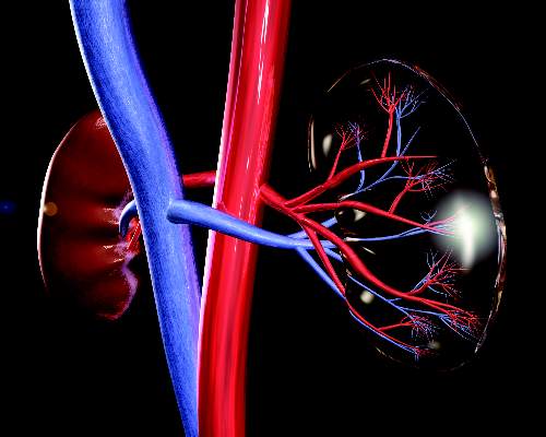
Acute kidney injury is a common complication after pediatric cardiac surgery, but measuring for a specific genetic protein immediately after cardiac surgery may improve cardiac surgeons’ ability to predict patients at higher risk of AKI, according to researchers from Brazil. The study results are in the July issue of the Journal of Thoracic and Cardiovascular Surgery (2016;152-178-86).
“Plasma syndecan-1 levels measured early in the postoperative period were independently associated with severe acute kidney injury,” wrote Candice Torres de Melo Bezerra Cavalcante, MD, of Heart Hospital of Messejana and Federal University of Ceará.
Their prospective cohort study involved 289 pediatric patients who had cardiac surgery at their institution between September 2013 and December 2014.

Dr. Cavalcante and colleagues acknowledged that the traditional biomarker for renal function, serum creatinine, only increases appreciably after the glomerular filtration rate declines 50%, impairing physicians’ ability to detect AKI early enough to treat it. “This delay can explain, in part the, negative results in AKI therapeutic clinical trials,” they wrote.
They evaluated two different endothelial biomarkers in addition to syndecan-1 with regard to their capacity for predicting severe AKI: plasma ICAM-1, a marker of endothelial cell activation; and E-selectin, an endothelial cell adhesion molecule. Syndecan-1 works as a biomarker of injury to the glycocalyx protein that surrounds endothelial cell membranes that acts as a permeability barrier and prevents the cells from adhering to blood. They found that median syndecan-1 levels soon after surgery were higher in patients with severe AKI, 103.6 vs. 42.3 ng/mL.
“Although syndecan-1 is not a renal-specific biomarker, there has been recent increasing evidence that endothelial injury has an important role in AKI pathophysiology,” the researchers noted.
Study results showed the higher the level of syndecan-1, the greater the adjusted odds ratio (OR) for severe AKI. Levels of less than 17 ng/mL were considered normal; 17.1-46.7 ng/mL carried an adjusted OR of 1.42; 47.4-93.1 ng/mL had an adjusted OR of 2.05; and levels 96.3 or greater had an OR of 8.87.
“Maintenance of endothelial glycocalyx integrity can be a therapeutic target to reduce AKI in this setting,” the researchers wrote.
The authors acknowledged that the study was done at a single center that had dialysis and death rates three and five times higher, respectively, than those of developed countries; and it measured syndecan-1 at only one time point almost immediately after the operation.
“Adding postoperative syndecan-1, even when using a clinical model that already incorporates variables from renal angina index, results in significant improvement in the capacity to predict severe AKI,” Dr. Cavalcante and colleagues concluded.
They had no financial relationships to disclose.
Results of AKI in heart surgery patients have been “sobering,” with up to 56% of these patients being diagnosed with AKI, but research such as that by Dr. Cavalcante and colleagues represents a new approach to improving outcomes by combining clinical risk factors with specific biomarkers to identify patients at risk, Petros V. Anagnostopoulos, MD, of American Family Children’s Hospital, University of Wisconsin, said in his invited commentary (J Thorac Cardiovasc Surg. 2016;152[1]:187-8).
Dr. Anagnostopoulos acknowledged problems with traditional markers for renal function. “An ideal biomarker should be sensitive, easy to measure, reproducible, and inexpensive,” he said. “Finally, when combined with clinical prediction models, it should potentiate the discrimination of these models.”
Syndecan-1 answers that call, he said. “It peaks early and is cheap, fast, and easy to measure with readily available methods, which makes it an ideal early biomarker of AKI,” Dr. Anagnostopoulos said. Even so, he pointed out potential shortcomings of syndecan-1: It is not renal specific and it does not increase before the operation.
But he applauded Dr. Cavalcante and colleagues for pursuing research to combine clinical risk factors with specific biomarkers. “It is very likely that this type of clinical research will become prevalent in the near future and will hopefully produce results that will allow better individual patient-specific risk stratification,” Dr. Anagnostopoulos said.
He had no financial relationships to disclose.
Acute kidney injury is a common complication after pediatric cardiac surgery, but measuring for a specific genetic protein immediately after cardiac surgery may improve cardiac surgeons’ ability to predict patients at higher risk of AKI, according to researchers from Brazil. The study results are in the July issue of the Journal of Thoracic and Cardiovascular Surgery (2016;152-178-86).
“Plasma syndecan-1 levels measured early in the postoperative period were independently associated with severe acute kidney injury,” wrote Candice Torres de Melo Bezerra Cavalcante, MD, of Heart Hospital of Messejana and Federal University of Ceará.
Their prospective cohort study involved 289 pediatric patients who had cardiac surgery at their institution between September 2013 and December 2014.
Dr. Cavalcante and colleagues acknowledged that the traditional biomarker for renal function, serum creatinine, only increases appreciably after the glomerular filtration rate declines 50%, impairing physicians’ ability to detect AKI early enough to treat it. “This delay can explain, in part the, negative results in AKI therapeutic clinical trials,” they wrote.
They evaluated two different endothelial biomarkers in addition to syndecan-1 with regard to their capacity for predicting severe AKI: plasma ICAM-1, a marker of endothelial cell activation; and E-selectin, an endothelial cell adhesion molecule. Syndecan-1 works as a biomarker of injury to the glycocalyx protein that surrounds endothelial cell membranes that acts as a permeability barrier and prevents the cells from adhering to blood. They found that median syndecan-1 levels soon after surgery were higher in patients with severe AKI, 103.6 vs. 42.3 ng/mL.
“Although syndecan-1 is not a renal-specific biomarker, there has been recent increasing evidence that endothelial injury has an important role in AKI pathophysiology,” the researchers noted.
Study results showed the higher the level of syndecan-1, the greater the adjusted odds ratio (OR) for severe AKI. Levels of less than 17 ng/mL were considered normal; 17.1-46.7 ng/mL carried an adjusted OR of 1.42; 47.4-93.1 ng/mL had an adjusted OR of 2.05; and levels 96.3 or greater had an OR of 8.87.
“Maintenance of endothelial glycocalyx integrity can be a therapeutic target to reduce AKI in this setting,” the researchers wrote.
The authors acknowledged that the study was done at a single center that had dialysis and death rates three and five times higher, respectively, than those of developed countries; and it measured syndecan-1 at only one time point almost immediately after the operation.
“Adding postoperative syndecan-1, even when using a clinical model that already incorporates variables from renal angina index, results in significant improvement in the capacity to predict severe AKI,” Dr. Cavalcante and colleagues concluded.
They had no financial relationships to disclose.
FROM THE JOURNAL OF THORACIC AND CARDIOVASCULAR SURGERY
Key clinical point: The biomarker syndecan-1 may aid in determining acute kidney injury risk for children having cardiac surgery.
Major finding: Children with elevated levels of syndecan-1 had a two- to ninefold greater risk of acute kidney injury.
Data source: Single-institution, prospective cohort study of 289 pediatric patients who had cardiac surgery from September 2013 to December 2014.
Disclosures: Dr. Cavalcante and coauthors had no financial relationships to disclose.
Making the case for CABG using bilateral thoracic arteries
Cardiac surgeons have been slow to embrace bilateral internal thoracic arteries (ITAs) for coronary artery bypass grafting (CABG) despite accumulating evidence that this technique achieves better long-term survival than the single-artery technique, perhaps because they think the bilateral technique is more difficult. However, investigators from Johns Hopkins University have found no difference in results between four different bilateral ITAs techniques regardless of complexity.
Their single-center study analyzed outcomes from 762 patients at Johns Hopkins who had CABG by way of one of four different bilateral ITA (BITA) techniques between 1997 and 2014. The results are in the July issue of the Journal of Thoracic and Cardiovascular Surgery (2016;152:120-7).
“We found no significant difference in terms of long-term survival or freedom from repeat revascularization between different configurations of BITA use,” wrote J. Trent Magruder, MD, and his colleagues.
Dr. Magruder and his coauthors cited 13 reports that found BITA achieved better graft patency and long-term survival than did single internal thoracic arteries, but they noted the lack of reports comparing different BITA techniques. “Given the paucity of comparative data on long-term outcomes of various BITA configurations, we sought to study differences in mortality and the need for repeat revascularization among patients receiving varying BITA graft configurations at our institution,” they said.
The four groups and types of BITA procedures they analyzed were:
LL/RL group, in situ left ITA (LITA) anastomosed to the left anterior descending artery (LAD) with in situ right ITA (RITA) anastomosed to the left coronary circulation (n = 239).
LL/RR group, in situ LITA-LAD and in situ RITA-right coronary circulation (n = 239 patients).
RL/LL group, in situ RITA-LAD with in situ LITA-left coronary circulation (n = 185 patients).
Y group, in situ LITA-LAD with a free RITA as a composite graft with inflow from the LITA or a saphenous vein graft (n = 99 patients).
BITA cases comprised 5.7% of all 14,502 CABG procedures Johns Hopkins cardiac surgeons performed through the study period (60 BITA cases were dropped from the analysis because of incomplete data). That rate is about in line with a previously reported use rate of 4% of CABG procedures in the United States (Circulation. 2009;120:935-40).
Among the reasons Dr. Magruder and his coauthors cited for the lack of uptake of BITA among cardiac surgeons are discrepancies in survival data, a perceived high risk of complications such as sternal surgical site infections in patients with diabetes or chronic obstructive pulmonary disease or in those who are obese, and increased operative time and risk of bleeding.
With regard to the operation itself, the mean cross-clamp and coronary bypass times and number of bypass grafts were similar among all four groups, the latter ranging from 3.0 for the RL/LL group to 3.4 for the Y group. However, the researchers did find appreciable differences in rates of transfusions during the operation and skeletonization of the RITA at harvest. The Y group had the highest rates for both – 57.6% had transfusions and 72.7% had skeletonized RITA at harvest – followed by the RL/LL with rates of 43.2% for transfusions and 31.4% with skeletonized RITA. Rates for both intraoperative transfusion and RITA skeletonization were 24.7% and 8%, respectively, in the LL/RL group; and 37.7% and 18%, respectively, in the LL/RR group.
In-hospital complications, including reintervention for bleeding, heart attack, stroke, inflammation of the mediastinum, and death, were similar among all four groups. There were no in-hospital heart attacks. The only statistically significant difference was in hospital stay, ranging from an average of 6.1 days for the LL/RL group to 7.4 for both the LL/RR and RL/LL groups.
Through the duration of follow-up, the overall rate for repeat percutaneous coronary intervention was 7.6% – highest among the RL/LL (9.2%) and Y groups (9%). Those in the LL/RR group had the highest rates of repeat CABG: 1.7% vs. 0.8% for LL/RL group, 0.5% for the RL/LL group and 0% for group Y. Rates of late cardiac death were around 5% for the first three groups, but none were reported in group Y.
Dr. Magruder and colleagues acknowledged their study used a limited sample size for each procedure, but that their findings show that cardiac surgeons should choose their BITA configuration based on individual patient factors. “In general, the technically simplest operations should be selected because more complex procedures offer no additional benefit,” they said.
Dr. Magruder and his coauthors had no financial disclosures.
The take-home message of the study by Dr. Magruder and colleagues is that using bilateral internal thoracic arteries (BITA) is more important than the specific configuration, Saswata Deb, MD, BSc, and Stephen E. Fremes, MD, MSc, BSc, of the University of Toronto wrote in their invited commentary (J Thorac Cardiovasc Surg. 2016;152:128-30). “In other words: BITA – just do it!” they wrote.
Because the survival advantage of BITA in CABG typically becomes apparent 10 years or more after the operation, the Johns Hopkins study, along with the Arterial Revascularization Trial that compares BITA with single ITA (Semin Thorac Cardiovasc Surg. 2014;26:76-94), can help redefine how cardiac surgeons select conduits for CABG, the commentators said.
“What does this particular study add?” they asked. “Point estimates for the adjusted hazard ratio of death or repeat revascularization were close to unity for each of the primary grafting hypothesis comparisons.”
Dr. Deb and Dr. Fremes had no financial relationships to disclose.
Cardiac surgeons have been slow to embrace bilateral internal thoracic arteries (ITAs) for coronary artery bypass grafting (CABG) despite accumulating evidence that this technique achieves better long-term survival than the single-artery technique, perhaps because they think the bilateral technique is more difficult. However, investigators from Johns Hopkins University have found no difference in results between four different bilateral ITAs techniques regardless of complexity.
Their single-center study analyzed outcomes from 762 patients at Johns Hopkins who had CABG by way of one of four different bilateral ITA (BITA) techniques between 1997 and 2014. The results are in the July issue of the Journal of Thoracic and Cardiovascular Surgery (2016;152:120-7).
“We found no significant difference in terms of long-term survival or freedom from repeat revascularization between different configurations of BITA use,” wrote J. Trent Magruder, MD, and his colleagues.
Dr. Magruder and his coauthors cited 13 reports that found BITA achieved better graft patency and long-term survival than did single internal thoracic arteries, but they noted the lack of reports comparing different BITA techniques. “Given the paucity of comparative data on long-term outcomes of various BITA configurations, we sought to study differences in mortality and the need for repeat revascularization among patients receiving varying BITA graft configurations at our institution,” they said.
The four groups and types of BITA procedures they analyzed were:
LL/RL group, in situ left ITA (LITA) anastomosed to the left anterior descending artery (LAD) with in situ right ITA (RITA) anastomosed to the left coronary circulation (n = 239).
LL/RR group, in situ LITA-LAD and in situ RITA-right coronary circulation (n = 239 patients).
RL/LL group, in situ RITA-LAD with in situ LITA-left coronary circulation (n = 185 patients).
Y group, in situ LITA-LAD with a free RITA as a composite graft with inflow from the LITA or a saphenous vein graft (n = 99 patients).
BITA cases comprised 5.7% of all 14,502 CABG procedures Johns Hopkins cardiac surgeons performed through the study period (60 BITA cases were dropped from the analysis because of incomplete data). That rate is about in line with a previously reported use rate of 4% of CABG procedures in the United States (Circulation. 2009;120:935-40).
Among the reasons Dr. Magruder and his coauthors cited for the lack of uptake of BITA among cardiac surgeons are discrepancies in survival data, a perceived high risk of complications such as sternal surgical site infections in patients with diabetes or chronic obstructive pulmonary disease or in those who are obese, and increased operative time and risk of bleeding.
With regard to the operation itself, the mean cross-clamp and coronary bypass times and number of bypass grafts were similar among all four groups, the latter ranging from 3.0 for the RL/LL group to 3.4 for the Y group. However, the researchers did find appreciable differences in rates of transfusions during the operation and skeletonization of the RITA at harvest. The Y group had the highest rates for both – 57.6% had transfusions and 72.7% had skeletonized RITA at harvest – followed by the RL/LL with rates of 43.2% for transfusions and 31.4% with skeletonized RITA. Rates for both intraoperative transfusion and RITA skeletonization were 24.7% and 8%, respectively, in the LL/RL group; and 37.7% and 18%, respectively, in the LL/RR group.
In-hospital complications, including reintervention for bleeding, heart attack, stroke, inflammation of the mediastinum, and death, were similar among all four groups. There were no in-hospital heart attacks. The only statistically significant difference was in hospital stay, ranging from an average of 6.1 days for the LL/RL group to 7.4 for both the LL/RR and RL/LL groups.
Through the duration of follow-up, the overall rate for repeat percutaneous coronary intervention was 7.6% – highest among the RL/LL (9.2%) and Y groups (9%). Those in the LL/RR group had the highest rates of repeat CABG: 1.7% vs. 0.8% for LL/RL group, 0.5% for the RL/LL group and 0% for group Y. Rates of late cardiac death were around 5% for the first three groups, but none were reported in group Y.
Dr. Magruder and colleagues acknowledged their study used a limited sample size for each procedure, but that their findings show that cardiac surgeons should choose their BITA configuration based on individual patient factors. “In general, the technically simplest operations should be selected because more complex procedures offer no additional benefit,” they said.
Dr. Magruder and his coauthors had no financial disclosures.
FROM THE JOURNAL OF THORACIC AND CARDIOVASCULAR SURGERY
Key clinical point: No specific technique for bilateral internal thoracic artery (BITA) graft in coronary artery bypass grafting (CABG) has an advantage over other techniques.
Major finding: Among four different BITA configurations, no in-hospital deaths occurred and the late cardiac death rate was around 5% for three of the four groups and 0% for the fourth group.
Data source: Single-center, retrospective study of 762 patients who had CABG with use of BITA from 1997 to 2014.
Disclosures: Dr. Magruder and colleagues reported having no financial disclosures.
Alendronate Therapy Lowers Risk of Hip Fractures
NEW YORK - Long-term use of the bisphosphonate alendronate substantially lowers the risk of hip fracture without increasing the risk of atypical fractures of the subtrochanteric femur or femoral shaft, according to a Danish study.
"These findings support an acceptable balance between benefit and risk with treatment with alendronate in terms of fracture outcomes, even for over 10 years of continuous use," the authors conclude in the BMJ online June 28.
Placebo-controlled trials have shown a reduction in hip fracture risk with alendronate treatment for three years in older women with osteoporosis, but observational studies have suggested that atypical femur fractures are more common in long-term users of bisphosphonates.
To investigate further, researchers analyzed data from Danish practice registries on nearly 62,000 alendronate users. A total of 1,428 people sustained a fracture of the subtrochanteric femur or femoral shaft (ST/FS), an incidence rate of 3.4 per 1,000 person years, and 6,784 sustained a hip fracture (incidence rate 16.2 per 1,000 person years).
"Long-term adherent use of alendronate in excess of 10 dose years was associated with an adjusted 30% lower risk of hip fracture and no increase in the risk of fractures of the subtrochanteric femur or femoral shaft," the investigators report in their paper.
"In addition, we have shown that even in the worst case scenario (assuming 100% of subtrochanteric and femoral shaft fractures are atypical and secondary to bisphosphonate use and making no allowance for the higher prevalence of comorbid conditions in these patients) the number of atypical femur fractures remains too low to offset the benefits on hip fracture in patients with long-term alendronate use up to 10 years," they say.
"Safety concerns have probably limited the adherence to anti-osteoporosis therapies," Dr. Daniel Preto-Alhambra, University of Oxford, Nuffield Orthopaedics Centre in the United Kingdom, who worked on the study, told Reuters Health by email.
"Our findings support a good risk-benefit for this treatment in terms of femur fracture risk reduction, even when prescribed for 10 years and over. These data should reassure physicians and patients on the bone-related risk-benefits of alendronate therapy," he added.
The study had no commercial funding. Several authors made disclosure statements.
SOURCE: http://bit.ly/29y33NQ
BMJ 2016.
PCR better at diagnosing fungal chronic rhinosinusitis than cultures
ITS1/ITS2 polymerase chain reaction sequencing was significantly better at identifying fungal chronic rhinosinusitis (CRS) than were cultures, according to Dr. Pauline Comacle and her associates.
A total of 70 samples from 61 patients with CRS were tested via the use of polymerase chain reaction (PCR) testing and cultures. Fungal CRS was identified in 42 patients, of whom 37 had a fungus ball, 3 had allergic fungal CRS, and 2 had unspecified fungal CRS. ITS1/ITS2 PCR sequencing successfully identified fungal CRS in all patients, while cultures were positive for fungal CRS in 20 of the 42 patients.
Aspergillus fumigatus was the most common fungus in the study, occurring in 69% of patients. Cladosporium cladosporoides occurred in 9.5% of patients, and A. nidulans, A. flavus, and Scedosporium species each occurred in 7% of patients.
“This study nicely shows that molecular methods are powerful tools for the diagnosis of chronic rhinosinusitis and help in characterizing the accurate epidemiology of fungal CRS,” the investigators concluded.
Find the full study in Medical Mycology (doi: 10.1093/mmy/myw041).
FROM MEDICAL MYCOLOGY
Four-branched arch replacement gets acceptable results
NEW YORK – A total aortic arch replacement approach that uses a four-branched graft with antegrade cerebral perfusion can be done with low rates of in-hospital death and complications, a large series from two institutions in Japan showed.
Kenji Minatoya, MD, of the National Cerebral and Cardiovascular Center in Osaka, Japan, reported that his institution’s approach for total arch replacement (TAR) had an in-hospital death rate of 5.2%.
Dr. Minatoya and his colleagues started using four-branch TAR in the 1980s, switching from retrograde to antegrade cerebral perfusion to protect the brain later on. “The study purpose was to investigate the results of total arch replacement using the four-branch graft as a benchmark in the endovascular era,” he said at the meeting, sponsored by the American Association for Thoracic Surgery.
The study involved 1,005 cases of total arch replacement performed at Dr. Minatoya’s center and at Tokyo Medical University from 2001 to 2016.
The study population included a cohort of 152 people in their 80s. The in-hospital death rate in this group was 11.8%, Dr. Minatoya said. The over-80 group mostly underwent thoracic endovascular aortic repair (TEVAR) beginning in 2008, he said, but in recent years some had open total arch replacement operations.
The univariate analysis showed that chronic kidney disease, long operation times, long durations for coronary bypass and circulatory arrest, and extended time on mechanical ventilation were risk factors for in-hospital death in octogenarians, Dr. Minatoya said. The multivariate analysis showed that male gender along with extended mechanical ventilation were risk factors for in-hospital death in this group, he said.
The overall population included 252 emergent operations, 224 of which were for acute aortic dissections, Dr. Minatoya said. The in-hospital death rate was 4.5% for elective operations and 7.1% for emergent cases, he said. The death rate for isolated, elective total arch replacement was 3.4%.
Focusing on acute aortic dissections, Dr. Minatoya said, “We have adopted an aggressive strategy for entry-site resection, including total arch replacement, in patients with arch tears.” Almost 50% of patients with acute aortic dissection had total arch replacement, he said, with identical 4.9% rates for in-hospital mortality rate and permanent neurological deficit in this group.
The leading overall causes of in-hospital death were low-output syndrome (38.5%), sepsis (25%), respiratory failure (21%) and rupture of the residual aneurysm (9%), Dr. Minatoya said.
Fifteen patients (1.5%) underwent second operations for arch grafts, he said: 11 for pseudoaneurysm; three for hemolysis and one for infection. Other overall measures in the analysis were a permanent neurological dysfunction rate of 3.6%, a temporary neurological dysfunction rate of 6.4%, and no spinal cord complications. Overall 5-year survival was 80.7% and 10-year survival was 63.1%, Dr. Minatoya said.
A total of 311 patients had concomitant procedures. They included aortic valve operations (64); aortic root replacement (38); mitral valve replacement (13); and coronary artery bypass grafting (196).
The typical operation in the study population took about 8 hours, Dr. Minatoya said (482 minutes). Timing of key operative steps were cardiopulmonary time of 254 minutes, cardiac arrest time of 146 minutes, antegrade cerebral perfusion time of 160 minutes and lower-body circulatory arrest time of 62 minutes.
“Since the mean age was 70 years old, we think the survival rate was acceptable,” Dr. Minatoya said, regarding overall study results. Overall risk factors for in-hospital death were short stature, long pump time, chronic kidney disease, and age of 80 and up, he said. Short stature was a risk factor for permanent neurological deficit, and males over age 80 had a higher risk for total arch replacement.
“Total arch replacement using the four-branched graft with antegrade cerebral perfusion could be accomplished with acceptable early and late results,” Dr. Minatoya said. “The branched-arch TEVAR may be a good option for octogenarians and patients with chronic kidney disease.”
Dr. Minatoya had no financial relationships to disclose.
AT THE AATS AORTIC SYMPOSIUM 2016
Key clinical point: Four-branched total aortic arch replacement can achieve acceptable early and late results.
Major finding: Overall in-hospital mortality was 5.2% and 5-year survival was 80.7%.
Data source: Consecutive series of 1,005 patients who had total arch replacement between 2001 and 2016 at two centers in Japan.
Disclosures: Dr. Minatoya reported having no financial disclosures.
First female-to-male sexual transmission of Zika virus
A suspected case of sexual transmission of the Zika virus from a female to a male has occurred in New York City, according to the New York City Department of Health and Mental Hygiene (DOHMH).
“This case represents the first reported occurrence of female-to-male sexual transmission of Zika virus,” Alexander Davidson of the DOHMH and his coauthors stated in the CDC’s Morbidity and Mortality Weekly Report. “Current guidance to prevent sexual transmission of Zika virus is based on the assumption that transmission occurs from a male partner to a receptive partner.”

The woman, reportedly in her twenties and not pregnant at the time of infection, had traveled to a region experiencing high volumes of Zika virus transmission. Upon returning home, the woman engaged in condomless vaginal intercourse with her male partner, and subsequently developed symptoms consistent with a Zika virus infection. Three days after symptom onset, her primary care provider took blood and urine samples, from which a Zika virus infection was confirmed. (MMWR Morb Mortal Wkly Rep. 2016 Jul 15. doi: 10.15585/mmwr.mm6528e2)
A week after the sexual encounter, her partner – also in his twenties – began experiencing symptoms of Zika virus infection. Three days after the onset of his symptoms, he went to the same primary care provider as the woman. He confirmed that he had not traveled outside of the United States in the last year, had engaged in condomless vaginal sex with just one individual (the aforementioned female), had no blood on his penis to indicate vaginal bleeding or the presence of open lesions, and had no mosquito bites in the previous week.
“The timing and sequence of events support female-to-male Zika virus transmission through condomless vaginal intercourse,” the coauthors conclude, adding that “virus present in either vaginal fluids or menstrual blood might have been transmitted during exposure to her male partner’s urethral mucosa or undetected abrasions on his penis.”
Both the female and male were tested via real-time reverse transcription–polymerase chain reaction (rRT-PCR), with serum testing done via the Zika immunoglobulin M antibody capture enzyme-linked immunosorbent assay (Zika MAC-ELISA). The rRT-PCR testing showed Zika virus RNA in the woman’s serum, despite being collected three days after the sexual encounter, meaning she was viremic at the time. Studies on nonhuman primates have shown that Zika virus RNA can remain present in vaginal fluid for up to a week, according to the report.
The CDC is cautioning pregnant women against travel to Zika-heavy areas, in particular the 2016 Summer Olympic Games in Rio de Janeiro. Health care providers who receive patients with Zika-like symptoms should ask if the patient has had sexual contact with someone who has traveled to an affected region, if the patient did not travel to such a region.
A suspected case of sexual transmission of the Zika virus from a female to a male has occurred in New York City, according to the New York City Department of Health and Mental Hygiene (DOHMH).
“This case represents the first reported occurrence of female-to-male sexual transmission of Zika virus,” Alexander Davidson of the DOHMH and his coauthors stated in the CDC’s Morbidity and Mortality Weekly Report. “Current guidance to prevent sexual transmission of Zika virus is based on the assumption that transmission occurs from a male partner to a receptive partner.”
The woman, reportedly in her twenties and not pregnant at the time of infection, had traveled to a region experiencing high volumes of Zika virus transmission. Upon returning home, the woman engaged in condomless vaginal intercourse with her male partner, and subsequently developed symptoms consistent with a Zika virus infection. Three days after symptom onset, her primary care provider took blood and urine samples, from which a Zika virus infection was confirmed. (MMWR Morb Mortal Wkly Rep. 2016 Jul 15. doi: 10.15585/mmwr.mm6528e2)
A week after the sexual encounter, her partner – also in his twenties – began experiencing symptoms of Zika virus infection. Three days after the onset of his symptoms, he went to the same primary care provider as the woman. He confirmed that he had not traveled outside of the United States in the last year, had engaged in condomless vaginal sex with just one individual (the aforementioned female), had no blood on his penis to indicate vaginal bleeding or the presence of open lesions, and had no mosquito bites in the previous week.
“The timing and sequence of events support female-to-male Zika virus transmission through condomless vaginal intercourse,” the coauthors conclude, adding that “virus present in either vaginal fluids or menstrual blood might have been transmitted during exposure to her male partner’s urethral mucosa or undetected abrasions on his penis.”
Both the female and male were tested via real-time reverse transcription–polymerase chain reaction (rRT-PCR), with serum testing done via the Zika immunoglobulin M antibody capture enzyme-linked immunosorbent assay (Zika MAC-ELISA). The rRT-PCR testing showed Zika virus RNA in the woman’s serum, despite being collected three days after the sexual encounter, meaning she was viremic at the time. Studies on nonhuman primates have shown that Zika virus RNA can remain present in vaginal fluid for up to a week, according to the report.
The CDC is cautioning pregnant women against travel to Zika-heavy areas, in particular the 2016 Summer Olympic Games in Rio de Janeiro. Health care providers who receive patients with Zika-like symptoms should ask if the patient has had sexual contact with someone who has traveled to an affected region, if the patient did not travel to such a region.
FROM MMWR
