User login
VIDEO: Smartphones could ‘democratize’ EEG
SAN DIEGO – Epilepsy is a common condition, but many people live in areas too poor or underserved to benefit from electroencephalogram (EEG) testing to confirm a diagnosis. Modern technology may be changing that. Smartphones wirelessly linked to headsets can collect EEG data and potentially serve as an alternate platform for diagnosis of epilepsy.
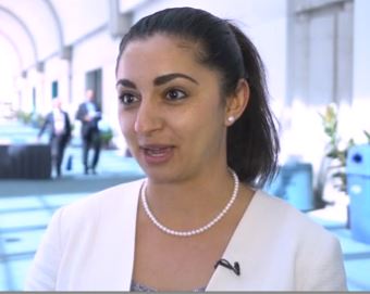
To test the efficacy of one such system, Farrah Mateen, MD, PhD, a neurologist at Massachusetts General Hospital, Boston, conducted a study in the remote, mountainous Kingdom of Bhutan, which lies between India and China. Her group compared the signals between a smartphone EEG and standard EEG.
The sensitivity was low, at around 40%, but the device’s specificity was 90%-95%. For now, the device performs better as a confirmatory test than as a screening device, but the researchers hope to adjust the lead location to improve sensitivity.
The headset used was designed for use by video gamers, and at a cost of less than $300, it represents a low-cost entry into portable EEG.
Dr. Mateen discussed the smartphone EEG study in a video interview at the annual meeting of the American Neurological Association.
SAN DIEGO – Epilepsy is a common condition, but many people live in areas too poor or underserved to benefit from electroencephalogram (EEG) testing to confirm a diagnosis. Modern technology may be changing that. Smartphones wirelessly linked to headsets can collect EEG data and potentially serve as an alternate platform for diagnosis of epilepsy.
To test the efficacy of one such system, Farrah Mateen, MD, PhD, a neurologist at Massachusetts General Hospital, Boston, conducted a study in the remote, mountainous Kingdom of Bhutan, which lies between India and China. Her group compared the signals between a smartphone EEG and standard EEG.
The sensitivity was low, at around 40%, but the device’s specificity was 90%-95%. For now, the device performs better as a confirmatory test than as a screening device, but the researchers hope to adjust the lead location to improve sensitivity.
The headset used was designed for use by video gamers, and at a cost of less than $300, it represents a low-cost entry into portable EEG.
Dr. Mateen discussed the smartphone EEG study in a video interview at the annual meeting of the American Neurological Association.
AT ANA 2017
Are we on the verge of a cocaine epidemic?

Barbara Vickrey, MD, MPH

VIDEO: Rapid influenza test obviates empiric antivirals
TORONTO – A test that only requires a maximum 2-hour wait for results was highly accurate at detecting influenza and respiratory syncytial virus infection in lung transplant patients, according to research presented at the CHEST annual meeting on Oct. 30.
This rapid and highly accurate test for detecting three common respiratory viruses has dramatically cut the need for empiric treatments and the risk for causing nosocomial infections in lung transplant patients who develop severe upper respiratory infections, Macé M. Schuurmans, MD, noted during the presentation.
This study involved 100 consecutive lung transplant patients who presented at Zurich University Hospital with signs of severe upper respiratory infection. The researchers ran the rapid and standard diagnostic tests for each patient and found that, relative to the standard test, the rapid test had positive and negative predictive values of 95%.
The number of empiric treatments with oseltamivir (Tamiflu) and ribavirin to treat a suspected influenza or respiratory syncytial virus infection (RSV) has “strongly diminished” by about two-thirds, noted Dr. Schuurmans, who is a pulmonologist at the hospital.
Until the rapid test became available, Dr. Shuurmans and his associates used a standard polymerase chain reaction test that takes 36-48 hours to yield a result. Using this test made treating patients empirically with oseltamivir and oral antibiotics for a couple of days a necessity, he said in a video interview. The older test also required isolating patients to avoid the potential spread of influenza or RSV in the hospital.
The rapid test, which became available for U.S. use in early 2017, covers influenza A and B and RSV in a single test with a single mouth-swab specimen.
“We now routinely use the rapid test and don’t prescribe empiric antivirals or antibiotics as often,” Dr. Schuurmans said. “There is much less drug cost and fewer potential adverse effects from empiric treatment.” Specimens still also undergo conventional testing, however, because that can identify eight additional viruses that the rapid test doesn’t cover.
Dr. Schuurmans acknowledged that further study needs to assess the cost-benefit of the rapid test to confirm that its added expense is offset by reduced expenses for empiric treatment and hospital isolation.
He had no disclosures. The study received no commercial support.
The video associated with this article is no longer available on this site. Please view all of our videos on the MDedge YouTube channel
mzoler@frontlinemedcom.com
On Twitter @mitchelzoler
AT CHEST 2017
Key clinical point:
Major finding: The rapid test had positive and negative predictive values of 95%.
Data source: A single-center observational study of 100 consecutive lung transplant recipients who presented with severe, acute respiratory infection.
Disclosures: Dr. Schuurmans had no disclosures. The study received no commercial support.

VIDEO: High-volume endoscopists, centers produced better ERCP outcomes
Endoscopists who performed endoscopic retrograde cholangiopancreatography (ERCP) at high-volume centers had a 60% greater odds of procedure success compared with those at low-volume centers, according to the results of a systematic review and meta-analysis.
High-volume endoscopists also had a 30% lower odds of performing ERCP that led to adverse events such as pancreatitis, perforation, and bleeding, reported Rajesh N. Keswani, MD, MS, of Northwestern University, Chicago, and his associates. High-volume centers themselves also were associated with a significantly higher odds of successful ERCP (odds ratio, 2.0; 95% CI, 1.6 to 2.5), although they were not associated with a significantly lower risk of adverse events, the reviewers wrote. The study was published in the December issue of Clinical Gastroenterology and Hepatology (doi: 10.1016/j.cgh.2017.06.002).
Diagnostic ERCP has fallen sevenfold in the past 30 years while therapeutic use has increased 30-fold, the researchers noted. Therapeutic use spans several complex pancreaticobiliary conditions, including chronic pancreatitis, malignant jaundice, and complications of liver transplantation. This shift from diagnostic to therapeutic has naturally increased the complexity of ERCP, the need for expert endoscopy, and the potential risk of adverse events. “As health care continues to shift toward rewarding value rather than volume, it will be increasingly important to deliver care that is effective and efficient,” the reviewers wrote. “Thus, understanding factors associated with unsuccessful interventions, such as a failed ERCP, will be of critical importance to payers and patients (Clin Gastroenterol Hepatol. 2017 Jun 7;218:237-45).
Therefore, they searched MEDLINE, EMBASE, and the Cochrane register of controlled trials for prospective and retrospective studies published through January 2017. In all, the researchers identified 13 studies that stratified outcomes by volume per endoscopist or center. These studies comprised 59,437 procedures and patients. Definitions of low volume varied by study, ranging from less than 25 to less than 156 annual ERCPs per endoscopist and from less than 87 to less than 200 annual ERCPs per center. Endoscopists who achieved this threshold were significantly more likely to perform successful ERCPs than were low-volume endoscopists (OR, 1.6; 95% CI, 1.2 to 2.1), and were significantly less likely to have patients develop pancreatitis, perforation, or bleeding after ERCP (OR, 0.7; 95% CI, 0.5 to 0.8).
SOURCE: AMERICAN GASTROENTEROLOGICAL ASSOCIATION
“Given these compelling findings, we propose that providers and payers consider consolidating ERCP to high-volume endoscopists and centers to improve ERCP outcomes and value,” the reviewers wrote. Minimum thresholds for endoscopists and centers to maintain ERCP skills and optimize outcomes have not been defined, they noted. Intuitively, there is no “critical volume threshold” at which “outcomes suddenly improve,” but the studies in this analysis used widely varying definitions of low volume, they added. It also remains unclear whether a low-volume endoscopist can achieve optimal outcomes at a high-volume center, or vice versa, they said. They recommended studies to better define procedure success and the appropriate use of ERCP in therapeutic settings.
One reviewer acknowledged support from the University of Colorado Department of Medicine Outstanding Early Career Faculty Program. The reviewers reported having no conflicts of interest.
With the increasing proportion of complex therapeutic ERCPs, the field is shifting toward performance of these procedures by those who have had advanced training and who make them the focus of their clinical practice. Consistent with this, the meta-analysis by Keswani et al. highlights benefits of higher-volume centers and endoscopists - improved ERCP success rate (at the provider and practice level) and reduced adverse events (provider level only). It is unclear, however, if higher-volume endoscopists received additional training that translated into better outcomes. Other variables, including case complexity and provider experience, could not be fully assessed in this study.

Overall, however, this large, well-performed meta-analysis adds to the growing chorus that endoscopists and endoscopic centers will have better results if the endoscopists are specially trained and routinely perform these procedures. Future studies are needed to more accurately define procedure success (significant variation in the meta-analysis) and assess other variables that affect outcomes for which volume may only be a proxy. In an era of reporting and demonstrating value in endoscopic care, quality metrics for ERCP performance may not be fully appreciated but eventually may become the driving force in consolidation of these procedures to particular centers or providers, regardless of volume.
Avinash Ketwaroo, MD, MSc, is assistant professor in the division of gastroenterology and hepatology at Baylor College of Medicine, Houston, and an associate editor of GI & Hepatology News. He has no relevant conflicts of interest.
With the increasing proportion of complex therapeutic ERCPs, the field is shifting toward performance of these procedures by those who have had advanced training and who make them the focus of their clinical practice. Consistent with this, the meta-analysis by Keswani et al. highlights benefits of higher-volume centers and endoscopists - improved ERCP success rate (at the provider and practice level) and reduced adverse events (provider level only). It is unclear, however, if higher-volume endoscopists received additional training that translated into better outcomes. Other variables, including case complexity and provider experience, could not be fully assessed in this study.
Overall, however, this large, well-performed meta-analysis adds to the growing chorus that endoscopists and endoscopic centers will have better results if the endoscopists are specially trained and routinely perform these procedures. Future studies are needed to more accurately define procedure success (significant variation in the meta-analysis) and assess other variables that affect outcomes for which volume may only be a proxy. In an era of reporting and demonstrating value in endoscopic care, quality metrics for ERCP performance may not be fully appreciated but eventually may become the driving force in consolidation of these procedures to particular centers or providers, regardless of volume.
Avinash Ketwaroo, MD, MSc, is assistant professor in the division of gastroenterology and hepatology at Baylor College of Medicine, Houston, and an associate editor of GI & Hepatology News. He has no relevant conflicts of interest.
Endoscopists who performed endoscopic retrograde cholangiopancreatography (ERCP) at high-volume centers had a 60% greater odds of procedure success compared with those at low-volume centers, according to the results of a systematic review and meta-analysis.
High-volume endoscopists also had a 30% lower odds of performing ERCP that led to adverse events such as pancreatitis, perforation, and bleeding, reported Rajesh N. Keswani, MD, MS, of Northwestern University, Chicago, and his associates. High-volume centers themselves also were associated with a significantly higher odds of successful ERCP (odds ratio, 2.0; 95% CI, 1.6 to 2.5), although they were not associated with a significantly lower risk of adverse events, the reviewers wrote. The study was published in the December issue of Clinical Gastroenterology and Hepatology (doi: 10.1016/j.cgh.2017.06.002).
Diagnostic ERCP has fallen sevenfold in the past 30 years while therapeutic use has increased 30-fold, the researchers noted. Therapeutic use spans several complex pancreaticobiliary conditions, including chronic pancreatitis, malignant jaundice, and complications of liver transplantation. This shift from diagnostic to therapeutic has naturally increased the complexity of ERCP, the need for expert endoscopy, and the potential risk of adverse events. “As health care continues to shift toward rewarding value rather than volume, it will be increasingly important to deliver care that is effective and efficient,” the reviewers wrote. “Thus, understanding factors associated with unsuccessful interventions, such as a failed ERCP, will be of critical importance to payers and patients (Clin Gastroenterol Hepatol. 2017 Jun 7;218:237-45).
Therefore, they searched MEDLINE, EMBASE, and the Cochrane register of controlled trials for prospective and retrospective studies published through January 2017. In all, the researchers identified 13 studies that stratified outcomes by volume per endoscopist or center. These studies comprised 59,437 procedures and patients. Definitions of low volume varied by study, ranging from less than 25 to less than 156 annual ERCPs per endoscopist and from less than 87 to less than 200 annual ERCPs per center. Endoscopists who achieved this threshold were significantly more likely to perform successful ERCPs than were low-volume endoscopists (OR, 1.6; 95% CI, 1.2 to 2.1), and were significantly less likely to have patients develop pancreatitis, perforation, or bleeding after ERCP (OR, 0.7; 95% CI, 0.5 to 0.8).
SOURCE: AMERICAN GASTROENTEROLOGICAL ASSOCIATION
“Given these compelling findings, we propose that providers and payers consider consolidating ERCP to high-volume endoscopists and centers to improve ERCP outcomes and value,” the reviewers wrote. Minimum thresholds for endoscopists and centers to maintain ERCP skills and optimize outcomes have not been defined, they noted. Intuitively, there is no “critical volume threshold” at which “outcomes suddenly improve,” but the studies in this analysis used widely varying definitions of low volume, they added. It also remains unclear whether a low-volume endoscopist can achieve optimal outcomes at a high-volume center, or vice versa, they said. They recommended studies to better define procedure success and the appropriate use of ERCP in therapeutic settings.
One reviewer acknowledged support from the University of Colorado Department of Medicine Outstanding Early Career Faculty Program. The reviewers reported having no conflicts of interest.
FROM CLINICAL GASTROENTEROLOGY AND HEPATOLOGY
Key clinical point: High endoscopic retrograde cholangiopancreatography (ERCP) volume predicted procedure success.
Major finding: High-volume endoscopists were significantly more likely to achieve success with ERCP than were low-volume endoscopists (odds ratio, 1.6; 95% confidence interval, 1.2 to 2.1). High-volume centers also had greater odds of successful ERCP than did low-volume centers (OR, 2; 95% CI, 1.6 to 2.5).
Data source: A systematic review and meta-analysis of 13 studies comprising 59,437 procedures and patients.
Disclosures: One coinvestigator acknowledged support from the University of Colorado Department of Medicine Outstanding Early Career Faculty Program. The researchers reported having no conflicts of interest.
VIDEO: Back off on screening colonoscopy after nonadvanced adenomas
ORLANDO – Evidence supports “backing off” from screening colonoscopies every 5 years for patients who had one or two nonadvanced adenomas removed during a prior colonoscopy, Thomas F. Imperiale, MD, AGAF, said at the World Congress of Gastroenterology at ACG 2017.
He reported findings from more than 66,000 U.S. veterans followed at any one of 13 Veterans Affairs medical centers for an average of more than 7 years. The 10,220 patients who underwent a second screening colonoscopy after an index colonoscopy that led to removal of one or two nonadvanced adenomas had 0.16% colorectal cancer mortality, compared with 0.13% among 8,718 patients with a similar history who did not receive follow-up colonoscopy. The rate of colorectal cancer death was 0.12% among 47,629 control veterans who had no adenomas removed during their index colonoscopy.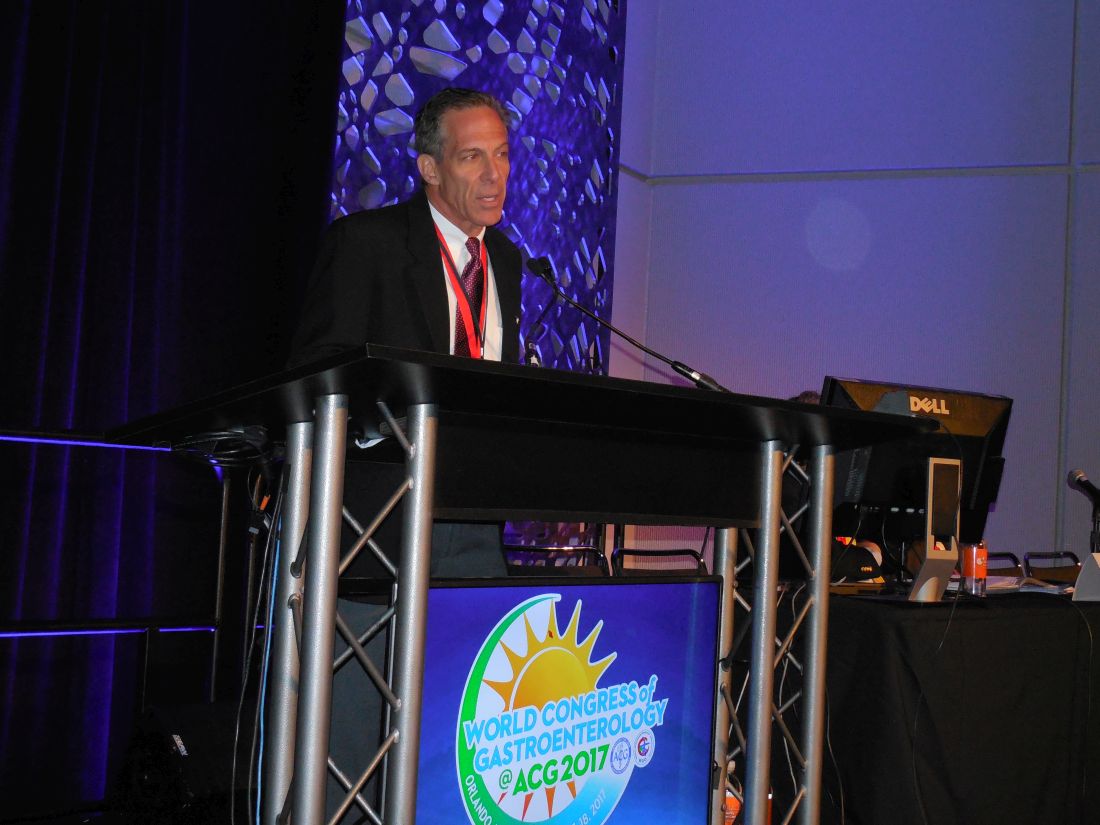
In current U.S. practice, many gastroenterologists perform follow-up colonoscopy about 5 years after removing one or two nonadvanced adenomas during a screening colonoscopy, Dr. Imperiale said during a video interview. Deferring follow-up colonoscopy in the absence of any clinical indication seems advisable, he said, especially for older patients with two or more comorbidities who had a high-quality index colonoscopy with good preparation and good colonic visibility.
“We just can’t do colonoscopy for surveillance on this subgroup continuously; it doesn’t make sense,” he said.
No randomized trial results have documented the need for stepped up colonoscopies in patients with a history of one or two nonadvanced adenomas, and these new observational findings are consistent with prior observational reports.
“These data need to be integrated with common sense,” he said. An extended interval before repeat surveillance seems particularly appropriate for patients with a higher risk for adverse effects from the colonoscopy preparation and for patients more likely to die from a cause other than colorectal cancer.
Backing off on repeat colonoscopy “minimizes the harm from surveillance. As patients get older they don’t tolerate the prep as well. It grows more onerous, and the returns diminish,” Dr. Imperiale said.
The patients included in the review had their index colonoscopy performed during 2002-2009, when they averaged about 61 years old, and about 95% were men. Their average Charlson comorbidity index was about 1.3. The incidence of colorectal cancer during follow-up after the index colonoscopy was 0.18% in patients with one or two nonadvanced adenomas in their index examination and no follow-up colonoscopy, 0.71% in those with nonadvanced adenomas who had one or more subsequent colonoscopies, and 0.31% in the people with no adenomas removed during the index procedure.
The rates of all-cause death during follow-up of the three subgroups were notably different: 34% in those with nonadvanced adenomas and no repeat colonoscopy, 13% in patients with nonadvanced adenomas and repeat colonoscopy, and 21% in those without nonadvanced adenomas. Dr. Imperiale discounted the significance of comparing rates of all-cause mortality, stressing that the most relevant primary endpoint is colorectal cancer mortality.
Dr. Imperiale reported having no disclosures.
mzoler@frontlinemedcom.com
On Twitter @mitchelzoler
The video associated with this article is no longer available on this site. Please view all of our videos on the MDedge YouTube channel
AT THE WORLD CONGRESS OF GASTROENTEROLOGY
Key clinical point:
Major finding: Colorectal cancer mortality was 0.13% in patients without follow-up colonoscopy and 0.16% in patients who had a second colonoscopy.
Data source: Review of 66,567 people at 13 U.S. VA Medical Centers.
Disclosures: Dr. Imperiale reported having no disclosures.
VIDEO: Burnout affects half of U.S. gastroenterologists
ORLANDO – Nearly half of U.S. gastroenterologists who responded to a recent survey had symptoms of burnout that seemed largely driven by work-life balance issues.
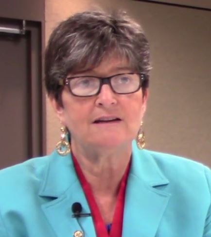
Burnout appeared to disproportionately affect younger gastroenterologists, those who spend more time on chores at home including caring for young children, physicians who were neutral toward or dissatisfied with a spouse or partner, and clinicians planning to soon leave their practice, Carol A. Burke, MD, said at the World Congress of Gastroenterology at ACG 2017.
Factors not linked with burnout included their type of practice, whether the gastroenterologists worked full or part time, their location, and their compensation, said Dr. Burke, director of the Center for Colon Polyp and Cancer Prevention at the Cleveland Clinic.
The life issues that appeared most strongly linked to burnout “speak to a problem for physicians to balance” their professional and personal lives, Dr. Burke said in a video interview. Several interventions exist that can potentially mitigate burnout, and the American College of Gastroenterology, which ran the survey, is taking steps to make information on these interventions available to members, noted Dr. Burke, the organization’s president.
Dr. Burke and her associates sent a 60-item survey to all 11,080 College members during 2014 and 2015 and received 1,021 replies including 754 fully completed responses. Their prespecified definition of burnout was a high score for emotional exhaustion or for depersonalization, or both, on the Maslach Burnout Inventory. The results showed that 45% of respondents had a high score for emotional exhaustion, 21% scored high on depersonalization, and overall 49% met the burnout criteria set by the investigators. The Inventory answers also showed that 18% had a low sense of personal accomplishment.
A multivariate analysis showed that significant links with burnout were younger age, more time spent on domestic chores, having a neutral or dissatisfying relationship with a spouse or partner, and plans for imminent retirement from gastroenterology practice, Dr. Burke reported.
The main reasons for planning imminent retirement were reimbursement, cited by 32% of this subgroup, regulations, cited by 21%, recertification, cited by 16%, and electronic medical records, cited by 10% as the main reason for leaving practice.
Strategies and resources aimed at better dealing with burnout were requested by 60% of all survey respondents, and the College is in the process of making these tools available, Dr. Burke said.
A recent study conducted by the AGA Institute Education and Training Committee and reported in AGA Perspectives supports these findings. Read more here and join the discussion on the AGA Community.
The video associated with this article is no longer available on this site. Please view all of our videos on the MDedge YouTube channel
mzoler@frontlinemedcom.com
On Twitter @mitchelzoler
ORLANDO – Nearly half of U.S. gastroenterologists who responded to a recent survey had symptoms of burnout that seemed largely driven by work-life balance issues.
Burnout appeared to disproportionately affect younger gastroenterologists, those who spend more time on chores at home including caring for young children, physicians who were neutral toward or dissatisfied with a spouse or partner, and clinicians planning to soon leave their practice, Carol A. Burke, MD, said at the World Congress of Gastroenterology at ACG 2017.
Factors not linked with burnout included their type of practice, whether the gastroenterologists worked full or part time, their location, and their compensation, said Dr. Burke, director of the Center for Colon Polyp and Cancer Prevention at the Cleveland Clinic.
The life issues that appeared most strongly linked to burnout “speak to a problem for physicians to balance” their professional and personal lives, Dr. Burke said in a video interview. Several interventions exist that can potentially mitigate burnout, and the American College of Gastroenterology, which ran the survey, is taking steps to make information on these interventions available to members, noted Dr. Burke, the organization’s president.
Dr. Burke and her associates sent a 60-item survey to all 11,080 College members during 2014 and 2015 and received 1,021 replies including 754 fully completed responses. Their prespecified definition of burnout was a high score for emotional exhaustion or for depersonalization, or both, on the Maslach Burnout Inventory. The results showed that 45% of respondents had a high score for emotional exhaustion, 21% scored high on depersonalization, and overall 49% met the burnout criteria set by the investigators. The Inventory answers also showed that 18% had a low sense of personal accomplishment.
A multivariate analysis showed that significant links with burnout were younger age, more time spent on domestic chores, having a neutral or dissatisfying relationship with a spouse or partner, and plans for imminent retirement from gastroenterology practice, Dr. Burke reported.
The main reasons for planning imminent retirement were reimbursement, cited by 32% of this subgroup, regulations, cited by 21%, recertification, cited by 16%, and electronic medical records, cited by 10% as the main reason for leaving practice.
Strategies and resources aimed at better dealing with burnout were requested by 60% of all survey respondents, and the College is in the process of making these tools available, Dr. Burke said.
A recent study conducted by the AGA Institute Education and Training Committee and reported in AGA Perspectives supports these findings. Read more here and join the discussion on the AGA Community.
The video associated with this article is no longer available on this site. Please view all of our videos on the MDedge YouTube channel
mzoler@frontlinemedcom.com
On Twitter @mitchelzoler
AT THE 13TH WORLD CONGRESS OF GASTROENTEROLOGY
Key clinical point:
Major finding: Forty-nine percent of surveyed U.S. gastroenterologists showed a high level of emotional exhaustion, depersonalization, or both.
Data source: Survey results from 754 members of the American College of Gastroenterology.
Disclosures: The American College of Gastroenterology funded the survey. Dr. Burke had no relevant disclosures.
MACRA Monday: Screening for hypertension
If you haven’t started reporting quality data for the Merit-Based Incentive Payment System (MIPS), there’s still time to avoid a 4% cut to your Medicare payments.
Under the Pick Your Pace approach being offered this year, the Centers for Medicare & Medicaid Services allows clinicians to test the system by reporting on one quality measure for one patient through paper-based claims. Be sure to append a Quality Data Code (QDC) to the claim form for care provided up to Dec. 31, 2017, in order to avoid a penalty in payment year 2019.
Consider this measure:
Measure #317: Preventive Care and Screening: Screening for High Blood Pressure and Follow-Up Documented
This measure is aimed at capturing the percentage of patients aged 18 years and older who were screened for high blood pressure and given a follow-up plan.
What you need to do: Check the patient’s blood pressure and recommend follow-up care – lifestyle modifications, additional testing, or medication, as appropriate – and document that plan.
Eligible cases include patients aged 18 years and older on the date of the encounter and a patient encounter during the performance period. Applicable codes include (CPT or HCPCS): 90791, 90792, 90832, 90834, 90837, 90839, 90845, 90880, 92002, 92004, 92012, 92014, 96118, 99201, 99202, 99203, 99204, 99205, 99212, 99213, 99214, 99281, 99282, 99283, 99284, 99285, 99215, 99304, 99305, 99306, 99307, 99308, 99309, 99310, 99318, 99324, 99325, 99326, 99327, 99328, 99334, 99335, 99336, 99337, 99341, 99342, 99343, 99344, 99345, 99347, 99348, 99349, 99350, D7140, D7210, G0101, G0402, G0438, G0439 without telehealth modifier: GQ or GT.
To get credit under MIPS, be sure to include a QDC that shows that you successfully performed the measure or had a good reason for not doing so. For instance, code G8783 indicates that a normal blood pressure reading was documented and follow-up is not required, while code G8950 indicates that the patient had a pre-hypertensive or hypertensive blood pressure reading documented and the appropriate follow-up was documented. Exclusion code G9744 should be used if the patient is not eligible due to an active diagnosis of hypertension.
CMS has a full list measures available for claims-based reporting at qpp.cms.gov. The American Medical Association has also created a step-by-step guide for reporting on one quality measure.
Certain clinicians are exempt from reporting and do not face a penalty under MIPS:
- Those who enrolled in Medicare for the first time during a performance period.
- Those who have Medicare Part B allowed charges of $30,000 or less.
- Those who have 100 or fewer Medicare Part B patients.
- Those who are significantly participating in an Advanced Alternative Payment Model (APM).

VIDEO: When surgery hurts the surgeon: Intervening to prevent ergonomic injuries
SAN DIEGO – Work-related musculoskeletal disorders are practically inevitable for surgeons, eventually occurring in more than 90%, no matter what type of surgery they practice.
At the annual clinical congress of the American College of Surgeons, this eyebrow-raising fact was presented with a sobering addendum: No one seems to be doing much about it.
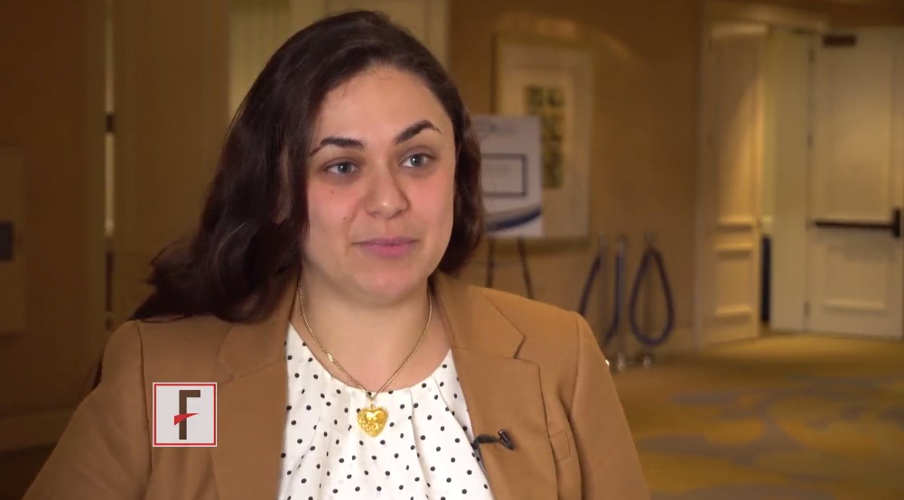
“There are some ergonomic guidelines for surgeons out there, but most surgeons don’t know about them,” said Tatiana Catanzarite, MD, who has conducted research on this topic. When she began looking into the problem of work-related injuries among surgeons, she was surprised at the dearth of published research. It’s no wonder then, said Dr. Catanzarite and other panel members, that most surgeons learn proper work posture on the fly and may or may not be using the most efficient and mechanically sound instrumentation angles when performing surgery.
Dr. Catanzarite, a female pelvic medicine and reconstructive surgery fellow at the University of California, San Diego, has just published a literature review on surgeon ergonomics. But reading about how to stand, how to hold instruments, and even how to sit at a robotic surgical console is no match for having an observer on the ground guiding and reinforcing work posture, she said. Unfortunately, that’s an unrealistic expectation for most surgeons, so Dr. Catanzarite is borrowing video-gaming technology to address the situation, she said in an interview.
She has adapted a popular video game motion-capture system that uses an infrared laser projector and a computer sensor to capture video data in three dimensions. The sensing range of the depth sensor is adjustable, and the software is capable of automatically calibrating the sensor based on the physical environment, accommodating the presence of obstacles and using infrared and depth cameras to capture a subject’s 3-D movements. The system doesn’t require bulky wearable components, “making it an ideal technology for the live operating room setting,” Dr. Catanzarite said. “In order to effectively assess surgical ergonomics, a less intrusive approach is needed, which can deliver precise reports on the body movements of the surgeons, as well as capturing the temporal distribution of different postures and limb angles.”
Dr. Catanzarite is using the system to launch an ergonomics assessment tool she calls Ergo-Kinect. The system will record surgeons’ movements in real time, gathering data about how they stand, move, and operate their instruments.
“Three-D interactive visualizations allow us to rotate and investigate specific motor activities from the collected data,” she said. The technology enables them to capture the movements of the surgeon and assign an ergonomic score for each movement. “Eventually we may be able to develop a system that can warn surgeons in real time if they are performing an activity which may be harmful from an ergonomics standpoint,” Dr. Catanzarite said.
The research is in its earliest phase – Dr. Catanzarite has only scanned a few surgeons. But she will continue to accrue data in order to eventually construct a system that could help surgeons of the future avoid the painful, and sometimes debilitating, physical costs of their career.
Dr. Catanzarite reported having no financial disclosures.
msullivan@frontlinemedcom.com
On Twitter @alz_gal
SAN DIEGO – Work-related musculoskeletal disorders are practically inevitable for surgeons, eventually occurring in more than 90%, no matter what type of surgery they practice.
At the annual clinical congress of the American College of Surgeons, this eyebrow-raising fact was presented with a sobering addendum: No one seems to be doing much about it.
“There are some ergonomic guidelines for surgeons out there, but most surgeons don’t know about them,” said Tatiana Catanzarite, MD, who has conducted research on this topic. When she began looking into the problem of work-related injuries among surgeons, she was surprised at the dearth of published research. It’s no wonder then, said Dr. Catanzarite and other panel members, that most surgeons learn proper work posture on the fly and may or may not be using the most efficient and mechanically sound instrumentation angles when performing surgery.
Dr. Catanzarite, a female pelvic medicine and reconstructive surgery fellow at the University of California, San Diego, has just published a literature review on surgeon ergonomics. But reading about how to stand, how to hold instruments, and even how to sit at a robotic surgical console is no match for having an observer on the ground guiding and reinforcing work posture, she said. Unfortunately, that’s an unrealistic expectation for most surgeons, so Dr. Catanzarite is borrowing video-gaming technology to address the situation, she said in an interview.
She has adapted a popular video game motion-capture system that uses an infrared laser projector and a computer sensor to capture video data in three dimensions. The sensing range of the depth sensor is adjustable, and the software is capable of automatically calibrating the sensor based on the physical environment, accommodating the presence of obstacles and using infrared and depth cameras to capture a subject’s 3-D movements. The system doesn’t require bulky wearable components, “making it an ideal technology for the live operating room setting,” Dr. Catanzarite said. “In order to effectively assess surgical ergonomics, a less intrusive approach is needed, which can deliver precise reports on the body movements of the surgeons, as well as capturing the temporal distribution of different postures and limb angles.”
Dr. Catanzarite is using the system to launch an ergonomics assessment tool she calls Ergo-Kinect. The system will record surgeons’ movements in real time, gathering data about how they stand, move, and operate their instruments.
“Three-D interactive visualizations allow us to rotate and investigate specific motor activities from the collected data,” she said. The technology enables them to capture the movements of the surgeon and assign an ergonomic score for each movement. “Eventually we may be able to develop a system that can warn surgeons in real time if they are performing an activity which may be harmful from an ergonomics standpoint,” Dr. Catanzarite said.
The research is in its earliest phase – Dr. Catanzarite has only scanned a few surgeons. But she will continue to accrue data in order to eventually construct a system that could help surgeons of the future avoid the painful, and sometimes debilitating, physical costs of their career.
Dr. Catanzarite reported having no financial disclosures.
msullivan@frontlinemedcom.com
On Twitter @alz_gal
AT THE ACS CLINICAL CONGRESS