User login
VIDEO: How to prevent, manage perinatal HIV infection
PARK CITY, UTAH – Despite notable progress in recent years, . Early recognition and treatment of HIV infection in pregnancy, prophylaxis for at-risk women, and retention of infected women in postpartum HIV care remain important goals, but they aren’t always met.
In a wide-ranging interview at the annual scientific meeting of the Infectious Diseases Society for Obstetrics and Gynecology, Gweneth Lazenby, MD, associate professor of obstetrics and gynecology at the Medical University of South Carolina, Charleston, explained what to do to improve the situation. Among the many pearls she shared: One HIV test isn’t enough for at-risk women.
AT IDSOG

VIDEO: Triple therapy study and new recommendations provide guidance on CAPS
MADRID – Catastrophic antiphospholipid syndrome (CAPS) is associated with a high mortality rate, but new research presented at the European Congress of Rheumatology shows that patient survival can be significantly improved by a triple therapy treatment approach.
Researchers at the Congress also presented clinical practice guidelines for the diagnosis and management of the rare disease, which accounts for just 1% of patients with antiphospholipid syndrome (APS).
CAPS is characterized by a fast onset of widespread thrombosis, mainly in the small vessels, and, often, microangiopathic hemolytic anemia is seen in the laboratory. If undiagnosed or left untreated, patients may present with multiorgan failure needing intensive care treatment, which can be fatal in up to 50% of cases.
At the Congress, Ignasi Rodríguez-Pintó, MD, presented new data from the CAPS Registry that looks at the combined effect of anticoagulation, corticosteroids, and plasma exchange or intravenous immunoglobulins on the survival of patients with CAPS as well as the new clinical practice guidelines.
CAPS Registry study
The aim of the study Dr. Rodríguez-Pintó presented on behalf of the CAPS Registry Project Group was to determine what, if any, survival benefit would be incurred from a triple therapy approach when compared with other different combinations of anticoagulation, corticosteroids, and plasma exchange or intravenous immunoglobulins, or none of these treatments.
Although the triple therapy treatment approach is already being used in practice, its use is largely empirical, Dr. Rodríguez-Pintó of the department of autoimmune disease at the Hospital Clinic, Barcelona, explained in a video interview.
The investigators derived their data from episodes of CAPS occurring in patients in the CAPS Registry from the European Forum on Antiphospholipid Antibodies. This international registry was set up in 2000 and has been assembling the clinical, laboratory, and therapeutic findings of patients with CAPS for almost 20 years.
“We observed 525 episodes of CAPS in 502 patients. That means that some patients had two to three episodes of CAPS,” Dr. Rodríguez-Pintó said. Data on 38 episodes of CAPS had to be excluded from the analysis because of missing information, which left 487 episodes occurring in 471 patients.
The mean age of the 471 patients included in the analysis was 38 years. The majority (67.9%) were female and had primary (68.8%) APS. Triple therapy was given to about 40% of patients who experienced CAPS, with about 57% receiving other combinations of drugs and 2.5% receiving no treatment for CAPS.
Overall, 177 of the 487 (36.3%) episodes of CAPS were fatal.
“Triple therapy was associated with a higher chance of survival when compared to other combinations or to none of these treatments,” Dr. Rodríguez-Pintó said.
While 28% of patients with CAPS died in the triple therapy group, mortality was 41% with other combinations of treatments and 75% with none of these treatments.
All-cause mortality was reduced by 47% with triple therapy, compared with none of these treatments. The adjusted odds ratio (aOR) when comparing survival between triple therapy and no treatment was 7.7, with a 95% confidence interval of 2.0 to 29.7. The aOR comparing other drug combinations versus none of these treatments was 6.8 (95% CI, 1.7-29.6).
“For a long time, we have been saying that triple therapy would probably be the best approach, but we had no firm evidence,” Dr. Rodríguez-Pintó said.
“So, this is the first time that we have clear clinical evidence of the benefit of these approaches, and I think that these results are important because they will give us more confidence in how we treat patients and help develop guidance on [the treatment’s] use in the future.”
Guidelines
A steering committee composed of representatives from the European Commission–funded RARE-BestPractices project and McMaster University in Hamilton, Ont., used GRADE methodology to develop the guidelines for CAPS diagnosis and management. The committee answered three diagnostic and seven treatment questions that originated from a panel of 19 international stakeholders, including Dr. Rodríguez-Pintó, through systematic reviews of the literature that used Cochrane criteria.
Although the review of studies did not include the study of CAPS Registry data that Dr. Rodríguez-Pintó and his colleagues conducted, he said that the recommendations still confirm the value of using a triple therapy approach to treatment.
The panel created three diagnostic recommendations for patients suspected of having CAPS, all of which were conditional and based on very low certainty of evidence: use preliminary CAPS classification criteria to diagnose CAPS; use or nonuse of biopsy, depending on the circumstances, because of its high specificity but possibly low sensitivity for thrombotic microangiopathy; and test for antiphospholipid antibodies, which should not delay initiation of treatment.
All seven first-line treatment recommendations that the panel developed relied on a very low certainty of evidence, and most were conditional:
- Triple therapy combination treatment with corticosteroids, heparin, and plasma exchange or intravenous immunoglobulins instead of a single agent or other combination treatments.
- Therapeutic dose anticoagulation was one of only two treatment recommendations to be considered “strong,” but use of direct oral anticoagulants is not advised.
- Therapeutic plasma exchange is recommended for use with other therapies and should be strongly considered for patients with microangiopathic hemolytic anemia.
- Intravenous immunoglobulin is advised for use in conjunction with other therapies and should be given special consideration for patients with immune thrombocytopenia or renal insufficiency.
- Antiplatelet agents are conditionally recommended as an add-on therapy, but their potential mortality benefit is tempered by increased risk of bleeding when used with anticoagulants. Strong consideration should be given to their use as an alternative therapy to anticoagulation when anticoagulation is contraindicated for a reason other than bleeding.
- Rituximab (Rituxan) should not be used because of little available data on its use, uncertainty regarding long-term consequences, and its expense – except for refractory cases where other therapies have been insufficient.
- Corticosteroids should not be used because of their lack of efficacy in CAPS when used alone and potential for adverse effects, except for certain circumstances where they may be indicated.
The authors of the guidelines emphasized that these recommendations are not meant to apply to every CAPS patient. They also noted that the available evidence did not allow for temporal analysis of treatments and that conclusions could not be drawn regarding “first-line” versus “second-line” therapies.
None of the authors of the registry study or the guidelines had relevant conflicts of interest to declare.
AT THE EULAR 2017 CONGRESS
Key clinical point:
Major finding: Mortality was 28% with triple therapy, 41% with other combinations of treatments, and 75% with none of these treatments.
Data source: A registry study of 471 CAPS patients and clinical practice guidelines for CAPS.
Disclosures: None of the authors of the registry study or the guidelines had relevant conflicts of interest to declare.
VIDEO: Lenvatinib’s real-world thyroid cancer performance matches trial
BOSTON – Lenvatinib’s real-world performance treating advanced, radio-iodine refractory, differentiated thyroid cancer closely followed the efficacy and adverse effect profiles the drug showed in its pivotal trial.
Lenvatinib showed good efficacy in 75 French registry patients, while also producing adverse effects in virtually every patient, but with the possibility to resolve the adverse effects with dose reductions or short-term treatment discontinuations, Martin Schlumberger, MD, said at the World Congress on Thyroid Cancer.
“Lenvatinib is toxic, but the toxicity can be managed in almost all patients by drug withholding or by reducing the dosage, and with symptomatic treatments,” Dr. Schlumberger said in a video interview. But adverse events are a “major problem” for the drug, so patients receiving lenvatinib “should be seen very frequently, and as soon as toxicity appears it should be treated,” said Dr. Schlumberger, professor of medicine and chairman of nuclear medicine and endocrine oncology at Gustave Roussy in Paris.
But lenvatinib’s efficacy makes it a first-line option despite the frequent adverse effects it causes.
“Without doubt it is the most effective drug” for treating advanced, rapidly progressing, radio-iodine refractory thyroid cancer, he said. “When patients really need systemic therapy they should get lenvatinib. It’s a balance of risk and benefit, and the risk from not being treated is higher than the risk from adverse effects.”
A similar pattern of adverse effects and efficacy was seen for lenvatinib in the pivotal Study of Lenvatinib in Differentiated Cancer of the Thyroid (SELECT) trial, which reported a median 18-month progression-free survival rate among patients treated with the drug compared with a median 4-month progression-free survival rate in placebo-treated patients (N Engl J Med. 2015 Feb 12;372[7]:621-30).
Among the 75 patients enrolled in the French registry, the median time of progression-free survival was 10 months, with 8 patients on continued therapy without progression. The response rate in the registry was 31% compared with 65% in the SELECT trial (and 2% in placebo-treated patients in SELECT), but the registry included many patients with advanced disease, comorbidities, and pretreatment, Dr. Schlumberger reported. Just 17 of the registry patients (23%) would have met the enrollment criteria for SELECT. Among this subset the response rate to lenvatinib was 47%.
A multivariate analysis identified three factors that significantly linked with drug responses, Dr. Schlumberger said: pretreatment, more advanced disease, and comorbidities.
Treatment-related adverse effects occurred in 71 of the registry patients (95%), with half of these grade 3 or higher. Twelve patients (16%) discontinued treatment because of an adverse effect. Hypertension was the most common adverse effect, occurring in 50 patients (67%), with 26 having grade 3 or higher hypertension. Other common adverse effects were fatigue, weight loss, diarrhea, and anorexia.
The 75 patients began treatment with lenvatinib for advanced thyroid cancer at any of 24 French centers during April 2015–June 2016. This marked the first year when lenvatinib was available in France for routine use, which roughly coincided with its U.S. introduction after lenvatinib received Food and Drug Administration marketing approval for advanced thyroid cancer in February 2015. Fifty-four patients (72%) began treatment on the labeled dosage of 24 mg/day; the remaining patients started the drug at a lower dosage.
mzoler@frontlinemedcom.com
On Twitter @mitchelzoler
Because of its efficacy lenvatinib is absolutely the top thymidine kinase inhibitor to use today to treat patients with radio-iodine-resistant, progressive, differentiated thyroid cancer. Although comparing drugs across trials is unreliable, the activity of lenvatinib in the SELECT trial (N Engl J Med. 2015 Feb 12;372[7]:621-30) was better than the activity of sorafenib in the DECISION trial (Lancet. 2014 July 26;384[9940]:319-28). There was enough of a difference between lenvatinib and sorafenib in the SELECT and DECISION trials to convince me that lenvatinib is the better drug.
Many of the patients enrolled in the French registry would not have qualified to enter the SELECT trial, so I’m not surprised that there was a lower response rate in the registry. We know that lenvatinib works better when the tumor burden is low, and some of the registry patients had a high tumor burden. In addition, a fraction of the registry patients did not receive a dosage of 24 mg/day, and data from the SELECT trial suggests that dosage size matters. The full dosage of 24 mg/day should be used as the starting dosage for lenvatinib, but that isn’t always possible for elderly patients or those with comorbidities.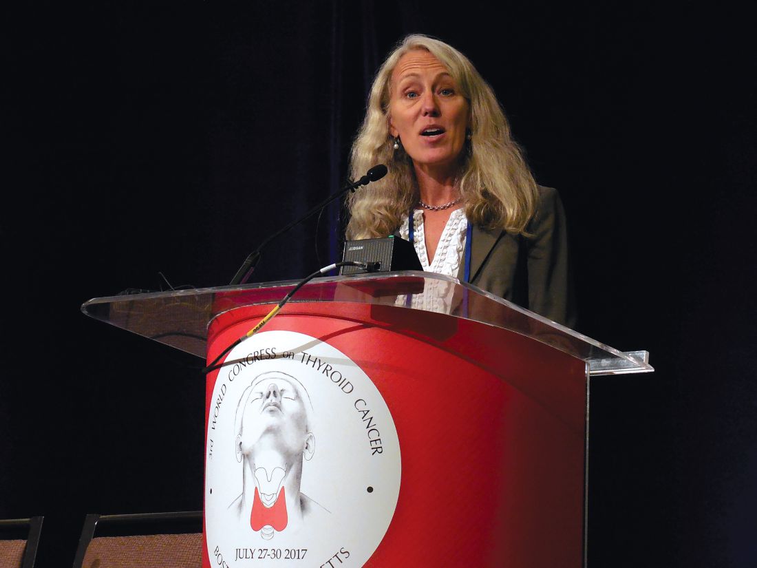
A rise in blood pressure with lenvatinib treatment is not a completely bad outcome, because our experience with lenvatinib shows that this adverse effect actually links with a survival benefit. A spike in a patient’s blood pressure in response to lenvatinib is a sign that the drug is working and the patient will have a good treatment response, an association that we’ve seen with other tumor types and with other thymidine kinase inhibitors.
Unfortunately, a good response to lenvatinib is usually not enough in the long run. Experience shows that even when advanced thyroid cancer responds to lenvatinib or to another thymidine kinase inhibitor, eventually the disease will progress despite this treatment.
Lori J. Wirth, MD , is medical director of the Center for Head and Neck Cancers at Massachusetts General Hospital in Boston. She has been a consultant to Eisai, Blueprint Medicines, Loxo, and Merck. She made these comments in an interview.
BOSTON – Lenvatinib’s real-world performance treating advanced, radio-iodine refractory, differentiated thyroid cancer closely followed the efficacy and adverse effect profiles the drug showed in its pivotal trial.
Lenvatinib showed good efficacy in 75 French registry patients, while also producing adverse effects in virtually every patient, but with the possibility to resolve the adverse effects with dose reductions or short-term treatment discontinuations, Martin Schlumberger, MD, said at the World Congress on Thyroid Cancer.
“Lenvatinib is toxic, but the toxicity can be managed in almost all patients by drug withholding or by reducing the dosage, and with symptomatic treatments,” Dr. Schlumberger said in a video interview. But adverse events are a “major problem” for the drug, so patients receiving lenvatinib “should be seen very frequently, and as soon as toxicity appears it should be treated,” said Dr. Schlumberger, professor of medicine and chairman of nuclear medicine and endocrine oncology at Gustave Roussy in Paris.
But lenvatinib’s efficacy makes it a first-line option despite the frequent adverse effects it causes.
“Without doubt it is the most effective drug” for treating advanced, rapidly progressing, radio-iodine refractory thyroid cancer, he said. “When patients really need systemic therapy they should get lenvatinib. It’s a balance of risk and benefit, and the risk from not being treated is higher than the risk from adverse effects.”
A similar pattern of adverse effects and efficacy was seen for lenvatinib in the pivotal Study of Lenvatinib in Differentiated Cancer of the Thyroid (SELECT) trial, which reported a median 18-month progression-free survival rate among patients treated with the drug compared with a median 4-month progression-free survival rate in placebo-treated patients (N Engl J Med. 2015 Feb 12;372[7]:621-30).
Among the 75 patients enrolled in the French registry, the median time of progression-free survival was 10 months, with 8 patients on continued therapy without progression. The response rate in the registry was 31% compared with 65% in the SELECT trial (and 2% in placebo-treated patients in SELECT), but the registry included many patients with advanced disease, comorbidities, and pretreatment, Dr. Schlumberger reported. Just 17 of the registry patients (23%) would have met the enrollment criteria for SELECT. Among this subset the response rate to lenvatinib was 47%.
A multivariate analysis identified three factors that significantly linked with drug responses, Dr. Schlumberger said: pretreatment, more advanced disease, and comorbidities.
Treatment-related adverse effects occurred in 71 of the registry patients (95%), with half of these grade 3 or higher. Twelve patients (16%) discontinued treatment because of an adverse effect. Hypertension was the most common adverse effect, occurring in 50 patients (67%), with 26 having grade 3 or higher hypertension. Other common adverse effects were fatigue, weight loss, diarrhea, and anorexia.
The 75 patients began treatment with lenvatinib for advanced thyroid cancer at any of 24 French centers during April 2015–June 2016. This marked the first year when lenvatinib was available in France for routine use, which roughly coincided with its U.S. introduction after lenvatinib received Food and Drug Administration marketing approval for advanced thyroid cancer in February 2015. Fifty-four patients (72%) began treatment on the labeled dosage of 24 mg/day; the remaining patients started the drug at a lower dosage.
mzoler@frontlinemedcom.com
On Twitter @mitchelzoler
AT WCTC 2017
Key clinical point:
Major finding: The median time of progression-free survival was 10 months in the registry and 18 months in the pivotal trial.
Data source: A retrospective review of the first 75 French patients with advanced differentiated thyroid cancer who received lenvatinib following its marketing approval.
Disclosures: Dr. Schlumberger has received research funding from Eisai, the company that markets lenvatinib (Lenvima). He has also received research support and honoraria from AstraZeneca, Bayer, and Excelixis.
VIDEO: What role does autoimmune dysfunction play in schizophrenia?
SAN DIEGO – Researchers are looking at the potential role that autoimmune reactions, such as inflammation, play in the pathogenesis of schizophrenia, both with and without comorbid substance use.
“What we see is evidence of autoimmune involvement through the lifespan in schizophrenia,” explained Brian Miller, MD, PhD, of the department of psychiatry at Augusta (Georgia) University. “We know there is a bidirectional association between schizophrenia and autoimmune disorders, [where] starting with either increases the chances of the other occurring comorbidly.”
Previous studies have shown that adding anti-inflammatory agents to second-generation antipsychotics has been associated with greater symptomatic improvement in schizophrenia than with second-generation antipsychotics alone. Now, Dr. Miller and his colleagues are conducting a randomized, controlled study using the systemic immunotherapy siltuximab to see if adjunct anti-inflammatory treatments can improve cognition in schizophrenia. They are using the monoclonal antibody instead of nonsteroidal anti-inflammatory drugs because of its precise ability to target specific autoimmune pathways, according to Dr. Miller.
In addition, Dr. Miller and his colleagues conducted a post-hoc analysis of data from the CATIE (Clinical Antipsychotic Trials of Intervention Effectiveness) study to determine if there was a difference between those patients with comorbid substance in schizophrenia, compared with those without. He and his coinvestigators found in a post-hoc analysis that the higher the level of certain inflammatory markers, the higher the Positive and Negative Syndrome Scale (PANSS) psychopathology score in people who’d tested positive for marijuana use at baseline.
The results, he said, beg the question about whether trial inclusion criteria for schizophrenia research need revisiting.
“What we really need to do is investigate, in a well-controlled, well-designed, prospective sample of patients who have this substance use comorbidity, [whether] we see alterations in these immune markers, and then how they vary over the course of illness,” Dr. Miller explained in a video interview at the International Congress on Schizophrenia Research. “It’s an important selection bias that as a field we’ve ignored. Maybe that’s a population that is enriched for some kind of immune dysfunction that might be targeted by some kind of anti-inflammatory or other immunotherapy strategy.”
The video associated with this article is no longer available on this site. Please view all of our videos on the MDedge YouTube channel
On Twitter @whitneymcknight
AT THE ICSR BIENNIAL MEETING
VIDEO: Study highlights risks of postponing cholecystectomy
Almost half of patients who underwent endoscopic retrograde cholangiopancreatography (ERCP) did not undergo cholecystectomy (CCY) within the next 60 days according to the results of a large, retrospective cohort study reported in the September issue of Gastroenterology (doi: 10.1053/j.gastro.2017.05.048).
“Although early and delayed CCY equally reduce the risk of subsequent recurrent biliary events, patients are at 10-fold higher risk of a recurrent biliary event while waiting for a delayed CCY, compared with patients who underwent early CCY,” wrote Robert J. Huang, MD, and his associates of Stanford (Calif.) University Medical Center. Delayed CCY is cost effective, but that benefit must be weighed against the risk of loss to follow-up, especially if patients have “little or no health insurance,” they said.
Source: American Gastroenterological Association
Gallstone disease affects up to 15% of adults in developed societies, including about 20-25 million Americans. Yearly costs of treatment tally at more than $6.2 billion and have risen by more than 20% in 3 decades, according to multiple studies. Approximately 20% of patients with gallstone disease have choledocholithiasis, mainly because gallstones can pass from the gallbladder into the common bile duct. After undergoing ERCP, such patients are typically referred for CCY, but there are no “societal guidelines” on timing the referral, the researchers said. Practice patterns remain “largely institution based and may be subject to the vagaries of surgeon availability and other institutional resource constraints.” One prior study linked a median 7-week wait time for CCY with a 20% rate of recurrent biliary events. To evaluate large-scale practice patterns, the researchers studied 4,516 patients who had undergone ERCP for choledocholithiasis in California (during 2009-2011), New York (during 2011-2013), and Florida (during 2012-2014) and calculated timing and rates of subsequent CCY, recurrent biliary events, and deaths. Patients were followed for up to 365 days after ERCP.
Of the 4,516 patients studied, 1,859 (41.2%) patients underwent CCY during their index hospital admission (early CCY). Of the 2,657 (58.8%) patients who were discharged without CCY, only 491 (18%) had a planned CCY within 60 days (delayed CCY), 350 (71.3%) of which were done in an outpatient setting. Of the patients in the study, 2,168 (48.0%) did not have a CCY (no CCY) during their index visit or within 60 days. Over 365 days of follow-up, 10% of patients who did not have a CCY had recurrent biliary events, compared with 1.3% of patients who underwent early or delayed CCY. The risk of recurrent biliary events for patients who underwent early or delayed CCY was about 88% lower than if they had had no CCY within 60 days of ERCP (P less than .001 for each comparison). Performing CCY during index admission cut the risk of recurrent biliary events occurring within 60 days by 92%, compared with delayed or no CCY (P less than .001).
In all, 15 (0.7%) patients who did not undergo CCY died after subsequent hospitalization for a recurrent biliary event, compared with 1 patient who underwent early CCY (0.1%; P less than .001). There were no deaths associated with recurrent biliary events in the delayed-CCY group. Rates of all-cause mortality over 365 days were 3.1% in the no-CCY group, 0.6% in the early-CCY group, and 0% in the delayed-CCY group. Thus, cumulative death rates were about seven times higher among patients who did not undergo CCY compared with those who did (P less than .001).
Patients who did not undergo CCY tended to be older than delayed- and early-CCY patients (mean ages 66 years, 58 years, and 52 years, respectively). No-CCY patients also tended to have more comorbidities. Nonetheless, having an early CCY retained a “robust” protective effect against recurrent biliary events after accounting for age, sex, comorbidities, stent placement, facility volume, and state of residence. Even after researchers adjusted for those factors, the protective effect of early CCY dropped by less than 5% (from 92% to about 87%), the investigators said.
They also noted that the overall cohort averaged 60 years of age and that 64% were female, which is consistent with the epidemiology of biliary stone disease. Just over half were non-Hispanic whites. Medicare was the single largest primary payer (46%), followed by private insurance (28%) and Medicaid (16%).
“A strategy of delayed CCY performed on an outpatient basis was least costly,” the researchers said. “Performance of early CCY was inversely associated with low facility volume. Hispanic race, Asian race, Medicaid insurance, and no insurance associated inversely with performance of delayed CCY.”
Funders included a seed grant from the Stanford division of gastroenterology and hepatology and the National Institutes of Health. The investigators had no conflicts of interest.
FROM GASTROENTEROLOGY
Key clinical point: Almost half of patients who underwent endoscopic retrograde cholangiopancreatography (ERCP) did not undergo cholecystectomy within 60 days.
Major finding: A total of 48% had no cholecystectomy within 60 days. Performing cholecystectomy during index admission cut the risk of recurrent biliary events within 60 days by 92%, compared with delayed or no cholecystectomy (P less than .001).
Data source: A multistate, retrospective study of 4,516 patients hospitalized with choledocholithiasis.
Disclosures: Funders included a Stanford division of gastroenterology and hepatology divisional seed grant and the National Institutes of Health. The investigators had no conflicts of interest.

Evaluating suicidality

VIDEO: Less follow-up proposed for low-risk thyroid cancer
BOSTON – , Bryan R. Haugen, MD, suggested in a keynote lecture during the World Congress on Thyroid Cancer.
Traditionally, thyroid cancer specialists have monitored these patients for persistent or recurrent disease as often as every 6 or 12 months. “But what we’ve realized with recent assessments of response to treatment is that some patients do well without a recurrence over many years; so, the concept of doing less monitoring and less imaging, especially in patients with an excellent response [to their initial treatment], is being studied,” Dr. Haugen said in a video interview following his talk.
He estimated that perhaps two-thirds or as many as three-quarters of patients with differentiated thyroid cancer fall into the category of having low- or intermediate-risk disease with an excellent or good response to treatment, and hence they are potential candidates for eventually transitioning to less frequent follow-up.
During his talk, Dr. Haugen suggested that after several years with no sign of disease recurrence, lower-risk patients with an excellent treatment response may be able to stop undergoing regular monitoring, and those with a good treatment response may be able to safely have their monitoring intervals extended.
According to the most recent (2015) guidelines for differentiated thyroid cancer management from the American Thyroid Association, lower-risk patients with an excellent treatment response should have their serum thyroglobulin measured every 12-24 months and undergo an ultrasound examination every 3-5 years, while patients with a good response are targeted for serum thyroglobulin measurement annually with an ultrasound every 1-3 years (Thyroid. 2016 Jan;26[1]:1-133). Dr. Haugen chaired the expert panel that wrote these guidelines.
In another provocative suggestion, Dr. Haugen proposed that once well-responsive, lower-risk patients have remained disease free for several years, their less frequent follow-up monitoring could be continued by a primary care physician or another less specialized clinician.
At some time in the future, “a patient’s primary care physician could follow a simple tumor marker, thyroglobulin, maybe once every 5 years,” said Dr. Haugen, professor of medicine and head of the division of endocrinology, metabolism, and diabetes at the University of Colorado in Aurora. “At the University of Colorado, we use advanced-practice providers to do long-term follow-up” for lower-risk, treatment-responsive patients, he said.
The video associated with this article is no longer available on this site. Please view all of our videos on the MDedge YouTube channel
On Twitter @mitchelzoler
AT WCTC 2017

VIDEO: How to discharge new pediatric diabetes cases in 2 days
NASHVILLE, TENN. – Pediatric endocrinologist Cassandra Brady, MD, caught the attention of her audience at Pediatric Hospital Medicine when she mentioned that children presenting with new-onset diabetes rarely spend more than 2 days at Vanderbilt University’s children’s hospital, even if they present in diabetic ketoacidosis.
In many places, children with new-onset diabetes spend quite a bit longer in the hospital – even if they are medically stable and feeling fine – for diabetes education.
That’s not the case at Vanderbilt, where Dr. Brady is an assistant professor. Once kids are stabilized, they and their parents undergo a 3-hour crash course – sometimes even in the PICU – on diabetes survival skills, and then they’re sent home with insulin. They learn the finer points about carbohydrate counting and tight glucose control at subsequent outpatient visits.
More and more payers are probably going to push for that model, Dr. Brady noted at the meeting, which was sponsored by the Society of Hospital Medicine, the American Academy of Pediatrics, and the Academic Pediatric Association.
For those interested in making the transition to outpatient eduction, she explained in an interview exactly how Vanderbilt’s been doing it safely for years.
The video associated with this article is no longer available on this site. Please view all of our videos on the MDedge YouTube channel
AT PHM 2017

VIDEO: Further CTE correlation found in deceased American football players
Evidence of a correlation between chronic traumatic encephalopathy (CTE) and playing American football was strengthened in the largest retrospective study cohort to date.
The progressive neurodegenerative disease associated with recurrent head trauma has been linked to football over the past decade. However, this new study took steps to solidify the connection by studying 202 deceased brains, more than double the size of a previous 2013 report.
First author Jesse Mez, MD, of Boston University, and his colleagues examined the brains donated to a brain bank jointly run by the VA Boston Healthcare System, Boston University, and the Concussion Legacy Foundation. They correlated their findings with interviews and questionnaires filled out in 2014 by those close to the donors (JAMA. 2017 Jul 25. doi: 10.1001/jama.2017.8334).
The donors were male, and a majority (79%) were white. The average age at death was 67 years. In the mild cases, there was an average of 13 years of play; in severe cases, the average was 15.8 years.
The researchers found evidence of CTE in 177 (88%) of the 202 brains of football players, with cases found in 3 (21%) who played only in high school, 48 (91%) who played in college, 9 (64%) semiprofessional players, 7 (88%) Canadian Football League players, and 110 (99%) NFL players, according to investigators.
“Nearly all of the former NFL players in this study had CTE pathology, and this pathology was frequently severe,” noted Dr. Mez and his coinvestigators. ”These findings suggest that CTE may be related to prior participation in football and that a high level of play may be related to substantial disease burden.”
Severity of CTE positively correlated with longer playing history and more competitive levels of play. According to the investigators, severe cases of CTE were found in none of the high school players, 27 college players (56%), 5 semiprofessional players (56%), 6 Canadian professional players (86%), and 95 NFL players (86%).
All participants with CTE commonly displayed certain symptoms regardless of severity level.
Impulsiveness occurred in 23 mild cases (89%) and 65 severe cases (80%). Depression was seen in 18 mild cases (67%) and 46 severe cases (56%). A total of 13 players (50%) with mild symptoms showed apathy, compared with 43 with severe symptoms (52%). Anxiety was reported for 14 mild cases (52%) and 41 severe cases (50%). Cognitive symptoms such as problems with memory and attention were also common at both levels of severity, with 19 cases (73%) and 18 cases (69%), respectively, among donors with mild CTE, and 76 (92%) and 67 (81%) among donors with severe CTE.
Those interviewed who were close to the donors also reported other behavior or mood symptoms that the investigators inferred to be related to CTE.
“Many of these participants had a substance use disorder, demonstrated suicidality, or had a family history of psychiatric illness,” the investigators wrote. “Behavior or mood symptoms may be the initial presentation for a subset of individuals with CTE, or alternatively, CTE [phosphorylated tau] pathology may lower the threshold for psychiatric manifestations in susceptible individuals.”
The investigators asserted that the correlation of CTE diagnosis and severity may be more nuanced than just how many times a player is hit.
“Several other football-related factors may influence CTE risk and disease severity, including but not limited to age at first exposure to football, duration of play, player position, cumulative hits, and linear and rotational acceleration of hits,” Dr. Mez and his associates wrote. “Recent work in living former football players has shown that age at first exposure may be related to impaired cognitive performance and altered corpus callosum white matter and that cumulative hits may be related to impairment on self-report and objective measures of cognition, mood, and behavior, although it is unclear if any of these outcomes are related to CTE pathology.”
This study was limited by the potential bias of donors who participated in the program. Researchers acknowledge that public awareness of the issue of CTE and head trauma may have influenced players with symptoms to donate their brains, thereby increasing the CTE frequency in the sample.
In addition, the brain bank where the sample was taken is not representative of the entire population of American football players, as most donors played at the college, semi-professional, and professional level, as opposed to only high school or youth leagues.
The senior author of the study, Ann C. McKee, MD, discussed the study further in a related video interview.
The National Institute of Neurological Disorders and Stroke, the National Institute of Aging, the U.S. Department of Defense, the U.S. Department of Veterans Affairs, the National Operating Committee on Standards for Athletic Equipment, the Alzheimer’s Association, the Concussion Legacy Foundation, the Andlinger Family Foundation, the WWE, and the NFL supported the study. Five authors reported receiving support from relevant sources, including the NFL.
The video associated with this article is no longer available on this site. Please view all of our videos on the MDedge YouTube channel
ezimmerman@frontlinemedcom.com
On Twitter @eaztweets
Traumatic brain injuries (TBIs) are a serious health concern that currently affect 10 million people per year worldwide.
While a majority of TBIs are caused by car crashes, contact sports are estimated to cause between 1.6-3.8 million concussions in the United States every year, making the root of this study – to learn more about CTE among those in contact sports – imperative.
This study used the largest CTE cohort to date, but it is important to read the results, which are concerning, within the context of the study.
Results from the informants who filled out the questionnaires – most commonly friends or family of the deceased – showed cognitive symptoms in 88% of donors and behavioral symptoms in 95%, compared with 5% and 20% prevalence, respectively, among a questionnaire-based study filled out by retired National Football League players. This may mean that rates of symptomatic CTE may be lower outside of a selected cohort of former players.
While early-stage CTE was found in some high school players, most individuals do not play football beyond a high school level, and the link between high school football and future neurological decline was not well established in this study.
This study is a good example of the progress that has been made in TBI research, but there are still many fundamental questions that need to be answered. The next step will be improving disease models and expanding the studies of at-risk populations.
It is of the utmost importance that these improvements are made immediately; increasing our knowledge and public awareness of TBIs is paramount.
Gil Rabinovici, MD, is associate professor of neurology at the University of California–San Francisco, and an associate editor for JAMA Neurology. He receives research support from Avid Radiopharmaceuticals, Eli Lilly, GE Healthcare, and Piramal. He has received consulting or speaking honoraria from Eisai, Genentech, Lundbeck, Merck, Putnam, and Roche. These comments were taken from an editorial (JAMA 2017 Jul 25. doi: 10.1001/jama.2017.9353 ).
Evidence of a correlation between chronic traumatic encephalopathy (CTE) and playing American football was strengthened in the largest retrospective study cohort to date.
The progressive neurodegenerative disease associated with recurrent head trauma has been linked to football over the past decade. However, this new study took steps to solidify the connection by studying 202 deceased brains, more than double the size of a previous 2013 report.
First author Jesse Mez, MD, of Boston University, and his colleagues examined the brains donated to a brain bank jointly run by the VA Boston Healthcare System, Boston University, and the Concussion Legacy Foundation. They correlated their findings with interviews and questionnaires filled out in 2014 by those close to the donors (JAMA. 2017 Jul 25. doi: 10.1001/jama.2017.8334).
The donors were male, and a majority (79%) were white. The average age at death was 67 years. In the mild cases, there was an average of 13 years of play; in severe cases, the average was 15.8 years.
The researchers found evidence of CTE in 177 (88%) of the 202 brains of football players, with cases found in 3 (21%) who played only in high school, 48 (91%) who played in college, 9 (64%) semiprofessional players, 7 (88%) Canadian Football League players, and 110 (99%) NFL players, according to investigators.
“Nearly all of the former NFL players in this study had CTE pathology, and this pathology was frequently severe,” noted Dr. Mez and his coinvestigators. ”These findings suggest that CTE may be related to prior participation in football and that a high level of play may be related to substantial disease burden.”
Severity of CTE positively correlated with longer playing history and more competitive levels of play. According to the investigators, severe cases of CTE were found in none of the high school players, 27 college players (56%), 5 semiprofessional players (56%), 6 Canadian professional players (86%), and 95 NFL players (86%).
All participants with CTE commonly displayed certain symptoms regardless of severity level.
Impulsiveness occurred in 23 mild cases (89%) and 65 severe cases (80%). Depression was seen in 18 mild cases (67%) and 46 severe cases (56%). A total of 13 players (50%) with mild symptoms showed apathy, compared with 43 with severe symptoms (52%). Anxiety was reported for 14 mild cases (52%) and 41 severe cases (50%). Cognitive symptoms such as problems with memory and attention were also common at both levels of severity, with 19 cases (73%) and 18 cases (69%), respectively, among donors with mild CTE, and 76 (92%) and 67 (81%) among donors with severe CTE.
Those interviewed who were close to the donors also reported other behavior or mood symptoms that the investigators inferred to be related to CTE.
“Many of these participants had a substance use disorder, demonstrated suicidality, or had a family history of psychiatric illness,” the investigators wrote. “Behavior or mood symptoms may be the initial presentation for a subset of individuals with CTE, or alternatively, CTE [phosphorylated tau] pathology may lower the threshold for psychiatric manifestations in susceptible individuals.”
The investigators asserted that the correlation of CTE diagnosis and severity may be more nuanced than just how many times a player is hit.
“Several other football-related factors may influence CTE risk and disease severity, including but not limited to age at first exposure to football, duration of play, player position, cumulative hits, and linear and rotational acceleration of hits,” Dr. Mez and his associates wrote. “Recent work in living former football players has shown that age at first exposure may be related to impaired cognitive performance and altered corpus callosum white matter and that cumulative hits may be related to impairment on self-report and objective measures of cognition, mood, and behavior, although it is unclear if any of these outcomes are related to CTE pathology.”
This study was limited by the potential bias of donors who participated in the program. Researchers acknowledge that public awareness of the issue of CTE and head trauma may have influenced players with symptoms to donate their brains, thereby increasing the CTE frequency in the sample.
In addition, the brain bank where the sample was taken is not representative of the entire population of American football players, as most donors played at the college, semi-professional, and professional level, as opposed to only high school or youth leagues.
The senior author of the study, Ann C. McKee, MD, discussed the study further in a related video interview.
The National Institute of Neurological Disorders and Stroke, the National Institute of Aging, the U.S. Department of Defense, the U.S. Department of Veterans Affairs, the National Operating Committee on Standards for Athletic Equipment, the Alzheimer’s Association, the Concussion Legacy Foundation, the Andlinger Family Foundation, the WWE, and the NFL supported the study. Five authors reported receiving support from relevant sources, including the NFL.
The video associated with this article is no longer available on this site. Please view all of our videos on the MDedge YouTube channel
ezimmerman@frontlinemedcom.com
On Twitter @eaztweets
FROM JAMA
Key clinical point:
Major finding: Of 202 deceased football players who played at levels ranging from high school to the NFL and who donated their brains, 177 (88%) were diagnosed with CTE.
Data source: A blinded, retrospective study of 202 donated brains, as well as questionnaires and interviews with informants close to the deceased.
Disclosures: The National Institute of Neurological Disorders and Stroke, the National Institute of Aging, U.S. Department of Defense, U.S. Department of Veterans Affairs, the National Operating Committee on Standards for Athletic Equipment, the Alzheimer’s Association, the Concussion Legacy Foundation, the Andlinger Family Foundation, the WWE, and the NFL supported the study. Five authors reported receiving support from relevant sources, including the NFL.

Algorithm-based trunk support system shows potential for recovery of walking ability
A multidirectional gravity-assist device that delivers precise trunk support to stroke and spinal cord injury (SCI) patients via an artificial intelligence algorithm has demonstrated significantly improved locomotor performance beyond treadmill-based systems in a new study.
The harness device used in the study adjusts patients’ balance while they stand still or walk by employing a unique, adaptive multidirectional gravity-assist (MGA) algorithm tailored to the specific needs of the patient, according to Jean-Baptiste Mignardot, PhD, of the Center for Neuroprosthetics and Brain Mind Institute, Swiss Federal Institute of Technology, Lausanne, Switzerland, and fellow investigators.
“The MGA establishes a safe and natural rehabilitation environment wherein individuals with neurological deficits can perform basic and skilled locomotor activities that would not be possible without robotic assistance,” according to the investigators. “The immediate and short-term ameliorations of gait performance during locomotion with MGA illustrate the potential of this environment to augment motor recovery.”
Current gait rehabilitation methods in stroke or SCI patients most commonly involves counterweight mechanisms or force-controlled equipment that apply upward support while walking on a treadmill. However Dr. Mignardot and his colleagues believe these methods are flawed.
The video associated with this article is no longer available on this site. Please view all of our videos on the MDedge YouTube channel
“Treadmill-restricted environments markedly differ from the rich repertoire of natural locomotor activities underlying daily living,” the investigators wrote. “Vertically restricted trunk support creates undesired forces that impede gait execution.”
To counteract these negative effects, the MGA adjusts both upward and forward forces on the patient’s body, re-creating a more naturally occurring gait posture, which investigators have likened to an inverted pendulum with a natural forward tilt. In order to create the algorithm, investigators ran through a series of procedures, starting with calibrations based on the gait of healthy subjects and adjusting for necessary upward and forward assistance for stroke and SCI patients.
The artificial neural network within the algorithm analyzes patients’ support needs, a job that therapists currently have to do based on visual observations. This opens a window to faster and more accurate estimations, according to the investigators (Sci Transl Med. 2017 Jul 19. doi: 10.1126/scitranslmed.aah3621).
Investigators tested the algorithm on 15 SCI patients and 12 stroke patients. The stroke patients had an average age of 51 years, with length of time after stroke varying from 8 to 235 months. The SCI patients had an average age of 47 years, with a length of time since injury ranging from 12 to 264 months. Most patients in both groups were male.
When tested, the algorithm showed varying success depending on the severity of the injury, according to the researchers.
“For example, the MGA enabled subjects who could not stand independently to walk overground with or without assistive device.” Subjects who were able to move around only with crutches or a walker progressed without the use of assistive devices and exhibited improved spatiotemporal gait features, according to Dr. Mignardot and fellow investigators. “Individuals with stroke exhibited similar or even superior amelioration of locomotor performance and showed that individuals who could only walk with crutches exhibited improved intralimb coordination.”
The video associated with this article is no longer available on this site. Please view all of our videos on the MDedge YouTube channel
After initial efficacy tests, the researchers tested the MGA’s effectiveness in five SCI patients immediately after 1 hour of training with the device and found that their gait speed increased during the training. However, the improvements were not evident in a similar test 1 week later. Similar tests using treadmill-restricted step training without the MGA device did not show any improvement during either week of testing.
Although the study’s small sample size limited the conclusions that could be reached, the investigators were encouraged by the overall effects of the algorithm. They noted that further tests are required to test the potential sensitivity and accuracy of the software.
The study was supported by the European Commission’s Seventh Framework Programme, various foundations, and the Swiss National Science Foundation.
Investigators reported holding patents on the step-by-step procedure and use of the MGA algorithm in this study.
ezimmerman@frontlinemedcom.com
On Twitter @eaztweets
FROM SCIENCE TRANSLATIONAL MEDICINE