User login
VIDEO: Childhood second-hand smoke boosts RA incidence
MADRID – Second-hand smoke exposure to children was about as potent a trigger for future rheumatoid arthritis as active smoking by an adult, based on an analysis of data collected from more than 70,000 French women followed for an average of more than 20 years
“This is the first demonstration of a rheumatoid arthritis risk associated with passive smoking,” Raphaèle Seror, MD, said at the European Congress of Rheumatology.
“This is an important finding because we can avoid passive smoke exposure,” Dr. Seror added in a video interview . The imperative to eliminate second-hand smoke exposure to children is particularly acute for those with a genetic risk for developing rheumatoid arthritis (RA), specifically children with a parent diagnosed with RA, suggested Dr. Seror, a professor of rheumatology at the University of Paris–South.
She and her associates used data collected in the E3N, a longitudinal French epidemiological study that enrolled nearly 100,000 women in 1990 when they were 40-65 years old and collected health data by questionnaire every 2-3 years for an average of 21 years. They identified from this cohort women with “confirmed” RA based on a self report of having incident RA during follow-up plus a coincident record of reimbursement for a prescription for an RA-specific treatment, such as methotrexate or a biological disease-modifying drug.
This identified 389 women with confirmed incident RA, including 350 with a complete smoking history that made the current analysis possible. The study also included 70,248 women who did not develop RA and who had provided a complete smoking history.
The analysis showed that women who reported a history of second-hand smoke exposure estimated at more than an hour daily as children but without a history of active smoking had a 43% higher rate of incident RA compared with never smoker women without a history of passive smoke exposure, Dr. Seror reported. This association just missed reaching statistical significance, a limitation that Dr. Seror attributed to a power issue as the analysis included only 30 women who had incident RA and a history of second-hand smoke exposure without adult smoke exposure. By comparison, women in the study with a history of active smoking without childhood exposure linked had a 37% increased incidence of RA, a finding that confirmed the well-known link between smoking and RA incidence.
The study also found that women with both second-hand smoke exposure as children and adult smoking linked with a 73% higher RA incidence, an indication that the contributions from second-hand smoke in children and active smoking by adults were not only similar in magnitude but also worked additively to promote RA development.
The video associated with this article is no longer available on this site. Please view all of our videos on the MDedge YouTube channel
mzoler@frontlinemedcom.com
On Twitter @mitchelzoler
AT THE EULAR 2017 CONGRESS
Key clinical point:
Major finding: Women with significant second-hand smoke exposure as children had a 43% increased rate of incident rheumatoid arthritis.
Data source: E3N, a prospective, longitudinal, observational study of nearly 100,000 French women begun in 1990.
Disclosures: Dr. Seror had no relevant disclosures.

VIDEO: Guselkumab bests placebo in psoriatic arthritis
MADRID – Guselkumab, an IL-23 blocker that has proved its mettle in psoriasis, also posted excellent results in an early-phase psoriatic arthritis trial.
The fully human monoclonal antibody, which is being developed by Janssen, targets the p19 subunit of interleukin-23, Atul Deodhar, MD, said at the European Congress of Rheumatology. The 52-week phase 2a study randomized 149 patients to 100 mg guselkumab or placebo given subcutaneously at baseline and week 4, then every 8 weeks, for 24 weeks. Patients who didn’t respond adequately in either arm could begin using ustekinumab. After 24 weeks, everyone remaining in the placebo group switched to guselkumab, and patients in both arms continued open-label treatment until 52 weeks.
Dr. Deodhar of the Oregon Health and Science University, Portland, reported 24-week outcomes. A full year of data will be presented at the American College of Rheumatology meeting in San Diego in November.
Guselkumab aced the study’s primary endpoint – ACR 20 response by week 24 (58% vs. 18% with the placebo, P less than .001). Improvement developed rapidly, with a significant separation between the groups by week 4 (21% vs. 0%; P less than .001). It also succeeded on the secondary endpoints of ACR 50 response (34% vs. 10%) and ACR 70 (14% vs. 2%). The antibody also significantly outperformed placebo on the Health Assessment Questionnaire Disability Index (HAQ-DI), 36-Item Short Form Health Survey (SF-36), and Minimal Disease Activity score. Skin clearance was strikingly good, Dr. Deodhar said: 39% achieved complete clearance, with a Psoriasis Area and Severity Index (PASI) score of 100; 66% achieved a PASI of 90; and 79% acheived a PASI of 75.
Guselkumab also significantly improved enthesitis – a symptom that can be terribly troubling for patients with psoriatic arthritis. Enthesitis was present in 106 at baseline. By 24 weeks, it had resolved in 56.6% of those taking the antibody and 29% of those taking the placebo (P = .012.).
A phase III trial in psoriatic arthritis will be forthcoming, Dr. Deodhar said.
Dr. Deodhar has received research funding and is a consultant for Janssen and other pharmaceutical companies.
The video associated with this article is no longer available on this site. Please view all of our videos on the MDedge YouTube channel
msullivan@frontlinemedcom.com
On Twitter @alz_gal
AT THE EULAR 2017 CONGRESS
Key clinical point:
Major finding: ACR 20 response at 24 weeks was 58% in the treated group, vs. 18% in the placebo group.
Data source: The 52-week 2a study randomized 149 patients to guselkumab or placebo.
Disclosures: Dr. Deodhar has received research monies and is a consultant for Janssen, which is developing guselkumab.
VIDEO: Troponin acts as atherosclerotic biomarker in patients with lupus
MADRID – Measuring levels of troponin, a well-known cardiac biomarker, could help identify patients with systemic lupus erythematosus (SLE) at particularly high risk for cardiovascular (CV) events, according to the results of a cross-sectional study presented at the European Congress of Rheumatology.
Karim Sacré, MD, presented the findings of the study that looked for possible biomarkers of atherosclerosis in patients with SLE and provide preliminary evidence that high-sensitivity troponin T (HS-cTnT) was predictive regardless of whether or not patients already had visible atherosclerotic plaques on vascular ultrasound.
“Patients with SLE have been known to be at risk for cardiovascular disease for at least a decade,” Dr. Sacré of Bichat Hospital, University of Paris-Diderot, France, said in an interview at the meeting. Today, SLE is considered an independent risk factor for CV events, much like diabetes, he added.
However, determining which patients with lupus will and which will not develop cardiac problems is still tricky in routine practice. This is because the traditional ways of assessing CV events do not fully account for the increased risk seen in lupus patients. Indeed, the Framingham risk score, which is based on several risk factors such as tobacco use, hypertension, and dyslipidemia, has been shown to underestimate the cardiovascular risk of lupus patients, he observed.
“So, we need something that will help clinicians to better define the real risk of cardiovascular disease in such populations,” he said at an earlier press conference.
Thus, the objective of the study he presented was to try to find a biomarker in the blood that might aid clinicians in identifying which patients who had SLE and no obvious cardiac symptoms might be at risk for future CV events.
The study involved 63 patients with SLE who were consecutively recruited and 18 individuals without SLE who were used as controls. None had any symptoms of cardiovascular disease at recruitment, and all were assessed prospectively by vascular ultrasound for the presence of atherosclerotic plaques in the carotid artery.
The concentration of HS-cTnT was measured in the serum by using an electrochemiluminescence method, which could detect a concentration level greater than 3 ng/L .
At recruitment, the Framingham risk score was low (2.1) in both patients and controls, none of whom showed any signs of already having cardiovascular disease. The results of the carotid ultrasound, however, showed a different story for the SLE patients, with 23 (36.5%) identified as having carotid plaques, compared with just 2 (11.1%) of the control group.
Serum HS-cTNT could be detected in more SLE patients than controls (58.7% vs. 33.3%; P = .057), and the SLE patients who had detectable levels were nine times more likely than controls to have a carotid plaque, Dr. Sacré reported, although the 95% confidence interval was wide (1.55 to 90.07; P = .033).
Interestingly, a higher percentage of SLE patients with carotid plaques than those without had detectable HS-cTNT (87% vs. 42.5%; P less than .001). Conversely, more patients with detectable HS-cTnT than without had a carotid plaque (54.5% vs. 11.5%; P less than .001).
In multivariate analyses, only SLE status and age were significantly associated with having carotid plaques, and body mass index and HS-cTnT (P = .033) were statistically associated with the presence of carotid plaques in SLE patients.
The research is, of course, preliminary, Dr. Sacré emphasized, and further investigation is needed. The study looked at subclinical disease rather than actual CV events, and that is something to look at next in a larger cohort of patients with a longer follow-up period, he said.
Dr. Sacré disclosed that he had received support for travel to the EULAR Congress from Roche Diagnostics France.
*This story was updated 6/20/2017.
The video associated with this article is no longer available on this site. Please view all of our videos on the MDedge YouTube channel
AT THE EULAR 2017 CONGRESS
VIDEO: Rheumatology biosimilars gain U.S. momentum
MADRID – With biosimilar infliximab on the U.S. market since November 2016 and producing an immediate, albeit modest, price drop for this tumor necrosis factor inhibitor (TNFi) and a second biosimilar infliximab now approved by the Food and Drug Administration and awaiting market entry, biosimilars are in a new phase of integration into U.S. practice.
“Physicians are willing to prescribe Inflectra,” the first biosimilar infliximab and the first TNFi to be sold in the United States last November, Jonathan Kay, MD, said in a video interview during the European Congress of Rheumatology. “Rheumatologists who were initially skeptical are now on the bandwagon and willing to prescribe biosimilars,” said Dr. Kay, a rheumatologist who has often consulted on biosimilar issues and has recently spoken to rheumatologists at various state society meetings to explain the U.S. biosimilar regulatory concepts and spread the message of the societal value of these agents.
“This is not a quick and casual drug evaluation” that produces “knockoff drugs,” but a “careful and extensive” FDA review that results in drugs that are equivalent in efficacy, safety, and immunogenicity to the reference drug and only compete on price, he explained.
When Pfizer began marketing Inflectra last Fall, it set the drug’s list price 15% lower than the list price at the time for Remicade, the reference-product infliximab. However, complex pricing and rebate strategies actually led to Remicade selling for a lower price than Inflectra, at least for some U.S. hospitals, including the University of Massachusetts in Worcester, where Dr. Kay is a professor of medicine.
“The effect of biosimilars is to reduce the cost to patients of an effective treatment. Whether that cost is for the reference drug or for the biosimilar drug doesn’t matter [from society’s perspective] as long as patients are able to receive an effective therapy at a [more] affordable cost, making the effective therapy available to more patients,” he said.
While Inflectra’s price impact my have been modest so far, the biosimilar effect on infliximab’s cost may soon intensify now that a second biosimilar of this TNFi, Renflexis – made by Samsung Bioepis and with U.S. marketing by Merck, received FDA approval on April 21, 2017. Until recently, U.S. pharmaceutical regulations had been understood to require a 180-day hiatus between FDA marketing approval for a biosimilar and the start of U.S. sales. But, on June 12, 2017, the U.S. Supreme Court, in a 9-0 decision, ruled that this 180-day wait was not required, making it possible for U.S. marketing of Renflexis to begin soon. (In mid-June, a statement on the Merck U.S. website for Renflexis says that the product is not currently available.)
Availability of a second biosimilar infliximab “is likely to drive the price down rapidly,” predicted Dr. Kay, citing what happened when multiple biosimilars for a reference drug came onto the European market.
Two other biosimilar TNFi have also received FDA marketing approvals but remain on hold as patent issues and litigation barriers play out. Erelzi – biosimilar etanercept – received FDA approval in August 2016, and Amjevita, biosimilar adalimumab, received FDA approval last September.
The efficacy and safety of Inflectra specifically, and by extension all biosimilars, received a recent boost with publication of findings from a randomized study with 482 patients that provided a real-world test of the core principle of biosimilar equivalence. After Inflectra came onto the Norwegian market, during July 2014 to August 2015, Norwegian researchers ran the NOR-SWTICH trial, which randomized patients who were on stable treatment with Remicade for a variety of indications (including 41% with a rheumatologic disease) to either stay on Remicade or to abruptly switch to treatment with Inflectra. During 1-year follow-up, the incidence of adverse effects and of episodes of disease worsening were virtually identical in the two treatment arms (Lancet. 2017 June 10;389[10086]:2304-16).
Dr. Kay has been a consultant to several companies that develop or market biosimilars, including Samsung Bioepis, Amgen, Pfizer, and Sandoz (Novartis), and to AbbVie, Boehringer Ingelheim, Bristol-Myers Squibb, Genentech, Janssen, Roche, and UCB.
The video associated with this article is no longer available on this site. Please view all of our videos on the MDedge YouTube channel
mzoler@frontlinemedcom.com
On Twitter @mitchelzoler
EXPERT ANALYSIS FROM THE EULAR 2017 CONGRESS
VIDEO: Adding ultrasound to treat to target doesn’t improve RA remission outcomes
MADRID – Adding ultrasound exams to a treat to target (T2T) protocol did not improve remission outcomes in patients with rheumatoid arthritis.
In fact, seven-joint ultrasound actually reduced the chance that patients would achieve clinical remission in several remission assessment tools, Alexandre Sepriano, MD, said at the European Congress of Rheumatology.
“We saw no advantage in using ultrasound of seven joints in addition to clinical examination, compared to clinical examination alone,” said Dr. Sepriano of Leiden (the Netherlands) University Medical Center. “We can speculate on the reasons why, but, in truth, this is the same message we have now seen in two other studies.”
Subclinical, ultrasound-detected synovitis has been shown to be predictive of disease flare in people with rheumatoid arthritis (RA), suggesting that ultrasound may have a role in defining treatment strategies, but recent trials integrating musculoskeletal ultrasound assessments into a T2T protocol have not shown better outcomes than when standard clinical definitions of remission are used.
Dr. Sepriano presented findings from BIODAM, a 2-year observational cohort of RA patients across 10 countries who are managed under a T2T protocol.
Several studies, including BIODAM, have helped to establish T2T – which intensifies treatment if patients are not in remission and eases treatment intensity when patients are in remission – as an optimal management strategy in RA. Dr. Sepriano and his colleagues set out to learn whether using ultrasound data in T2T would result in better outcomes, by creating a combined new strategy using both ultrasound and clinical measures, than does use of the established T2T strategy that uses only clinical data.
To do this, they looked at a subgroup of 130 patients from six countries who were treated at the BIODAM centers that had expertise in ultrasound. Patients’ clinical and ultrasound data were collected every 3 months through 2 years (for 1,037 visits in total) and were managed by rheumatologists under established T2T protocols. These patients were a mean of 55 years old, with a mean disease duration of 6 years.
As in the broader BIODAM study, the researchers used multiple clinical definitions of remission, including 28-joint and 44-joint Disease Activity Scores and the European League against Rheumatism/American College of Rheumatology–Boolean criteria. For the ultrasound measure, they used the previously validated US-7, which looks at seven joints for signs of synovitis.
In general, the proportion of patients in clinical remission rose over the study period, no matter what assessment tool was used. However, Dr. Sepriano and his colleagues found that the combined clinical and ultrasound benchmark for T2T decreased the likelihood of DAS-44 clinical remission after 3 months by 41% when compared with the conventional strategy. The story was similar for other assessments: The reduction was 49% on the DAS-28, 55% on Boolean remission, and 66% on Simple Disease Activity Index remission.
The reasons for this finding are difficult to discern, Dr. Sepriano said, and are complicated by the fact that this study was not a randomized, controlled trial but a longitudinal cohort in real-world practice settings.
Given the many variables involved, Dr. Sepriano said, “it may be not entirely linear to have an explanation as to why, when we used ultrasound, we actually got worse results.”
But, he noted, results from two randomized trials in more restricted populations of RA patients have also shown no benefit from adding ultrasound.
“What the data are telling us is that the clinician should be encouraged to use clinical data in his or her decisions – so we stress the importance of following a T2T strategy according to clinical data,” Dr. Sepriano said. “Adding ultrasound may not be an advantage in this scenario.”
Additional studies may end up changing this outlook on ultrasound, but, for now, the evidence does not exist, he said in a video interview.
Dr. Sepriano and his associates had no conflicts of interest to declare.
The video associated with this article is no longer available on this site. Please view all of our videos on the MDedge YouTube channel
AT THE EULAR 2017 CONGRESS
Key clinical point:
Major finding: Adding ultrasound to the treatment protocol actually reduced the likelihood of patients achieving remission by up to 66%, depending on the remission assessment used.
Data source: The observational study comprised 130 patients and more than 1,000 clinical visits.
Disclosures: Dr. Sepriano had no financial disclosures.
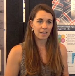
VIDEO: Social networking offers coping help for endometriosis patients
VANCOUVER – Nearly half of the discussion topics on the social networking site www.myendometriosisteam.com are about pain, but other uncontrolled symptoms are popular topics, including fatigue and depression.
The online social support network includes 30,000 women with endometriosis and offers them a chance to connect with other women with the condition, find a provider, and research treatments. Elise-Marie Menke, director of alliance management at MyHealthTeams, which runs the site, presented data at the World Congress on Endometriosis. Among the findings she presented was how symptoms mapped to a woman’s cycle.
In a video interview, Ms. Menke described how this type of patient-generated data can play a role in the management of disease and serve to highlight unmet needs to physicians.
The video associated with this article is no longer available on this site. Please view all of our videos on the MDedge YouTube channel
AT WCE 2017
VIDEO: No cancer risk found from biological DMARDs
MADRID – Additional real-world evidence confirmed that biological disease modifying drugs used to treat rheumatoid arthritis produced no spikes in new cancers or in cancer recurrences in registry data from tens of thousands of Swedish patients.
Among rheumatoid arthritis patients with a history of cancer, patients treated with a tumor necrosis factor inhibitor (TNFi) were not at an increased risk for cancer recurrence, Johan Askling, MD, said at the European Congress of Rheumatology. In a second study, patients with rheumatoid arthritis (RA) treated with a non-TNFi, biological, disease-modifying drug, specifically abatacept, rituximab, or tocilizumab, had no significantly different rate of new cancer onset when compared with RA patients who never received a biological disease modifying drug nor when compared with the general Swedish adult population, said Dr. Askling, a professor of clinical epidemiology at the Karolinska Institute in Stockholm.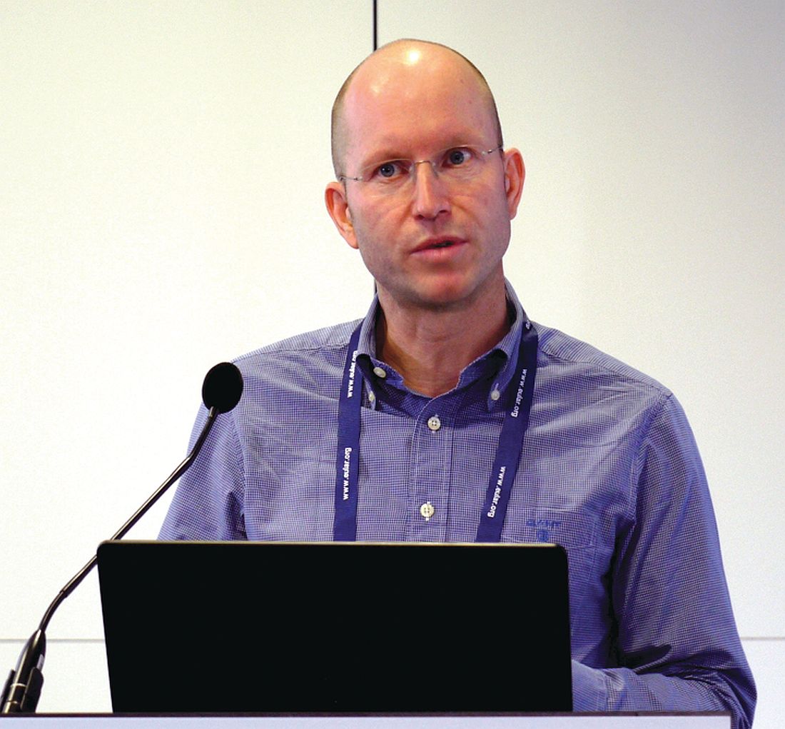
“Five-year data are a good start, but we need data on 30-year risk,” Dr. Askling said in an interview.
The cancer-recurrence risk study with TNFi treatment used data collected by the Swedish national outpatient care registry on nearly 62,000 people, the Swedish cancer registry, and a rheumatology treatment registry called ARTIS. It also included patients treated during 2001-2014. From these sources, the researchers identified 446 RA patients with a history of at least one cancer who then began treatment with any type of TNFi and matched these cases with 1,278 similar RA patients with a cancer history who had never received a biologic drug. On average, the patients were nearly 10 years removed from their initial cancer diagnoses, and the average duration on TNFi treatment was nearly 5 years.
The adjusted hazard ratio for cancer recurrence among the TNFi recipients was reduced by a nominal 30%, compared with that of the controls, a difference that was not statistically significant, Dr. Askling reported.
The second study used data from similar sources for patients treated during 2006-2014 and included nearly 100,000 Swedes from the general population, more than 42,000 RA patients who did not receive a biological drug, more than 14,000 treated with either a first or second TNFi drug, and 1,693 patients treated with tocilizumab (Actemra), 1,894 on abatacept (Orencia), and 3,119 on rituximab (Rituxan).
The rates of new onset cancer in any of these treatment groups, including the patients on tocilizumab, abatacept, or rituximab, was not significantly different from the rate among RA patients who never received a biologic drug, nor from the general Swedish population rate, Dr. Askling said.
This is “one of the first large-scale assessments” of the cancer risk posed by non-TNFi biological drugs, aside from what was reported from the pivotal trials for these drugs, Dr. Askling said.
Dr. Askling has received research support from AbbVie, Lilly, MSD, Pfizer, Roche, and UCB.
mzoler@frontlinemedcom.com
On Twitter @mitchelzoler
Rheumatologists began having concerns about the possible impact of biological drugs on cancer when these types of drugs first became available 20 or more years ago. Registries have allowed us to follow these patients, and, so far, we have consistently seen that the risk for cancer is very low. The major adverse effect from treatment with biological drugs is infection.
The most confirmed finding has been that biologic drugs do not cause new cancers. We have known less about the risk patients with a history of cancer face for recurrence by taking a biological drug. The data on this have so far been scarce. Most guidelines advise that, when patients have had cancer, the possible use of a biologic drug should be the subject of a shared-decision discussion with the patient. The new data reported by Dr. Askling add to the risk information we have available to discuss with patients.
The risk that biologic drugs poses for infections is more complex. The infection risk also depends on a patient’s use of glucocorticoids, their age, and their comorbidities. The infection risk faced by a patient from treatment with a biological drug requires an individualized discussion that takes into account the severity of all the relevant risk factors.
The video associated with this article is no longer available on this site. Please view all of our videos on the MDedge YouTube channel
João E. Fonseca, MD is a professor of rheumatology at the University of Lisbon. He has been a speaker for or has received research funding from Abbvie, MSD, Pfizer, Roche, and UCB. He made these comments in a video interview.
MADRID – Additional real-world evidence confirmed that biological disease modifying drugs used to treat rheumatoid arthritis produced no spikes in new cancers or in cancer recurrences in registry data from tens of thousands of Swedish patients.
Among rheumatoid arthritis patients with a history of cancer, patients treated with a tumor necrosis factor inhibitor (TNFi) were not at an increased risk for cancer recurrence, Johan Askling, MD, said at the European Congress of Rheumatology. In a second study, patients with rheumatoid arthritis (RA) treated with a non-TNFi, biological, disease-modifying drug, specifically abatacept, rituximab, or tocilizumab, had no significantly different rate of new cancer onset when compared with RA patients who never received a biological disease modifying drug nor when compared with the general Swedish adult population, said Dr. Askling, a professor of clinical epidemiology at the Karolinska Institute in Stockholm.
“Five-year data are a good start, but we need data on 30-year risk,” Dr. Askling said in an interview.
The cancer-recurrence risk study with TNFi treatment used data collected by the Swedish national outpatient care registry on nearly 62,000 people, the Swedish cancer registry, and a rheumatology treatment registry called ARTIS. It also included patients treated during 2001-2014. From these sources, the researchers identified 446 RA patients with a history of at least one cancer who then began treatment with any type of TNFi and matched these cases with 1,278 similar RA patients with a cancer history who had never received a biologic drug. On average, the patients were nearly 10 years removed from their initial cancer diagnoses, and the average duration on TNFi treatment was nearly 5 years.
The adjusted hazard ratio for cancer recurrence among the TNFi recipients was reduced by a nominal 30%, compared with that of the controls, a difference that was not statistically significant, Dr. Askling reported.
The second study used data from similar sources for patients treated during 2006-2014 and included nearly 100,000 Swedes from the general population, more than 42,000 RA patients who did not receive a biological drug, more than 14,000 treated with either a first or second TNFi drug, and 1,693 patients treated with tocilizumab (Actemra), 1,894 on abatacept (Orencia), and 3,119 on rituximab (Rituxan).
The rates of new onset cancer in any of these treatment groups, including the patients on tocilizumab, abatacept, or rituximab, was not significantly different from the rate among RA patients who never received a biologic drug, nor from the general Swedish population rate, Dr. Askling said.
This is “one of the first large-scale assessments” of the cancer risk posed by non-TNFi biological drugs, aside from what was reported from the pivotal trials for these drugs, Dr. Askling said.
Dr. Askling has received research support from AbbVie, Lilly, MSD, Pfizer, Roche, and UCB.
mzoler@frontlinemedcom.com
On Twitter @mitchelzoler
AT THE EULAR 2017 CONGRESS
Key clinical point:
Major finding: Former cancer patients on a TNFi had no increased cancer recurrences, compared with patients on other rheumatic treatments.
Data source: Data from the Swedish national registries.
Disclosures: Dr. Askling has received research support from AbbVie, Lilly, MSD, Pfizer, Roche, and UCB.
Mandar Jog, MD
The video associated with this article is no longer available on this site. Please view all of our videos on the MDedge YouTube channel
The video associated with this article is no longer available on this site. Please view all of our videos on the MDedge YouTube channel
The video associated with this article is no longer available on this site. Please view all of our videos on the MDedge YouTube channel

For chronic abdominal pain, THC resembled placebo
Seven weeks of treatment with delta-9-atetrahydrocannabinol (THC) did not improve chronic abdominal pain in a placebo-controlled trial of 65 adults.
Treatment “was safe and well tolerated,” but did not significantly reduce pain scores or secondary efficacy outcomes, Marjan de Vries, MSc, and her associates wrote in the July issue of Clinical Gastroenterology and Hepatology (doi: 10.1016/j.cgh.2016.09.147). Studies have not clearly shown that THC improves central pain sensitization, a key mechanism in chronic abdominal pain, they noted. Future studies of THC and central sensitization include quantitative sensory testing or electroencephalography, they added.
Source: American Gastroenterological Association
Treatment-refractory chronic abdominal pain is common after abdominal surgery or in chronic pancreatitis, wrote Ms. de Vries of Radboud University Medical Center, Nijmegen, the Netherlands. Affected patients tend to develop central sensitization, or hyper-responsive nociceptive central nervous system pathways. When this happens, pain no longer couples reliably with peripheral stimuli, and therapy targeting central nociceptive pathways is indicated. The main psychoactive compound of Cannabis sativa is THC, which interacts with CB1 receptors in the central nervous system, including in areas of the brain that help regulate emotions, such as the amygdala. Emotion-processing circuits are often overactive in chronic pain, and disrupting them might help modify pain perception, the investigators hypothesized. Therefore, they randomly assigned 65 adults with at least 3 months of abdominal pain related to chronic pancreatitis or abdominal surgery to receive oral placebo or THC tablets three times daily for 50-52 days. The 31 patients in the THC group received step-up dosing (3 mg per dose for 5 days, followed by 5 mg per dose for 5 days) followed by stable dosing at 8 mg. Both groups continued other prescribed analgesics as usual, including oxycontin, fentanyl, morphine, codeine, tramadol, paracetamol, anti-epileptics, and nonsteroidal anti-inflammatories. All but two study participants were white, 25 were male, and 24 were female.
At baseline, all patients reported pain of at least 3 on an 11-point visual analogue scale (VAS). By days 50-52, average VAS scores decreased by 1.6 points (40%) in the THC group and by 1.9 points (37%) in the placebo group (P = .9). Although a strong placebo effect is common in studies of visceral pain, that did not prevent pregabalin from significantly outperforming placebo in another similarly designed randomized clinical trial of patients from this study group with chronic pancreatitis, the investigators noted.
The THC and placebo groups also resembled each other on various secondary outcome measures, including patient global impression of change, pain catastrophizing, pain-related anxiety, measures of depression and generalized anxiety, and subjective impressions of alertness, mood, feeling “high,” drowsiness, and difficulties in controlling thoughts. The only exception was that the THC group showed a trend toward improvement on the Short Form 36, compared with the placebo group (P = .051).
Pharmacokinetic analysis showed good oral absorption of THC. Dizziness, somnolence, and headache were common in both groups, but were more frequent with THC than placebo, as was nausea, dry mouth, and visual impairment. There were no serious treatment-related adverse events, although seven patients stopped THC because they could not tolerate the maximum dose.
Some evidence suggests that the shift from acute to chronic pain entails a transition from nociceptive to cognitive, affective, and autonomic sensitization, the researchers noted. “Therefore, an agent targeting particular brain areas related to the cognitive emotional feature of chronic pain, such as THC, might be efficacious in our chronic pain population, but might be better measured by using affective outcomes of pain,” they concluded.
The trial was supported by a grant from the European Union, the European Fund for Regional Development, and the Province of Gelderland. The THC was provided by Echo Pharmaceuticals, Nijmegen, the Netherlands. The investigators reported having no conflicts of interest.
FROM CLINICAL GASTROENTEROLOGY AND HEPATOLOGY
Key clinical point: Tetrahydrocannabinol did not improve chronic abdominal pain more than did placebo.
Major finding: By days 50-52, average VAS scores decreased by 1.6 points (40%) in the THC group and by 1.9 points (37%) in the placebo group (P = .9).
Data source: A phase II, placebo-controlled study of 65 patients with chronic abdominal pain for at least 3 months who received either placebo or delta-9-atetrahydrocannabinol (THC), 8 mg three times daily.
Disclosures: The trial was supported by a grant from the European Union, the European Fund for Regional Development, and the Province of Gelderland. The THC was provided by Echo Pharmaceuticals, Nijmegen, the Netherlands. The investigators reported having no conflicts of interest.
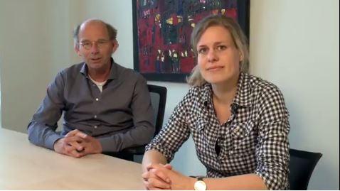
Improved adenoma detection rate found protective against interval cancers, death
An improved annual adenoma detection rate was associated with a significantly decreased risk of interval colorectal cancer (ICRC) and subsequent death in a national prospective cohort study published in the July issue of Gastroenterology (doi: 10.1053/j.gastro.2017.04.006).

Source: American Gastroenterological Association
This is the first study to show a significant inverse relationship between an improved annual adenoma detection rate (ADR) and ICRC or subsequent death, Michal F. Kaminski MD, PhD, of the Institute of Oncology, Warsaw, wrote with his associates.
The rates of these outcomes were lowest when endoscopists achieved and maintained ADRs above 24.6%, which supports the currently recommended performance target of 25% for a mixed male-female population, they reported (Am J Gastroenterol. 2015;110:72-90).
This study included 294 endoscopists and 146,860 individuals who underwent screening colonoscopy as part of a national cancer prevention program in Poland between 2004 and 2008. Endoscopists received annual feedback based on quality benchmarks to spur improvements in colonoscopy performance, and all participated for at least 2 years. For each endoscopist, investigators categorized annual ADRs based on quintiles for the entire data set. “Improved ADR” was defined as keeping annual ADR within the highest quintile (above 24.6%) or as increasing annual ADR by at least one quintile, compared with baseline.
Based on this definition, 219 endoscopists (75%) improved their ADR during a median of 5.8 years of follow-up (interquartile range, 5-7.2 years). In all, 168 interval CRCs were diagnosed, of which 44 cases led to death. After age, sex, and family history of colorectal cancer were controlled for, patients whose endoscopists improved their ADRs were significantly less likely to develop (adjusted hazard ratio, 0.6; 95% confidence interval, 0.5-0.9; P = .006) or to die of interval CRC (95% CI, 0.3-0.95; P = .04) than were patients whose endoscopists did not improve their ADRs.
Maintaining ADR in the highest quintile (above 24.6%) throughout follow-up led to an even lower risk of interval CRC (HR, 0.3; 95% CI, 0.1-0.6; P = .003) and death (HR, 0.2; 95% CI, 0.1-0.6; P = .003), the researchers reported. In absolute numbers, that translated to a decrease from 25.3 interval CRCs per 100,000 person-years of follow-up to 7.1 cases when endoscopists eventually reached the highest ADR quintile or to 4.5 cases when they were in the highest quintile throughout follow-up. Rates of colonic perforation remained stable even though most endoscopists upped their ADRs.
Together, these findings “prove the causal relationship between endoscopists’ ADRs and the likelihood of being diagnosed with, or dying from, interval CRC,” the investigators concluded. The national cancer registry in Poland is thought to miss about 10% of cases, but the rate of missing cases was not thought to change over time, they noted. However, they also lacked data on colonoscope withdrawal times, and had no control group to definitively show that feedback based on benchmarking was responsible for improved ADRs.
Funders included the Foundation of Polish Science, the Innovative Economy Operational Programme, the Polish Foundation of Gastroenterology, the Polish Ministry of Health, and the Polish Ministry of Science and Higher Education. The investigators reported having no relevant conflicts of interest.
The U.S. Multi-Society Task Force on Colorectal Cancer proposed the adenoma detection rate (ADR) as a colonoscopy quality measure in 2002. The rationale for a new measure was emerging evidence of highly variable adenoma detection and cancer prevention among colonoscopists. Highly variable performance, consistently verified in subsequent studies, casts a pall of severe operator dependence over colonoscopy. In landmark studies from Kaminski et al. and Corley et al. in 2010 and 2014, respectively, it was shown that doctors with higher ADRs provide patients with much greater protection against interval colorectal cancer (CRC).

Now Kaminski and colleagues from Poland have delivered a second landmark study, demonstrating for the first time that improving ADR prevents CRCs. We now have strong evidence that ADR predicts the level of cancer prevention, that ADR improvement is achievable, and that improving ADR further prevents CRCs and CRC deaths. Thanks to this study, ADR has come full circle. Measurement of and improvement in detection is now a fully validated concept that is essential to modern colonoscopy. In 2017, ADR measurement is mandatory for all practicing colonoscopists who are serious about CRC prevention. The tools to improve ADR that are widely accepted include ADR measurement and reporting, split or same-day preparations, lesion recognition and optimal technique, high-definition imaging, double examination (particularly for the right colon), patient rotation during withdrawal, chromoendoscopy, mucosal exposure devices (caps, cuffs, balloons, etc.), and water exchange. Tools for ADR improvement that are emerging or under study are brighter forms of electronic chromoendoscopy, and videorecording.
Douglas K. Rex, MD, is professor of medicine, division of gastroenterology/hepatology, at Indiana University, Indianapolis.* He has no relevant conflicts of interest.
Correction, 6/20/17: An earlier version of this article misstated Dr. Rex's affiliation.
The U.S. Multi-Society Task Force on Colorectal Cancer proposed the adenoma detection rate (ADR) as a colonoscopy quality measure in 2002. The rationale for a new measure was emerging evidence of highly variable adenoma detection and cancer prevention among colonoscopists. Highly variable performance, consistently verified in subsequent studies, casts a pall of severe operator dependence over colonoscopy. In landmark studies from Kaminski et al. and Corley et al. in 2010 and 2014, respectively, it was shown that doctors with higher ADRs provide patients with much greater protection against interval colorectal cancer (CRC).
Now Kaminski and colleagues from Poland have delivered a second landmark study, demonstrating for the first time that improving ADR prevents CRCs. We now have strong evidence that ADR predicts the level of cancer prevention, that ADR improvement is achievable, and that improving ADR further prevents CRCs and CRC deaths. Thanks to this study, ADR has come full circle. Measurement of and improvement in detection is now a fully validated concept that is essential to modern colonoscopy. In 2017, ADR measurement is mandatory for all practicing colonoscopists who are serious about CRC prevention. The tools to improve ADR that are widely accepted include ADR measurement and reporting, split or same-day preparations, lesion recognition and optimal technique, high-definition imaging, double examination (particularly for the right colon), patient rotation during withdrawal, chromoendoscopy, mucosal exposure devices (caps, cuffs, balloons, etc.), and water exchange. Tools for ADR improvement that are emerging or under study are brighter forms of electronic chromoendoscopy, and videorecording.
Douglas K. Rex, MD, is professor of medicine, division of gastroenterology/hepatology, at Indiana University, Indianapolis.* He has no relevant conflicts of interest.
Correction, 6/20/17: An earlier version of this article misstated Dr. Rex's affiliation.
An improved annual adenoma detection rate was associated with a significantly decreased risk of interval colorectal cancer (ICRC) and subsequent death in a national prospective cohort study published in the July issue of Gastroenterology (doi: 10.1053/j.gastro.2017.04.006).
Source: American Gastroenterological Association
This is the first study to show a significant inverse relationship between an improved annual adenoma detection rate (ADR) and ICRC or subsequent death, Michal F. Kaminski MD, PhD, of the Institute of Oncology, Warsaw, wrote with his associates.
The rates of these outcomes were lowest when endoscopists achieved and maintained ADRs above 24.6%, which supports the currently recommended performance target of 25% for a mixed male-female population, they reported (Am J Gastroenterol. 2015;110:72-90).
This study included 294 endoscopists and 146,860 individuals who underwent screening colonoscopy as part of a national cancer prevention program in Poland between 2004 and 2008. Endoscopists received annual feedback based on quality benchmarks to spur improvements in colonoscopy performance, and all participated for at least 2 years. For each endoscopist, investigators categorized annual ADRs based on quintiles for the entire data set. “Improved ADR” was defined as keeping annual ADR within the highest quintile (above 24.6%) or as increasing annual ADR by at least one quintile, compared with baseline.
Based on this definition, 219 endoscopists (75%) improved their ADR during a median of 5.8 years of follow-up (interquartile range, 5-7.2 years). In all, 168 interval CRCs were diagnosed, of which 44 cases led to death. After age, sex, and family history of colorectal cancer were controlled for, patients whose endoscopists improved their ADRs were significantly less likely to develop (adjusted hazard ratio, 0.6; 95% confidence interval, 0.5-0.9; P = .006) or to die of interval CRC (95% CI, 0.3-0.95; P = .04) than were patients whose endoscopists did not improve their ADRs.
Maintaining ADR in the highest quintile (above 24.6%) throughout follow-up led to an even lower risk of interval CRC (HR, 0.3; 95% CI, 0.1-0.6; P = .003) and death (HR, 0.2; 95% CI, 0.1-0.6; P = .003), the researchers reported. In absolute numbers, that translated to a decrease from 25.3 interval CRCs per 100,000 person-years of follow-up to 7.1 cases when endoscopists eventually reached the highest ADR quintile or to 4.5 cases when they were in the highest quintile throughout follow-up. Rates of colonic perforation remained stable even though most endoscopists upped their ADRs.
Together, these findings “prove the causal relationship between endoscopists’ ADRs and the likelihood of being diagnosed with, or dying from, interval CRC,” the investigators concluded. The national cancer registry in Poland is thought to miss about 10% of cases, but the rate of missing cases was not thought to change over time, they noted. However, they also lacked data on colonoscope withdrawal times, and had no control group to definitively show that feedback based on benchmarking was responsible for improved ADRs.
Funders included the Foundation of Polish Science, the Innovative Economy Operational Programme, the Polish Foundation of Gastroenterology, the Polish Ministry of Health, and the Polish Ministry of Science and Higher Education. The investigators reported having no relevant conflicts of interest.
FROM GASTROENTEROLOGY
Key clinical point: An improved adenoma detection rate was associated with a significantly reduced risk of interval colorectal cancer and subsequent death.
Major finding: Adjusted hazard ratios were 0.6 for developing ICRC (95% CI, 0.5-0.9; P = .006) and 0.50 for dying of ICRC (95% CI, 0.3-0.95; P = .04).
Data source: A prospective registry study of 294 endoscopists and 146,860 individuals who underwent screening colonoscopy as part of a national screening program between 2004 and 2008.
Disclosures: Funders included the Foundation of Polish Science, the Innovative Economy Operational Programme, the Polish Foundation of Gastroenterology, the Polish Ministry of Health, and the Polish Ministry of Science and Higher Education. The investigators reported having no relevant conflicts of interest.