User login
Micro-focused ultrasound improves appearance of the décolletage
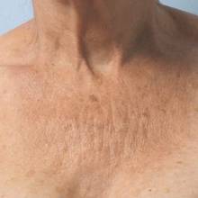
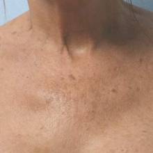
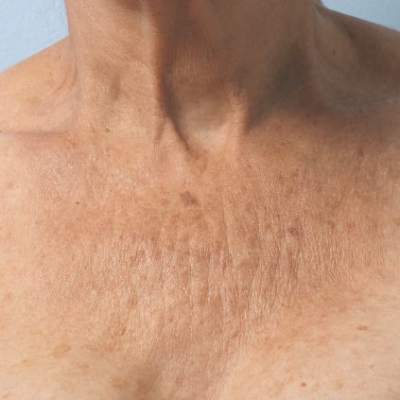
NEW ORLEANS – Micro-focused ultrasound with visualization can effectively treat lines and wrinkles of the décolletage by penetrating deeply enough into the skin to promote tissue restructuring, according to Dr. Sabrina Guillen Fabi.
When treating women with sagging skin, lines, and wrinkles who want to invest in rejuvenating their facial skin, don’t overlook the skin of the décolleté, Dr. Fabi stressed at the annual meeting of the American Academy of Cosmetic Surgery.
Various devices are currently available to combat components of skin aging, including rhytids, but they are only able to treat the superficial layers of skin with wavelengths possessing limited energy penetration capabilities, said Dr. Fabi of Goldman Butterwick Fitzpatrick Groff & Fabi Cosmetic Laser Dermatology in San Diego. Ultrasound, however, can be focused to specific depths, penetrating deep enough to cause thermal coagulation and restructuring of tissue. “Micro-focused ultrasound combines the ability to actually see what you’re treating – similar to how we’ve used ultrasound to visualize babies for over 50 years – with focused ultrasound waves at predetermined depths,” she explained.
To evaluate the effectiveness of using micro-focused ultrasound with visualization (MFU-V) to demonstrate aesthetic improvement in lines and wrinkles of the décolleté, Dr. Fabi and colleagues conducted a prospective multicenter trial of 125 patients with an average age of 57 years.
Based on masked assessments, 70.5% of patients showed some level of improvement at 90 days, and at 180 days it was relatively maintained at 67%. Physician Global Aesthetic Improvement Scale (PGAIS) scores showed that 75.0% of subjects improved at 90 days and 66% at 180 days.
At the 90-day visit, 84% of subjects noted an improvement in their skin and 65.5% were satisfied with their results. Day 180 results showed a similar trend, with 83% of subjects noting improvement and 62.7% satisfied. “Although they reported improvement in their wrinkles, they weren’t satisfied,” noted Dr. Fabi. “Patients with that much wrinkle severity have a lot of other problems on their chest such as redness and brown spots, which need to be addressed separately. So 65% of subjects were satisfied even though 84% reported improvement,” she added.
Each patient received a single MFU-V treatment on the chest using three transducers: 4-4.5 mm (1.2J), 7-3.0 mm (0.45 J), and 10-1.5 mm (0.20 J). All participants were rated as a grade of 4 or higher on the Fabi/Bolton Chest Wrinkle 5-point scale. Grade 4 is characterized by deep rhytids, well-defined lines, and dyschromia; grade 5 is defined by very deep rhytids with redundant folds, also accompanied by dyschromia. “This treatment takes less than 20 minutes with no downtime,” said Dr. Fabi.

Using a validated scale (0-10), subjects reported their average level of discomfort during the treatment, and pain was managed with oral pretreatment medications such as ibuprofen. Photographs were taken prior to treatment, immediately after treatment, and at each follow-up visit (day 90 and day 180).
Efficacy was determined by using both the PGAIS and the Subject Global Aesthetic Improvement Scale, Patient Satisfaction Questionnaire responses, masked assessments of photographs compared with baseline, and the Fabi/Bolton chest wrinkle scale, all tabulated at 90 and 180 days post treatment.
No serious adverse events were reported. The most frequent adverse event was soreness or tenderness, which was experienced by 38% of subjects; 22% showed some level of redness after treatment, but that usually dissipated within about 24 hours, Dr. Fabi noted. The mean pain score at each depth was 6.2 at 4.5 mm, 5.9 at 3.0 mm, and 4.8 at 1.5 mm.
All adverse events were mild except two (1.9%) that were moderate, one of which was not device related. All events were resolved.
Agreement was noted across all measures of efficacy at days 90 and 180, suggesting that post-treatment improvements in lines and wrinkles of the décolleté were clinically significant, she said.
Significant reduction of wrinkles and lines, as well as tightening and toning of the décolleté, was observed at 3 and 6 months after a single treatment using MFU-V; Dr. Fabi and her colleagues are seeking FDA clearance of MFU-V treatment based on these results. “This modality helps patients at least have their décolletage match their face after some sort of facial rejuvenation procedure, whether it be nonsurgical or surgical,” said Dr. Fabi. “Severely wrinkled patients may need additional treatments, but more importantly, I think if you address the dyschromia in addition to the wrinkles, you may have greater patient satisfaction,” she suggested.
Dr. Fabi reported serving as a consultant and advisory board member for device manufacturer Ulthera.
NEW ORLEANS – Micro-focused ultrasound with visualization can effectively treat lines and wrinkles of the décolletage by penetrating deeply enough into the skin to promote tissue restructuring, according to Dr. Sabrina Guillen Fabi.
When treating women with sagging skin, lines, and wrinkles who want to invest in rejuvenating their facial skin, don’t overlook the skin of the décolleté, Dr. Fabi stressed at the annual meeting of the American Academy of Cosmetic Surgery.
Various devices are currently available to combat components of skin aging, including rhytids, but they are only able to treat the superficial layers of skin with wavelengths possessing limited energy penetration capabilities, said Dr. Fabi of Goldman Butterwick Fitzpatrick Groff & Fabi Cosmetic Laser Dermatology in San Diego. Ultrasound, however, can be focused to specific depths, penetrating deep enough to cause thermal coagulation and restructuring of tissue. “Micro-focused ultrasound combines the ability to actually see what you’re treating – similar to how we’ve used ultrasound to visualize babies for over 50 years – with focused ultrasound waves at predetermined depths,” she explained.
To evaluate the effectiveness of using micro-focused ultrasound with visualization (MFU-V) to demonstrate aesthetic improvement in lines and wrinkles of the décolleté, Dr. Fabi and colleagues conducted a prospective multicenter trial of 125 patients with an average age of 57 years.
Based on masked assessments, 70.5% of patients showed some level of improvement at 90 days, and at 180 days it was relatively maintained at 67%. Physician Global Aesthetic Improvement Scale (PGAIS) scores showed that 75.0% of subjects improved at 90 days and 66% at 180 days.
At the 90-day visit, 84% of subjects noted an improvement in their skin and 65.5% were satisfied with their results. Day 180 results showed a similar trend, with 83% of subjects noting improvement and 62.7% satisfied. “Although they reported improvement in their wrinkles, they weren’t satisfied,” noted Dr. Fabi. “Patients with that much wrinkle severity have a lot of other problems on their chest such as redness and brown spots, which need to be addressed separately. So 65% of subjects were satisfied even though 84% reported improvement,” she added.
Each patient received a single MFU-V treatment on the chest using three transducers: 4-4.5 mm (1.2J), 7-3.0 mm (0.45 J), and 10-1.5 mm (0.20 J). All participants were rated as a grade of 4 or higher on the Fabi/Bolton Chest Wrinkle 5-point scale. Grade 4 is characterized by deep rhytids, well-defined lines, and dyschromia; grade 5 is defined by very deep rhytids with redundant folds, also accompanied by dyschromia. “This treatment takes less than 20 minutes with no downtime,” said Dr. Fabi.
Using a validated scale (0-10), subjects reported their average level of discomfort during the treatment, and pain was managed with oral pretreatment medications such as ibuprofen. Photographs were taken prior to treatment, immediately after treatment, and at each follow-up visit (day 90 and day 180).
Efficacy was determined by using both the PGAIS and the Subject Global Aesthetic Improvement Scale, Patient Satisfaction Questionnaire responses, masked assessments of photographs compared with baseline, and the Fabi/Bolton chest wrinkle scale, all tabulated at 90 and 180 days post treatment.
No serious adverse events were reported. The most frequent adverse event was soreness or tenderness, which was experienced by 38% of subjects; 22% showed some level of redness after treatment, but that usually dissipated within about 24 hours, Dr. Fabi noted. The mean pain score at each depth was 6.2 at 4.5 mm, 5.9 at 3.0 mm, and 4.8 at 1.5 mm.
All adverse events were mild except two (1.9%) that were moderate, one of which was not device related. All events were resolved.
Agreement was noted across all measures of efficacy at days 90 and 180, suggesting that post-treatment improvements in lines and wrinkles of the décolleté were clinically significant, she said.
Significant reduction of wrinkles and lines, as well as tightening and toning of the décolleté, was observed at 3 and 6 months after a single treatment using MFU-V; Dr. Fabi and her colleagues are seeking FDA clearance of MFU-V treatment based on these results. “This modality helps patients at least have their décolletage match their face after some sort of facial rejuvenation procedure, whether it be nonsurgical or surgical,” said Dr. Fabi. “Severely wrinkled patients may need additional treatments, but more importantly, I think if you address the dyschromia in addition to the wrinkles, you may have greater patient satisfaction,” she suggested.
Dr. Fabi reported serving as a consultant and advisory board member for device manufacturer Ulthera.
AT THE AACS ANNUAL MEETING
Kaempferol
Kaempferol (3,5,7,4’-tetrahydroxyflavone; C15H10O6) is among the natural flavonols found in green tea, broccoli, cabbage, kale, endive, beans, leeks, tomatoes, grapes, apples, grapefruit, berries, and propolis, as well as myriad other plant sources, including Brassica and species (J. Agric. Food Chem. 2006;54:2951-6; Cancer Prev. Res. (Phila) 2014;7:958-67; Biochem. Pharmacol. 2010;80:2042-9; Chem. Pharm. Bull. (Tokyo) 2012;60:1171-5; J. Eur. Acad. Dermatol. Venereol. 2013 June 27 [doi:10.1111/jdv.12204]).
It is one of the most commonly found dietary flavonoids and is also present in beer, particularly hops (Carcinogenesis 2010;31:1338-43; J. Eur. Acad. Dermatol. Venereol. 2013 June 27 [doi:10.1111/jdv.12204]). Significantly, kaempferol is known to exhibit anticancer, anti-inflammatory, antioxidant, cytoprotective, and antiapoptotic activity (Cancer Prev. Res. (Phila) 2014;7:958-67; Biochem. Pharmacol. 2010;80:2042-9; Exp. Mol. Med. 2008;40:208-19), and is believed to play a role in protecting plants from ultraviolet (UV)-induced damage (J. Agric. Food Chem. 2012;60:6966-76).
Skin protection: antioxidant and anti-inflammatory activity
Among 35 flavonoids tested by Cos et al. in 2001 for lipid peroxidation-inhibiting activity, kaempferol was identified as having the highest antioxidant selectivity index (Planta Med. 2001;67:515-9).
Work by Kim et al. in 2002 revealed that four kaempferol glycosides are key active ingredients in the flowers of Prunus persica, which has long been used in traditional Chinese medicine to treat skin disorders (J. Cosmet. Sci. 2002;53:27-34). Kim and colleagues have also shown in animal studies that the topical application of P. persica may be effective at thwarting UVB-induced skin damage (J. Cosmet. Sci. 2002;53:27-34).
In addition, kaempferol is a key component in Punica granatum, which has been found to act as an effective protector against UVB-induced photodamage and aging in cultured skin fibroblasts (Int. J. Dermatol. 2010;49:276-82).
In various tests on the effects of natural flavonoids on matrix metalloproteinase (MMP)-1 activity and expression, Lim et al. reported in 2007 that kaempferol and quercetin potently inhibited recombinant human MMP-1, and both flavonols along with apigenin and wogonin were found to be strong inhibitors of MMP-1 induction in 12-O-tetradecanoylphorbol-13-acetate–treated human dermal fibroblasts. All four flavonoids also suppressed the activation of activator protein (AP)-1. Kaempferol also hindered p38 mitogen-activated protein kinase c-Jun N-terminal kinase (JNK) activation. The investigators concluded that kaempferol is among the flavonoids or plant extracts containing them that may be useful as an agent to protect against photoaging and to treat some cutaneous inflammatory conditions (Planta Med. 2007;73:1267-74).
In 2010, Park et al. demonstrated that kaempferol alleviated burn injuries in mice and that expression of tumor necrosis factor–alpha (TNF-alpha) induced by burn injuries was reduced by kaempferol. They concluded that their findings suggest the possible application of kaempferol to treat thermal burn–induced skin injuries (BMB Rep. 2010;43:46-51).
Anti-inflammatory as well as depigmenting activity was found by Rho et al. in 2011 to be associated with kaempferol and kaempferol rhamnosides isolated from Hibiscus cannabinus (Molecules 2011;16:3338-44).
In 2014, Kim et al. found that extracts of Aceriphyllum rossii (native to Korea and China) and its active constituents, quercetin and kaempferol, blocked secretion of beta-hexosaminidase and histamine; lowered the production and mRNA expression of interleukin-4 and TNF-alpha; and reduced prostaglandin E2 and leukotriene B4 synthesis as well as the expression of cyclooxygenase-2 (COX-2) and 5-lipoxygenase. These and other findings led the investigators to conclude that A. rossii and its active ingredients kaempferol and quercetin may be effective agents for the treatment of immediate-type hypersensitivity (J. Agric. Food Chem. 2014;62:3750-8).
Anticancer activity
Lee et al. reported in 2010 that the inhibition by kaempferol of phosphatidylinositol 3-kinase (PI3K) activity, a key factor in carcinogenesis, and its concomitant effects may account for the chemopreventive activity of the flavonol (Carcinogenesis 2010;31:1338-43).
At the end of that year, Lee et al. found that kaempferol inhibited UVB-induced COX-2 protein expression in mouse skin epidermal JB6 P+ cells, by blocking Src kinase activity and attenuated the UVB-induced transcriptional activities of COX-2 gene and the transcription factor AP-1. They concluded that kaempferol exerts robust chemopreventive activity against skin cancer by suppressing Src (Biochem. Pharmacol. 2010;80:2042-9).
In 2014, Yao et al. found that kaempferol acted as a safe and potent inhibitor of solar ultraviolet-induced mouse skin carcinogenesis that acted by targeting RSK2 and MSK1 (Cancer Prev. Res. (Phila) 2014;7:958-67).
Significantly, in terms of topical delivery, Chao et al. recently showed that submicron emulsions are effective carriers for the transdermal delivery of kaempferol (Chem. Pharm. Bull. (Tokyo) 2012;60:1171-5).
Conclusion
Kaempferol is one among the many natural flavonols found to exert significant salutary effects. Evidence suggests reasons for confidence that kaempferol can play a role in skin health. More research is necessary to determine the effectiveness of topical products intended to harness the benefits of this flavonoid as proper formulation is challenging.
Dr. Baumann is chief executive officer of the Baumann Cosmetic & Research Institute in the Design District in Miami. She founded the Cosmetic Dermatology Center at the University of Miami in 1997. Dr. Baumann wrote the textbook, “Cosmetic Dermatology: Principles and Practice” (New York: McGraw-Hill, 2002), and a book for consumers, “The Skin Type Solution” (New York: Bantam Dell, 2006). She has contributed to the Cosmeceutical Critique column in Dermatology News since January 2001. Her latest book, “Cosmeceuticals and Cosmetic Ingredients,” was published in November 2014. Dr. Baumann has received funding for clinical grants from Allergan, Aveeno, Avon Products, Evolus, Galderma, GlaxoSmithKline, Kythera, Mary Kay, Medicis Pharmaceuticals, Neutrogena, Philosophy, Topix Pharmaceuticals, and Unilever.

Manage Your Dermatology Practice: Managing Difficult Patient Encounters
Difficult patient encounters in the dermatology office can be navigated through honest physician-patient communication regarding problems within the office and insurance coverage. Dr. Gary Goldenberg provides tips on communicating with patients about cosmetic procedures that may be noncovered services as well as diagnoses such as melanoma and psoriasis. He also advises how to work through a long list of questions patients may bring to their visit.
Difficult patient encounters in the dermatology office can be navigated through honest physician-patient communication regarding problems within the office and insurance coverage. Dr. Gary Goldenberg provides tips on communicating with patients about cosmetic procedures that may be noncovered services as well as diagnoses such as melanoma and psoriasis. He also advises how to work through a long list of questions patients may bring to their visit.
Difficult patient encounters in the dermatology office can be navigated through honest physician-patient communication regarding problems within the office and insurance coverage. Dr. Gary Goldenberg provides tips on communicating with patients about cosmetic procedures that may be noncovered services as well as diagnoses such as melanoma and psoriasis. He also advises how to work through a long list of questions patients may bring to their visit.

Hormones loom large in female pseudofolliculitis barbae
Pseudofolliculitis barbae, a disorder better known as razor bumps, is poorly studied in women, but may have a different etiology than in men and could be indicative of underlying hormonal comorbidities, based on data from 62 men and 62 women.
T.A. Nguyen, a medical student at Montefiore Medical Center – Albert Einstein College of Medicine, New York, and colleagues sought to characterize pseudofolliculitis barbae (PFB) in women by reviewing patient files from men and women presenting with unspecified acne or diseases of hair and hair follicles. They collected information on patients’ age, laboratory findings, treatments, and comorbidities. The findings were published in the British Journal of Dermatology (2015 [doi:10.1111/bjd.13644]) .
Overall, the researchers found that only differences in hormonal disorders, such as polycystic ovarian syndrome (PCOS), hirsutism, infertility, or hypo/hypertestosteronism, and acne were statistically different between men and women.
“These results point toward hyperandrogenism, a common cause of hirsutism, acne, and virilization, as a potential mitigating factor for PFB in women,” the researchers wrote, adding that the higher prevalence of hormonal disorders in the women’s cohort “demonstrates the importance of considering hormonal influences when working-up a female patient with PFB.”
Of the 62 women, 5 had known PCOS at diagnosis, and 4 were later diagnosed. Two women saw improvement of PFB severity after starting oral contraceptives or antiandrogen agents under the care of endocrinologists.
“It is possible that these agents may provide some benefit for a subset of patients,” the researchers wrote. A quarter of the female patients in the study were treated with eflornithine hydrochloride, which inhibits hair cell growth, while fewer than 2% of males received this treatment. The investigators noted that few women were treated with oral contraceptive pills, despite their use as a first-line strategy for acne in women.
“Our study demonstrates that female patients with PFB are empirically different from their male counterparts. In women with PFB, acne and hormonal disorders are significantly more common. Despite this fact, antiandrogen agents were not considered for treatment. Further studies are needed to evaluate the use of these agents in women with PFB and to identify optimal clinical work-up and treatment algorithms,” the researchers noted.
Dr. Adam Friedman, who was the lead author, disclosed financial relationships with Onset, GSK, Galderma, Amgen, L’Oreal, Johnson & Johnson, MicroCures, Liquidia, Salvona, and Valent.
Patient-led teledermoscopy appears feasible and effective
Patient-administered teledermoscopy using an iPhone-based mobile dermatoscope attachment and app is an effective and feasible method for short-term monitoring of clinically atypical nevi, with the added benefit of improving patient and physician convenience, based on data from a pilot study of 29 patients.
Researchers found a high level of diagnostic concordance (0.87) between dermatoscope images taken and assessed by an office-based dermatologist and those taken by the patient – albeit in the clinic setting – using the mobile dermatoscope and assessed by a teledermatologist.
All but one of the 29 patients with clinically atypical nevi who completed the study were able to acquire evaluable baseline and follow-up images, the researchers noted. In addition, most of the patients reported that the device was easy to use and that it saved them a trip to the doctor’s office. The study findings were published online Jan. 28 in JAMA Dermatology (doi:10.1001/jamadermatol.2014.3837).
“Under our modality of care, patients needing short-term monitoring will have an established relationship with their dermatologists, who will be the ones identifying concerning lesions that need to be monitored and the ones who evaluate the lesions via teledermoscopy and communicate treatment options directly with the patients,” wrote Xinyuan Wu of Memorial Sloan Kettering Cancer Center, New York, and colleagues.
The authors of an accompanying editorial wrote that recommendations for screening and follow-up for melanoma placed considerable burdens on patients, physicians, and the health care system, and that the patient-led mobile teledermoscopy described in the study was one of a number of options being considered to reduce that burden.
“The study by Wu and colleagues in this issue adds significantly to the discussion on whether regular follow-up visits with clinicians could be replaced by patient self-monitoring with remote feedback by a teledermatologist,” wrote Monika Janda, Ph.D., of the Queensland University of Technology in Brisbane, Australia, and colleagues.
One editorial author reported shares and consultancies with e-derm-consult GmbH and MoleMap, but there were no other conflicts of interest declared.
FROM JAMA DERMATOLOGY
Key clinical point: Patient-administered teledermoscopy using an iPhone-based mobile dermatoscope attachment and app is an effective and feasible method for short-term monitoring of clinically atypical nevi.
Major finding: Researchers found a high level of diagnostic concordance (0.87) between dermatoscope images taken and assessed by the office-based dermatologist and those taken by the patient using an iPhone.
Data source:A prospective cohort study in 34 patients – 29 of whom completed follow-up – with clinically atypical nevi.
Disclosures: One editorial author reported shares and consultancies with e-derm-consult GmbH and MoleMap. No other conflicts of interest were declared.
Consider 3-week treatment intervals for laser tattoo removal
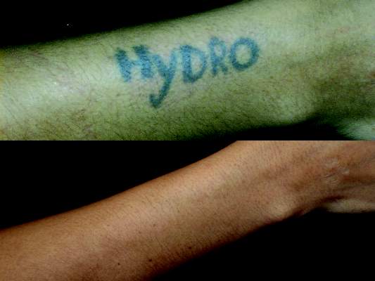
NEW ORLEANS – Laser tattoo removal using a 3-week interval between treatments may be feasible for most patients, based on data from a pilot study conducted by Dr. Robert Burke.
The demand for tattoo removal has increased along with tattoo prevalence in the United States, Dr. Burke said at the annual meeting of the American Academy of Cosmetic Surgery. The treatment protocol for laser tattoo removal depends on the patient’s skin type, as well as on the type and density of inks used, said Dr. Burke, founder and director of the Michigan Center for Cosmetic Surgery in Ann Arbor.

Some methods have been tested to accelerate the laser tattoo removal process, but have resulted in skin damage such as scarring and hypopigmentation, he added.
No standard recommendation currently exists for time intervals between laser tattoo removal treatments, Dr. Burke said. Intervals of 3, 4, 6, and 8 weeks or longer have been recommended. “It has been speculated that 3-week intervals can interfere with macrophage activity, but long-term studies have shown no clinical or other interference whether you treat at 3, 4, 6, or 8 weeks,” he said.
Previous recommendations were based mainly on tissue reactions observed with older lasers; this thermal damage was likely because of laser beam irregularities and nonuniform energy application in round beam lasers, he added.
Laser tattoo removal involves selective photothermolysis, Dr. Burke noted, and the laser light can destroy the target without damaging the surrounding tissue only if the thermal relaxation time (defined as the time necessary for the target to cool 50% through diffusion of heat to surrounding tissues) is greater than the pulse duration. “Fluence sufficient to reach a damaging temperature in target chromophores without damaging surrounding tissue structures is the critical element of selective photothermolysis,” he explained.
The study involved the treatment of nine tattoos on seven adults (two women and five men) with an average age of 33 years. The tattoos were of all treatable colors including black, blue-green, and red-yellow-orange.

Dr. Burke used a laser with three wavelengths that included Q-switched Nd:YAG and true ruby lasers. The laser’s square beam with a uniform power distribution is designed to decrease thermal damage to the uninvolved surrounding tissue. “I looked specifically into doing this pilot study after I saw this new laser,” he said. Laser beam selection was based on the major tattoo colors observed on the day of treatment, and wavelengths of 1,064-nm, 694-nm, and 532-nm with a consistent 3-mm spot size were used. Dark colors were treated first, followed by blue-green. Red was treated in the same session as the other colors only if the red areas were well defined, separate, and distinct in location; otherwise they were treated in subsequent sessions. The patients underwent an average of 3.5 treatments, with an average of 22 days between each treatment. The majority of patients responded favorably to the 3-week treatment interval, and there were no significant adverse effects. The tattoos continued to clear throughout the study period, and all patients reported satisfaction with their treatments, although the tattoos had not completely cleared by the end of the study period, Dr. Burke said.
One minor, self-limiting, increased healing interval occurred in a patient with a red tattoo treated with a 532-nm wavelength. “Interestingly enough, in this case, the red tattoo cleared completely after one treatment,” he added. A 3-week interval between treatments could result in more rapid tattoo removal, shortening the overall treatment time by 63%, compared with the existing average treatment time, Dr. Burke said. The treatment interval may be adjusted upward if skin requires a longer healing time following a particular treatment, he added, although larger, statistically valid studies are needed to support the results.
Dr. Burke reported no relevant financial disclosures.
NEW ORLEANS – Laser tattoo removal using a 3-week interval between treatments may be feasible for most patients, based on data from a pilot study conducted by Dr. Robert Burke.
The demand for tattoo removal has increased along with tattoo prevalence in the United States, Dr. Burke said at the annual meeting of the American Academy of Cosmetic Surgery. The treatment protocol for laser tattoo removal depends on the patient’s skin type, as well as on the type and density of inks used, said Dr. Burke, founder and director of the Michigan Center for Cosmetic Surgery in Ann Arbor.
Some methods have been tested to accelerate the laser tattoo removal process, but have resulted in skin damage such as scarring and hypopigmentation, he added.
No standard recommendation currently exists for time intervals between laser tattoo removal treatments, Dr. Burke said. Intervals of 3, 4, 6, and 8 weeks or longer have been recommended. “It has been speculated that 3-week intervals can interfere with macrophage activity, but long-term studies have shown no clinical or other interference whether you treat at 3, 4, 6, or 8 weeks,” he said.
Previous recommendations were based mainly on tissue reactions observed with older lasers; this thermal damage was likely because of laser beam irregularities and nonuniform energy application in round beam lasers, he added.
Laser tattoo removal involves selective photothermolysis, Dr. Burke noted, and the laser light can destroy the target without damaging the surrounding tissue only if the thermal relaxation time (defined as the time necessary for the target to cool 50% through diffusion of heat to surrounding tissues) is greater than the pulse duration. “Fluence sufficient to reach a damaging temperature in target chromophores without damaging surrounding tissue structures is the critical element of selective photothermolysis,” he explained.
The study involved the treatment of nine tattoos on seven adults (two women and five men) with an average age of 33 years. The tattoos were of all treatable colors including black, blue-green, and red-yellow-orange.
Dr. Burke used a laser with three wavelengths that included Q-switched Nd:YAG and true ruby lasers. The laser’s square beam with a uniform power distribution is designed to decrease thermal damage to the uninvolved surrounding tissue. “I looked specifically into doing this pilot study after I saw this new laser,” he said. Laser beam selection was based on the major tattoo colors observed on the day of treatment, and wavelengths of 1,064-nm, 694-nm, and 532-nm with a consistent 3-mm spot size were used. Dark colors were treated first, followed by blue-green. Red was treated in the same session as the other colors only if the red areas were well defined, separate, and distinct in location; otherwise they were treated in subsequent sessions. The patients underwent an average of 3.5 treatments, with an average of 22 days between each treatment. The majority of patients responded favorably to the 3-week treatment interval, and there were no significant adverse effects. The tattoos continued to clear throughout the study period, and all patients reported satisfaction with their treatments, although the tattoos had not completely cleared by the end of the study period, Dr. Burke said.
One minor, self-limiting, increased healing interval occurred in a patient with a red tattoo treated with a 532-nm wavelength. “Interestingly enough, in this case, the red tattoo cleared completely after one treatment,” he added. A 3-week interval between treatments could result in more rapid tattoo removal, shortening the overall treatment time by 63%, compared with the existing average treatment time, Dr. Burke said. The treatment interval may be adjusted upward if skin requires a longer healing time following a particular treatment, he added, although larger, statistically valid studies are needed to support the results.
Dr. Burke reported no relevant financial disclosures.
AT THE AACS ANNUAL MEETING
Teledermoscopy referrals surpass paper for managing skin cancer patients
Smartphone teledermoscopy referrals were faster and allowed for more efficient management of patients with skin cancer, compared with paper referrals, according to Dr. Alexander Börve of the University of Gothenburg, Sweden, and his associates.
The waiting time was significantly shorter using teledermoscopy for patients with various melanomas and carcinomas when surgical treatment was necessary. “Triage decisions were also more reliable with teledermoscopy, and over 40% of the teledermoscopy patients could potentially have avoided face-to-face visits,” the researchers noted (Acta. Derm. Venereol. 2015;95:186-90).
Less than 1% of teledermoscopy referrals were excluded because of poor image quality, they said.
Read the full article at Acta Dermato-Venereologica (doi:10.2340/00015555-1906).
Lobular keloids effectively treated by fistulectomy
Fistulectomy is an effective surgical approach for treating keloids on the earlobe, according to Dr. Zhenghua Zhu and associates.
Fistulectomies were sucessfully performed on 11 earlobe keloids in 9 patients over 4 years. All of the patients were followed for at least 12 months without recurrence, the researchers reported.
Read the full article at Dermatologic Surgery (doi: 10.1097/DSS.0000000000000214).
Botulinum toxin safe and effective treatment for rosacea-induced facial erythema
Intradermal injection of botulinum toxin was an effective and safe method of treating facial erythema of rosacea, according to Dr. Bradley Bloom, of the Laser & Skin Surgery Center of New York, and his associates.
Of 15 patients, the mean baseline erythema grade was 1.8, and the mean erythema grade at 3 months after treatment was 1. The treatment resulted in statistically significant improvement in erythema at 1, 2, and 3 months after treatment when compared with baseline, according to the researchers. The patients were of Fitzpatrick skin Types I to III with a mean age of 54 years, and 80% were women
Because of the promising results, further and more extensive trials are recommended, but further investigation is needed to elucidate the mechanism of action by which botulinum toxin improves facial flushing of rosacea, the researchers said.
Read the full article at Dermatologic Surgery (doi: 10.1097/DSS.0000000000000277).
Combination MSU-AFL treatment effective at treating skin aging
The combination of microfocused ultrasound and ablative fractionated laser is a safe and effective means of targeting facial and neck skin aging, according to Dr. Julie Woodward and her associates.
There was significant improvement in skin laxity and photodamage after MSU-AFL treatment, and except for increased facial swelling in a small number of patients, post-op recovery and side effects were similar to those obtained from individual treatments, judging from the findings of a retrospective analysis of 100 combination face and neck treatments from 3 centers, according to the researchers.
The MSU-AFL treatment can be safely performed in a single session, the researchers reported.
Read the full article at Dermatologic Surgery (doi: 10.1097/DSS.0000000000000228).