User login
Von Willebrand disease screening is low prior to hysterectomy
The number of women being screened for von Willebrand disease (VWD) before undergoing hysterectomy for heavy menstrual bleeding is “very low” and appears not to align with current recommendations, according to a retrospective analysis.
“One essential reason to identify females with VWD prior to hysterectomy or hysterectomy alternative is to avoid peri‐ and postoperative complications,” Amanda E. Jacobson‐Kelly, MD, of Ohio State University in Columbus and her colleagues wrote in a letter to the editor published in Haemophilia.
Current recommendations from the American College of Obstetricians and Gynecologists and other groups call for consideration of underlying bleeding disorders in women with heavy menstrual bleeding and additional risks factors, such as family history of abnormal bleeding.
In this study, researchers retrospectively analyzed 8,998 women under the age of 40 years who had a hysterectomy for heavy menstrual bleeding without a known bleeding disorder. Patient data was collected from a national insurance claims database representative of the entire U.S. population.
“We hypothesized that the overall frequency of VWD screening would be low, despite [more than] 15 years of expert recommendations,” the team wrote.
Study participants were included if they had a diagnosis of anemia or heavy menstrual bleeding before surgery, while women with certain genital malignancies and fibroids were excluded. Other information, such as age, geographical factors, and insurance-related factors were also included to evaluate whether these parameters affected disease screening.
After analysis, the researchers found that just 0.6% (n = 57) of women were screened for VWD in the 12 months leading up to surgery. Additionally, they reported that 14% (n = 1,276) of women underwent other forms of coagulation testing before surgery, such as partial thromboplastin time and prothrombin time.
“We found that greater distance to a [hemophilia treatment center] was strongly predictive of decreased likelihood of undergoing VWD screening,” Dr. Jacobson‐Kelly and her colleagues wrote.
The authors acknowledged that a key limitation of the study was the inability to account for factors not recorded in the database, including drug use, family history of bleeding disorders, and ethnicity.
“This study brings to light the need for the hematology community to improve education and awareness among women’s health providers,” they wrote.
The study was supported by grant funding from the National Heart, Lung, and Blood Institute. The authors reported having no conflicts of interest.
SOURCE: Jacobson-Kelly AE et al. Haemophilia. 2019 Feb 28. doi: 10.1111/hae.13709.
FROM HAEMOPHILIA
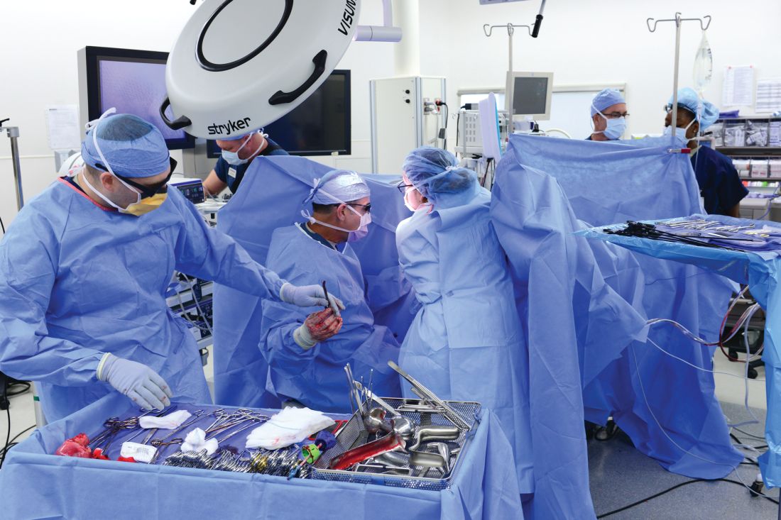
Minilaparotomy: Minimally invasive approach to abdominal myomectomy
A minilaparotomy is loosely defined as a laparotomy measuring between 4 cm and 6 cm. For the appropriate surgical candidate, a minilaparotomy is a useful alternative to laparotomy or laparoscopy, especially for large pathology.1 Benefits of minilaparotomy include improved pain management and postoperative recovery, as well as improved cosmetic outcome, with comparable blood loss and operative time.2,3

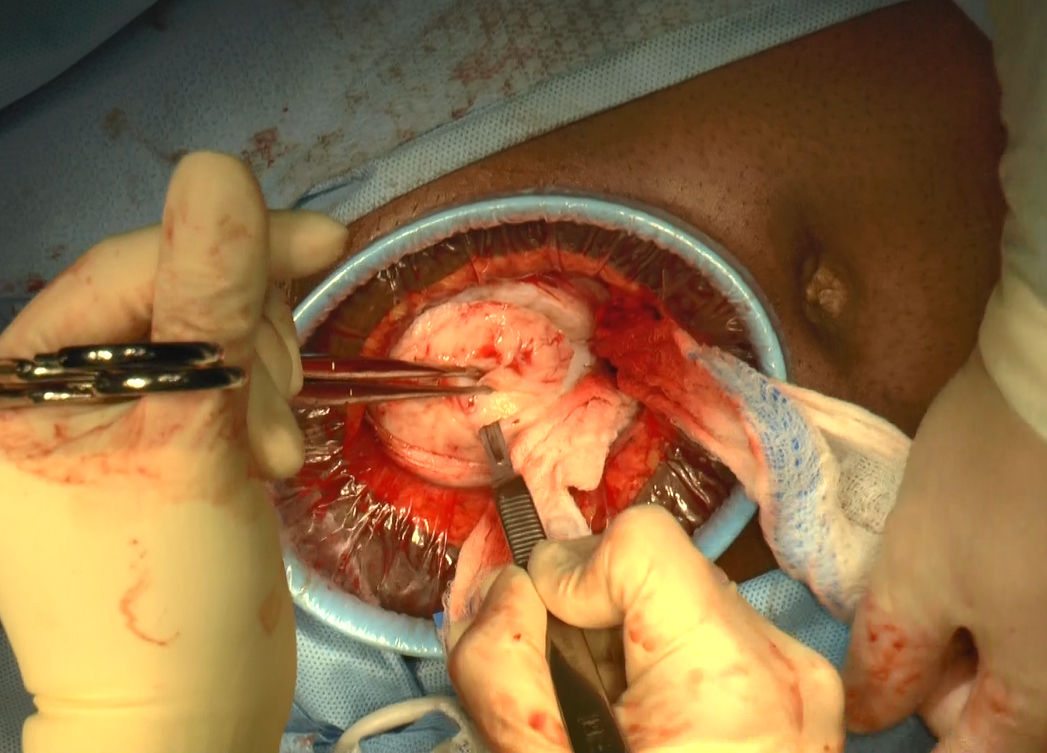
In this video, we illustrate the key surgical steps of a minilaparotomy for the removal of large fibroids. These steps include:
- strategic vertical skin incision
- use of a self-retaining retractor
- infiltrate myometrium with dilute vasopressin
- strategic hysterotomy
- use of tenaculum for upward traction
- 10# blade scalpels for the “lemon wedge” coring technique
- layered closure.
Minilaparotomy myomectomy can be an excellent minimally invasive alternative to a traditional “full laparotomy” for women with large fibroids.
We hope that you find this video beneficial to your clinical practice.
>> Arnold P. Advincula, MD
Laparoscopic bilateral salpingo-oophorectomy via minilaparotomy assistance for the massively enlarged adnexal mass
- Pelosi MA 2nd, Pelosi MA 3rd. Pelosi minilaparotomy hysterectomy: a non-endoscopic minimally invasive alternative to laparoscopy and laparotomy. Surg Technol Int. 2004;13:157-167.
- Fanafani F, Fagotti A, Longo R. Minilaparotomy in the management of benign gynecologic disease. Eur J Obstet Gynecol Reprod Biol. 2005;119:232-236.
- Glasser MH. Minilaparotomy: a minimally invasive alternative for major gynecologic abdominal surgery. Perm J. 2005;9:41-45.
A minilaparotomy is loosely defined as a laparotomy measuring between 4 cm and 6 cm. For the appropriate surgical candidate, a minilaparotomy is a useful alternative to laparotomy or laparoscopy, especially for large pathology.1 Benefits of minilaparotomy include improved pain management and postoperative recovery, as well as improved cosmetic outcome, with comparable blood loss and operative time.2,3
In this video, we illustrate the key surgical steps of a minilaparotomy for the removal of large fibroids. These steps include:
- strategic vertical skin incision
- use of a self-retaining retractor
- infiltrate myometrium with dilute vasopressin
- strategic hysterotomy
- use of tenaculum for upward traction
- 10# blade scalpels for the “lemon wedge” coring technique
- layered closure.
Minilaparotomy myomectomy can be an excellent minimally invasive alternative to a traditional “full laparotomy” for women with large fibroids.
We hope that you find this video beneficial to your clinical practice.
>> Arnold P. Advincula, MD
Laparoscopic bilateral salpingo-oophorectomy via minilaparotomy assistance for the massively enlarged adnexal mass
- Pelosi MA 2nd, Pelosi MA 3rd. Pelosi minilaparotomy hysterectomy: a non-endoscopic minimally invasive alternative to laparoscopy and laparotomy. Surg Technol Int. 2004;13:157-167.
- Fanafani F, Fagotti A, Longo R. Minilaparotomy in the management of benign gynecologic disease. Eur J Obstet Gynecol Reprod Biol. 2005;119:232-236.
- Glasser MH. Minilaparotomy: a minimally invasive alternative for major gynecologic abdominal surgery. Perm J. 2005;9:41-45.
Mucinous ovarian tumor survival rates stress correct diagnosis
Women with invasive, well-differentiated mucinous ovarian cancer are more likely to die from their disease within 10 years of diagnosis than women with mucinous borderline ovarian tumors, according to a retrospective study of more than 2,700 cases.
The analysis also revealed different clinical characteristics, reported lead author Koji Matsuo, MD, PhD of the University of Southern California, Los Angeles, and his colleagues. For example, compared with borderline ovarian tumors (BOT), patients with ovarian cancer (OC) were usually older and had undergone hysterectomy.
“Our study endorses the importance of making the proper diagnosis for invasive cancer when the ovarian tumor is of mucinous histology,” the investigators wrote in Gynecologic Oncology.
Using Surveillance, Epidemiology, and End Results data from 1988-2000, the analysis included 581 cases of stage I, invasive, well-differentiated mucinous OC and 2,130 cases of stage I mucinous BOT. The investigators noted “histological misclassification of BOT as OC or OC as BOT is not uncommon,” because of similar histopathologic characteristics.
Multivariable analysis showed that, compared with cases of BOT, women with OC were more often from the eastern United States (22.0% vs. 13.6%), older (51.9 vs. 48.0 years), and had a history of lymphadenectomy (47.0% vs. 23.2%) or hysterectomy (64.4% vs. 35.8%). Women with OC were also more likely to have tumors smaller than 4 cm (12.9% vs. 8.9%) and stage T1c disease (15.7% vs. 7.3%). Rates of OC declined over time, from 34.7% during 1988-1991 to 22.0% during 1997-2000. Following propensity score matching, multivariable analysis showed that 10-year cause-specific survival rates for OC and BOT were 92.7% and 97.5%, respectively, giving OC a hazard ratio of 2.03 (P = .007). Overall survival showed a similar disparity, of 76.1% for OC, compared with 83.6% for BOT. The investigators concluded that “survival of these two diseases is completely different.”
Regarding histopathologic misclassification, the investigators proposed “a standardized specimen sectioning protocol and diagnostic criteria for mucinous ovarian tumors.”
The study was funded by Ensign Endowment for Gynecologic Cancer Research. The investigators reported financial relationships with KIYATEC, BioPath, M-Trap, and Chugai.
SOURCE: Matsuo K et al. Gynecol Oncol. 2019 Feb 20. doi: 10.1016/j.ygyno.2019.02.003.
FROM GYNECOLOGIC ONCOLOGY
Antipsychotics show no link to increased risk of major congenital malformations
LAS VEGAS – Assessing the risk of major congenital malformations related to antipsychotic exposure requires detailed assessment of other sources of risk, including those related to the diagnosis and associated behaviors, according to Jonathan M. Meyer, MD.

However, the largest study to date showed no significant difference in rates of major congenital malformations for those with one or more prescriptions for atypical antipsychotics in the first trimester, compared with pregnancies with no first trimester antipsychotic exposure.
In the U.S. general population, the estimated risk of major birth defects is 2%-4%, Dr. Meyer, a clinical professor of psychiatry at the University of California, San Diego, said at an annual psychopharmacology update held by the Nevada Psychiatric Association.
Medications represent one source of risk for major congenital malformations in patients with psychiatric illness. Other factors include lifestyle factors such substance abuse and smoking, diet and physical activity, adherence with medical/prenatal care regimens, general medical disease burden, and unknown genetic risk because of the illness itself.
Until recently, published studies examining antipsychotic exposure and the risk for congenital malformations have been flawed because of numerous factors, Dr. Meyer said, including the small sample size of live births, absence of systematic collection of risk data prior to and during pregnancy, and failure to examine all possible covariates that might moderate the risk potentially attributable to the medication itself.
For example, researchers led by Frank Habermann, PhD, prospectively evaluated three cohorts who were followed in a psychiatry consultation in Freiburg, Germany: 453 women who received atypical antipsychotics in the first trimester of pregnancy (group A); 238 women who received typical antipsychotics in the first trimester of pregnancy (group B); and 1,104 women who had no records of treatment with medications associated with harmful fetal effects (group C).
Covariates included maternal age, alcohol consumption, smoking habits, number of previous spontaneous abortions, number of previous malformed children, and gestational week at delivery (J Clin Psychopharmacol. 2013;33[4]:453-62).
The researchers found that 5.2% of women in group A gave birth to a child with a major congenital malformation, compared with 5% in group B and 2.5% in group C. Nonsignificant associations were observed between group A vs. B (adjusted odds ratio, 1.26; 95% confidence interval, 0.57-2.82) and group B vs. C (adjusted OR, 1.71; 95% CI, 0.78-3.76). The only significant association noted was between group A and C (adjusted OR, 2.17; 95% CI, 1.20-3.91). However, Dr. Meyer emphasized limitations of the study, including its small sample size and certain missing covariates, including illegal substance use.
In addition, since subjects were enrolled in a consultation clinic, surveillance bias might have detected a higher number of CV malformations. “I don’t have a lot of confidence in this study because there were enormous sources of risk that were not controlled for,” said Dr. Meyer, who is also a psychopharmacology consultant for the California State Department of Hospitals.
In a separate study, researchers led by Lee S. Cohen, MD, assessed data from 487 women in the National Pregnancy Registry for Atypical Antipsychotics based at the Massachusetts General Hospital Center for Women’s Health, Boston. Of the 487 women, 353 were on atypical antipsychotics and 134 served as controls (Am J Psychiatry. 2016;73[3]:263-70). Medical records were obtained at baseline, month 7, and postpartum for 82% of subjects, which left 303 women in the final analysis. Covariates included demographic characteristics, medication use and dosage changes, social habits including smoking, use of alcohol and illicit drugs, medical and psychiatric history, and family history of birth defects.
Of 214 live births with first-trimester exposure to atypical antipsychotics, three major malformations were confirmed, while among the control group of 89 women, one major malformation was confirmed. The absolute risk of major malformations was 1.4% for exposed infants and 1.1% for unexposed infants. Meanwhile, the OR for major malformations comparing exposed infants was 1.25, which did not reach statistical significance (95% CI, 0.13-12.19). Limitations of the study include the small sample size of live births and the low overall rate of malformations, said Dr. Meyer. “This group of women living in the Boston area might not be representative of the general population based on the extremely low rates of congenital malformations for both cohorts in this study,” he commented.
In what Dr. Meyer said was the most robust study of its kind to date, researchers led by Krista F. Huybrechts, MS, PhD, drew from Medicaid data from 2000-2010 and included only women who were enrolled from 3 months before their last menstrual period through at least 1 month after delivery (JAMA Psychiatry. 2016;73[9]:938-46). The sample consisted of 1,341,715 pregnancies. Among those pregnancies, 9,258 women filled at least one prescription for an atypical antipsychotic, and 733 filled at least one prescription for a typical antipsychotic, for a total of 9,991 pregnancies. The researchers used propensity score matching to match for risk of antipsychotic exposure. They also balanced the antipsychotic-exposed and nonexposed groups for covariates that might be related to the outcome of interest (major congenital malformations), including (but not limited to) calendar year, age, race, smoking history, multiple gestation, indications for antipsychotic use, other maternal morbidity, concomitant medication use, and general markers of illness burden in the 3 months prior to pregnancy.
The atypical antipsychotics used included quetiapine (n = 4,221), followed by aripiprazole (n = 1,756), risperidone (n = 1,566), olanzapine (n = 1,394), and ziprasidone (n = 697). The absolute risks for congenital malformations per 1,000 live-born infants was 38.2 (95% CI, 26.6-54.7) for those treated with typicals and 44.5 (95% CI, 40.5-48.9) for those treated with atypicals versus 32.7 (95% CI, 32.4-33.0) for untreated women. In the fully adjusted analysis, the risk ratio was not statistically different for those exposed to atypical antipsychotics, compared with the control group, for malformations overall (relative risk, 1.05; 95% CI, 0.96-1.16) nor for cardiac malformations (RR, 1.06; 95% CI, 0.90-1.24). However, the risk remained elevated for risperidone for overall malformations (RR, 1.26; 1.02-1.56) and cardiac malformations (RR, 1.26; 95% CI, 0.88-1.81).
When Dr. Huybrechts and her colleagues redefined exposure as having filled two more prescriptions or having at least a 1-day supply in the first trimester, the results did not meaningfully change. However, the association appeared to strengthen somewhat for risperidone when filling two or more prescriptions (RR, 1.46 for any malformation; 95% CI, 1.01-2.10; RR, 1.87 for cardiac malformations; 95% CI, 1.09-3.19). The researchers observed no evidence of a dose-response relationship for any of the individual antipsychotics except risperidone. Risperidone dosages of 2 mg/day or more were associated with an increased risk for cardiac malformation (RR, 2.08; 95% CI, 1.32-3.28).
“The small increase in absolute risk and RR for malformations observed with risperidone should be interpreted with caution because no apparent biological mechanism can readily explain this outcome and the possibility of a chance finding cannot be ruled out,” the authors wrote in their study. “This finding should therefore be interpreted as a potential safety signal that will require follow-up in other studies.”
If the finding in this study is replicated, Dr. Meyer said, a number needed to harm analysis suggests that compared with no antipsychotic use. “Given the risks of bad outcomes that might occur related to medication switching or nonadherence, the risk-benefit ratio may tilt towards continuing risperidone, especially when a long-acting injectable [LAI] antipsychotic is needed in someone who responds to risperidone and either doesn’t respond to or tolerate the medications available in other LAI preparations such aripiprazole, haloperidol, or fluphenazine,” he said. “This need to balance the risks of exacerbating the mental disorder and the extremely small chance of an adverse pregnancy outcome is part of a clinical discussion one should have with the patient and her family.”
Dr. Meyer reported having received speaking or advising fees from Acadia, Alkermes, Allergan, Intracellular Therapies, Merck, Neurocrine, Otsuka America, Sunovion, and Teva.
LAS VEGAS – Assessing the risk of major congenital malformations related to antipsychotic exposure requires detailed assessment of other sources of risk, including those related to the diagnosis and associated behaviors, according to Jonathan M. Meyer, MD.
However, the largest study to date showed no significant difference in rates of major congenital malformations for those with one or more prescriptions for atypical antipsychotics in the first trimester, compared with pregnancies with no first trimester antipsychotic exposure.
In the U.S. general population, the estimated risk of major birth defects is 2%-4%, Dr. Meyer, a clinical professor of psychiatry at the University of California, San Diego, said at an annual psychopharmacology update held by the Nevada Psychiatric Association.
Medications represent one source of risk for major congenital malformations in patients with psychiatric illness. Other factors include lifestyle factors such substance abuse and smoking, diet and physical activity, adherence with medical/prenatal care regimens, general medical disease burden, and unknown genetic risk because of the illness itself.
Until recently, published studies examining antipsychotic exposure and the risk for congenital malformations have been flawed because of numerous factors, Dr. Meyer said, including the small sample size of live births, absence of systematic collection of risk data prior to and during pregnancy, and failure to examine all possible covariates that might moderate the risk potentially attributable to the medication itself.
For example, researchers led by Frank Habermann, PhD, prospectively evaluated three cohorts who were followed in a psychiatry consultation in Freiburg, Germany: 453 women who received atypical antipsychotics in the first trimester of pregnancy (group A); 238 women who received typical antipsychotics in the first trimester of pregnancy (group B); and 1,104 women who had no records of treatment with medications associated with harmful fetal effects (group C).
Covariates included maternal age, alcohol consumption, smoking habits, number of previous spontaneous abortions, number of previous malformed children, and gestational week at delivery (J Clin Psychopharmacol. 2013;33[4]:453-62).
The researchers found that 5.2% of women in group A gave birth to a child with a major congenital malformation, compared with 5% in group B and 2.5% in group C. Nonsignificant associations were observed between group A vs. B (adjusted odds ratio, 1.26; 95% confidence interval, 0.57-2.82) and group B vs. C (adjusted OR, 1.71; 95% CI, 0.78-3.76). The only significant association noted was between group A and C (adjusted OR, 2.17; 95% CI, 1.20-3.91). However, Dr. Meyer emphasized limitations of the study, including its small sample size and certain missing covariates, including illegal substance use.
In addition, since subjects were enrolled in a consultation clinic, surveillance bias might have detected a higher number of CV malformations. “I don’t have a lot of confidence in this study because there were enormous sources of risk that were not controlled for,” said Dr. Meyer, who is also a psychopharmacology consultant for the California State Department of Hospitals.
In a separate study, researchers led by Lee S. Cohen, MD, assessed data from 487 women in the National Pregnancy Registry for Atypical Antipsychotics based at the Massachusetts General Hospital Center for Women’s Health, Boston. Of the 487 women, 353 were on atypical antipsychotics and 134 served as controls (Am J Psychiatry. 2016;73[3]:263-70). Medical records were obtained at baseline, month 7, and postpartum for 82% of subjects, which left 303 women in the final analysis. Covariates included demographic characteristics, medication use and dosage changes, social habits including smoking, use of alcohol and illicit drugs, medical and psychiatric history, and family history of birth defects.
Of 214 live births with first-trimester exposure to atypical antipsychotics, three major malformations were confirmed, while among the control group of 89 women, one major malformation was confirmed. The absolute risk of major malformations was 1.4% for exposed infants and 1.1% for unexposed infants. Meanwhile, the OR for major malformations comparing exposed infants was 1.25, which did not reach statistical significance (95% CI, 0.13-12.19). Limitations of the study include the small sample size of live births and the low overall rate of malformations, said Dr. Meyer. “This group of women living in the Boston area might not be representative of the general population based on the extremely low rates of congenital malformations for both cohorts in this study,” he commented.
In what Dr. Meyer said was the most robust study of its kind to date, researchers led by Krista F. Huybrechts, MS, PhD, drew from Medicaid data from 2000-2010 and included only women who were enrolled from 3 months before their last menstrual period through at least 1 month after delivery (JAMA Psychiatry. 2016;73[9]:938-46). The sample consisted of 1,341,715 pregnancies. Among those pregnancies, 9,258 women filled at least one prescription for an atypical antipsychotic, and 733 filled at least one prescription for a typical antipsychotic, for a total of 9,991 pregnancies. The researchers used propensity score matching to match for risk of antipsychotic exposure. They also balanced the antipsychotic-exposed and nonexposed groups for covariates that might be related to the outcome of interest (major congenital malformations), including (but not limited to) calendar year, age, race, smoking history, multiple gestation, indications for antipsychotic use, other maternal morbidity, concomitant medication use, and general markers of illness burden in the 3 months prior to pregnancy.
The atypical antipsychotics used included quetiapine (n = 4,221), followed by aripiprazole (n = 1,756), risperidone (n = 1,566), olanzapine (n = 1,394), and ziprasidone (n = 697). The absolute risks for congenital malformations per 1,000 live-born infants was 38.2 (95% CI, 26.6-54.7) for those treated with typicals and 44.5 (95% CI, 40.5-48.9) for those treated with atypicals versus 32.7 (95% CI, 32.4-33.0) for untreated women. In the fully adjusted analysis, the risk ratio was not statistically different for those exposed to atypical antipsychotics, compared with the control group, for malformations overall (relative risk, 1.05; 95% CI, 0.96-1.16) nor for cardiac malformations (RR, 1.06; 95% CI, 0.90-1.24). However, the risk remained elevated for risperidone for overall malformations (RR, 1.26; 1.02-1.56) and cardiac malformations (RR, 1.26; 95% CI, 0.88-1.81).
When Dr. Huybrechts and her colleagues redefined exposure as having filled two more prescriptions or having at least a 1-day supply in the first trimester, the results did not meaningfully change. However, the association appeared to strengthen somewhat for risperidone when filling two or more prescriptions (RR, 1.46 for any malformation; 95% CI, 1.01-2.10; RR, 1.87 for cardiac malformations; 95% CI, 1.09-3.19). The researchers observed no evidence of a dose-response relationship for any of the individual antipsychotics except risperidone. Risperidone dosages of 2 mg/day or more were associated with an increased risk for cardiac malformation (RR, 2.08; 95% CI, 1.32-3.28).
“The small increase in absolute risk and RR for malformations observed with risperidone should be interpreted with caution because no apparent biological mechanism can readily explain this outcome and the possibility of a chance finding cannot be ruled out,” the authors wrote in their study. “This finding should therefore be interpreted as a potential safety signal that will require follow-up in other studies.”
If the finding in this study is replicated, Dr. Meyer said, a number needed to harm analysis suggests that compared with no antipsychotic use. “Given the risks of bad outcomes that might occur related to medication switching or nonadherence, the risk-benefit ratio may tilt towards continuing risperidone, especially when a long-acting injectable [LAI] antipsychotic is needed in someone who responds to risperidone and either doesn’t respond to or tolerate the medications available in other LAI preparations such aripiprazole, haloperidol, or fluphenazine,” he said. “This need to balance the risks of exacerbating the mental disorder and the extremely small chance of an adverse pregnancy outcome is part of a clinical discussion one should have with the patient and her family.”
Dr. Meyer reported having received speaking or advising fees from Acadia, Alkermes, Allergan, Intracellular Therapies, Merck, Neurocrine, Otsuka America, Sunovion, and Teva.
EXPERT ANALYSIS FROM NPA 2019
FDA panel tackles mesh for anterior repair of POP
Patient-reported outcomes should be the priority consideration for determining whether the three synthetic mesh devices currently available for transvaginal repair of pelvic organ prolapse (POP) in the anterior vaginal compartment should remain on the market, according to the Food and Drug Administration Obstetrics and Gynecology Devices panel.

The panel was convened in February 2019 to advise the Food and Drug Administration on how it should evaluate the safety and effectiveness of the three currently marketed devices – each of which has ongoing postmarket surveillance studies – as well as any other similar devices that come up for premarket approval in the future.
The panel’s main messages: Subjective outcomes are what really matter – even more so than anatomic or objective outcomes – as does long-term follow-up.
“ “compared to native tissue repair,” said panel chair Keith Isaacson, MD, medical director of the Newton-Wellesley Hospital in Newton, Mass. “But we feel that, if we had to score [each category of outcome], about 75% should be subjective.”
The three devices currently marketed for transvaginal repair of POP (Boston Scientific’s Uphold LITE and Xenform, as well as Coloplast’s Restorelle DirectFix Anterior) are being scrutinized under a new regulatory paradigm and amid a charged backdrop of safety warnings and years of lawsuits regarding debilitating complications following surgeries that involved the implantation of synthetic vaginal mesh.
The two manufacturers of the currently available devices launched postmarket surveillance studies, called 522 studies, after the FDA issued postmarket surveillance study orders in 2012 to all manufacturers of surgical mesh for transvaginal repair of POP. (Most companies chose at the time to pull their products from the market.) This FDA action, along with a reclassification of the devices from class II to the high-risk class III, had been recommended at a 2011 meeting of the Obstetrics and Gynecology Devices panel.
In anticipation of a future reclassification, the 522 studies were designed at the time to support future premarket approval (PMA) applications, as advised by the FDA. Now, as a result of the 2016 reclassification of surgical mesh for transvaginal POP repair to class III – and the companies’ subsequent PMA applications – the FDA is reviewing the ongoing postmarket study results with a PMA lens to determine each device’s benefit/risk profile.
It’s a challenging assessment to make, FDA officials said.
The agency reported to the panel that a search of medical device reports from 2008 to 2018 identified 11,274 adverse events associated with mesh placed in the anterior vaginal compartment to treat POP. These included 10,391 reports of serious injury, 806 reports of device malfunctions, and 77 reports of death.
Findings from an FDA literature review covering the same period and also focusing on anterior and/or apical repair show that synthetic mesh may have some advantage over native tissue repair for objective effectiveness outcomes – but not necessarily subjective outcomes – over 1-3 years of follow-up. And the risks of using mesh are greater, particularly with respect to reoperation for recurrence and mesh complications, the latter of which continued beyond the first year of follow-up and through 5 years, the agency said.
Although the review may help the FDA frame its questions moving forward, it has limited utility beyond that, according to urogynecologic surgeons who testified on behalf of three professional societies. The review does not delineate differences between the newer materials used today and older mesh materials that were of heavier weight/higher mesh density and often placed using more invasive delivery systems. Nor does it offer any insight on the use of mesh for secondary repair.
“Much of the existing data on the use of transvaginal mesh in POP surgery comes from low to moderate quality, short-term studies of synthetic mesh that is no longer used in clinical practice,” said Cheryl Iglesia, MD, a Washington-based ob.gyn. who spoke to the advisory panel on behalf of the American College of Obstetricians and Gynecologists. “There’s a critical need for data from high-quality studies on the use of the newer, lightweight type 1 transvaginal meshes used in POP surgery.”
The FDA’s 522 orders requested that manufacturers conduct a randomized, controlled study or parallel cohort study comparing their device to native tissue repair. Requested effectiveness endpoints included anatomic success, subjective success, and retreatment for prolapse. For safety endpoints, the agency requested all device- and procedure-related adverse events, as well as the rate of individual adverse events, such as mesh erosion and de novo dyspareunia and urinary dysfunction. The FDA asked for all endpoints at 6-month intervals out to 24 months and at 36 months.
The panel advised that superiority should be the standard for the general population of women with POP – that mesh used in the anterior/apical vaginal compartment should be shown to be superior to native tissue repair at each time point. In specific patient populations for whom native tissue repair is not deemed feasible or appropriate, demonstrating equivalence is sufficient, they advised.
They called for “more diligent” presurgical assessments of sexual function and activity, as well as other symptoms that will be assessed later. And the panel agreed with the FDA that concomitant procedures (for example, hysterectomy and sling placement) and certain preexisting medical conditions and patient characteristics (such as obesity and diabetes) can affect outcomes and should be delineated and considered in the FDA’s evaluations and interpretation of study results.
Regarding surgeon characteristics, the panel’s biostatisticians and physicians (largely urogynecologists, but also one community ob.gyn.) advised the FDA to pay attention to surgeon training, experience, and volume, but they declined to offer any specific recommendations. Discussions often came back to the value of a registry that would capture both surgeon data and patient experience. And throughout the panel’s discussion, surgeons stepped away from the main questions at hand and emphasized the individualized nature of risk-benefit ratios and decision making.
Registries have been successfully used for cardiology and orthopedic implants and, within obstetrics and gynecology, for assisted reproductive technologies, Dr. Iglesia said in an interview after the meeting. “We have models … we just need to make it easy for physicians, using our EMRs. But I’m hopeful.”
The American Urogynecologic Society (AUGS) operates a quality improvement registry (AQUIRE) that is collecting information on surgical and nonsurgical treatment of POP and stress urinary incontinence – including surgical complications – from a diverse group of physicians, not just those at academic medical centers. AUGS is growing its registry this year to include device identifiers and patient-reported outcomes that are sent directly to the registry by the patient.
The panel generally agreed that postmarket follow-up of synthetic mesh for transvaginal anterior repair of POP should extend up to 5 years, Dr. Isaacson said, though “from the patients’ perspective, 10 years of experience [is meaningful].”
Geoffrey Cundiff, MD, who is AUGS president, told the committee that there are lessons to be gleaned from the CARE trial, which looked at outcomes up to 7 years after abdominal sacrocolpopexy (JAMA. 2013 May 15;309[19]:2016-24). “At 7 years, the complications [including rates of mesh erosion] had increased,” he said. “It’s a different procedure, but it’s a good example.”
Prior to its deliberations, the panel heard preliminary results of the ongoing SUPeR trial (Female Pelvic Med Reconstr Surg. 2016 Jul-Aug;22[4]:182-9), a randomized, controlled superiority trial of vaginal hysterectomy with suture apical suspension versus uterine conservation with vaginal mesh (Boston Scientific’s Uphold LITE) hysteropexy for uterovaginal prolapse. Researchers have found comparable rates of primary outcome success – no objective prolapse beyond the hymen, no retreatment, and no bulge symptoms – through 36 months and no differences in patient-reported outcomes thus far.
Hysteroplexy mesh exposure rates were approximately 8% at 36 months, and suture exposure and excessive granulation were 11%-20% in the hysterectomy group. None of these exposure cases has required reoperation. Both groups have shown improvements in sexual function and decreases in dyspareunia, said Charles W. Nager, MD, a San Diego ob.gyn. who is primary investigator of the trial.
The trial is sponsored by the Pelvic Floor Disorders Network of the National Institute of Child Health and Human Development, as was the CARE trial of abdominal sacrocolpopexy. It is following patients for 60 months and collecting data every 6 months, including data from validated functional and quality of life assessments. Patients were masked to their treatment assignment to eliminate patient reporting bias. At 36 months, approximately three-quarters of the patients in each group remained masked.
In addition to the ongoing 522 studies for anterior/apical prolapse, there is another 522 study underway of a mesh device designed for transvaginal repair of total prolapse (the Acell Matristem Pelvic Floor Repair Matrix). In addition, Coloplast is studying a mesh device designed for posterior/apical prolapse (Restorelle DirectFix) as part of its 522 study. Neither device is being marketed currently, however.
Patient-reported outcomes should be the priority consideration for determining whether the three synthetic mesh devices currently available for transvaginal repair of pelvic organ prolapse (POP) in the anterior vaginal compartment should remain on the market, according to the Food and Drug Administration Obstetrics and Gynecology Devices panel.
The panel was convened in February 2019 to advise the Food and Drug Administration on how it should evaluate the safety and effectiveness of the three currently marketed devices – each of which has ongoing postmarket surveillance studies – as well as any other similar devices that come up for premarket approval in the future.
The panel’s main messages: Subjective outcomes are what really matter – even more so than anatomic or objective outcomes – as does long-term follow-up.
“ “compared to native tissue repair,” said panel chair Keith Isaacson, MD, medical director of the Newton-Wellesley Hospital in Newton, Mass. “But we feel that, if we had to score [each category of outcome], about 75% should be subjective.”
The three devices currently marketed for transvaginal repair of POP (Boston Scientific’s Uphold LITE and Xenform, as well as Coloplast’s Restorelle DirectFix Anterior) are being scrutinized under a new regulatory paradigm and amid a charged backdrop of safety warnings and years of lawsuits regarding debilitating complications following surgeries that involved the implantation of synthetic vaginal mesh.
The two manufacturers of the currently available devices launched postmarket surveillance studies, called 522 studies, after the FDA issued postmarket surveillance study orders in 2012 to all manufacturers of surgical mesh for transvaginal repair of POP. (Most companies chose at the time to pull their products from the market.) This FDA action, along with a reclassification of the devices from class II to the high-risk class III, had been recommended at a 2011 meeting of the Obstetrics and Gynecology Devices panel.
In anticipation of a future reclassification, the 522 studies were designed at the time to support future premarket approval (PMA) applications, as advised by the FDA. Now, as a result of the 2016 reclassification of surgical mesh for transvaginal POP repair to class III – and the companies’ subsequent PMA applications – the FDA is reviewing the ongoing postmarket study results with a PMA lens to determine each device’s benefit/risk profile.
It’s a challenging assessment to make, FDA officials said.
The agency reported to the panel that a search of medical device reports from 2008 to 2018 identified 11,274 adverse events associated with mesh placed in the anterior vaginal compartment to treat POP. These included 10,391 reports of serious injury, 806 reports of device malfunctions, and 77 reports of death.
Findings from an FDA literature review covering the same period and also focusing on anterior and/or apical repair show that synthetic mesh may have some advantage over native tissue repair for objective effectiveness outcomes – but not necessarily subjective outcomes – over 1-3 years of follow-up. And the risks of using mesh are greater, particularly with respect to reoperation for recurrence and mesh complications, the latter of which continued beyond the first year of follow-up and through 5 years, the agency said.
Although the review may help the FDA frame its questions moving forward, it has limited utility beyond that, according to urogynecologic surgeons who testified on behalf of three professional societies. The review does not delineate differences between the newer materials used today and older mesh materials that were of heavier weight/higher mesh density and often placed using more invasive delivery systems. Nor does it offer any insight on the use of mesh for secondary repair.
“Much of the existing data on the use of transvaginal mesh in POP surgery comes from low to moderate quality, short-term studies of synthetic mesh that is no longer used in clinical practice,” said Cheryl Iglesia, MD, a Washington-based ob.gyn. who spoke to the advisory panel on behalf of the American College of Obstetricians and Gynecologists. “There’s a critical need for data from high-quality studies on the use of the newer, lightweight type 1 transvaginal meshes used in POP surgery.”
The FDA’s 522 orders requested that manufacturers conduct a randomized, controlled study or parallel cohort study comparing their device to native tissue repair. Requested effectiveness endpoints included anatomic success, subjective success, and retreatment for prolapse. For safety endpoints, the agency requested all device- and procedure-related adverse events, as well as the rate of individual adverse events, such as mesh erosion and de novo dyspareunia and urinary dysfunction. The FDA asked for all endpoints at 6-month intervals out to 24 months and at 36 months.
The panel advised that superiority should be the standard for the general population of women with POP – that mesh used in the anterior/apical vaginal compartment should be shown to be superior to native tissue repair at each time point. In specific patient populations for whom native tissue repair is not deemed feasible or appropriate, demonstrating equivalence is sufficient, they advised.
They called for “more diligent” presurgical assessments of sexual function and activity, as well as other symptoms that will be assessed later. And the panel agreed with the FDA that concomitant procedures (for example, hysterectomy and sling placement) and certain preexisting medical conditions and patient characteristics (such as obesity and diabetes) can affect outcomes and should be delineated and considered in the FDA’s evaluations and interpretation of study results.
Regarding surgeon characteristics, the panel’s biostatisticians and physicians (largely urogynecologists, but also one community ob.gyn.) advised the FDA to pay attention to surgeon training, experience, and volume, but they declined to offer any specific recommendations. Discussions often came back to the value of a registry that would capture both surgeon data and patient experience. And throughout the panel’s discussion, surgeons stepped away from the main questions at hand and emphasized the individualized nature of risk-benefit ratios and decision making.
Registries have been successfully used for cardiology and orthopedic implants and, within obstetrics and gynecology, for assisted reproductive technologies, Dr. Iglesia said in an interview after the meeting. “We have models … we just need to make it easy for physicians, using our EMRs. But I’m hopeful.”
The American Urogynecologic Society (AUGS) operates a quality improvement registry (AQUIRE) that is collecting information on surgical and nonsurgical treatment of POP and stress urinary incontinence – including surgical complications – from a diverse group of physicians, not just those at academic medical centers. AUGS is growing its registry this year to include device identifiers and patient-reported outcomes that are sent directly to the registry by the patient.
The panel generally agreed that postmarket follow-up of synthetic mesh for transvaginal anterior repair of POP should extend up to 5 years, Dr. Isaacson said, though “from the patients’ perspective, 10 years of experience [is meaningful].”
Geoffrey Cundiff, MD, who is AUGS president, told the committee that there are lessons to be gleaned from the CARE trial, which looked at outcomes up to 7 years after abdominal sacrocolpopexy (JAMA. 2013 May 15;309[19]:2016-24). “At 7 years, the complications [including rates of mesh erosion] had increased,” he said. “It’s a different procedure, but it’s a good example.”
Prior to its deliberations, the panel heard preliminary results of the ongoing SUPeR trial (Female Pelvic Med Reconstr Surg. 2016 Jul-Aug;22[4]:182-9), a randomized, controlled superiority trial of vaginal hysterectomy with suture apical suspension versus uterine conservation with vaginal mesh (Boston Scientific’s Uphold LITE) hysteropexy for uterovaginal prolapse. Researchers have found comparable rates of primary outcome success – no objective prolapse beyond the hymen, no retreatment, and no bulge symptoms – through 36 months and no differences in patient-reported outcomes thus far.
Hysteroplexy mesh exposure rates were approximately 8% at 36 months, and suture exposure and excessive granulation were 11%-20% in the hysterectomy group. None of these exposure cases has required reoperation. Both groups have shown improvements in sexual function and decreases in dyspareunia, said Charles W. Nager, MD, a San Diego ob.gyn. who is primary investigator of the trial.
The trial is sponsored by the Pelvic Floor Disorders Network of the National Institute of Child Health and Human Development, as was the CARE trial of abdominal sacrocolpopexy. It is following patients for 60 months and collecting data every 6 months, including data from validated functional and quality of life assessments. Patients were masked to their treatment assignment to eliminate patient reporting bias. At 36 months, approximately three-quarters of the patients in each group remained masked.
In addition to the ongoing 522 studies for anterior/apical prolapse, there is another 522 study underway of a mesh device designed for transvaginal repair of total prolapse (the Acell Matristem Pelvic Floor Repair Matrix). In addition, Coloplast is studying a mesh device designed for posterior/apical prolapse (Restorelle DirectFix) as part of its 522 study. Neither device is being marketed currently, however.
Tranexamic acid shows improvements in heavy menstrual bleeding
new research suggests.
Writing in the Journal of Pediatric & Adolescent Gynecology, Sarah H. O’Brien, MD, from Nationwide Children’s Hospital and the Ohio State University, both in Columbus, and her coauthors presented the results of an open-label efficacy study of the competitive plasminogen inhibitor in 25 adolescent girls aged 10-19 years who attended pediatric hematology clinics for evaluation or management of heavy menstrual bleeding. The study participants were instructed to take 1,300 mg of tranexamic acid (two tablets) three times a day for up to 5 days during their monthly menstruation for three cycles.
The study found a significant improvement in mean menstrual impact questionnaire (MIQ) scores, which improved from a mean of 3 at baseline to 1.91 (P less than .001). Two-thirds of patients reported at least a one-point improvement from baseline, and all reported that this was clinically meaningful. At baseline, 84% of patients reported heavy to very heavy blood loss, but this decreased to 23% after treatment with tranexamic acid (P less than .001).
The study population included ten individuals (40%) with bleeding disorders. However, the researchers did not see a significant difference in response between those with bleeding disorders and those without.
While the treatment did not significantly affect school attendance (only 24% reported that their heavy bleeding limited school attendance), researchers did see a significant improvement in limitations on physical activities and on social and leisure activities. Patients who reported at baseline that their menstrual bleeding significantly affected their social and leisure activities had an average score improvement of 1.74, a greater than or equal to one point improvement. Participants also reported significant improvements in their Pictorial Blood Assessment Chart scores, which dropped from an average of 255 to 155 (P less than .001).
The treatment did not show any significant effects on hemoglobin or ferritin. The most common adverse events were sinonasal symptoms, such as nasal congestion, headache, and sinus pain, but no thrombotic or ocular adverse events were seen.
Dr. O’Brien and her coauthors wrote that one limitation of their study was using the MIQ score as their primary endpoint as opposed to a more objective measure, such as change in measured blood loss.
“However, a major factor that motivates patients with heavy menstrual bleeding to seek medical care is the negative impact of heavy menstrual bleeding on daily life,” they wrote.
The study drug was supplied by Ferring pharmaceuticals, and the study was supported by the Hemostasis and Thrombosis Research Society. One author disclosed receiving the Joan Fellowship in Pediatric Hemostasis and Thrombosis at Nationwide Children’s Hospital; no other authors said they had relevant financial disclosures.
SOURCE: O’Brien SH et al. J Pediatr Adol Gynec. 2019 Feb 4. doi: 10.1016/j.jpag.2019.01.009.
FROM THE JOURNAL OF PEDIATRIC & ADOLESCENT GYNECOLOGY
Key clinical point: Tranexamic acid appears to improve quality of life for adolescents with heavy menstrual bleeding.
Major finding: Patients treated with tranexamic acid reported significant improvements in mean menstrual impact questionnaire scores.
Study details: Open-label efficacy study in 25 adolescent girls with heavy menstrual bleeding.
Disclosures: The study drug was supplied by Ferring pharmaceuticals, and the study was supported by the Hemostasis and Thrombosis Research Society. One author disclosed receiving the Joan Fellowship in Pediatric Hemostasis and Thrombosis at Nationwide Children’s Hospital; no other authors said they had relevant financial disclosures.
Source: O’Brien SH et al. J Pediatr Adol Gynec. 2019 Feb 4. doi: 10.1016/j.jpag.2019.01.009.
Riboflavin helps visualize urine flow during cystoscopy
By increasing urine color, 400 mg of oral riboflavin the night before gynecologic surgery makes it easier to see and confirm urine flow during intraoperative cystoscopy, according to results of a randomized, blinded, placebo-controlled trial.
The traditional go-to for that purpose, intravenous indigo carmine, has been in short supply, if possible to get at all, so surgeons have been looking for other options. Alternatives include intravenous methylene blue, intravenous fluorescein, and oral phenazopyridine, but each have their own problems, including cost, contraindications, and anaphylaxis.
So the study team turned to riboflavin – vitamin B2 – which, in excess, turns the urine bright, sometimes almost neon yellow. It’s “safe, readily available without prescription, and inexpensive ... and should be considered for routine use,” wrote investigators led by Michael L. Stitely, MD, an ob.gyn. at the University of Otago in Dunedin, New Zealand.
The team randomized 33 women to four 100-mg capsules of riboflavin the night before surgery and 33 to four 1,000-IU capsules of vitamin D3, which served as the placebo. Participants, clinicians, researchers, and study staff all were blinded to group allocation, the investigators noted in Obstetrics & Gynecology.
Surgeons rated urine color a median of 2 (slight yellow) in the riboflavin group, compared with 1 (clear) in the placebo arm, on a 3-point scale (P less than .001). About 13 women on riboflavin got a rating of 3 – strong yellow – versus 1 woman in the placebo arm.
The operating surgeons also said it was easier to visualize urine flow in the riboflavin group, giving a median of 5, compared with 4 in the placebo group, on a 5-point scale (P less than .013). They gave a score of 5 to 19 women in the riboflavin group but only to 8 placebo women, meaning that they “strongly agreed” that it was easy to see urine flow; a score of 4 meant that they simply agreed with the statement.
Overall, surgeons confirmed bilateral urine flow in 30 women (91%) in the riboflavin group, compared with 28 women (85%) in the placebo group (P = .71). When a blinded investigator checked the videos, their assessments of the same parameters correlated with those of the surgeons.
No significant differences were found between the groups in age, height, weight, body mass index, or ethnicity. The most common procedure was a midurethral sling (10 in the riboflavin group; 4 in the placebo arm), followed by cystoscopy with botox (4 in the riboflavin group; 7 in the placebo group). None of the women required intervention for urinary tract injury.
Among the limitations cited was the use of subjective and nonvalidated measures of urine color.
The work was funded by the Healthcare Otago Charitable Trust and the Australasian Gynaecological Endoscopy and Surgery Society. The authors reported no conflicts of interest.
SOURCE: Stitely ML et al. Obstet Gynecol. 2019 Jan 8. doi: 10.1097/AOG.0000000000003063.
FROM OBSTETRICS & GYNECOLOGY
Key clinical point: Oral riboflavin the night before gynecologic surgery is a sound alternative to indigo carmine for cystoscopy visualization.
Major finding: Surgeons rated urine color a median of 2 (slight yellow) in the riboflavin group, compared with 1 (clear) in the placebo arm on a 3-point scale (P less than .001). About 13 women on riboflavin got a rating of 3 – strong yellow – versus 1 woman in the placebo arm.
Study details: A randomized trial with 66 women.
Disclosures: The work was funded by the Healthcare Otago Charitable Trust and the Australasian Gynaecological Endoscopy and Surgery Society. The authors reported no conflicts of interest.
Source: Stitely ML et al. Obstet Gynecol. 2019 Jan 8. doi: 10.1097/AOG.0000000000003063.
Preoperative acetaminophen does not improve prolapse surgery pain
Preoperative intravenous acetaminophen does not appear to reduce postoperative pain in women undergoing surgery for pelvic organ prolapse, new research suggests.
“Owing to its lack of clinical benefit, routine use of IV acetaminophen as the sole preemptive analgesia agent is not supported by this study,” reported Lindsay C. Turner, MD, of the department of obstetrics and gynecology at the Allegheny Health Network in Pittsburgh, and her coauthors.
In a double-blind placebo-controlled trial, published in Obstetrics & Gynecology, Dr. Turner and her coauthors randomized 202 women undergoing laparoscopic or vaginal surgery to either 1,000 mg intravenous acetaminophen or saline.
Among the 162 patients for whom pain score data were available at baseline and 24 hours after surgery, there were no significant differences in the mean change in pain scores from baseline to 24 hours between the acetaminophen group and the placebo group, or at any other time point in the 24 hours, regardless of surgical route.
, which was similar between the intervention and placebo groups, irrespective of surgical route.
The women in the study also were asked to complete pain diaries for the 7 days after their operation. This revealed no significant difference between the acetaminophen and placebo groups in narcotic pain relief use during that week. Patients used a median of 30 of the narcotic tablets prescribed at discharge, which represented less than one-quarter of the total tablets prescribed.
“Although preoperative IV acetaminophen is included in many extended recovery pathways owing to its improvement in pain scores and reduction in narcotic use after many other types of procedures, the use of IV acetaminophen alone before surgery for [pelvic organ prolapse] is not supported,” wrote Dr. Turner and her coauthors.
The rates of side effects, such as nausea, drowsiness, itching, or dizziness also were similar between the two groups. However, Dr. Turner and her coauthors noted a significantly higher rate of urinary retention among women who underwent surgery by the vaginal route and received intravenous acetaminophen, compared with those who received placebo (59.2% vs. 33.3%; P = .01).
“The increased rates of urinary retention with IV acetaminophen in women undergoing vaginal surgery are surprising, because more women in the placebo group underwent posterior repair, which historically has been thought to increase risks of postoperative urinary retention,” the authors wrote. They suggested that the medium in which the IV acetaminophen was suspended might have had a diuretic effect but said that further study was needed to explore the observation.
Dr. Turner and her coauthors cited as a limitation the absence of primary outcome data for 40 of the patients. They said the inconsistency found in the patients’ completion of postoperative and pain diaries was a limitation.
The study was supported by the American Urogynecologic Society Pelvic Floor Disorders Research Foundation. No conflicts of interest were declared.
SOURCE: Turner LC et al. Obstet Gynecol. 2019;133:492-502.
FROM OBSTETRICS & GYNECOLOGY
Key clinical point: “Routine use of IV acetaminophen as the sole preemptive analgesia agent is not supported by this study.”
Major finding: Pain scores and narcotic use were similar in women given preoperative IV acetaminophen and those given placebo.
Study details: Prospective randomized, controlled trial in 202 women undergoing prolapse surgery.
Disclosures: The study was supported by the American Urogynecologic Society Pelvic Floor Disorders Research Foundation. No conflicts of interest were declared.
Source: Turner LC et al. Obstet Gynecol. 2019;133:492-502.
Woman loses hands and feet after cystectomy

Woman loses hands and feet after cystectomy: $109M award

On November 1, a 45-year-old woman underwent laparoscopic excision of a benign ovarian cyst performed by a minimally invasive gynecologic (MIG) surgeon. After surgery, the patient’s blood pressure (BP) declined. She was given fluids, but her BP remained low. The next day, she became incoherent and her BP could not be stabilized. Twenty-seven hours after surgery, the 5-cm umbilical incision opened while the patient was attempting to stand up from the commode. A large amount of bloody discharge drained.
At 11:00
At 4:30
The patient remained unconscious from the time of the exploratory operation until the end of January. She required additional surgeries to control the bacteria as well as amputation of both hands above the wrists and both feet above the ankles due to gangrene. Because she no longer had an abdominal wall, a skin sac was created to hold her intestines outside of her body. When a fistula developed, a colostomy was performed.
She went to a Maryland hospital for rehab, where she learned to walk with prosthetic feet and to use her prosthetic hands. Currently, she has constant abdominal pain, can walk a short distance, and uses a wheelchair. She requires 24/7 assistance for everyday tasks. She can no longer work and is on disability.
PATIENT’S CLAIM: The patient sued the university health system that employed the MIG surgeon. During the cystectomy, he almost completely transected her small intestine, but did not find the injury during surgery. This allowed bacteria to enter the abdominal cavity, causing sepsis and necrotizing fasciitis. The trauma surgeon referred to the injury as an enterotomy, not a tear.
During the procedure, the surgeon used ADEPT, a solution to prevent the formation of adhesions. The patient’s ObGyn expert concluded that ADEPT created an environment that allowed the necrotizing fasciitis to flourish.
The ICU physicians concluded that the patient was stable enough to be transported for a CT scan, but the surgeon repeatedly delayed the procedure and did not call for a surgical consult until 12 hours later. Had the CT scan or exploratory surgery occurred earlier, the diagnosis would have been discovered, and the bacteria would have been prevented from spreading. She would not have required extensive doses of vasopressors, which increase BP by cutting off blood circulation to the 4 extremities. In this case, use of vasopressors led to gangrene and the subsequent amputations.
Continue to: DEFENDANTS’ DEFENSE...
DEFENDANTS’ DEFENSE: The defendants denied all allegations. The expert witness for the defense opined that the surgeon had only nicked the intestine and that the main injury was a tear that had occurred on its own. The defense also claimed that the surgeon did not call for a CT scan because it would not have shown the source of the patient’s condition.
VERDICT: After 2 trials ended with hung juries, a $109 million Florida verdict was returned against the university health system. Under Florida’s sovereign immunity statute, the patient must seek recovery of all but $100,000 of the award through the Florida legislature in a separate claims bill.
Child has hypoxic brain injury: $7.75M settlement
At 41 weeks’ gestation, a mother presented to the emergency department (ED) for delivery after an unremarkable pregnancy. During the last 90 minutes of labor, fetal heart-rate (FHR) monitoring showed nonreassuring findings. After a vaginal delivery, the infant was found to have a hypoxic brain injury.
PARENT’S CLAIM: Even though nonreassuring FHR monitoring findings occurred, the physicians did not offer cesarean delivery (CD). The pediatrician and ED physician were negligent in failing to provide proper neonatal resuscitation and in recognizing a problem with the infants’ intubation. The delay in delivery and poor resuscitation procedure caused the child’s injury.
DEFENDANTS’ DEFENSE: All allegations were denied. There was no deviation from the standard of care.
VERDICT: A $7.75 million Massachusetts settlement was reached.
Kidney failed after hysterectomy
A 46-year-old woman underwent a hysterectomy performed by her ObGyn. Surgery went well but the patient continued to report symptoms. A year later, she underwent an oophorectomy. Two years later, the patient reported blood in her urine and underwent a computed tomography scan, which revealed an obstructed left ureter that had caused injury to the left kidney. Seven months later, the kidney was removed.
PATIENT’S CLAIM: Her kidney loss was a direct result of the ObGyn’s initial surgical procedure. He had placed several clips near the ureter and did not verify their position or protect the ureter. He also failed to address her reported symptoms in a timely manner.
PHYSICIAN’S DEFENSE: The damage to the ureter is a known risk of hysterectomy and oophorectomy. The obstruction developed over time, not as an immediate result of the surgery.
VERDICT: A Kentucky defense verdict was returned.
History of shoulder dystocia, Erb's palsy: $1.2M settlement
An obese mother was admitted to the hospital at 39 weeks’ gestation with signs of labor. She requested a CD and was advised that she had progressed too far for that to be an option, and that vaginal delivery would be safe. During the second stage of labor, shoulder dystocia was encountered. The ObGyn made several attempts to deliver using downward traction, but was unsuccessful. A second ObGyn swept the shoulder with an internal maneuver of his hand and delivered the baby. The child has a severe brachial plexus injury at multiple spinal levels resulting in Erb’s palsy.
PARENT’S CLAIM: A CD should have been performed. The first ObGyn failed to provide a CD and repeatedly applied excessive downward traction, causing the infant’s injury.
Continue to: PHYSICIAN’S DEFENSE...
PHYSICIAN’S DEFENSE: Shoulder dystocia is unpredictable and an unpreventable obstetric emergency. The ObGyn used proper maneuvers to release the shoulder dystocia.
VERDICT: A $1.2 million Virginia settlement was reached.
Ureter injured during hysterectomy
When a patient was found to have multiple, symptomatic fibroids and an enlarged uterus, her gynecologist suggested a total laparoscopic hysterectomy. During the procedure, when he inspected the pelvis and found multiple fibroids in and around the uterus, the gynecologist converted to a supracervical hysterectomy. Surgery was difficult because of a large myoma on the right broad ligament.
The patient tolerated surgery well and was released home the next day. At follow-up one week later, she had no signs or symptoms of ureter injury. Later that same evening, she experienced sharp flank pain and nausea. When she called the gynecologist, he sent her to the emergency department. A computed tomography scan showed extravasation of the right ureter. She underwent months of stent placements and replacements, nephrostomies, and ultimately ureteral reimplantation surgery.
PATIENT’S CLAIM: The gynecologist caused a thermal injury to her right ureter during the hysterectomy by misusing an electrocautery device. There was a delay in timely diagnosis postsurgery.
PHYSICIAN’S DEFENSE: The gynecologist contended that he employed proper surgical technique, and that he reacted properly when the patient reported the pain.
VERDICT: A Virginia defense verdict was returned.
Woman loses hands and feet after cystectomy: $109M award
On November 1, a 45-year-old woman underwent laparoscopic excision of a benign ovarian cyst performed by a minimally invasive gynecologic (MIG) surgeon. After surgery, the patient’s blood pressure (BP) declined. She was given fluids, but her BP remained low. The next day, she became incoherent and her BP could not be stabilized. Twenty-seven hours after surgery, the 5-cm umbilical incision opened while the patient was attempting to stand up from the commode. A large amount of bloody discharge drained.
At 11:00
At 4:30
The patient remained unconscious from the time of the exploratory operation until the end of January. She required additional surgeries to control the bacteria as well as amputation of both hands above the wrists and both feet above the ankles due to gangrene. Because she no longer had an abdominal wall, a skin sac was created to hold her intestines outside of her body. When a fistula developed, a colostomy was performed.
She went to a Maryland hospital for rehab, where she learned to walk with prosthetic feet and to use her prosthetic hands. Currently, she has constant abdominal pain, can walk a short distance, and uses a wheelchair. She requires 24/7 assistance for everyday tasks. She can no longer work and is on disability.
PATIENT’S CLAIM: The patient sued the university health system that employed the MIG surgeon. During the cystectomy, he almost completely transected her small intestine, but did not find the injury during surgery. This allowed bacteria to enter the abdominal cavity, causing sepsis and necrotizing fasciitis. The trauma surgeon referred to the injury as an enterotomy, not a tear.
During the procedure, the surgeon used ADEPT, a solution to prevent the formation of adhesions. The patient’s ObGyn expert concluded that ADEPT created an environment that allowed the necrotizing fasciitis to flourish.
The ICU physicians concluded that the patient was stable enough to be transported for a CT scan, but the surgeon repeatedly delayed the procedure and did not call for a surgical consult until 12 hours later. Had the CT scan or exploratory surgery occurred earlier, the diagnosis would have been discovered, and the bacteria would have been prevented from spreading. She would not have required extensive doses of vasopressors, which increase BP by cutting off blood circulation to the 4 extremities. In this case, use of vasopressors led to gangrene and the subsequent amputations.
Continue to: DEFENDANTS’ DEFENSE...
DEFENDANTS’ DEFENSE: The defendants denied all allegations. The expert witness for the defense opined that the surgeon had only nicked the intestine and that the main injury was a tear that had occurred on its own. The defense also claimed that the surgeon did not call for a CT scan because it would not have shown the source of the patient’s condition.
VERDICT: After 2 trials ended with hung juries, a $109 million Florida verdict was returned against the university health system. Under Florida’s sovereign immunity statute, the patient must seek recovery of all but $100,000 of the award through the Florida legislature in a separate claims bill.
Child has hypoxic brain injury: $7.75M settlement
At 41 weeks’ gestation, a mother presented to the emergency department (ED) for delivery after an unremarkable pregnancy. During the last 90 minutes of labor, fetal heart-rate (FHR) monitoring showed nonreassuring findings. After a vaginal delivery, the infant was found to have a hypoxic brain injury.
PARENT’S CLAIM: Even though nonreassuring FHR monitoring findings occurred, the physicians did not offer cesarean delivery (CD). The pediatrician and ED physician were negligent in failing to provide proper neonatal resuscitation and in recognizing a problem with the infants’ intubation. The delay in delivery and poor resuscitation procedure caused the child’s injury.
DEFENDANTS’ DEFENSE: All allegations were denied. There was no deviation from the standard of care.
VERDICT: A $7.75 million Massachusetts settlement was reached.
Kidney failed after hysterectomy
A 46-year-old woman underwent a hysterectomy performed by her ObGyn. Surgery went well but the patient continued to report symptoms. A year later, she underwent an oophorectomy. Two years later, the patient reported blood in her urine and underwent a computed tomography scan, which revealed an obstructed left ureter that had caused injury to the left kidney. Seven months later, the kidney was removed.
PATIENT’S CLAIM: Her kidney loss was a direct result of the ObGyn’s initial surgical procedure. He had placed several clips near the ureter and did not verify their position or protect the ureter. He also failed to address her reported symptoms in a timely manner.
PHYSICIAN’S DEFENSE: The damage to the ureter is a known risk of hysterectomy and oophorectomy. The obstruction developed over time, not as an immediate result of the surgery.
VERDICT: A Kentucky defense verdict was returned.
History of shoulder dystocia, Erb's palsy: $1.2M settlement
An obese mother was admitted to the hospital at 39 weeks’ gestation with signs of labor. She requested a CD and was advised that she had progressed too far for that to be an option, and that vaginal delivery would be safe. During the second stage of labor, shoulder dystocia was encountered. The ObGyn made several attempts to deliver using downward traction, but was unsuccessful. A second ObGyn swept the shoulder with an internal maneuver of his hand and delivered the baby. The child has a severe brachial plexus injury at multiple spinal levels resulting in Erb’s palsy.
PARENT’S CLAIM: A CD should have been performed. The first ObGyn failed to provide a CD and repeatedly applied excessive downward traction, causing the infant’s injury.
Continue to: PHYSICIAN’S DEFENSE...
PHYSICIAN’S DEFENSE: Shoulder dystocia is unpredictable and an unpreventable obstetric emergency. The ObGyn used proper maneuvers to release the shoulder dystocia.
VERDICT: A $1.2 million Virginia settlement was reached.
Ureter injured during hysterectomy
When a patient was found to have multiple, symptomatic fibroids and an enlarged uterus, her gynecologist suggested a total laparoscopic hysterectomy. During the procedure, when he inspected the pelvis and found multiple fibroids in and around the uterus, the gynecologist converted to a supracervical hysterectomy. Surgery was difficult because of a large myoma on the right broad ligament.
The patient tolerated surgery well and was released home the next day. At follow-up one week later, she had no signs or symptoms of ureter injury. Later that same evening, she experienced sharp flank pain and nausea. When she called the gynecologist, he sent her to the emergency department. A computed tomography scan showed extravasation of the right ureter. She underwent months of stent placements and replacements, nephrostomies, and ultimately ureteral reimplantation surgery.
PATIENT’S CLAIM: The gynecologist caused a thermal injury to her right ureter during the hysterectomy by misusing an electrocautery device. There was a delay in timely diagnosis postsurgery.
PHYSICIAN’S DEFENSE: The gynecologist contended that he employed proper surgical technique, and that he reacted properly when the patient reported the pain.
VERDICT: A Virginia defense verdict was returned.

FDA: 246 new reports on breast implant-associated lymphoma
The Food and Drug Administration has identified 457 unique cases of breast implant–associated anaplastic large cell lymphoma (BIA-ALCL) and 9 related deaths since 2010, and received 246 new medical device reports (MDRs) regarding BIA-ALCL between September 2017 and September 2018, according to an update from the agency’s Center for Devices and Radiological Health.

That brings the total number of reports to 660; however, that number reflects duplicative cases, Binita Ashar, MD, a general surgeon and the director of the division of surgical devices at the center, said in a statement.
“These types of increases in the MDRs are to be expected and may include past cases that were not previously reported to the FDA,” Dr. Ashar said, addressing the high number of new reports. “The increased number of MDRs contributes to our evolving understanding of BIA-ALCL and represents a more thorough and comprehensive analysis.”
BIA-ALCL is a type of non-Hodgkin lymphoma and a known risk from breast implants that was first communicated by the FDA in 2011. Regular updates have been provided with respect to related medical device reports, cases, deaths, and known risks.
“We hope that this information prompts providers and patients to have important, informed conversations about breast implants and the risk of BIA-ALCL. At the same time, we remain committed to working in partnership with all stakeholders to continue to study, understand, and provide updates about this important public health issue,” Dr. Ashar said.
To that end, the center also issued a Letter to Health Care Providers to “encourage those who regularly treat patients, including primary care physicians and gynecologists, to learn about BIA-ALCL in patients with breast implants.”
Patients and providers are encouraged to file MDRs with the FDA via MedWatch, the FDA Safety Information and Adverse Event Reporting program, she said.
The Food and Drug Administration has identified 457 unique cases of breast implant–associated anaplastic large cell lymphoma (BIA-ALCL) and 9 related deaths since 2010, and received 246 new medical device reports (MDRs) regarding BIA-ALCL between September 2017 and September 2018, according to an update from the agency’s Center for Devices and Radiological Health.
That brings the total number of reports to 660; however, that number reflects duplicative cases, Binita Ashar, MD, a general surgeon and the director of the division of surgical devices at the center, said in a statement.
“These types of increases in the MDRs are to be expected and may include past cases that were not previously reported to the FDA,” Dr. Ashar said, addressing the high number of new reports. “The increased number of MDRs contributes to our evolving understanding of BIA-ALCL and represents a more thorough and comprehensive analysis.”
BIA-ALCL is a type of non-Hodgkin lymphoma and a known risk from breast implants that was first communicated by the FDA in 2011. Regular updates have been provided with respect to related medical device reports, cases, deaths, and known risks.
“We hope that this information prompts providers and patients to have important, informed conversations about breast implants and the risk of BIA-ALCL. At the same time, we remain committed to working in partnership with all stakeholders to continue to study, understand, and provide updates about this important public health issue,” Dr. Ashar said.
To that end, the center also issued a Letter to Health Care Providers to “encourage those who regularly treat patients, including primary care physicians and gynecologists, to learn about BIA-ALCL in patients with breast implants.”
Patients and providers are encouraged to file MDRs with the FDA via MedWatch, the FDA Safety Information and Adverse Event Reporting program, she said.