User login
MitraClip improves survival and health status for at least 2 years
, based on results from a substudy of the COAPT trial.
Significant improvements seen at 1 month in the TMVr group had waned only slightly by the 2-year time point, reported lead author Suzanne V. Arnold, MD, of Saint Luke’s Mid America Heart Institute and University of Missouri–Kansas City, who presented the findings at the annual meeting of the American College of Cardiology. The study was simultaneously published in the Journal of the American College of Cardiology.
“Considering the previously reported benefits of TMVr on survival and heart failure hospitalization, these health status findings further support the device as a valuable treatment option for heart failure patients with severe secondary mitral regurgitation who remain symptomatic despite maximally-tolerated guideline-directed medical therapy,” Dr. Arnold and her colleagues concluded.
Primary findings from the COAPT (Cardiovascular Outcomes Assessment of the MitraClip Percutaneous Therapy for Heart Failure Patients with Functional Mitral Regurgitation) trial showed that TMVr reduced hospitalizations due to heart failure and all-cause mortality over 2 years, leading the Food and Drug Administration to grant an extended indication to MitraClip. With the present substudy, the investigators sought to learn more about impacts of TMVr on overall health.
“Beyond prolonging survival and reducing hospitalizations, improving patients’ health status (i.e., symptoms, functional status, quality of life) is a key treatment goal of TMVr,” the investigators wrote. “In fact, among older patients with comorbidities and high symptom burden, health status improvement may be of greater importance to patients than improved survival.”
To measure these outcomes, the investigators employed the Kansas City Cardiomyopathy Questionnaire (KCCQ) and the SF-36 health status survey, which they administered to 302 patients in the TMVr group and 312 patients in the standard care group. The primary endpoint was the KCCQ overall summary score (KCCQ-OS), which ranges from 0 to 100, with higher scores indicating better health status.
Across all patients, the average baseline KCCQ-OS score was 52.4 ± 23.0. After 1 month, the average KCCQ-OS score rose 2.1 points in the standard care group, while the TMVr group saw a 16.9-point increase, most heavily through the questionnaire’s quality of life domain. These figures translate to a mean between-group difference of 15.9 points, a value that decreased only slightly after 2 years, to 12.8 points. Further suggesting that TMVr had beneficial and lasting effects, a significantly greater percentage of patients in the TMVr group than in the standard care group were alive with a moderately large health improvement after 2 years (36.4% vs 16.6%; P less than .001).
The study was funded by Abbott Vascular. Several of the investigators reported financial relationships with Abbott as well as Novartis, Bayer, V-wave, Corvia, and others.
SOURCE: Arnold et al. J Am Coll Cardiol. 2019 Mar 17.
FROM JOURNAL OF THE AMERICAN COLLEGE OF CARDIOLOGY
Real-world efficacy with intravascular lithotripsy
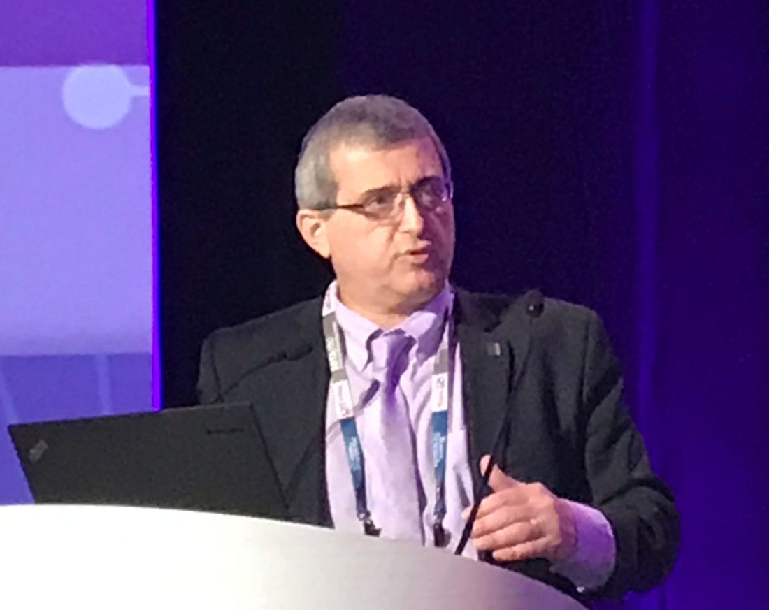
washington, dc – A real-world case series suggests intravascular lithotripsy (IVL) is safe and effective when used selectively to treat coronary arterial calcifications, according to data presented at the 2019 Transcatheter Cardiovascular Therapeutics (CRT) meeting.
Relative to other options, “IVL offers a more controlled means of calcium modification and it avoids the no-reflow phenomenon common to atherectomy in patients with a high calcium burden,” reported Julian Yeoh, MBBS, an interventional cardiologist affiliated with King’s College Hospital, London, UK.
On the basis of the DISRUPT CAD study, presented at the 2016 TCT meeting, IVL was approved in Europe for calcified coronary artery disease in May 2018. The Shockwave IVL device (Shockwave Medical) is currently approved in the U.S. only for treatment of calcified lesions associated with peripheral artery disease (PAD).
In what was characterized as a “real-world series,” 14 procedures were performed at Dr. Yeoh’s institution as part of a clinical study, but 40 procedures were completed on an all-comer basis. Many were performed for indications, such as multivessel disease, that would have been excluded from the DISRUPT CAD study.
“We included elderly patients, patients in cardiogenic shock, and patients with chronic total occlusions,” Dr. Yeoh reported. Presenting specific cases, he described using IVL to permit venous access for a transcatheter aortic valve replacement (TAVR), a failed rotational atherectomy, and to salvage a percutaneous angioplasty thwarted by residual calcium calcification.
“Total procedural success in this series was 91% with 100% facilitation of stent delivery,” Dr. Yeoh said. “There have been no cases of coronary perforation and no reflow or 30-day target lesion failure.”
In this series, the mean age of the patients was 75.9 years. On optical coherence tomography (OCT), which was employed in about half of the cases, the mean residual stenosis was approximately 20%.
IVL involves passing a balloon into the target lesion with the same guidewire used for other percutaneous interventions. Once in position, sonic pressure waves fracture the calcium deposit “with no injury to the intimal soft tissue,” according to Dr. Yeoh, who said that there were no serious adverse events associated with IVL in the series he presented.
In DISRUPT CAD, which enrolled 60 patients, procedural success was 95% with a reduction in mean stenosis from 68.1% to 13.1%. The rate of major adverse cardiovascular event (MACE) events was 5% at 30 days.
While DISRUPT CAD-II is an on-going post-market registry collecting data in Europe and other areas of the world where IVL is approved for treatment of coronary artery disease, a pivotal trial called DISRUPT CAD III has been launched to gain an indication for treatment of coronary calcifications in the U.S. The prospective global trial has a planned enrollment of nearly 400 patients with expected completion in August 2020.
SOURCE: Yeoh J et al. 2019 Cardiovascular Research Technologies (CRT) Meeting abstract.
REPORTING FROM CRT 2019
FDA advises alternatives to paclitaxel-coated devices for PAD, pending review
“Alternative treatment options should generally be used for most patients,” rather than paclitaxel-coated balloons and stents for peripheral arterial disease (PAD), pending an ongoing safety review, according to the Food and Drug Administration.

The FDA conducted a preliminary analysis of long-term follow-up data (up to 5 years in some studies) of the pivotal premarket randomized trials for paclitaxel-coated products indicated for peripheral arterial disease (PAD). In a Letter to Healthcare providers issued March 15, the FDA reported that their preliminary review of these data found “a potentially concerning signal of increased long-term mortality in study subjects treated with paclitaxel-coated products, compared to patients treated with uncoated devices.”
The three trials (totaling 975 patients) that had 5-year follow-up data demonstrated an approximately 50% increased risk of mortality in subjects treated with paclitaxel-coated devices vs. those treated with control devices (20.1% vs. 13.4% crude risk of death at 5 years), according to the agency.
The FDA indicated that these data “should be interpreted with caution for several reasons.” They cited a large variability in the risk estimate of mortality because of the limited amount of long-term data and pointed out that the studies were not designed to be pooled. In addition, the specific cause and mechanism of the increased mortality was unknown.
The FDA also announced that they are planning on convening an Advisory Committee meeting of the Circulatory System Devices Panel to address this issue, including plausible mechanisms for this mortality effect, a re-examination of the benefit-risk profile, modifications of current and future clinical trials regarding these devices, and guidance to any regulatory action, as needed. The timing of this meeting is to be announced within the upcoming weeks.
The FDA letter further stated that the agency intends to conduct additional analyses “to determine whether the benefits continue to outweigh the risks for approved paclitaxel-coated balloons and paclitaxel-eluting stents when used in accordance with their indications for use.”
mlesney@mdedge.com
SOURCE: Food and Drug Administration Letter to Healthcare Providers. 2019 Mar 15.
“Alternative treatment options should generally be used for most patients,” rather than paclitaxel-coated balloons and stents for peripheral arterial disease (PAD), pending an ongoing safety review, according to the Food and Drug Administration.
The FDA conducted a preliminary analysis of long-term follow-up data (up to 5 years in some studies) of the pivotal premarket randomized trials for paclitaxel-coated products indicated for peripheral arterial disease (PAD). In a Letter to Healthcare providers issued March 15, the FDA reported that their preliminary review of these data found “a potentially concerning signal of increased long-term mortality in study subjects treated with paclitaxel-coated products, compared to patients treated with uncoated devices.”
The three trials (totaling 975 patients) that had 5-year follow-up data demonstrated an approximately 50% increased risk of mortality in subjects treated with paclitaxel-coated devices vs. those treated with control devices (20.1% vs. 13.4% crude risk of death at 5 years), according to the agency.
The FDA indicated that these data “should be interpreted with caution for several reasons.” They cited a large variability in the risk estimate of mortality because of the limited amount of long-term data and pointed out that the studies were not designed to be pooled. In addition, the specific cause and mechanism of the increased mortality was unknown.
The FDA also announced that they are planning on convening an Advisory Committee meeting of the Circulatory System Devices Panel to address this issue, including plausible mechanisms for this mortality effect, a re-examination of the benefit-risk profile, modifications of current and future clinical trials regarding these devices, and guidance to any regulatory action, as needed. The timing of this meeting is to be announced within the upcoming weeks.
The FDA letter further stated that the agency intends to conduct additional analyses “to determine whether the benefits continue to outweigh the risks for approved paclitaxel-coated balloons and paclitaxel-eluting stents when used in accordance with their indications for use.”
mlesney@mdedge.com
SOURCE: Food and Drug Administration Letter to Healthcare Providers. 2019 Mar 15.
Key clinical point: FDA advises that alternatives to paclitaxel-coated devices for PAD should be used for most patients.
Major finding: Five-year data demonstrated an approximately 50% increased risk of mortality in patients with paclitaxel-coated devices, compared with uncoated ones.
Study details: Preliminary FDA review of three trials with 975 patients.
Disclosures: Study is funded and performed by the FDA.
Source: Food and Drug Administration. Letter to Healthcare Providers. 2019 Mar 15.
ACC to offer new certification for transcatheter valve repair, replacement
The American College of Cardiology announced at its ACC Quality Summit in New Orleans that it will offer a Transcatheter Valve Certification to assist hospitals that perform transcatheter valve repair and replacement.
The certification is an external review process that will allow hospitals to meet standards for multidisciplinary teams, formalized training, shared decision making, and registry performance. During the certification process, hospitals will learn best practices for implementing evidence-based medicine in the care of individual patients and identify quality improvement opportunities.
The Transcatheter Valve Certification will be launched in mid-2019. To earn the certification, hospitals must already participate in an established national clinical database.
“. This certification incorporates recent guidelines and expert consensus statements regarding the care of patients requiring transcatheter valve therapies,” Phillip D. Levy, MD, chair of the ACC accreditation management board, said in a press release.
Find the full press release on the ACC website.
Registry supports efficacy of coated balloon for peripheral artery stenosis
WASHINGTON – After patients with symptomatic peripheral arterial disease (PAD) were treated with a paclitaxel-coated balloon for 1 year, 89.5% remain free of target lesion restenosis (TLR), according to real-world registry data presented as a late-breaker at CRT 2019 sponsored by MedStar Heart & Vascular Institute.
Freedom from TLR is the primary endpoint of this registry, which will continue to accrue data for 2 more years, according to Nicolas W. Shammas, MD, medical director of Midwest Cardiovascular Research Foundation, Davenport, Iowa.
The nearly 90% rate of freedom from TLR at 1 year was achieved “despite the fact that over 50% of the patients had diabetes, 29% had severe calcification, 35% had critical limb ischemia, and 25% had complete total occlusions,” said Dr. Shammas, an interventional cardiologist.
The registry, called SAFE-DCB, was created to evaluate long-term outcomes after treatment with the Lutonix (Bard Medical) paclitaxel-coated balloon catheter, which is employed in percutaneous angioplasty to treat stenotic lesions in the peripheral vasculature. Over an 18-month period, 1,005 patients were enrolled at 74 treatment centers. Dr. Shammas presented data on 766 of these patients, who have completed 12-months of follow-up. There are 835 patients enrolled in the ongoing study.
In a review of characteristics prior to treatment, Dr. Shammas reported that the average target lesion stenosis was 86.7% and the average target lesion length was 75 mm. Endovascular treatments prior to angioplasty were permitted in the registry protocol. Half of the patients underwent directional atherectomy.
After treatment, the residual stenosis was 11.54%. Even though the recommended protocol called for balloon inflations of 30 seconds each at a pressure of 7 atmospheres, the mean balloon inflation times were 35 seconds at 8 atmospheres. The mean total time for balloon inflations per patient was 152 seconds against the protocol recommendation of 140 seconds.
The primary safety endpoint was freedom from periprocedural mortality, limb amputation, and TLR at 30 days, which was achieved in 98.2% of patients.
Mortality at 1 year was 7.1%. Cardiovascular deaths, such as those due to myocardial infarction, were the most common, but there were noncardiovascular deaths, including those due to sepsis, respiratory failure, and kidney disease.
Women represented 43% of the study population. When compared with men, women achieved the primary outcome at a numerically lower rate, but the difference was not statistically significant. Dr. Shammas reported similar findings for those without complete total occlusions relative to those with complete total occlusions and those treated within the study protocol relative to those who were not. In each case, the differences in the proportion that achieved the primary outcome did not reach statistical significance.
Following his presentation, Dr. Shammas was asked to respond to the criticism that TLR is a soft endpoint. Since some proportion of patients might have had a return of symptoms due to restenosis but elected not to have a second procedure, TLR at 1 year is not equivalent to patency at 1 year.
While acknowledging the accuracy of this criticism, Dr. Shammas reported that TLR was a practical surrogate in the absence of imaging or another objective method of target lesion assessment. Noting that this endpoint has been employed before for long-term follow-up in trials of percutaneous therapies, he said that the TLR rates in this SAFE-DCB registry “are well within previously reported data” for 1-year outcomes with other treatments of symptomatic PAD.
SOURCE: Shammas N. CRT 2019 Mar 5.
REPORTING FROM CRT 2019
Interventional cardiology pioneer reflects on field’s past, future
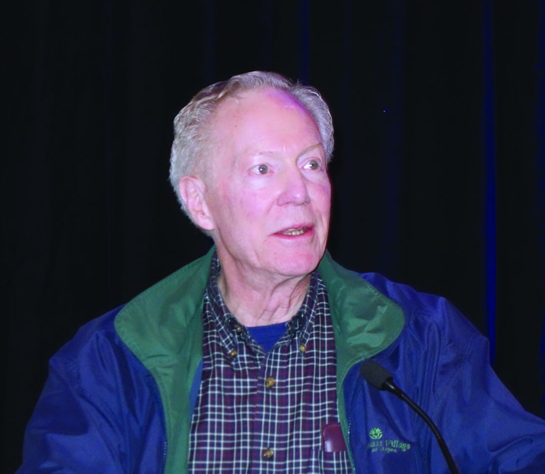
SNOWMASS, COLO. – When Spencer B. King III, MD, shared his thoughts about the future of interventional cardiology at the Annual Cardiovascular Conference at Snowmass sponsored by the American College of Cardiology, he felt compelled to offer a cautionary note about his past accuracy as a prognosticator.
It was way back at a poster session during the 1976 annual meeting of the American Heart Association in Miami Beach that he first met Andreas Gruentzig, MD, the father of percutaneous coronary intervention (PCI), who was presenting his initial revolutionary work on what he called “coronary transluminal angioplasty” in dogs.
“I looked at the poster and told him it would never work,” recalled Dr. King, professor emeritus of medicine at Emory University in Atlanta.
He soon changed his mind, however, because, to great acclaim, Dr. Gruentzig performed his successful first in-human coronary angioplasty the next year.
He noted that the Snowmass conference has played a significant role in the development of interventional cardiology in the United States. Dr. Gruentzig attended the conference in 1980, and Dr. King and others took that opportunity to persuade him to leave the bureaucratic confines of Zurich and join him at Emory later that year. The two cardiologists worked closely thereafter, refining angioplasty and conducting clinical trials until Dr. Gruentzig’s death in an airplane crash in Georgia in 1985 at age 46 years.
Turning to the future, Dr. King addressed a number of recent developments in interventional cardiology and rated their chances of significantly improving outcomes in patients with stable ischemic heart disease. He graded the innovations’ potential with use of a four-bar schema, akin to the WiFi signal power rating on a cell phone.
Noninvasive diagnostics to assess anatomy and physiology
“I think coronary CT angiography [CTA] will become the new diagnostic angiogram,” he predicted. “CTA has gotten much better. Outside the United States, in Europe and particularly in Japan and increasingly in China, CTA is becoming extremely common.”
Dr. King cited a recent multicenter study of blinded heart team treatment decision making on the basis of either CTA or conventional invasive angiography in 223 patients with left main or triple-vessel coronary artery disease (CAD). The level of agreement was impressively high: Coronary artery bypass grafting (CABG) was recommended for 28% of patients on the basis of CTA and 26% with conventional angiography, which suggests the feasibility of treatment decision making based solely on noninvasive imaging, history, and clinical examination (Eur Heart J. 2018 Nov 1;39[41]:3689-98).
“The other thing I like about the potential for noninvasive imaging to guide our interventions is that it may [replace] the diagnostic angiogram, which has largely become extinct,” the cardiologist continued. “If you think about it, patients are referred for an angiogram, and as far as informed consent is concerned, the patient is told to pack his bags, go off to some other city, get in the cath lab, and take the family because of what they might do to you. They might put stents in you, they might operate on you. We don’t have any idea because we don’t know what you have. And the patient has to buy into this. With CTA, the potential is there for people to actually know what you’re going to do to them before you do it.”
Coronary artery calcium scoring for primary risk assessment has taken on a prominent role in the latest practice guidelines. “I think it’s mostly helpful in getting people out of the system because they don’t have any calcium,” in Dr. King’s view.
PET and MRI will remain secondary noninvasive technologies. They will be used mostly to diagnose microvascular disease, but that’s information that doesn’t have much influence on whether interventional procedures are performed.
Overall, he gave noninvasive diagnostic tools high marks for their potential to improve outcomes in patients with stable ischemic heart disease.
“I give it a pretty robust three bars. Maybe you could give it four,” he said.
New pharmacologic therapies
Citing in particular the proprotein convertase subtilisin/kexin type 9 (PCSK9) inhibitors and the sodium-glucose cotransporter 2 (SGLT2) inhibitors, Dr. King declared, “It may be that the biggest, newest device in interventional cardiology going forward is not a device at all, it’s medical therapy.”
Interventional cardiologists either need to become expert in advanced medical therapies or else have access to someone in their group who prescribes those medications deftly.
“The future care of our patients will require more than percutaneous coronary intervention,” he emphasized.
So, four bars for the new medical therapies.
PCI and coronary artery bypass surgery
Both get one bar.
“PCI will be a partner of advanced antiatherosclerotic therapies, but will not be replaced by limiting antianginal therapy to medical treatment only,” Dr. King predicted.
Regarding CABG, he highlighted a recent systematic review and pooled analysis of 11,518 patients with stable ischemic heart disease randomized to CABG or PCI using drug-eluting or bare-metal stents in 11 clinical trials. CABG demonstrated a significant mortality benefit over PCI in patients with multivessel disease, particularly among those with diabetes or a higher degree of coronary disease complexity. However, there was no benefit in terms of 5-year all-cause mortality for CABG over PCI in those with left main disease (Lancet. 2018 Mar 10;391[10124]:9399-48).
“CABG will not go away. I predict that about 25% of revascularizations will continue to be done by surgery,” the cardiologist said. “For patients who can have complete revascularization by PCI, it’ll be done with advanced technology, but probably only by a subset of operators. We have a huge number of interventional cardiologists in this country, and some of them do a lot of these kinds of cases and some don’t.”
Endovascular imaging to optimize stent deployment and characterize plaque
Studies suggest that the use of intravascular ultrasound and other endovascular imaging technologies ends up providing better results than when they’re not employed.
“We see greatly increased use of IVUS [intravascular ultrasound], and not so much of optical coherence tomography, because of technical problems. So I give this at least two bars as far as moving practice forward,” according to Dr. King.
Bioresorbable scaffolds
This technology, which he noted “was supposed to solve all of our problems,” has tripped and fallen because of its associated increased risk of scaffold thrombosis. He cited a recent network meta-analysis of 91 randomized, controlled trials comparing bioresorbable scaffolds to current-generation metallic drug-eluting stents in more than 105,000 patients. The bioresorbable scaffolds had a significantly higher rate of scaffold thrombosis in the first 30 days after implantation, as well as from 31 days through 1 year and also beyond 1 year. In fact, there was a rising trend for scaffold thrombosis in the bioresorbable device group after the 1 year mark through a mean 3.7 years of follow-up (EuroIntervention. 2018 Mar 20;13[16]:1904-13).
“The overall impact of bioresorbable scaffolds has been nil. We don’t have them. Bioresorbable scaffolds may become noninferior to the best metal stents, but to become mainstream, they should show superiority,” the cardiologist said.
One bar, based on the uncertain possibility that new bioresorbable scaffolds now in early stages of development ultimately pan out.
Future training needs
PCI operator volumes are low, and that raises a host of issues regarding future training needs. Should fewer interventionalists be trained? Should training in endovascular imaging be a mandatory part of PCI training? Should interventional cardiology be divided into distinct coronary, structural heart, and peripheral vascular subspecialty domains involving different people, a change that is already informally underway in many places? How are operators who are interested in becoming experts in PCI for chronic total occlusion, diffuse disease, left main disease, or other complex cases going to get enough experience to be able to concentrate in those areas?
These are questions that will need to be addressed in the coming years. The answers will surely affect the delivery of interventional cardiology care.
Dr. King reported having no financial conflicts regarding his presentation.
SOURCE: King SB.
EXPERT ANALYSIS FROM ACC SNOWMASS 2019
Eisenmenger syndrome is a minefield for unwary physicians
SNOWMASS, COLO. – Interventions that are simple and straightforward for other patients – such as therapeutic phlebotomy, bronchoscopy, anticoagulation, administration of IV antibiotics – can quickly have catastrophic results in patients with Eisenmenger syndrome, Carole A. Warnes, MD, cautioned at the Annual Cardiovascular Conference at Snowmass sponsored by the American College of Cardiology.
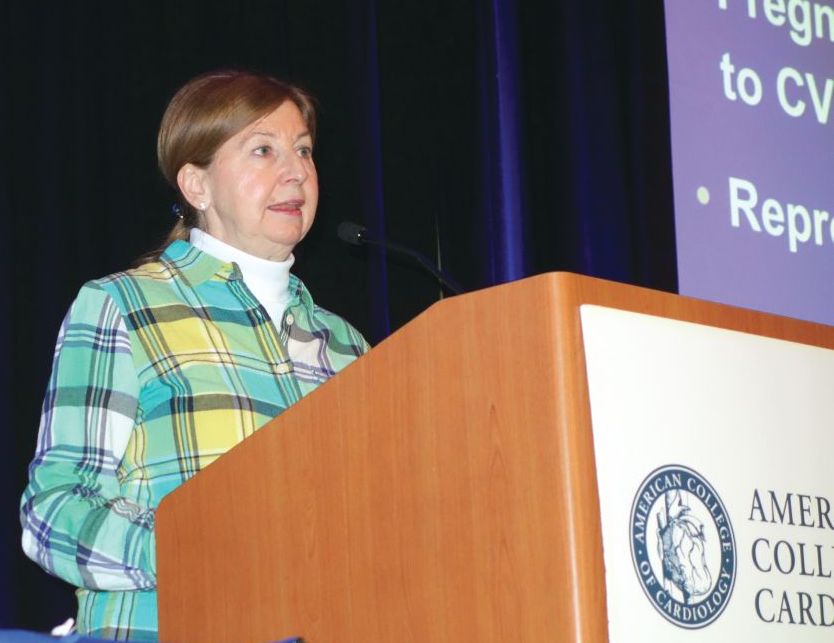
This is why the 2018 ACC/American Heart Association guidelines on the management of patients with adult congenital heart disease recommend as a Class I indication that most patients with ACHD, including all those with Eisenmenger syndrome, be managed in collaboration with a cardiologist at a specialized ACHD center (Circulation. 2018 Aug 16. doi: 10.1161/CIR.0000000000000603), noted Dr. Warnes, professor of medicine at the Mayo Clinic in Rochester, Minn., and director of the Snowmass conference.
“There are lots of mistakes in diagnosis and management because so many physicians are unfamiliar with the kinds of problems these patients face,” the cardiologist said.
She cited as a real-world example a patient with Eisenmenger syndrome admitted for pneumonia. Twenty minutes after being placed on intravenous antibiotics she had a stroke. Why?
“She didn’t have an air filter on her IV line. Any air going into a cyanotic’s blood stream will go to the head and give them a stroke. Something that is simple – routine for other patients – may kill a patient with cyanotic heart disease,” Dr. Warnes said.
Another illustrative case: a patient with Eisenmenger syndrome presents with hemoptysis, an infiltrate in her right lung, a hemoglobin of 19.8 g/dL, and anemia. Should she undergo therapeutic phlebotomy? How about urgent bronchoscopy?
No and no, Dr. Warnes emphasized.
“You do not phlebotomize cyanotic patients unless they have symptoms of hyperviscosity, meaning terrible headache or poor concentration, along with a hemoglobin greater than 20 g/dL. They need that high hemoglobin. They may be blue, and you need those red cells to carry the oxygen around. Otherwise you may make them worse. And if you give them iron for their anemia, you will increase their risk of stroke,” she explained.
Hemoptysis in patients with Eisenmenger syndrome is a life-threatening warning sign. By the time Eisenmenger syndrome patients reach age 40 years, nearly all of them have experienced episodes of hemoptysis. And more and more patients with the syndrome are moving well beyond that milestone and surviving into their 60s and beyond.
“Many Eisenmenger syndrome patients are not dying when they’re 20 or 30. We can get them to a good old age,” according to Dr. Warnes, who founded the ACHD center at the Mayo Clinic.
Most of the time the cause of hemoptysis in patients with Eisenmenger syndrome is an intrapulmonary hemorrhage. Anticoagulation can turn that hemorrhage catastrophic. So can bronchoscopy. Indeed, the cause of death in roughly 30% of patients with this form of congenital heart disease is catastrophic hemoptysis.
“You bar the door from your friendly pulmonologist who wants to bronchoscope these patients. Bronchoscopy never does any good unless the patient is so hypoxic that you need to suck the blood out of their lungs,” Dr. Warnes emphasized.
Anticoagulation is ordinarily to be avoided in patients with Eisenmenger syndrome because of their bleeding risk, even when pulmonary thrombosis is suspected, she added.
So, to summarize: These patients basically need stabilizing, with no anticoagulation, no bronchoscopy, and no phlebotomy, she said.
Another danger zone for patients with Eisenmenger syndrome is pregnancy. The condition is associated with a 30% maternal mortality rate.
Dr. Warnes noted that, even at the Mayo Clinic, which has had a pioneering ACHD center for decades, awareness of key elements of management of affected patients is low among nonspecialists.
“As I’ve done my hospital services in the last couple of months, I sort of felt I was on patrol, snatching these patients from problems,” she recalled.
Dr. Warnes reported having no financial conflicts regarding her presentation.
SNOWMASS, COLO. – Interventions that are simple and straightforward for other patients – such as therapeutic phlebotomy, bronchoscopy, anticoagulation, administration of IV antibiotics – can quickly have catastrophic results in patients with Eisenmenger syndrome, Carole A. Warnes, MD, cautioned at the Annual Cardiovascular Conference at Snowmass sponsored by the American College of Cardiology.
This is why the 2018 ACC/American Heart Association guidelines on the management of patients with adult congenital heart disease recommend as a Class I indication that most patients with ACHD, including all those with Eisenmenger syndrome, be managed in collaboration with a cardiologist at a specialized ACHD center (Circulation. 2018 Aug 16. doi: 10.1161/CIR.0000000000000603), noted Dr. Warnes, professor of medicine at the Mayo Clinic in Rochester, Minn., and director of the Snowmass conference.
“There are lots of mistakes in diagnosis and management because so many physicians are unfamiliar with the kinds of problems these patients face,” the cardiologist said.
She cited as a real-world example a patient with Eisenmenger syndrome admitted for pneumonia. Twenty minutes after being placed on intravenous antibiotics she had a stroke. Why?
“She didn’t have an air filter on her IV line. Any air going into a cyanotic’s blood stream will go to the head and give them a stroke. Something that is simple – routine for other patients – may kill a patient with cyanotic heart disease,” Dr. Warnes said.
Another illustrative case: a patient with Eisenmenger syndrome presents with hemoptysis, an infiltrate in her right lung, a hemoglobin of 19.8 g/dL, and anemia. Should she undergo therapeutic phlebotomy? How about urgent bronchoscopy?
No and no, Dr. Warnes emphasized.
“You do not phlebotomize cyanotic patients unless they have symptoms of hyperviscosity, meaning terrible headache or poor concentration, along with a hemoglobin greater than 20 g/dL. They need that high hemoglobin. They may be blue, and you need those red cells to carry the oxygen around. Otherwise you may make them worse. And if you give them iron for their anemia, you will increase their risk of stroke,” she explained.
Hemoptysis in patients with Eisenmenger syndrome is a life-threatening warning sign. By the time Eisenmenger syndrome patients reach age 40 years, nearly all of them have experienced episodes of hemoptysis. And more and more patients with the syndrome are moving well beyond that milestone and surviving into their 60s and beyond.
“Many Eisenmenger syndrome patients are not dying when they’re 20 or 30. We can get them to a good old age,” according to Dr. Warnes, who founded the ACHD center at the Mayo Clinic.
Most of the time the cause of hemoptysis in patients with Eisenmenger syndrome is an intrapulmonary hemorrhage. Anticoagulation can turn that hemorrhage catastrophic. So can bronchoscopy. Indeed, the cause of death in roughly 30% of patients with this form of congenital heart disease is catastrophic hemoptysis.
“You bar the door from your friendly pulmonologist who wants to bronchoscope these patients. Bronchoscopy never does any good unless the patient is so hypoxic that you need to suck the blood out of their lungs,” Dr. Warnes emphasized.
Anticoagulation is ordinarily to be avoided in patients with Eisenmenger syndrome because of their bleeding risk, even when pulmonary thrombosis is suspected, she added.
So, to summarize: These patients basically need stabilizing, with no anticoagulation, no bronchoscopy, and no phlebotomy, she said.
Another danger zone for patients with Eisenmenger syndrome is pregnancy. The condition is associated with a 30% maternal mortality rate.
Dr. Warnes noted that, even at the Mayo Clinic, which has had a pioneering ACHD center for decades, awareness of key elements of management of affected patients is low among nonspecialists.
“As I’ve done my hospital services in the last couple of months, I sort of felt I was on patrol, snatching these patients from problems,” she recalled.
Dr. Warnes reported having no financial conflicts regarding her presentation.
REPORTING FROM ACC SNOWMASS 2019
Low Risk TAVR trial shows 3% mortality at 1 year
WASHINGTON –Anticipating two pivotal trials scheduled for presentation at the 2019 annual meeting of the American College of Cardiology (ACC), an investigator-led study of transaortic valve replacement (TAVR) for aortic stenosis presented as a latebreaker at 2019 CRT meeting produced excellent results.
Not least impressive, “our mortality rates are the lowest ever reported in any TAVR study at one year,” said Ronald Waksman, MD, Associate Director, Division of Cardiology, Medstar Heart Institute, Washington, DC.
In a population of patients with a median age of 71.1 years, all-cause mortality was just 3% at one year while the rate of deaths due to cardiovascular causes was only 1%, according to results of the 200-patient Low Risk TAVR study (LRT 1.0, NCT02628899) that Dr. Waksman presented.
In addition, there were low rates at one year for stroke (2.1%, none of which was deemed disability), myocardial infarction (1%), new onset atrial fibrillation (6.2%), and pacemaker placement (7.3%). The rate of rehospitalization for any cause was 20.4% but only 3.1% were considered related to TAVR. Rehospitalization for any cardiovascular cause at one year occurred in 6.8%.
Although leaflet thickening was observed at one year with imaging in 14%, this has not had any identifiable clinical consequences so far, and hemodynamics have remained stable, according to Dr. Waksman, who presented the interim 30-day outcomes at the 2018 CRT meeting.
These findings are raising expectations for two phase 3 TAVR trials in low-risk patients that are being presented as latebreakers at the 2019 ACC annual meeting. Both are large randomized trials comparing TAVR to surgical aortic valve replacement (SAVR) in low risk patients. Each trial is testing a single type of value and is funded by the valve manufacturers.
In the PARTNER-3 trial, patients randomized to TAVR received the Sapien 3 valve (Edwards Lifesciences). In the other latebreaking trial, patients randomized to TAVR received an Evolut valve (Medtronic Cardiovascular). Both are comparing TAVR to SAVR with a composite primary outcome that includes mortality and stroke measured at 30 days and one year.
In contrast to these trials, LRT 1.0 was conducted with no funding from a third party, according to Dr. Waksman. The eleven centers participated in the study at their own cost. Also, the choice of TAVR device was left to the discretion of the interventional cardiologist. Finally, most of the participating centers, although experienced in TAVR, did not have a high-volume case load. In general, with the exception of Dr. Waksman’s center, most performed 100 to 150 TAVRs per year.
“We were struck by the excellence of the performance of these sites,” said Dr. Waksman, noting that a comparison of outcomes at his center relative to the lower volume centers showed no significant differences in outcome.
This real-world experience raises the bar for the pivotal phase 3 trials, which, if positive, are expected to lead the FDA to grant an indication for TAVR in low-risk patients, according to Dr. Waksman. He announced that an LRT 2.0 trial, which will again include centers performing TAVRs at moderate volumes, is now enrolling.
SOURCE: 2019 Cardiovascular Research Technologies (CRT) Meeting.
REPORTING FROM CRT 2019
Infective endocarditis isn’t what it used to be
SNOWMASS, COLO. – Infective endocarditis in 2019 is very different from the disease most physicians encountered in training, both in terms of epidemiology and clinical presentation, Patrick T. O’Gara, MD, observed at the Annual Cardiovascular Conference at Snowmass sponsored by the American College of Cardiology.
The classic description of infective endocarditis provided by Sir William Osler, MD, was of a subacute bacterial infection characterized by a long latent phase of low-grade fever, back pain, weight loss, and night sweats. It was mainly a right-heart disease of younger individuals with an infected native valve, and the predominant pathogens were streptococci, Dr. O’Gara said.
“I think in the current era endocarditis is more often characterized by an acute illness with toxic features in the context of adults with a high burden of degenerative diseases – for example, patients with rheumatoid arthritis or psoriatic arthritis on immunosuppressive therapy, or diabetes, end-stage renal disease, and risk factors for hospital-acquired infection. Injectable drug use is through the roof, there’s a wider prevalence of cardiac implanted electronic devices, which are a wonderful place for bacteria to hide, and Staphylococcus aureus has certainly become the leading pathogen with regard to endocarditis in the United States, especially MRSA, often multidrug resistant,” said Dr. O’Gara, professor of medicine at Harvard Medical School, Boston.
“Also, no talk about endocarditis is sufficient without paying some attention to the opioid crisis in which we find ourselves. It’s one of the top three causes of death among young men in the United States, along with accidents and gun violence. No region of the country is spared. This has completely inundated our ER and hospitalist services and our inpatient cardiology services with folks who are often repeat offenders when it comes to the difficulty in being able to give up an injectable drug use habit. They have multiple infections and hospitalizations, tricuspid valve involvement, and depending upon the aggressiveness of the Staphylococcus organism, typically they have left-sided disease with multiple complications, including aortic regurgitation and heart failure,” the cardiologist continued.
This description underscored one of Dr. O’Gara’s major points about the challenges posed by infective endocarditis in contemporary practice: “Expect the unexpected,” he advised. “When you’ve seen one case of infective endocarditis, you’ve seen one case of infective endocarditis.”
Outcomes are ‘sobering’
In the current era, outcomes are “sobering,” the cardiologist noted. Infective endocarditis carries a 6-month mortality rate of 20%-25% despite early surgery being performed during the index hospitalization in up to 60% of patients, with a relatively high perioperative mortality rate of about 10%. However, the risk of reinfection occurring in a newly implanted cardiac valve is impressively low at about 2%.
Refer early for multimodality imaging and surgical consultation
Transesophageal echocardiography is valuable in assessment of the infected valve. However, when extravalvular extension of the infection is suspected and the echo assessment is nondiagnostic or indeterminate, it’s time to quickly move on to advanced imaging, such as PET-CT.
The ACC/American Heart Association class I recommendations for early surgery in infected native valves haven’t changed substantially in over a decade. Based largely on observational data, there is an association between early surgery and lower in-hospital mortality (Lancet. 2012 Mar 10;379[9819]:965-975).
Class IIa recommendations for native valve surgery include recurrent emboli and a persistent vegetation despite appropriate antibiotic therapy. A “very controversial” class IIb recommendation for surgery because of weak supporting data is the identification of a mobile vegetation larger than 10 mm, particularly if it’s located on an anterior mitral valve leaflet, he said.
If the decision is made to forgo early surgery, be sure to repeat transesophageal echocardiography on day 7-10 to reassess the size of the patient’s vegetation.
“There is an association between size of vegetation and 1-year mortality, with a cut point of greater than 15 mm. Some would argue this constitutes a reasonable indication for early surgery,” Dr. O’Gara noted.
The embolization rate in patients with infective endocarditis is highest during the day before presentation, the day of presentation, and through the first 2 days afterward. The rate drops precipitously within 2 weeks after initiation of appropriate antibiotic therapy. Thus, to utilize early surgery to maximum effect in order to decrease the risk of embolization, it makes sense to operate within the first several days following presentation, before antibiotics have had sufficient time to catch up with the evolving disease process.
Don’t use half measures when it comes to removal of cardiac implanted electronic devices
The guidelines are clear regarding infected pacemakers, implanted cardioverter-defibrillators, and cardiac resynchronization devices: “It all needs to come out,” Dr. O’Gara emphasized. That includes all leads and the generator in patients with documented infection of only one portion of the device system, as a class I, level of evidence B recommendation. Moreover, complete removal of a pacemaker or defibrillator system is deemed “reasonable” as a class IIa recommendation in all patients with valvular infection caused by S. aureus or fungi even in the absence of evidence of device infection.
“I think we as general cardiologists have become increasingly impressed about how sick and festering these kinds of patients can become, even when we’re not able to prove that the lead is infected. The lead looks okay on transesophageal echo or PET-CT, blood cultures are negative, the valvular heart disease is really not that advanced, but several days go by and the patient is just not responding. We should have a high index of suspicion that there’s an infection we cannot appreciate. But obviously, you make these difficult decisions in consultation with your electrophysiology colleagues,” he added.
Know when the cardiologist should say ‘no’ to early aggressive surgery
While an aggressive early surgical approach often pays off in terms of prevention of embolic sequelae and a reduction in heart failure, the timing of surgery in the 20%-40% of patients with infective endocarditis who present with stroke or other neurologic complications remains controversial. An international group of Canadian and French cardiac surgeons and neurologists developed a useful algorithm regarding the types of neurologic complications for which early cardiac surgery is a poor idea because of the high risk of neurologic exacerbation. For example, a mycotic neuroaneurysm is grounds for postponement of cardiac surgery for at least 4 weeks (Circulation. 2016 Oct 25;134[17]:1280-92).
Dr. O’Gara reported receiving funding from the National Heart, Lung and Blood Institute, the National Institute of Dental and Craniofacial Research, from Medtronic in conjunction with the ongoing pivotal APOLLO transcatheter mitral valve replacement trial, and from Edwards Lifesciences for the ongoing EARLY TAVR trial.
REPORTING FROM ACC SNOWMASS 2019
Commentary: Should AVFs be ligated after kidney transplant?
Hemodynamic complications of arteriovenous (AV) access are uncommon but can be potentially life threatening. Fistulas and grafts can cause a decrease in systemic vascular resistance and secondary increase in cardiac output in patients who may already have myocardial dysfunction secondary to their end-stage renal disease.1 This increased cardiac output is usually insignificant but in rare cases can result in clinically significant cardiac failure. Patients with high-output fistulas with volume flow greater than 2 L/min may be at increased risk of heart failure but volume flow less than 2 L/min does not preclude this complication.2

In patients with AV access–related heart failure, optimal medical management and reduction of fistula flow or ligation of the dialysis access should be considered. If continued hemodialysis is necessary, loss of a functioning dialysis access is problematic and difficult management decisions must be made. Following successful renal transplantation, ligation of vascular access in the presence of symptomatic heart failure may represent a straightforward decision. Nonetheless, there is no clear consensus of how to manage patent fistulas or grafts in patients following renal transplantation in the absence of significant cardiac symptoms with particular concern to the important issues of transplant survival and long-term cardiac prognosis. Yaffe and Greenstein3 recommend preservation of almost all fistulas after transplantation in the absence of significant complications such as venous hypertension, pseudoaneurysm, significant high-output cardiac failure or hand ischemia. They recommend taking into account the 10-year adjusted renal transplantation graft survival rates and the relative paucity of donors, recognizing the possibility that the patient may have to return to dialysis at some point in the future. They also reference the lack of information regarding the beneficial impact of fistula ligation on cardiac morphology and function as a rationale for access preservation.
A recent presentation at the American Heart Association Scientific Sessions by Michael B. Stokes, MD,4 from the department of cardiology at Royal Adelaide Hospital in Australia, suggests that cardiovascular disease is responsible for 40% of deaths among kidney transplant recipients and that left ventricular (LV) mass is strongly associated with cardiovascular mortality.
He states that, although there is no guideline consensus on the management of an AV fistula following successful renal transplantation, the fistula continues to contribute adversely to cardiac remodeling and function. The lack of previous randomized controlled trials in this area led Dr. Stokes and his colleagues to randomly assign 64 patients at least 1 year following successful kidney transplantation with stable renal function and a functioning AV fistula to either fistula ligation or no intervention. All patients underwent cardiac MRI at baseline and 6 months.
The primary endpoint of decrease in LV mass at 6 months was significant in the ligation group but not in the control group. The ligation group also had significant decrease in LV end diastolic volume, LV end systolic volume, and multiple other parameters. In addition, NT-proBNP levels and left atrial volume were significantly reduced in the ligation group when compared with the control group. Complications in the ligation group included six patients with thrombosis of their fistula vein and two infections, all of which resolved with outpatient anti-inflammatory or antimicrobial therapy.
Dr. Stokes believes that control patients in his study face “persisting and substantial deleterious cardiac remodeling” and that “further investigation would clarify the impact of AV fistula ligation on clinical outcomes following kidney transplantation.”
I believe this is important information and represents the first randomized controlled data regarding the long-term adverse cardiac effects of a patent fistula after renal transplantation. Unfortunately, information regarding baseline fistula volume flow is not provided in this abstract. As discussed earlier, patients with high-flow fistulas may be at increased risk of heart failure and hemodynamic data can be critical in establishing an algorithm for managing these challenging patients.
Ligation of a functioning and asymptomatic access in a patient with a successful renal transplant should be recommended only after informed discussion with the patient weighing the ongoing potential negative effects on cardiac function of continued access patency versus the potential need for future hemodialysis. Dr. Stokes presents interesting data that must be considered in this controversy. I believe that, in the absence of a universally applicable algorithm, the clinical decision to recommend AV fistula ligation after successful kidney transplantation should be individualized and based on ongoing assessment of cardiac and renal function and fistula complications and hemodynamics.
References
1. Eur Heart J 2017;38:1913-23.
2. Nephrol Dial Transplant 2008;23:282-7.
3. J Vasc Access 2012;13:405-8.
4. Stokes MB, et al. LBS.05 – Late Breaking Clinical Trial: Hot News in HF. Presented at American Heart Association Scientific Sessions. 2018 Nov 10-12. Chicago.
Larry A. Scher, MD, is a vascular surgeon at the Montefiore Greene Medical Arts Pavilion, New York, and an associate medical editor for Vascular Specialist.
Hemodynamic complications of arteriovenous (AV) access are uncommon but can be potentially life threatening. Fistulas and grafts can cause a decrease in systemic vascular resistance and secondary increase in cardiac output in patients who may already have myocardial dysfunction secondary to their end-stage renal disease.1 This increased cardiac output is usually insignificant but in rare cases can result in clinically significant cardiac failure. Patients with high-output fistulas with volume flow greater than 2 L/min may be at increased risk of heart failure but volume flow less than 2 L/min does not preclude this complication.2
In patients with AV access–related heart failure, optimal medical management and reduction of fistula flow or ligation of the dialysis access should be considered. If continued hemodialysis is necessary, loss of a functioning dialysis access is problematic and difficult management decisions must be made. Following successful renal transplantation, ligation of vascular access in the presence of symptomatic heart failure may represent a straightforward decision. Nonetheless, there is no clear consensus of how to manage patent fistulas or grafts in patients following renal transplantation in the absence of significant cardiac symptoms with particular concern to the important issues of transplant survival and long-term cardiac prognosis. Yaffe and Greenstein3 recommend preservation of almost all fistulas after transplantation in the absence of significant complications such as venous hypertension, pseudoaneurysm, significant high-output cardiac failure or hand ischemia. They recommend taking into account the 10-year adjusted renal transplantation graft survival rates and the relative paucity of donors, recognizing the possibility that the patient may have to return to dialysis at some point in the future. They also reference the lack of information regarding the beneficial impact of fistula ligation on cardiac morphology and function as a rationale for access preservation.
A recent presentation at the American Heart Association Scientific Sessions by Michael B. Stokes, MD,4 from the department of cardiology at Royal Adelaide Hospital in Australia, suggests that cardiovascular disease is responsible for 40% of deaths among kidney transplant recipients and that left ventricular (LV) mass is strongly associated with cardiovascular mortality.
He states that, although there is no guideline consensus on the management of an AV fistula following successful renal transplantation, the fistula continues to contribute adversely to cardiac remodeling and function. The lack of previous randomized controlled trials in this area led Dr. Stokes and his colleagues to randomly assign 64 patients at least 1 year following successful kidney transplantation with stable renal function and a functioning AV fistula to either fistula ligation or no intervention. All patients underwent cardiac MRI at baseline and 6 months.
The primary endpoint of decrease in LV mass at 6 months was significant in the ligation group but not in the control group. The ligation group also had significant decrease in LV end diastolic volume, LV end systolic volume, and multiple other parameters. In addition, NT-proBNP levels and left atrial volume were significantly reduced in the ligation group when compared with the control group. Complications in the ligation group included six patients with thrombosis of their fistula vein and two infections, all of which resolved with outpatient anti-inflammatory or antimicrobial therapy.
Dr. Stokes believes that control patients in his study face “persisting and substantial deleterious cardiac remodeling” and that “further investigation would clarify the impact of AV fistula ligation on clinical outcomes following kidney transplantation.”
I believe this is important information and represents the first randomized controlled data regarding the long-term adverse cardiac effects of a patent fistula after renal transplantation. Unfortunately, information regarding baseline fistula volume flow is not provided in this abstract. As discussed earlier, patients with high-flow fistulas may be at increased risk of heart failure and hemodynamic data can be critical in establishing an algorithm for managing these challenging patients.
Ligation of a functioning and asymptomatic access in a patient with a successful renal transplant should be recommended only after informed discussion with the patient weighing the ongoing potential negative effects on cardiac function of continued access patency versus the potential need for future hemodialysis. Dr. Stokes presents interesting data that must be considered in this controversy. I believe that, in the absence of a universally applicable algorithm, the clinical decision to recommend AV fistula ligation after successful kidney transplantation should be individualized and based on ongoing assessment of cardiac and renal function and fistula complications and hemodynamics.
References
1. Eur Heart J 2017;38:1913-23.
2. Nephrol Dial Transplant 2008;23:282-7.
3. J Vasc Access 2012;13:405-8.
4. Stokes MB, et al. LBS.05 – Late Breaking Clinical Trial: Hot News in HF. Presented at American Heart Association Scientific Sessions. 2018 Nov 10-12. Chicago.
Larry A. Scher, MD, is a vascular surgeon at the Montefiore Greene Medical Arts Pavilion, New York, and an associate medical editor for Vascular Specialist.