User login
Bringing you the latest news, research and reviews, exclusive interviews, podcasts, quizzes, and more.
Trained interpreters essential for treating non–English-speaking patients
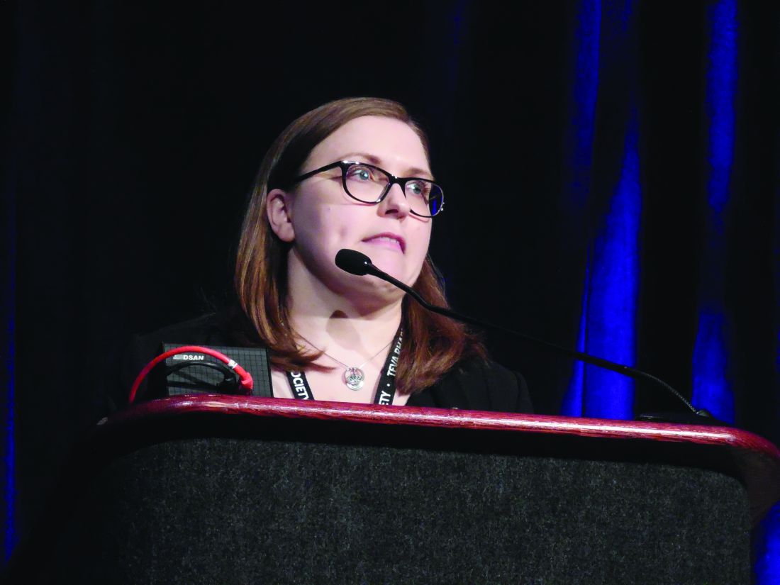
NEW YORK – Clinicians should resist the temptation to use untrained interpreters, such as a child, another relative, or their own limited language skills, when treating patients who cannot communicate in English, according to an expert reviewing this issue at the American Academy of Dermatology summer meeting.

In clinical encounters with patients who have limited English proficiency, “ reported Amy Y.Y. Chen, MD, who is affiliated with Central Connecticut Dermatology in Canton.
In many situations, interpreter services are required by law. This includes a provision of the 1963 Civil Rights Act that specifies these services should be made available to any individual with limited English proficiency receiving federal financial assistance (with the exception of Medicare Part B).
In reviewing this and other laws, Dr. Chen explained that many prohibitions are explicit. For example, it is against the law for clinicians to communicate with the patient through children, whether or not they are related to the patient. A patient’s adult companions are also prohibited from interpreting unless the patient has provided express permission.
Despite the rules, some clinicians might be tempted to forgo a translator when none is readily available, opting for an improvised solution. Dr. Chen said that this is ill advised even when it is not illegal.
“There are a lot of potential problems with using nonprofessional interpreters, starting with the issue of confidentiality,” Dr. Chen warned.
As defined by the Department of Health & Human Services, a qualified interpreter establishes competency by developing familiarity with specialized terminology; by communicating accurately, effectively, and impartially; and by recognizing the ethical issues, including confidentiality, essential to their role.
By itself, language fluency might not be sufficient. Many physicians have conversational fluency in one or more languages other than English, but Dr. Chen pointed out that complex and nuanced clinical descriptions might be difficult to follow for a nonnative speaker. Moreover, many individuals who have no problem posing questions in a foreign language don’t do nearly as well in following the answers.
As interpreters, family members can be particularly problematic. In addition to the issues of confidentiality and medical terminology, a family member might have his or her own agenda that influences how questions and answers are conveyed.
Moreover, family members and others untrained in translating might edit answers based on their own sense of relevance. Many clinicians working through an interpreter will recognize the experience of receiving a yes or no answer after a lengthy discussion between a nontrained interpreter and patient. In such situations, the clinician can reasonably worry that important information was lost.
Typically, major hospitals already offer a systematic approach to providing interpreters when needed, but physicians working in private practice or other smaller practice settings might not. According to Dr. Chen, who recently collaborated on review of this issue (J Am Acad Dermatol. 2019 Mar;80:829-31), they should.
Interpreter services are available by telephone or Internet. Fees typically fall in the range of $2-$5 per minute. In offices with bilingual staff members, formal medical interpreter training might make sense. The Certification Commission for Healthcare Interpreters and the National Board of Certification for Medical Interpreters can help in this process.
When using a medical interpreter, Dr. Chen had some tips.
“Maintain eye contact and talk to the patient,” said Dr. Chen, suggesting that the interpreter, if present in the room, be seated next to or behind the patient. Whether the interpreter is in the room or participating remotely, Dr. Chen advised against speaking through the interpreter with such phases as “tell her that.” Rather, she advised speaking directly to the patient with the interpreter providing the translation.
More practically, Dr. Chen recommended speaking slowly and posing only one question at a time. She also recommended strategies to elicit reassurance that the patient has understood what was communicated. Not least, she recommended a “show me” approach in which a patient can repeat or demonstrate what he or she has learned.
Citing evidence that poor and incomplete translation contributes to medical errors and patient dissatisfaction, Dr. Chen reiterated that engaging unbiased trained translators is advisable for good clinical care even if it were not mandated by law.
NEW YORK – Clinicians should resist the temptation to use untrained interpreters, such as a child, another relative, or their own limited language skills, when treating patients who cannot communicate in English, according to an expert reviewing this issue at the American Academy of Dermatology summer meeting.
In clinical encounters with patients who have limited English proficiency, “ reported Amy Y.Y. Chen, MD, who is affiliated with Central Connecticut Dermatology in Canton.
In many situations, interpreter services are required by law. This includes a provision of the 1963 Civil Rights Act that specifies these services should be made available to any individual with limited English proficiency receiving federal financial assistance (with the exception of Medicare Part B).
In reviewing this and other laws, Dr. Chen explained that many prohibitions are explicit. For example, it is against the law for clinicians to communicate with the patient through children, whether or not they are related to the patient. A patient’s adult companions are also prohibited from interpreting unless the patient has provided express permission.
Despite the rules, some clinicians might be tempted to forgo a translator when none is readily available, opting for an improvised solution. Dr. Chen said that this is ill advised even when it is not illegal.
“There are a lot of potential problems with using nonprofessional interpreters, starting with the issue of confidentiality,” Dr. Chen warned.
As defined by the Department of Health & Human Services, a qualified interpreter establishes competency by developing familiarity with specialized terminology; by communicating accurately, effectively, and impartially; and by recognizing the ethical issues, including confidentiality, essential to their role.
By itself, language fluency might not be sufficient. Many physicians have conversational fluency in one or more languages other than English, but Dr. Chen pointed out that complex and nuanced clinical descriptions might be difficult to follow for a nonnative speaker. Moreover, many individuals who have no problem posing questions in a foreign language don’t do nearly as well in following the answers.
As interpreters, family members can be particularly problematic. In addition to the issues of confidentiality and medical terminology, a family member might have his or her own agenda that influences how questions and answers are conveyed.
Moreover, family members and others untrained in translating might edit answers based on their own sense of relevance. Many clinicians working through an interpreter will recognize the experience of receiving a yes or no answer after a lengthy discussion between a nontrained interpreter and patient. In such situations, the clinician can reasonably worry that important information was lost.
Typically, major hospitals already offer a systematic approach to providing interpreters when needed, but physicians working in private practice or other smaller practice settings might not. According to Dr. Chen, who recently collaborated on review of this issue (J Am Acad Dermatol. 2019 Mar;80:829-31), they should.
Interpreter services are available by telephone or Internet. Fees typically fall in the range of $2-$5 per minute. In offices with bilingual staff members, formal medical interpreter training might make sense. The Certification Commission for Healthcare Interpreters and the National Board of Certification for Medical Interpreters can help in this process.
When using a medical interpreter, Dr. Chen had some tips.
“Maintain eye contact and talk to the patient,” said Dr. Chen, suggesting that the interpreter, if present in the room, be seated next to or behind the patient. Whether the interpreter is in the room or participating remotely, Dr. Chen advised against speaking through the interpreter with such phases as “tell her that.” Rather, she advised speaking directly to the patient with the interpreter providing the translation.
More practically, Dr. Chen recommended speaking slowly and posing only one question at a time. She also recommended strategies to elicit reassurance that the patient has understood what was communicated. Not least, she recommended a “show me” approach in which a patient can repeat or demonstrate what he or she has learned.
Citing evidence that poor and incomplete translation contributes to medical errors and patient dissatisfaction, Dr. Chen reiterated that engaging unbiased trained translators is advisable for good clinical care even if it were not mandated by law.
EXPERT ANALYSIS FROM SUMMER AAD 2019
As patients, physicians fare nearly the same as everyone else
For patients, including patients who are physicians, knowledge isn’t power, according to investigators.

A literature review and retrospective analysis of more than 35,000 physicians treated as patients revealed minimal associations between level of medical knowledge and quality of health outcomes, reported Michael D. Frakes, PhD, of Duke University, Durham, N.C., and colleagues. The study findings stand in opposition to the “widely prevailing view” that information and medical knowledge among patients are integral to realizing high-quality, low-cost health care, the investigators noted.
“[This] research is particularly relevant to modern discussions and debates about the consumer-driven health care movement and the use of plans with high deductibles and high copayments to encourage greater patient and consumer involvement in health care decision making,” Dr. Frakes said in an interview. “Recent research has suggested that the financial incentives created by such structures discourage the use of both low-value care and high-value care. Some have argued that greater disclosure of information to patients may address this concern and steer patients towards high-value decisions. Our results cast doubt on the potential for information initiatives alone to meet this aim.”
The study is one of the first of its kind, the investigators noted in the National Bureau of Economic Research working paper. Other than a 2016 publication that found that physician mothers were less likely to have cesarean sections (Am Econ J: Econ Policy. 2016;8[1]:115-41), “there is no work which has been able to study the role of physicians as patients,” they wrote.
To fill this gap, the investigators turned to a unique data source: The Military Health System, which provides insurance to active and retired military personnel and their families. Military Health System spending exceeds $50 billion per year, constituting a major portion of American health care expenditures, and with more than 35,000 military physicians treated as patients, the dataset is highly relevant and powerful. The investigators objectively evaluated health outcomes by focusing on evidence-based, measurable clinical decisions deemed “high value” or “low value,” comparing how the frequency of these choices related with physician versus nonphysician patient status.
Coauthor Jonathan Gruber, PhD, of the Massachusetts Institute of Technology in Cambridge, Mass., explained this methodology in an interview. “The literature is clear that high-value care has positive health outcomes with relatively small increases in health care spending, and that low-value care has no impact on health outcomes with large increases in spending.”
“One concern with this analysis, of course, is that physicians may be of different health statuses and have different tastes for medical interventions than nonphysicians,” the investigators wrote. They addressed this problem in five ways, by focusing on widely accepted medical standards that apply to all patients; examining both high- and low-value care to eliminate one-sided bias; controlling for underlying health differences across groups; comparing physicians with other military officers to account for underlying tastes; and evaluating military officer dependents in comparison with physician dependents, the latter of whom may benefit from medical knowledge by virtue of personal relationship.
“Our results suggest that physicians do only slightly better than nonphysicians,” the investigators wrote, “but not by much and not always.” Low-value care was slightly less common among physicians, but this difference was described as “modest.” Analysis of high-value care was more mixed, with some results supporting equivalence between groups and others pointing to a slightly higher rate of high-value care among physician patients.
“These results provide a rough boundary on the extent to which additional information disclosure [beyond prevailing levels] can be expected to improve the delivery of health care in the U.S.,” the investigators wrote. “[M]ost of the explanation behind the over- and underutilization of low- and high-value services likely arises from factors other than informational deficiencies of patients.”
“Perhaps one interpretation of these findings is that patients remain generally deferential to the care recommendations of their treating physicians, even in the case of near fully informed patients,” the investigators wrote, noting that this interpretation aligns with a recent working paper that found that physicians play a greater role in selecting the site of MRI scans than patient cost-sharing factors.
Looking to the future, Dr. Gruber said that he and his colleagues plan on exploring “what drives this lack of response among physicians [as patients].”
The study was funded by the National Institute on Aging. The investigators reported no conflicts of interest.
SOURCE: Frakes MD et al. Natl Bur Econ Res. 2019 Jul. doi: 10.3386/w26038.
This article was updated 8/6/19.
For patients, including patients who are physicians, knowledge isn’t power, according to investigators.
A literature review and retrospective analysis of more than 35,000 physicians treated as patients revealed minimal associations between level of medical knowledge and quality of health outcomes, reported Michael D. Frakes, PhD, of Duke University, Durham, N.C., and colleagues. The study findings stand in opposition to the “widely prevailing view” that information and medical knowledge among patients are integral to realizing high-quality, low-cost health care, the investigators noted.
“[This] research is particularly relevant to modern discussions and debates about the consumer-driven health care movement and the use of plans with high deductibles and high copayments to encourage greater patient and consumer involvement in health care decision making,” Dr. Frakes said in an interview. “Recent research has suggested that the financial incentives created by such structures discourage the use of both low-value care and high-value care. Some have argued that greater disclosure of information to patients may address this concern and steer patients towards high-value decisions. Our results cast doubt on the potential for information initiatives alone to meet this aim.”
The study is one of the first of its kind, the investigators noted in the National Bureau of Economic Research working paper. Other than a 2016 publication that found that physician mothers were less likely to have cesarean sections (Am Econ J: Econ Policy. 2016;8[1]:115-41), “there is no work which has been able to study the role of physicians as patients,” they wrote.
To fill this gap, the investigators turned to a unique data source: The Military Health System, which provides insurance to active and retired military personnel and their families. Military Health System spending exceeds $50 billion per year, constituting a major portion of American health care expenditures, and with more than 35,000 military physicians treated as patients, the dataset is highly relevant and powerful. The investigators objectively evaluated health outcomes by focusing on evidence-based, measurable clinical decisions deemed “high value” or “low value,” comparing how the frequency of these choices related with physician versus nonphysician patient status.
Coauthor Jonathan Gruber, PhD, of the Massachusetts Institute of Technology in Cambridge, Mass., explained this methodology in an interview. “The literature is clear that high-value care has positive health outcomes with relatively small increases in health care spending, and that low-value care has no impact on health outcomes with large increases in spending.”
“One concern with this analysis, of course, is that physicians may be of different health statuses and have different tastes for medical interventions than nonphysicians,” the investigators wrote. They addressed this problem in five ways, by focusing on widely accepted medical standards that apply to all patients; examining both high- and low-value care to eliminate one-sided bias; controlling for underlying health differences across groups; comparing physicians with other military officers to account for underlying tastes; and evaluating military officer dependents in comparison with physician dependents, the latter of whom may benefit from medical knowledge by virtue of personal relationship.
“Our results suggest that physicians do only slightly better than nonphysicians,” the investigators wrote, “but not by much and not always.” Low-value care was slightly less common among physicians, but this difference was described as “modest.” Analysis of high-value care was more mixed, with some results supporting equivalence between groups and others pointing to a slightly higher rate of high-value care among physician patients.
“These results provide a rough boundary on the extent to which additional information disclosure [beyond prevailing levels] can be expected to improve the delivery of health care in the U.S.,” the investigators wrote. “[M]ost of the explanation behind the over- and underutilization of low- and high-value services likely arises from factors other than informational deficiencies of patients.”
“Perhaps one interpretation of these findings is that patients remain generally deferential to the care recommendations of their treating physicians, even in the case of near fully informed patients,” the investigators wrote, noting that this interpretation aligns with a recent working paper that found that physicians play a greater role in selecting the site of MRI scans than patient cost-sharing factors.
Looking to the future, Dr. Gruber said that he and his colleagues plan on exploring “what drives this lack of response among physicians [as patients].”
The study was funded by the National Institute on Aging. The investigators reported no conflicts of interest.
SOURCE: Frakes MD et al. Natl Bur Econ Res. 2019 Jul. doi: 10.3386/w26038.
This article was updated 8/6/19.
FROM THE NATIONAL BUREAU OF ECONOMIC RESEARCH

NIH launches 5-year, $10 million study on acute flaccid myelitis
Researchers at the University of Alabama at Birmingham will lead a 5-year, federally-funded study of acute flaccid myelitis (AFM) – a rare pediatric neurologic disease.

The National Institute of Allergy and Infectious Diseases (NIAID) awarded the $10 million grant to primary investigator David Kimberlin, MD, a UAB professor of pediatrics. Carlos Pardo-Villamizar, MD, professor of neurology and pathology at Johns Hopkins University, Baltimore, is the co-principal investigator.
The university will organize and implement the international, multisite study. Its primary goal is to examine the incidence and distribution of AFM, and its pathogenesis and progression. Enrollment is expected to commence next fall. Investigators will enroll children with symptoms of AFM and follow them for 1 year. Household contacts of the subjects will serve as comparators.
In addition to collecting data about risk factors and disease progression, the researchers will collect clinical specimens, including blood and cerebrospinal fluid. More details about the design and study sites will be released then, according to a press statement issued by NIAID.
AFM targets spinal nerves and often develops after a mild respiratory illness. The disease mounted a global epidemic comeback in 2014, primarily affecting children; it has occurred concurrently with enterovirus outbreaks.
“Growing epidemiological evidence suggests that enterovirus-D68 [EV-D68] could play a role,” the statement noted. “Most people who become infected with EV-D68 are asymptomatic or experience mild, cold-like symptoms. Researchers and physicians are working to understand if there is a connection between these viral outbreaks and AFM, and if so, why some children but not others experience this sudden muscle weakness and paralysis.”
The study will draw on the expertise of the AFM Task Force, established last fall. The group comprises physicians, scientists, and public health experts from diverse disciplines and institutions who will assist in the ongoing investigation.
The AFM natural history study is funded under contract HHSN272201600018C.
Researchers at the University of Alabama at Birmingham will lead a 5-year, federally-funded study of acute flaccid myelitis (AFM) – a rare pediatric neurologic disease.
The National Institute of Allergy and Infectious Diseases (NIAID) awarded the $10 million grant to primary investigator David Kimberlin, MD, a UAB professor of pediatrics. Carlos Pardo-Villamizar, MD, professor of neurology and pathology at Johns Hopkins University, Baltimore, is the co-principal investigator.
The university will organize and implement the international, multisite study. Its primary goal is to examine the incidence and distribution of AFM, and its pathogenesis and progression. Enrollment is expected to commence next fall. Investigators will enroll children with symptoms of AFM and follow them for 1 year. Household contacts of the subjects will serve as comparators.
In addition to collecting data about risk factors and disease progression, the researchers will collect clinical specimens, including blood and cerebrospinal fluid. More details about the design and study sites will be released then, according to a press statement issued by NIAID.
AFM targets spinal nerves and often develops after a mild respiratory illness. The disease mounted a global epidemic comeback in 2014, primarily affecting children; it has occurred concurrently with enterovirus outbreaks.
“Growing epidemiological evidence suggests that enterovirus-D68 [EV-D68] could play a role,” the statement noted. “Most people who become infected with EV-D68 are asymptomatic or experience mild, cold-like symptoms. Researchers and physicians are working to understand if there is a connection between these viral outbreaks and AFM, and if so, why some children but not others experience this sudden muscle weakness and paralysis.”
The study will draw on the expertise of the AFM Task Force, established last fall. The group comprises physicians, scientists, and public health experts from diverse disciplines and institutions who will assist in the ongoing investigation.
The AFM natural history study is funded under contract HHSN272201600018C.
Summary: American Medical Society for Sports Medicine position statement on concussion in sport
An estimated 1-1.8 million sport-related concussions (SRC) occur per year in patients younger than 18 years of age. Concussion is defined as “a traumatically induced transient disturbance of brain function.” More than 50% of concussions among high school youth are not related to organized sports and between 2% and 15% of athletes in organized sports will sustain a concussion during a season of play.

which will be described in this article. The guidelines include recommendations for imaging, treatment, and decision making regarding when as well as whether to return to play. Here is a brief summary of those recommendations.
Preseason: Preseason evaluation includes a preparticipation physical evaluation and discussion of concussion history as well as risk factors associated with prolonged concussion recovery. Neurocognitive tests are available for baseline evaluation. While these may assist with diagnosis and return-to-play decisions, there can be considerable variation in an individual’s baseline score as well as the possibility of changes in that baseline over time. Because of this potential for variability, these tests are not required or accepted as the standard of care.

Sideline assessment: Familiarity with the athlete is the best way to detect subtle changes in personality or performance. Looking at symptoms is still the most sensitive way to diagnose a concussion. Loss of consciousness, seizure, tonic posturing, lack of motor coordination, confusion, amnesia, difficulty with balance, or any cognitive difficulty should prompt removal from play for possible concussion. Once a potential injury is identified, how the athlete responds to the elements of orientation, memory, concentration, speech pattern, and balance should be evaluated. If an athlete has a probable or definite concussion, the athlete needs to be removed from play and cannot return to same-day play, and a more detailed evaluation needs to be done.

Office assessment: It is not unusual for symptoms and testing to normalize by the time an office visit occurs. If this is the case, the visit should focus on recommendations for safe return to school and sport. A standard office evaluation should include taking a history with details of the mechanism of injury and preexisting conditions – such as depression and prior concussion – that can affect concussion recovery. The history should focus on detecting symptoms that typically cause impairment from concussion: headache, ocular-vestibular issues leading to problems with balance, and cognitive issues with difficulty concentrating and remembering, as well as fatigue and mood issues such as anxiety, irritability, and depression. The physical exam should include assessment of ocular and vestibular function, gait, and balance in addition to a neurological exam.
Imaging: Head CT or MRI are rarely indicated. Intracranial bleeds are rare in the context of SRC but can occur. If there is concern for a bleed, then CT scan is the imaging test of choice. MRI may have value for evaluation for atypical or prolonged recovery.
Recovery time: The large majority (80%-90%) of concussed older adolescents and adults return to preinjury levels of function within 2 weeks; in younger athletes, clinical recovery may take up to 4 weeks. The best predictor of recovery from SRC is the number and severity of symptoms.
Treatment: For decades, cognitive and physical rest has been the standard of treatment. However, this is no longer the “gold standard” as it has been shown that strict rest (“cocoon therapy”) after SRC slows recovery and leads to an increased chance of prolonged symptoms. Current consensus guidelines support 24-48 hours of symptom-limited rest, both cognitive and physical, followed by a gradual increase in activity, staying below symptom-exacerbation thresholds. Activity, along with good sleep hygiene, appears to be helpful in facilitating recovery from SRC. In athletes with persistent post concussive symptoms that continue beyond the expected recovery time frame, activities of daily living, school, and exercise that do not significantly exacerbate symptoms are recommended.
Return to learning/play: A concussion can cause temporary deficits in attention, cognitive processing, short-term memory, and executive functioning. School personnel should be informed of the injury and assist in employing an individualized return to learn plan, including academic accommodations. Ultimately, return to sports activities should follow a successful return to the classroom. Return to play involves a stepwise increase in physical demands/activity without symptoms before a student is allowed to participate in full contact play.
Concussion-related risks: Continuing to participate in sports before resolution of concussion can worsen and prolong symptoms of SRC. Returning too early after concussion, before full recovery, increases the risk of recurrent SRC. During the initial post-injury period, returning to sports too early increases the risk for a rare but devastating possibility of second impact syndrome that can be a life-threatening repeat head injury. Studies of long-term mental health diagnoses are conflicting and inconsistent. Chronic traumatic encephalopathy has been described in athletes with a long history of concussions and repetitive sub-symptom head impacts. The degree of exposure needed appears to be variable and dependent on the individual.
Disqualification from play: Because each athlete is individually assessed after SRC, there are no evidence-based studies indicating how many concussions are “safe” for an athlete to have in a lifetime. The decision to stop playing sports is both serious and difficult for most athletes and requires shared decision making between clinician, the athlete, and the athlete’s parents. Factors to consider when determining if disqualification from play is warranted include:
- The total number of concussions experienced by a patient.
- Whether a patient has sustained subsequent concussions with progressively less forceful blows to the head.
- If a patient has sustained multiple concussions,whether the time to complete a full recovery after each concussion event increased.
The bottom line: “Cocoon therapy” is no longer recommended. Consensus guidelines endorse 24-48 hours of symptom-limited cognitive and physical rest followed by a gradual increase in activity, including noncontact physical activity that does not provoke symptoms.
Dr. Belogorodsky is a second-year resident and Dr. Fidler is an associate director in the Family Medicine Residency Program at Abington (Pa.) Jefferson Health. Dr. Skolnik is professor of family and community medicine at Jefferson Medical College, Philadelphia, and an associate director of the family medicine residency program at Abington Jefferson Health.
Reference
Harmon KG et al. American Medical Society for Sports Medicine position statement on concussion in sport. Br J Sports Med. 2019;53:213-25.
An estimated 1-1.8 million sport-related concussions (SRC) occur per year in patients younger than 18 years of age. Concussion is defined as “a traumatically induced transient disturbance of brain function.” More than 50% of concussions among high school youth are not related to organized sports and between 2% and 15% of athletes in organized sports will sustain a concussion during a season of play.
which will be described in this article. The guidelines include recommendations for imaging, treatment, and decision making regarding when as well as whether to return to play. Here is a brief summary of those recommendations.
Preseason: Preseason evaluation includes a preparticipation physical evaluation and discussion of concussion history as well as risk factors associated with prolonged concussion recovery. Neurocognitive tests are available for baseline evaluation. While these may assist with diagnosis and return-to-play decisions, there can be considerable variation in an individual’s baseline score as well as the possibility of changes in that baseline over time. Because of this potential for variability, these tests are not required or accepted as the standard of care.
Sideline assessment: Familiarity with the athlete is the best way to detect subtle changes in personality or performance. Looking at symptoms is still the most sensitive way to diagnose a concussion. Loss of consciousness, seizure, tonic posturing, lack of motor coordination, confusion, amnesia, difficulty with balance, or any cognitive difficulty should prompt removal from play for possible concussion. Once a potential injury is identified, how the athlete responds to the elements of orientation, memory, concentration, speech pattern, and balance should be evaluated. If an athlete has a probable or definite concussion, the athlete needs to be removed from play and cannot return to same-day play, and a more detailed evaluation needs to be done.
Office assessment: It is not unusual for symptoms and testing to normalize by the time an office visit occurs. If this is the case, the visit should focus on recommendations for safe return to school and sport. A standard office evaluation should include taking a history with details of the mechanism of injury and preexisting conditions – such as depression and prior concussion – that can affect concussion recovery. The history should focus on detecting symptoms that typically cause impairment from concussion: headache, ocular-vestibular issues leading to problems with balance, and cognitive issues with difficulty concentrating and remembering, as well as fatigue and mood issues such as anxiety, irritability, and depression. The physical exam should include assessment of ocular and vestibular function, gait, and balance in addition to a neurological exam.
Imaging: Head CT or MRI are rarely indicated. Intracranial bleeds are rare in the context of SRC but can occur. If there is concern for a bleed, then CT scan is the imaging test of choice. MRI may have value for evaluation for atypical or prolonged recovery.
Recovery time: The large majority (80%-90%) of concussed older adolescents and adults return to preinjury levels of function within 2 weeks; in younger athletes, clinical recovery may take up to 4 weeks. The best predictor of recovery from SRC is the number and severity of symptoms.
Treatment: For decades, cognitive and physical rest has been the standard of treatment. However, this is no longer the “gold standard” as it has been shown that strict rest (“cocoon therapy”) after SRC slows recovery and leads to an increased chance of prolonged symptoms. Current consensus guidelines support 24-48 hours of symptom-limited rest, both cognitive and physical, followed by a gradual increase in activity, staying below symptom-exacerbation thresholds. Activity, along with good sleep hygiene, appears to be helpful in facilitating recovery from SRC. In athletes with persistent post concussive symptoms that continue beyond the expected recovery time frame, activities of daily living, school, and exercise that do not significantly exacerbate symptoms are recommended.
Return to learning/play: A concussion can cause temporary deficits in attention, cognitive processing, short-term memory, and executive functioning. School personnel should be informed of the injury and assist in employing an individualized return to learn plan, including academic accommodations. Ultimately, return to sports activities should follow a successful return to the classroom. Return to play involves a stepwise increase in physical demands/activity without symptoms before a student is allowed to participate in full contact play.
Concussion-related risks: Continuing to participate in sports before resolution of concussion can worsen and prolong symptoms of SRC. Returning too early after concussion, before full recovery, increases the risk of recurrent SRC. During the initial post-injury period, returning to sports too early increases the risk for a rare but devastating possibility of second impact syndrome that can be a life-threatening repeat head injury. Studies of long-term mental health diagnoses are conflicting and inconsistent. Chronic traumatic encephalopathy has been described in athletes with a long history of concussions and repetitive sub-symptom head impacts. The degree of exposure needed appears to be variable and dependent on the individual.
Disqualification from play: Because each athlete is individually assessed after SRC, there are no evidence-based studies indicating how many concussions are “safe” for an athlete to have in a lifetime. The decision to stop playing sports is both serious and difficult for most athletes and requires shared decision making between clinician, the athlete, and the athlete’s parents. Factors to consider when determining if disqualification from play is warranted include:
- The total number of concussions experienced by a patient.
- Whether a patient has sustained subsequent concussions with progressively less forceful blows to the head.
- If a patient has sustained multiple concussions,whether the time to complete a full recovery after each concussion event increased.
The bottom line: “Cocoon therapy” is no longer recommended. Consensus guidelines endorse 24-48 hours of symptom-limited cognitive and physical rest followed by a gradual increase in activity, including noncontact physical activity that does not provoke symptoms.
Dr. Belogorodsky is a second-year resident and Dr. Fidler is an associate director in the Family Medicine Residency Program at Abington (Pa.) Jefferson Health. Dr. Skolnik is professor of family and community medicine at Jefferson Medical College, Philadelphia, and an associate director of the family medicine residency program at Abington Jefferson Health.
Reference
Harmon KG et al. American Medical Society for Sports Medicine position statement on concussion in sport. Br J Sports Med. 2019;53:213-25.
Atogepant shows safety, efficacy for migraine prevention
PHILADELPHIA – Atogepant, an oral small-molecule migraine drug from the “gepant” class, showed safety and efficacy for preventing migraine headaches in a phase 2/3, dose-ranging trial with 825 evaluable patients.

Atogepant is the first drug from this novel class to undergo efficacy testing in an advanced-phase trial for prevention of migraine headaches, David W. Dodick, MD, said at the annual meeting of the American Headache Society. Two other agents from the gepant class, rimegepant and ubrogepant, have undergone phase 3 testing for acute treatment of migraine headache, and both drugs are now under Food and Drug Administration consideration for an acute-treatment indication.
Three monoclonal antibodies to the calcitonin gene–related peptide (CGRP) receptor have received FDA marketing approval since 2018 for migraine headache prevention. The small-molecule antagonists to the CGRP receptor in the gepant class have an effect similar to the monoclonal antibodies, but with different dosage schedules, an oral route of administration, and with half-lives measured in hours, compared with days and weeks.
“Historically the [gepant] class of drugs was designed for acute treatment, and a couple of drugs from this class didn’t make it because of liver toxicity, but the liver toxicity is not a class effect.” The more recent candidate agents – atogepant, ubrogepant, and rimegepant – have shown hepatic safety as well as overall safety, noted Dr. Dodick, a neurologist and headache specialist at the Mayo Clinic in Phoenix.
If these gepant agents eventually receive FDA approval, which seems likely based on the evidence collected so far, they would collectively form “the only class to have both acute and preventive indications” for migraine, Dr. Dodick said in an interview. The preventive efficacy seen with atogepant in the current study is likely a class effect that has not yet been tested in ubrogepant and rimegepant.
In the multicenter study he reported, 834 patients, 86% of them women, were randomized to placebo or to any of five dosages of atogepant, ranging from 10 mg once daily to 60 mg b.i.d. Study participants had a history of episodic migraine with an average of nearly eight migraine-headache days per month and an average disease duration of about 19 years. The patients’ average age was about 40 years, and 72% had never before used a migraine-preventive treatment. Half had migraine without aura, about a quarter had migraine with aura, and a quarter had migraines of both types.
The researchers had safety data for 825 patients, and efficacy data for 795.
The study’s primary efficacy endpoint was the reduction from baseline in average migraine headache days per month after 12 weeks on treatment. Average migraine headache days fell by 2.85 days in the placebo group and by 3.55-4.23 days in the atogepant treatment groups, a significant difference. The reduction depended on the dosage patients received, but there was no clear dose-response relationship.
At least a 50% drop in average monthly headache day totals was seen in 40% of the placebo patients and in 52%-62% of the patients on atogepant, depending on their dosage. In this case, a signal appeared for a dose-response relationship as only the two subgroups with the patients who received the largest atogepant dosages showed statistically significant improvements in response, compared with placebo.
All patients on atogepant, regardless of their dosage, also had statistically significant reductions in their use of acute migraine medications, compared with placebo patients.
The level of benefit beyond placebo seen in these results was “clinically meaningful,” and roughly comparable with the preventive benefit seen with both the CGRP receptor antagonist monoclonal antibodies, as well as with the three conventional agents most commonly used for migraine headache prevention in current U.S. practice: topiramate, amitriptyline, and propranolol, Dr. Dodick said. Amitriptyline is not approved for this indication.
The adverse event profile among patients who received atogepant was about the same as it was among the placebo recipients. Seven study patients had serious treatment-related adverse effects; two of these patients were in the placebo arm of 186 patients. The most common adverse events were nausea, constipation, and fatigue, and were seen mostly at the highest dosage of atogepant. The incidence of elevated liver enzymes was low and similar in the placebo and drug-treated patients. Two patients, one on placebo and one on atogepant, had their liver enzymes reach at least five times the upper limit of normal. None had their enzymes reach at least 10 times the upper limit of normal, and no patients in the study had a response that fulfilled “Hy’s law,” which flags drug-induced liver injury as patients who develop the combination of elevated liver enzymes, elevated bilirubin, and depressed alkaline phosphatase.
The study was sponsored by Allergan, the company developing atogepant and ubrogepant. Dr. Dodick has been a consultant to Allergan and to several other drug companies.
PHILADELPHIA – Atogepant, an oral small-molecule migraine drug from the “gepant” class, showed safety and efficacy for preventing migraine headaches in a phase 2/3, dose-ranging trial with 825 evaluable patients.
Atogepant is the first drug from this novel class to undergo efficacy testing in an advanced-phase trial for prevention of migraine headaches, David W. Dodick, MD, said at the annual meeting of the American Headache Society. Two other agents from the gepant class, rimegepant and ubrogepant, have undergone phase 3 testing for acute treatment of migraine headache, and both drugs are now under Food and Drug Administration consideration for an acute-treatment indication.
Three monoclonal antibodies to the calcitonin gene–related peptide (CGRP) receptor have received FDA marketing approval since 2018 for migraine headache prevention. The small-molecule antagonists to the CGRP receptor in the gepant class have an effect similar to the monoclonal antibodies, but with different dosage schedules, an oral route of administration, and with half-lives measured in hours, compared with days and weeks.
“Historically the [gepant] class of drugs was designed for acute treatment, and a couple of drugs from this class didn’t make it because of liver toxicity, but the liver toxicity is not a class effect.” The more recent candidate agents – atogepant, ubrogepant, and rimegepant – have shown hepatic safety as well as overall safety, noted Dr. Dodick, a neurologist and headache specialist at the Mayo Clinic in Phoenix.
If these gepant agents eventually receive FDA approval, which seems likely based on the evidence collected so far, they would collectively form “the only class to have both acute and preventive indications” for migraine, Dr. Dodick said in an interview. The preventive efficacy seen with atogepant in the current study is likely a class effect that has not yet been tested in ubrogepant and rimegepant.
In the multicenter study he reported, 834 patients, 86% of them women, were randomized to placebo or to any of five dosages of atogepant, ranging from 10 mg once daily to 60 mg b.i.d. Study participants had a history of episodic migraine with an average of nearly eight migraine-headache days per month and an average disease duration of about 19 years. The patients’ average age was about 40 years, and 72% had never before used a migraine-preventive treatment. Half had migraine without aura, about a quarter had migraine with aura, and a quarter had migraines of both types.
The researchers had safety data for 825 patients, and efficacy data for 795.
The study’s primary efficacy endpoint was the reduction from baseline in average migraine headache days per month after 12 weeks on treatment. Average migraine headache days fell by 2.85 days in the placebo group and by 3.55-4.23 days in the atogepant treatment groups, a significant difference. The reduction depended on the dosage patients received, but there was no clear dose-response relationship.
At least a 50% drop in average monthly headache day totals was seen in 40% of the placebo patients and in 52%-62% of the patients on atogepant, depending on their dosage. In this case, a signal appeared for a dose-response relationship as only the two subgroups with the patients who received the largest atogepant dosages showed statistically significant improvements in response, compared with placebo.
All patients on atogepant, regardless of their dosage, also had statistically significant reductions in their use of acute migraine medications, compared with placebo patients.
The level of benefit beyond placebo seen in these results was “clinically meaningful,” and roughly comparable with the preventive benefit seen with both the CGRP receptor antagonist monoclonal antibodies, as well as with the three conventional agents most commonly used for migraine headache prevention in current U.S. practice: topiramate, amitriptyline, and propranolol, Dr. Dodick said. Amitriptyline is not approved for this indication.
The adverse event profile among patients who received atogepant was about the same as it was among the placebo recipients. Seven study patients had serious treatment-related adverse effects; two of these patients were in the placebo arm of 186 patients. The most common adverse events were nausea, constipation, and fatigue, and were seen mostly at the highest dosage of atogepant. The incidence of elevated liver enzymes was low and similar in the placebo and drug-treated patients. Two patients, one on placebo and one on atogepant, had their liver enzymes reach at least five times the upper limit of normal. None had their enzymes reach at least 10 times the upper limit of normal, and no patients in the study had a response that fulfilled “Hy’s law,” which flags drug-induced liver injury as patients who develop the combination of elevated liver enzymes, elevated bilirubin, and depressed alkaline phosphatase.
The study was sponsored by Allergan, the company developing atogepant and ubrogepant. Dr. Dodick has been a consultant to Allergan and to several other drug companies.
REPORTING FROM AHS 2019
Moderately high dietary riboflavin linked to fewer migraines
PHILADELPHIA – People with moderately high levels of riboflavin consumption from food – two to three times the recommended dietary allowance – had a significantly lower prevalence of a recent severe or migraine headache in a study of more than 3,600 younger U.S. adults.
Adults 20-50 years old who consumed 2.07-2.87 mg riboflavin (vitamin B2) in food a day based on a 24-hour recall questionnaire had an adjusted, statistically significant 27% reduced prevalence of a recent severe or migraine headache, compared with people in the lowest quartile of dietary riboflavin intake, 1.45 mg/day or less, Margaret Slavin, Ph.D., said at the annual meeting of the American Headache Society. Foods particularly high in riboflavin include eggs, milk, and meat.
Dietary riboflavin intakes greater than 2.87 mg/day were not linked to a difference in the prevalence of a recent history of severe or migraine headache, compared with lowest-quartile consumption. Additionally, riboflavin intake from supplements alone at any level of consumption also showed no statistically significant link with the prevalence of a recent, severe headache, said Dr. Slavin, a nutrition and food studies researcher at George Mason University in Fairfax, Va.
The “vast majority” of people in the study had a riboflavin intake that at least matched the U.S. recommended dietary allowance (RDA),1.3 mg/ day for men and 1.1 mg/day for women), “but it’s possible that people with migraine headaches need more riboflavin,” Dr. Slavin suggested. Professional societies in the United States (Neurology. 2012 Apr;78[17]: 1346-53) and Canada (Can J Neurol Sci. 2012 Mar;39[Suppl 2]S8-S28) have gone on record with some level of recommendation for a daily riboflavin supplement of 400 mg to prevent migraine headaches, she said.
A U.S. guideline that included riboflavin has been “retired” because of an issue unrelated to riboflavin, according to the Neurology website.
The new study ran data collected in the biennial National Health and Nutrition Examination Survey (NHANES), specifically the surveys from 2001-2002 and 2003-2004. The combined data included 5,528 adults 20-50 years old, and 3,634 with complete data and without an excluding condition such as pregnancy, diabetes, or menopause. Among the study participants 884 reported having “severe headaches or migraines,” during the 3 months preceding the survey and the remaining 2,750 people served as controls. People who reported recent severe headache or migraine overall had a significantly lower average amount of vitamin B2 in their diet than did the controls, but the two subgroups showed no significant differences in their levels of riboflavin intake from supplements, or from both diet and supplements combined.
The researchers calculated odds ratios for people having severe headaches or migraines relative to their riboflavin-intake quartile, and they adjusted the findings for age, sex, body mass index, and alcohol intake.
Further analysis that looked at total riboflavin intake, from both food and supplements, showed that the two middle quartiles for this metric, with a combined riboflavin intake of 1.6-3.8 mg/day, had a significantly reduced prevalence of recent severe or migraine headaches, compared with the lowest-intake quartile, with an odds ratio that roughly matched the dietary riboflavin analysis.
Dr. Slavin has received research funding from the Egg Nutrition Center, the Maryland Soybean Board, the McCormick Science Institute, and PepsiCo.
SOURCE: Slavin M. Headache. 2019 June;59[S1]:1-208, Abstract LBOR04.
REPORTING FROM AHS 2019
Key clinical point: Higher dietary riboflavin intake may reduce the risk for severe or migraine headaches.
Major finding: People with dietary riboflavin levels 2-3 times the RDA had a 27% lower prevalence of severe or migraine headaches compared with the lowest riboflavin quartile.
Study details: Review of NHANES data from 2001-2004 for 3,634 adults 20-50 years old.
Disclosures: Dr. Slavin has received research funding from the Egg Nutrition Center, the Maryland Soybean Board, the McCormick Science Institute, and PepsiCo.
Source: Slavin M. Headache. 2019 June;59[S1]:1-208, Abstract LBOR04.
Pharmacist stigma a barrier to rural buprenorphine access
SAN ANTONIO – Most attention paid to barriers for medication-assisted treatment of opioid use disorder has focused on prescribers and patients, but pharmacists are “a neglected link in the chain,” according to Hannah Cooper, ScD, an assistant professor of behavioral sciences and health education at Emory University, Atlanta.

“Pharmacy-based dispensing of buprenorphine is one of the medication’s major advances over methadone,” Dr. Cooper told attendees at the annual meeting of the College on Problems of Drug Dependence. Yet, early interviews she and her colleagues conducted with rural Kentucky pharmacist colleagues in the CARE2HOPE study “revealed that pharmacy-level barriers might also curtail access to buprenorphine.”
Little research has examined those barriers, but Dr. Cooper noted. Further, anecdotal evidence has suggested that wholesaler concerns about Drug Enforcement Administration restrictions on dispensing buprenorphine has caused shortages at pharmacies.
Dr. Cooper and colleagues, therefore, designed a qualitative study aimed at learning about pharmacists’ attitudes and dispensing practices related to buprenorphine. They also looked at whether DEA limits actually exist on dispensing the drug. They interviewed 14 pharmacists operating 15 pharmacies across all 12 counties in two rural Kentucky health districts. Eleven of the pharmacists worked in independent pharmacies; the others worked at chains. Six pharmacies dispensed more than 100 buprenorphine prescriptions a month, five dispensed only several dozen a month, and four refused to dispense it at all.
Perceptions of federal restrictions
“Variations in buprenorphine dispensing did not solely reflect underlying variations in local need or prescribing practices,” Dr. Cooper said. At 12 of the 15 pharmacies, limits on buprenorphine resulted from a perceived DEA “cap” on dispensing the drug or “because of distrust in buprenorphine itself, its prescribers and its patients.”
The perceived cap from the DEA was shrouded in uncertainty: 10 of the pharmacists said the DEA capped the percentage of controlled substances pharmacists could dispense that were opioids, yet the pharmacists did not know what that percentage was.
Five of those interviewed said the cap often significantly cut short how many buprenorphine prescriptions they would dispense. Since they did not know how much the cap was, they internally set arbitrary limits, such as dispensing two prescriptions per day, to avoid risk of the DEA investigating their pharmacy.
Yet, those limits could not meet patient demand, so several pharmacists rationed buprenorphine only to local residents or long-term customers, causing additional problems. That practice strained relationships with prescribers, who then had to call multiple pharmacies to find one that would dispense the drug to new patients. It also put pharmacy staff at risk when a rejection angered a customer and “undermined local recovery efforts,” Dr. Cooper said.
Five other pharmacists, however, did not ration their buprenorphine and did not worry about exceeding the DEA cap.
No numerical cap appears to exist, but DEA regulations and the SUPPORT for Patients and Communities Act do require internal opioid surveillance systems at wholesalers that flag suspicious orders of controlled substances, including buprenorphine. And they enforce it: An $80 million settlement in 2013 resulted from the DEA’s charge that Walgreens distribution centers did not report suspicious drug orders.
Stigma among some pharmacists
Six of the pharmacists had low trust in buprenorphine and in those who prescribed it and used it, Dr. Cooper reported. Three would not dispense the drug at all, and two would not take new buprenorphine patients.
One such pharmacist told researchers: “It is supposed to be the drug to help them [recover.] They want Suboxone worse than they do the hydrocodone. … It’s not what it’s designed to be.”
Those pharmacists also reported believing that malpractice was common among prescribers, who, for example, did not provide required counseling to patients or did not quickly wean them off buprenorphine. The pharmacists perceived the physicians prescribing buprenorphine as doing so only to make more money, just as they had done by prescribing opioids in the first place.
Those pharmacists also believed the patients themselves sold buprenorphine to make money and that opioid use disorder was a choice. They told researchers that dispensing buprenorphine would bring more drug users to their stores and subsequently hurt business.
Yet, those beliefs were not universal among the pharmacists. Eight believed buprenorphine was an appropriate opioid use disorder treatment and had positive attitudes toward patients. Unlike those who viewed the disorder as a choice, those pharmacists saw it as a disease and viewed the patients admirably for their commitment to recovery.
Though a small, qualitative study, those findings suggest a need to more closely examine how pharmacies affect access to medication to treat opioid use disorder, Dr. Cooper said.
“In an epicenter of the U.S. opioid epidemic, policies and stigma curtail access to buprenorphine,” she told attendees. “DEA regulations, the SUPPORT Act, and related lawsuits have led wholesalers to develop proprietary caps that force some pharmacists to ration the number of buprenorphine prescriptions they filled.” Some pharmacists will not dispense the drug at all, while others “limited dispensing to known or local patients and prescribers, a practice that pharmacists recognized hurt patients who had to travel far to reach prescribers.”
The research was funded by the National Institutes of Health through CARE2HOPE, Rural Health Project, and the Emory Center for AIDS Research. The authors reported no disclosures.
SAN ANTONIO – Most attention paid to barriers for medication-assisted treatment of opioid use disorder has focused on prescribers and patients, but pharmacists are “a neglected link in the chain,” according to Hannah Cooper, ScD, an assistant professor of behavioral sciences and health education at Emory University, Atlanta.
“Pharmacy-based dispensing of buprenorphine is one of the medication’s major advances over methadone,” Dr. Cooper told attendees at the annual meeting of the College on Problems of Drug Dependence. Yet, early interviews she and her colleagues conducted with rural Kentucky pharmacist colleagues in the CARE2HOPE study “revealed that pharmacy-level barriers might also curtail access to buprenorphine.”
Little research has examined those barriers, but Dr. Cooper noted. Further, anecdotal evidence has suggested that wholesaler concerns about Drug Enforcement Administration restrictions on dispensing buprenorphine has caused shortages at pharmacies.
Dr. Cooper and colleagues, therefore, designed a qualitative study aimed at learning about pharmacists’ attitudes and dispensing practices related to buprenorphine. They also looked at whether DEA limits actually exist on dispensing the drug. They interviewed 14 pharmacists operating 15 pharmacies across all 12 counties in two rural Kentucky health districts. Eleven of the pharmacists worked in independent pharmacies; the others worked at chains. Six pharmacies dispensed more than 100 buprenorphine prescriptions a month, five dispensed only several dozen a month, and four refused to dispense it at all.
Perceptions of federal restrictions
“Variations in buprenorphine dispensing did not solely reflect underlying variations in local need or prescribing practices,” Dr. Cooper said. At 12 of the 15 pharmacies, limits on buprenorphine resulted from a perceived DEA “cap” on dispensing the drug or “because of distrust in buprenorphine itself, its prescribers and its patients.”
The perceived cap from the DEA was shrouded in uncertainty: 10 of the pharmacists said the DEA capped the percentage of controlled substances pharmacists could dispense that were opioids, yet the pharmacists did not know what that percentage was.
Five of those interviewed said the cap often significantly cut short how many buprenorphine prescriptions they would dispense. Since they did not know how much the cap was, they internally set arbitrary limits, such as dispensing two prescriptions per day, to avoid risk of the DEA investigating their pharmacy.
Yet, those limits could not meet patient demand, so several pharmacists rationed buprenorphine only to local residents or long-term customers, causing additional problems. That practice strained relationships with prescribers, who then had to call multiple pharmacies to find one that would dispense the drug to new patients. It also put pharmacy staff at risk when a rejection angered a customer and “undermined local recovery efforts,” Dr. Cooper said.
Five other pharmacists, however, did not ration their buprenorphine and did not worry about exceeding the DEA cap.
No numerical cap appears to exist, but DEA regulations and the SUPPORT for Patients and Communities Act do require internal opioid surveillance systems at wholesalers that flag suspicious orders of controlled substances, including buprenorphine. And they enforce it: An $80 million settlement in 2013 resulted from the DEA’s charge that Walgreens distribution centers did not report suspicious drug orders.
Stigma among some pharmacists
Six of the pharmacists had low trust in buprenorphine and in those who prescribed it and used it, Dr. Cooper reported. Three would not dispense the drug at all, and two would not take new buprenorphine patients.
One such pharmacist told researchers: “It is supposed to be the drug to help them [recover.] They want Suboxone worse than they do the hydrocodone. … It’s not what it’s designed to be.”
Those pharmacists also reported believing that malpractice was common among prescribers, who, for example, did not provide required counseling to patients or did not quickly wean them off buprenorphine. The pharmacists perceived the physicians prescribing buprenorphine as doing so only to make more money, just as they had done by prescribing opioids in the first place.
Those pharmacists also believed the patients themselves sold buprenorphine to make money and that opioid use disorder was a choice. They told researchers that dispensing buprenorphine would bring more drug users to their stores and subsequently hurt business.
Yet, those beliefs were not universal among the pharmacists. Eight believed buprenorphine was an appropriate opioid use disorder treatment and had positive attitudes toward patients. Unlike those who viewed the disorder as a choice, those pharmacists saw it as a disease and viewed the patients admirably for their commitment to recovery.
Though a small, qualitative study, those findings suggest a need to more closely examine how pharmacies affect access to medication to treat opioid use disorder, Dr. Cooper said.
“In an epicenter of the U.S. opioid epidemic, policies and stigma curtail access to buprenorphine,” she told attendees. “DEA regulations, the SUPPORT Act, and related lawsuits have led wholesalers to develop proprietary caps that force some pharmacists to ration the number of buprenorphine prescriptions they filled.” Some pharmacists will not dispense the drug at all, while others “limited dispensing to known or local patients and prescribers, a practice that pharmacists recognized hurt patients who had to travel far to reach prescribers.”
The research was funded by the National Institutes of Health through CARE2HOPE, Rural Health Project, and the Emory Center for AIDS Research. The authors reported no disclosures.
REPORTING FROM CPDD 2019
Study refines ALS risk among first-degree relatives of patients with disease
, according to a recent Irish population–based study.

Among first-degree relatives of individuals with ALS whose genetic status was unknown, the lifetime risk of developing ALS was 1.4%, compared with 0.3% in the general population (P less than .001).
Overall, lifetime heritability for the 1,117 patients in the heritability study cohort was 52.3% (95% confidence interval, 42.9%-61.7%). The highest heritability was seen in mother-daughter dyads; here, the concordance rate for ALS was 2.6% and the heritability estimate was 66.2% (95% CI, 58.5%-73.9%), “pointing to a previously unrecognized sex-mediated association with disease heritability,” Marie Ryan, MD, and coauthors at Trinity College Dublin wrote in JAMA Neurology.
For the purposes of this study, heritability was defined as “the proportion of variance in liability that is attributable to additive genetic factors,” explained the investigators. Their model was applied to 1,123 Irish patients with incident ALS over a 10-year study period, drawing on a national database. Complete medical histories were available for both parents of every individual in the cohort, and the investigators identified 32 parent-offspring dyads with ALS.
In their assessment of “liability” for ALS, Dr. Ryan and colleagues accounted for age-specific and sex-specific ALS incidence rates, using a well-delineated United States reference population. They also took into account competing mortality risks that might intervene before an individual developed ALS.
Although mother-daughter dyads yielded the highest heritability estimates, heritability was also elevated among father-son dyads, at 50.1% (95% CI, 42.0%-59.4%). Opposite-sex dyads (mother-son and father-daughter) yielded similar, and lower, heritability estimates of 34%-38%. Dr. Ryan and coinvestigators acknowledged that female sex hormones might play a role in mediating risk for ALS, but the contribution and mechanism are currently unknown.
The investigators identified 674 patients in the cohort who had been tested for C9orf72; large repeat expansions in the GGGGCC segment of this gene are known to cause ALS. In all, 69 patients were C9orf72 positive, and 14 of these individuals (20.3%) reported that one parent had ALS. Among the entire cohort, 23 of the 32 patients with parents who also had ALS had C9orf72 testing, and 14 of 23 (60.9%) were positive. This discrepancy, wrote Dr. Ryan and coauthors, suggests that “genetic anticipation or pleiotropic effects may have masked the clinical phenotype in their parents.”
Parents were a mean 69.6 years old at the time of diagnosis, and offspring were a mean 52.0 years old; 56% of patients in the study cohort were male.
“[I]n our study, ALS heritability was assessed for the first time in a population in whom genetic mutations have been excluded,” wrote Dr. Ryan and coinvestigators. “We have found that, in the absence of known ALS-associated genetic mutations in the proband, first-degree relatives of individuals with ALS remain at increased risk of developing ALS compared with the general population.”
Dr. Ryan and one coauthor reported receiving funding from Science Foundation Ireland, and one coauthor reported receiving book royalties from Taylor & Francis, and fees from Cytokinetics and Wave Pharmaceuticals. The study was funded by Science Foundation Ireland and the Motor Neurone Disease Association.
SOURCE: Ryan M et al. JAMA Neurol. 2019 Jul 22. doi: 10.1001/jamaneurol.2019.2044.
, according to a recent Irish population–based study.
Among first-degree relatives of individuals with ALS whose genetic status was unknown, the lifetime risk of developing ALS was 1.4%, compared with 0.3% in the general population (P less than .001).
Overall, lifetime heritability for the 1,117 patients in the heritability study cohort was 52.3% (95% confidence interval, 42.9%-61.7%). The highest heritability was seen in mother-daughter dyads; here, the concordance rate for ALS was 2.6% and the heritability estimate was 66.2% (95% CI, 58.5%-73.9%), “pointing to a previously unrecognized sex-mediated association with disease heritability,” Marie Ryan, MD, and coauthors at Trinity College Dublin wrote in JAMA Neurology.
For the purposes of this study, heritability was defined as “the proportion of variance in liability that is attributable to additive genetic factors,” explained the investigators. Their model was applied to 1,123 Irish patients with incident ALS over a 10-year study period, drawing on a national database. Complete medical histories were available for both parents of every individual in the cohort, and the investigators identified 32 parent-offspring dyads with ALS.
In their assessment of “liability” for ALS, Dr. Ryan and colleagues accounted for age-specific and sex-specific ALS incidence rates, using a well-delineated United States reference population. They also took into account competing mortality risks that might intervene before an individual developed ALS.
Although mother-daughter dyads yielded the highest heritability estimates, heritability was also elevated among father-son dyads, at 50.1% (95% CI, 42.0%-59.4%). Opposite-sex dyads (mother-son and father-daughter) yielded similar, and lower, heritability estimates of 34%-38%. Dr. Ryan and coinvestigators acknowledged that female sex hormones might play a role in mediating risk for ALS, but the contribution and mechanism are currently unknown.
The investigators identified 674 patients in the cohort who had been tested for C9orf72; large repeat expansions in the GGGGCC segment of this gene are known to cause ALS. In all, 69 patients were C9orf72 positive, and 14 of these individuals (20.3%) reported that one parent had ALS. Among the entire cohort, 23 of the 32 patients with parents who also had ALS had C9orf72 testing, and 14 of 23 (60.9%) were positive. This discrepancy, wrote Dr. Ryan and coauthors, suggests that “genetic anticipation or pleiotropic effects may have masked the clinical phenotype in their parents.”
Parents were a mean 69.6 years old at the time of diagnosis, and offspring were a mean 52.0 years old; 56% of patients in the study cohort were male.
“[I]n our study, ALS heritability was assessed for the first time in a population in whom genetic mutations have been excluded,” wrote Dr. Ryan and coinvestigators. “We have found that, in the absence of known ALS-associated genetic mutations in the proband, first-degree relatives of individuals with ALS remain at increased risk of developing ALS compared with the general population.”
Dr. Ryan and one coauthor reported receiving funding from Science Foundation Ireland, and one coauthor reported receiving book royalties from Taylor & Francis, and fees from Cytokinetics and Wave Pharmaceuticals. The study was funded by Science Foundation Ireland and the Motor Neurone Disease Association.
SOURCE: Ryan M et al. JAMA Neurol. 2019 Jul 22. doi: 10.1001/jamaneurol.2019.2044.
FROM JAMA NEUROLOGY
MIPS: Nearly all eligible clinicians got a bonus for 2018
Nearly all clinicians who are eligible to participate in the Merit-Based Incentive Payment System (MIPS) track of the Quality Payment Program did so in 2018; most scored above the performance threshold and got a bonus.

According to the most recent data released this month by the Centers for Medicare & Medicaid Services, 98.37% of MIPS-eligible clinicians participated in the program. In the small/solo practice space, 89.20% of MIPS-eligible clinicians participated.
But more importantly, the clinicians are performing better one year later with the program, even though fewer are participating.
In 2018, 97.63% of clinicians scored above the performance threshold, up from 93.12% in 2017. There also were fewer clinicians performing at the threshold (0.42% in 2018, down from 2.01% in the previous year) and fewer clinicians scoring below the threshold (1.95%, down from 4.87%).
Exceeding the performance threshold resulted in a bonus to fee schedule payments in 2018, although the agency did not disclose how much money was paid out in performance bonuses.
MIPS scored “improved across performance categories, with the biggest gain in the Quality performance category, which highlights the program’s effectiveness in measuring outcomes for beneficiaries,” CMS Administrator Seema Verma wrote in a blog post.
The total number of eligible clinicians decreased in 2018 to 916,058, down from 1,057,824 in 2017 because CMS broadened the low-volume threshold to exempt providers from participation requirements.
Participants in a MIPS alternative payment model saw even more success in 2018. Participation increased from 341,220 clinicians in 2017 to 356,828 clinicians in 2018, while virtually all performed above the performance threshold (100% in 2017 and 99.99% in 2018). The 0.01% that was not above the threshold still met it, while no clinicians in either year that participated in a MIPS alternative payment model performed below the threshold.
Participation in the advanced alternative payment model track increased as well, going from 99,026 in 2017 to 183,306 in 2018.
Nearly all clinicians who are eligible to participate in the Merit-Based Incentive Payment System (MIPS) track of the Quality Payment Program did so in 2018; most scored above the performance threshold and got a bonus.
According to the most recent data released this month by the Centers for Medicare & Medicaid Services, 98.37% of MIPS-eligible clinicians participated in the program. In the small/solo practice space, 89.20% of MIPS-eligible clinicians participated.
But more importantly, the clinicians are performing better one year later with the program, even though fewer are participating.
In 2018, 97.63% of clinicians scored above the performance threshold, up from 93.12% in 2017. There also were fewer clinicians performing at the threshold (0.42% in 2018, down from 2.01% in the previous year) and fewer clinicians scoring below the threshold (1.95%, down from 4.87%).
Exceeding the performance threshold resulted in a bonus to fee schedule payments in 2018, although the agency did not disclose how much money was paid out in performance bonuses.
MIPS scored “improved across performance categories, with the biggest gain in the Quality performance category, which highlights the program’s effectiveness in measuring outcomes for beneficiaries,” CMS Administrator Seema Verma wrote in a blog post.
The total number of eligible clinicians decreased in 2018 to 916,058, down from 1,057,824 in 2017 because CMS broadened the low-volume threshold to exempt providers from participation requirements.
Participants in a MIPS alternative payment model saw even more success in 2018. Participation increased from 341,220 clinicians in 2017 to 356,828 clinicians in 2018, while virtually all performed above the performance threshold (100% in 2017 and 99.99% in 2018). The 0.01% that was not above the threshold still met it, while no clinicians in either year that participated in a MIPS alternative payment model performed below the threshold.
Participation in the advanced alternative payment model track increased as well, going from 99,026 in 2017 to 183,306 in 2018.
Liberalized low–glycemic-index diet effective for seizure reduction
BANGKOK – in a randomized, double-blind, 24-week, noninferiority study.

The low–glycemic-index diet (LGID) was introduced as a kinder, gentler, variant of the classic ketogenic diet for seizure frequency reduction. The ketogenic diet’s efficacy for this purpose is well established, but compliance is a problem and discontinuation rates are high. Yet even though the LGID was designed to be less onerous than the ketogenic diet, many children and parents also find the 7-days-a-week LGID to be excessively burdensome. This was the impetus for pitting the daily LGID against an intermittent version – 5 days on, 2 days off – in a randomized trial, Prateek K. Panda, MD, explained at the International Epilepsy Congress.
The hypothesis of this noninferiority trial was that adherence to the liberalized LGID would be similar to or better than that with the daily LGID regimen, with resultant similar reductions in seizure frequency. And further, that patients on the intermittent LGID would feel better because it would help improve depleted glycogen stores important for daily activity and that the liberalized diet would also be rated more favorably by caregivers, Dr. Panda said at the congress sponsored by the International League Against Epilepsy.
The 24-week, single-center trial included 122 children ages 1-15 years with drug-resistant epilepsy. At baseline they averaged 99 seizures per week by parental diary despite being on a median of four antiepileptic drugs. A total of 88% of participants had some form of structural epilepsy; the rest had a probable or confirmed genetic cause for their seizure disorder, according to Dr. Panda of the All-India Institute of Medical Sciences in New Delhi.
The standard daily LGID was comprised of 10% carbohydrate, 30% protein, and 60% fat, with only low–glycemic-index foods permitted. The cohort randomized to the liberalized diet ate that way on weekdays; however, on Saturdays and Sundays their diet was 20% carbohydrate, 30% protein, and 50% fat, with both medium- and low–glycemic-index foods allowed.
The primary outcome was the mean reduction in seizures per week by caregiver records at 24 weeks. The reduction from baseline was 54% in the strict LGID group and not significantly different at 49% in the intermittent LGID patients. Overall, 54% of patients in the strict LGID arm experienced a greater than 50% reduction in weekly seizure frequency, as did 50% on the liberalized diet, a nonsignificant difference.
There were five study dropouts in the strict LGID group and three in the liberalized LGID cohort. The two groups showed similar improvements over baseline in measures of social function, behavior, and cognition. Parents of children in the liberalized LGID group rated that diet as significantly less difficult to administer than those randomized to the strict LGID therapy.
Mean hemoglobin A1c improved in the strict LGID patients from 5.7% at baseline to 5.1% at both 12 and 24 weeks. The intermittent LGID group went from 5.6% to 5.0% and then to 5.2%. There was no correlation between HbA1c and reduction in seizure frequency. In contrast, serum beta-hydroxybutyrate levels showed a moderate correlation with seizure frequency, a novel finding which if confirmed might render beta-hydroxybutyrate useful as a biomarker, according to Dr. Panda.
Adverse events – mostly dyslipidemia and GI complaints such as vomiting or constipation – occurred in 25% of the strict LGID group and 13% with the intermittent LGID. All adverse events were mild.
Dr. Panda reported having no financial conflicts regarding the study, sponsored by the All-India Institute of Medical Sciences.
SOURCE: Panda PK et al. IEC 2019, Abstract P056.
BANGKOK – in a randomized, double-blind, 24-week, noninferiority study.
The low–glycemic-index diet (LGID) was introduced as a kinder, gentler, variant of the classic ketogenic diet for seizure frequency reduction. The ketogenic diet’s efficacy for this purpose is well established, but compliance is a problem and discontinuation rates are high. Yet even though the LGID was designed to be less onerous than the ketogenic diet, many children and parents also find the 7-days-a-week LGID to be excessively burdensome. This was the impetus for pitting the daily LGID against an intermittent version – 5 days on, 2 days off – in a randomized trial, Prateek K. Panda, MD, explained at the International Epilepsy Congress.
The hypothesis of this noninferiority trial was that adherence to the liberalized LGID would be similar to or better than that with the daily LGID regimen, with resultant similar reductions in seizure frequency. And further, that patients on the intermittent LGID would feel better because it would help improve depleted glycogen stores important for daily activity and that the liberalized diet would also be rated more favorably by caregivers, Dr. Panda said at the congress sponsored by the International League Against Epilepsy.
The 24-week, single-center trial included 122 children ages 1-15 years with drug-resistant epilepsy. At baseline they averaged 99 seizures per week by parental diary despite being on a median of four antiepileptic drugs. A total of 88% of participants had some form of structural epilepsy; the rest had a probable or confirmed genetic cause for their seizure disorder, according to Dr. Panda of the All-India Institute of Medical Sciences in New Delhi.
The standard daily LGID was comprised of 10% carbohydrate, 30% protein, and 60% fat, with only low–glycemic-index foods permitted. The cohort randomized to the liberalized diet ate that way on weekdays; however, on Saturdays and Sundays their diet was 20% carbohydrate, 30% protein, and 50% fat, with both medium- and low–glycemic-index foods allowed.
The primary outcome was the mean reduction in seizures per week by caregiver records at 24 weeks. The reduction from baseline was 54% in the strict LGID group and not significantly different at 49% in the intermittent LGID patients. Overall, 54% of patients in the strict LGID arm experienced a greater than 50% reduction in weekly seizure frequency, as did 50% on the liberalized diet, a nonsignificant difference.
There were five study dropouts in the strict LGID group and three in the liberalized LGID cohort. The two groups showed similar improvements over baseline in measures of social function, behavior, and cognition. Parents of children in the liberalized LGID group rated that diet as significantly less difficult to administer than those randomized to the strict LGID therapy.
Mean hemoglobin A1c improved in the strict LGID patients from 5.7% at baseline to 5.1% at both 12 and 24 weeks. The intermittent LGID group went from 5.6% to 5.0% and then to 5.2%. There was no correlation between HbA1c and reduction in seizure frequency. In contrast, serum beta-hydroxybutyrate levels showed a moderate correlation with seizure frequency, a novel finding which if confirmed might render beta-hydroxybutyrate useful as a biomarker, according to Dr. Panda.
Adverse events – mostly dyslipidemia and GI complaints such as vomiting or constipation – occurred in 25% of the strict LGID group and 13% with the intermittent LGID. All adverse events were mild.
Dr. Panda reported having no financial conflicts regarding the study, sponsored by the All-India Institute of Medical Sciences.
SOURCE: Panda PK et al. IEC 2019, Abstract P056.
REPORTING FROM IEC 2019