User login
Bringing you the latest news, research and reviews, exclusive interviews, podcasts, quizzes, and more.
Study examines utility of repeat outpatient electrodiagnostic testing
AUSTIN, TEX. – During a 3-year period, 5.7% of patients referred to an electromyography laboratory returned for at least one additional electrodiagnostic study, according to research presented at the annual meeting of the American Association of Neuromuscular and Electrodiagnostic Medicine. A preliminary analysis suggests that repeat testing for the same indication does not change symptom or disease management in about one-third of cases.

Physicians may request repeat electrodiagnostic studies to monitor previous, new, or progressing symptoms in the same or different body segments. “While the utility of [electrodiagnostic] studies for clinical care has been established, testing is associated with some patient risk, time, and cost,” the researchers wrote. “To date, there have been no known studies investigating the utility of repeat [electrodiagnostic] testing in the outpatient setting.”
To study referral patterns and outcomes following repeat electrodiagnostic testing, Aimee K. Boegle, MD, PhD, an instructor in neurology at Beth Israel Deaconess Medical Center in Boston, and colleagues examined all outpatient electromyography and nerve conduction studies performed between 2015 and 2017 in the neurology department at their institution. The investigators excluded patients who underwent inpatient electrodiagnostic studies from their analysis.
Approximately 4,800 patients underwent electrodiagnostic testing, 276 of whom underwent testing more than once.
Among patients who underwent two studies, 55% were referred by a different physician for the second study. Median neuropathy was the most common referring and final diagnosis among patients who underwent repeat electrodiagnostic testing, Dr. Boegle said. This finding was not surprising because carpal tunnel syndrome is among the most common reasons for referral overall.
Median neuropathy was the referring diagnosis in 31% and the final diagnosis in 30%, cervical radiculopathy in 15% and 14%, ulnar neuropathy in 14% and 17%, lumbosacral radiculopathy in 12% and 10%, and polyneuropathy in 8% and 10%.
The neurology and orthopedics departments made the most referrals for repeat electrodiagnostic studies (49.4% and 29.3%, respectively), followed by primary care physicians/internal medicine (13%).
About 24% of the returning patients underwent testing for the same indication as their initial referral.
A preliminary analysis of 26 patients who underwent a repeat study for the same indication found no change in treatment in 34%. When a study prompted intervention, a conservative course of management such as a splint or physical therapy was used in 42%. About 8% received a pharmacologic intervention, such as a medication change or steroid injections. Another 8% received a surgical intervention and about 8% received further work-up.
The researchers had no relevant disclosures.
SOURCE: Boegle AK et al. AANEM 2019, Abstract 85.
AUSTIN, TEX. – During a 3-year period, 5.7% of patients referred to an electromyography laboratory returned for at least one additional electrodiagnostic study, according to research presented at the annual meeting of the American Association of Neuromuscular and Electrodiagnostic Medicine. A preliminary analysis suggests that repeat testing for the same indication does not change symptom or disease management in about one-third of cases.
Physicians may request repeat electrodiagnostic studies to monitor previous, new, or progressing symptoms in the same or different body segments. “While the utility of [electrodiagnostic] studies for clinical care has been established, testing is associated with some patient risk, time, and cost,” the researchers wrote. “To date, there have been no known studies investigating the utility of repeat [electrodiagnostic] testing in the outpatient setting.”
To study referral patterns and outcomes following repeat electrodiagnostic testing, Aimee K. Boegle, MD, PhD, an instructor in neurology at Beth Israel Deaconess Medical Center in Boston, and colleagues examined all outpatient electromyography and nerve conduction studies performed between 2015 and 2017 in the neurology department at their institution. The investigators excluded patients who underwent inpatient electrodiagnostic studies from their analysis.
Approximately 4,800 patients underwent electrodiagnostic testing, 276 of whom underwent testing more than once.
Among patients who underwent two studies, 55% were referred by a different physician for the second study. Median neuropathy was the most common referring and final diagnosis among patients who underwent repeat electrodiagnostic testing, Dr. Boegle said. This finding was not surprising because carpal tunnel syndrome is among the most common reasons for referral overall.
Median neuropathy was the referring diagnosis in 31% and the final diagnosis in 30%, cervical radiculopathy in 15% and 14%, ulnar neuropathy in 14% and 17%, lumbosacral radiculopathy in 12% and 10%, and polyneuropathy in 8% and 10%.
The neurology and orthopedics departments made the most referrals for repeat electrodiagnostic studies (49.4% and 29.3%, respectively), followed by primary care physicians/internal medicine (13%).
About 24% of the returning patients underwent testing for the same indication as their initial referral.
A preliminary analysis of 26 patients who underwent a repeat study for the same indication found no change in treatment in 34%. When a study prompted intervention, a conservative course of management such as a splint or physical therapy was used in 42%. About 8% received a pharmacologic intervention, such as a medication change or steroid injections. Another 8% received a surgical intervention and about 8% received further work-up.
The researchers had no relevant disclosures.
SOURCE: Boegle AK et al. AANEM 2019, Abstract 85.
REPORTING FROM AANEM 2019
Neurologists consider flu shot safe in most patients with autoimmune neuromuscular disorders
AUSTIN, TEX. – (CIDP), according to a survey presented at the annual meeting of the American Association of Neuromuscular and Electrodiagnostic Medicine. They are more conservative in recommending immunization for patients with a history of Guillain-Barré syndrome, however. Temporally associated disease relapses may be a risk factor for relapse with subsequent immunization, according to the investigators.

Influenza vaccination of patients with autoimmune neuromuscular disorders such as myasthenia gravis, CIDP, or Guillain-Barré syndrome is controversial, and no clear guideline helps clinicians to decide whether vaccination for such patients is appropriate. Tess Litchman, a medical student at Yale University, New Haven, Conn., and colleagues conducted a web-based survey of neurologists throughout the United States to examine current practices for recommending influenza vaccination for patients with myasthenia gravis, CIDP, and Guillain-Barré syndrome.
The researchers received 184 survey responses, with the highest proportions of responses coming from California (8.8%), Connecticut (8.8%), and Texas (8.3%). On average, respondents had been in practice for 15.5 years. Their reported practice specialties were neuromuscular medicine in 50%, general neurology in 20%, mixed specialties in 20%, and other in 10%.
Across practice settings, neurologists followed 6,448 patients with myasthenia gravis, 2,310 patients with CIDP, and 1,907 patients with Guillain-Barré syndrome. Approximately 83% of respondents reported recommending influenza vaccination for all of their patients with myasthenia gravis, 59% reported recommending vaccination for all of their patients with CIDP, and 43% of respondents reported recommending vaccination for all of their patients with Guillain-Barré syndrome. About 2%, 8%, and 15% of respondents reported that they do not recommend influenza vaccination for any of their patients with myasthenia gravis, CIDP, and Guillain-Barré syndrome, respectively.
A temporal association between disease relapse and influenza vaccination was reported in 1.5% of patients with myasthenia gravis, 3.7% of patients with CIDP, and 8.7% of patients with Guillain-Barré syndrome. Recurrent relapses occurred in 87% (26 of 30) of patients with myasthenia gravis, 92% (23 of 25) of patients with CIDP, and 74% (26 of 35) of patients with Guillain-Barré syndrome who received another influenza vaccination.
“According to existing guidelines per the Centers for Disease Control and Prevention Advisory Committee on Immunization Practices, all patients with myasthenia gravis and CIDP should be vaccinated, and patients with Guillain-Barré syndrome who did not develop the syndrome due to a flu shot should be vaccinated,” said Richard J. Nowak, MD, director of the program in clinical and translational neuromuscular research at Yale and one of the senior investigators on the study. “This survey demonstrates that clearer guidelines and education from a professional academic neurology society is an unmet need and would be helpful to better inform the neurology community about the possible risks and benefits of immunization in myasthenia gravis, CIDP, and Guillain-Barré syndrome patients. We hope to utilize these initial results to stimulate a larger scale study, and identify whether this topic represents a knowledge gap in the community or an area in which we can improve on the best-practice standard.”
Dr. Nowak had no relevant disclosures. The study was supported by the department of neurology at Yale University; there was no external funding.
SOURCE: Litchman T et al. AANEM 2019, Abstract 16.
AUSTIN, TEX. – (CIDP), according to a survey presented at the annual meeting of the American Association of Neuromuscular and Electrodiagnostic Medicine. They are more conservative in recommending immunization for patients with a history of Guillain-Barré syndrome, however. Temporally associated disease relapses may be a risk factor for relapse with subsequent immunization, according to the investigators.
Influenza vaccination of patients with autoimmune neuromuscular disorders such as myasthenia gravis, CIDP, or Guillain-Barré syndrome is controversial, and no clear guideline helps clinicians to decide whether vaccination for such patients is appropriate. Tess Litchman, a medical student at Yale University, New Haven, Conn., and colleagues conducted a web-based survey of neurologists throughout the United States to examine current practices for recommending influenza vaccination for patients with myasthenia gravis, CIDP, and Guillain-Barré syndrome.
The researchers received 184 survey responses, with the highest proportions of responses coming from California (8.8%), Connecticut (8.8%), and Texas (8.3%). On average, respondents had been in practice for 15.5 years. Their reported practice specialties were neuromuscular medicine in 50%, general neurology in 20%, mixed specialties in 20%, and other in 10%.
Across practice settings, neurologists followed 6,448 patients with myasthenia gravis, 2,310 patients with CIDP, and 1,907 patients with Guillain-Barré syndrome. Approximately 83% of respondents reported recommending influenza vaccination for all of their patients with myasthenia gravis, 59% reported recommending vaccination for all of their patients with CIDP, and 43% of respondents reported recommending vaccination for all of their patients with Guillain-Barré syndrome. About 2%, 8%, and 15% of respondents reported that they do not recommend influenza vaccination for any of their patients with myasthenia gravis, CIDP, and Guillain-Barré syndrome, respectively.
A temporal association between disease relapse and influenza vaccination was reported in 1.5% of patients with myasthenia gravis, 3.7% of patients with CIDP, and 8.7% of patients with Guillain-Barré syndrome. Recurrent relapses occurred in 87% (26 of 30) of patients with myasthenia gravis, 92% (23 of 25) of patients with CIDP, and 74% (26 of 35) of patients with Guillain-Barré syndrome who received another influenza vaccination.
“According to existing guidelines per the Centers for Disease Control and Prevention Advisory Committee on Immunization Practices, all patients with myasthenia gravis and CIDP should be vaccinated, and patients with Guillain-Barré syndrome who did not develop the syndrome due to a flu shot should be vaccinated,” said Richard J. Nowak, MD, director of the program in clinical and translational neuromuscular research at Yale and one of the senior investigators on the study. “This survey demonstrates that clearer guidelines and education from a professional academic neurology society is an unmet need and would be helpful to better inform the neurology community about the possible risks and benefits of immunization in myasthenia gravis, CIDP, and Guillain-Barré syndrome patients. We hope to utilize these initial results to stimulate a larger scale study, and identify whether this topic represents a knowledge gap in the community or an area in which we can improve on the best-practice standard.”
Dr. Nowak had no relevant disclosures. The study was supported by the department of neurology at Yale University; there was no external funding.
SOURCE: Litchman T et al. AANEM 2019, Abstract 16.
REPORTING FROM AANEM 2019
High burden of mental health symptoms in teens with insomnia
Adolescents diagnosed with insomnia have a high prevalence of concurrent mental health disorders and should be screened for them, according to new research.

For a study published in the Journal of Clinical Sleep Medicine, Tori R. Van Dyk, PhD, of Loma Linda (Calif.) University, and colleagues, enrolled 376 adolescents aged 11-18 years (mean age 14.5, 55% female) diagnosed with primary insomnia and referred to a sleep clinic. Subjects were evaluated using two validated questionnaires used to measure sleep disorders in adolescents, while caregivers reported and mental health diagnoses and symptoms using a standard behavioral checklist for adolescents.
Dr. Van Dyk and colleagues found that 75% of subjects had at least one or more parent-reported mental health diagnosis, most commonly anxiety, mood disorders, and ADHD. Some 64% had a clinical elevation of mental health symptoms on evaluation, most commonly affective disorders, with 40% of the cohort having two or more elevations. Specific mental health symptoms were seen linked with particular sleep symptoms. A greater burden of ADHD symptoms, for example, was significantly associated with more difficulties falling asleep, maintaining sleep, and reinitiating sleep after waking at night.
A total of 15% of subjects were reported by caregivers to engage in deliberate self-harming behaviors or talking about or attempting suicide – a higher rate than in the general adolescent population. “Because youth presenting for insomnia treatment may be even more likely to engage in self-harm behavior or to be suicidal, particular attention should be paid to directly assessing for these high-risk behaviors within the context of behavioral sleep medicine evaluations,” Dr. Van Dyk and colleagues wrote in their analysis.
Although mental health symptoms have been linked to sleep problems in other studies of children and adults, “associations identified in younger youths and/or adults should not be assumed to hold true among adolescents,” the researchers wrote, adding that adolescence “is a distinctive developmental period characterized by increases in both psychopathology and sleep problems, changing biology, increasing independence, and unique social and societal demands.” The investigators noted that because pediatric sleep specialists are relatively rare, the management of adolescent sleep problems and related mental health symptoms is likely to fall on primary care and other providers who “would benefit in recognizing the relationship between sleep problems and mental health symptoms in this population.”
Dr. Van Dyk and colleagues noted among the weaknesses of their study its cross-sectional design, use of parent-reported mental health symptoms only, lack of information on medication use or mental health treatment, and the potential for selection bias toward more severe cases.
The authors disclosed no outside funding or conflicts of interest related to their study.
SOURCE: Van Dyk TR et al. J Clin Sleep Med. 2019 Sep 6. doi: 10.5664/jcsm.7970.
Adolescents diagnosed with insomnia have a high prevalence of concurrent mental health disorders and should be screened for them, according to new research.
For a study published in the Journal of Clinical Sleep Medicine, Tori R. Van Dyk, PhD, of Loma Linda (Calif.) University, and colleagues, enrolled 376 adolescents aged 11-18 years (mean age 14.5, 55% female) diagnosed with primary insomnia and referred to a sleep clinic. Subjects were evaluated using two validated questionnaires used to measure sleep disorders in adolescents, while caregivers reported and mental health diagnoses and symptoms using a standard behavioral checklist for adolescents.
Dr. Van Dyk and colleagues found that 75% of subjects had at least one or more parent-reported mental health diagnosis, most commonly anxiety, mood disorders, and ADHD. Some 64% had a clinical elevation of mental health symptoms on evaluation, most commonly affective disorders, with 40% of the cohort having two or more elevations. Specific mental health symptoms were seen linked with particular sleep symptoms. A greater burden of ADHD symptoms, for example, was significantly associated with more difficulties falling asleep, maintaining sleep, and reinitiating sleep after waking at night.
A total of 15% of subjects were reported by caregivers to engage in deliberate self-harming behaviors or talking about or attempting suicide – a higher rate than in the general adolescent population. “Because youth presenting for insomnia treatment may be even more likely to engage in self-harm behavior or to be suicidal, particular attention should be paid to directly assessing for these high-risk behaviors within the context of behavioral sleep medicine evaluations,” Dr. Van Dyk and colleagues wrote in their analysis.
Although mental health symptoms have been linked to sleep problems in other studies of children and adults, “associations identified in younger youths and/or adults should not be assumed to hold true among adolescents,” the researchers wrote, adding that adolescence “is a distinctive developmental period characterized by increases in both psychopathology and sleep problems, changing biology, increasing independence, and unique social and societal demands.” The investigators noted that because pediatric sleep specialists are relatively rare, the management of adolescent sleep problems and related mental health symptoms is likely to fall on primary care and other providers who “would benefit in recognizing the relationship between sleep problems and mental health symptoms in this population.”
Dr. Van Dyk and colleagues noted among the weaknesses of their study its cross-sectional design, use of parent-reported mental health symptoms only, lack of information on medication use or mental health treatment, and the potential for selection bias toward more severe cases.
The authors disclosed no outside funding or conflicts of interest related to their study.
SOURCE: Van Dyk TR et al. J Clin Sleep Med. 2019 Sep 6. doi: 10.5664/jcsm.7970.
FROM THE JOURNAL OF CLINICAL SLEEP MEDICINE
Placebo response in negative rituximab BeatMG trial provides important lessons
AUSTIN, TEX. – Among patients with acetylcholine receptor (AChR) antibody-positive generalized myasthenia gravis, rituximab and placebo may have a similar glucocorticoid-sparing effect regardless of disease severity, according to research presented at the annual meeting of the American Association of Neuromuscular and Electrodiagnostic Medicine.
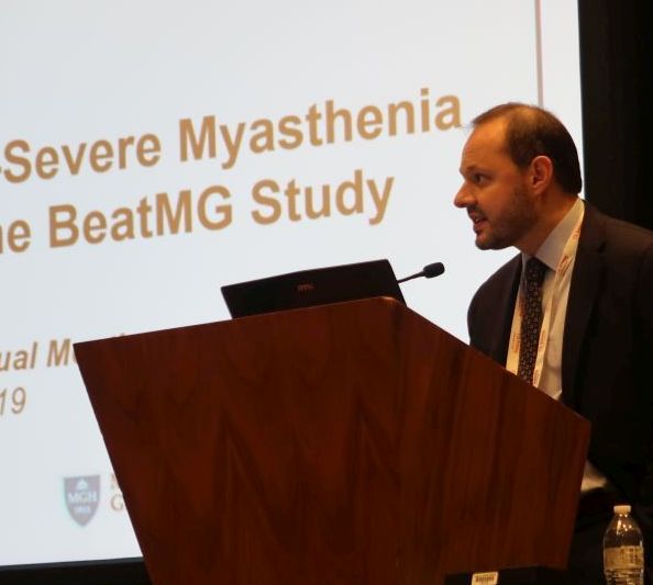
B Cell Targeted Treatment In Myasthenia Gravis (BeatMG) was a 52-week, randomized, double-blind, placebo-controlled clinical trial. The phase 2 study’s primary outcomes were safety and the glucocorticoid-sparing effect assessed by the percentage of patients who reduced their mean daily prednisone dose by at least 75% and maintained clinical stability. Secondary outcomes were change in Myasthenia Gravis Composite (MGC) score and change in Quantitative Myasthenia Gravis (QMG) score from baseline to 52 weeks.
Investigators randomized 52 participants 1:1 to rituximab (Rituxan) or placebo. Patients were taking at least 15 mg/day of prednisone and were a mean of about age 50 years. About two-thirds were treated with glucocorticoids alone at baseline, and nearly two-thirds had a Myasthenia Gravis Foundation of America (MGFA) clinical classification of II. “It was a mildly symptomatic group of individuals in terms of disease severity,” said Richard Nowak, MD, assistant professor of neurology and director of the Yale Myasthenia Gravis Clinic in New Haven, Conn.
During the study, 60% of patients who received rituximab had a 75% or greater reduction in their mean daily prednisone dose and maintained clinical stability. “However, what surprised us is that we had a significantly high placebo response rate of 56%,” he said. The difference between groups was not significant.
Patients who received rituximab had “directionally favorable reductions” in MGC and QMG scores over 52 weeks, compared with patients who received placebo. Nevertheless, “after correcting for baseline differences, there was no significant difference between the two groups,” Dr. Nowak said.
Rituximab had good safety and tolerability, and the placebo group had a threefold higher rate of clinical relapse requiring IV immunoglobulin or plasmapheresis. “While the placebo arm did achieve a similar rate of reduction in their steroid dose, at 52 weeks the patients may have been doing less well, reflected by the higher rate of relapse,” he said.
Subgroup analysis
To explore whether rituximab might benefit patients who were treatment resistant or had more symptomatic disease, Dr. Nowak and his colleagues conducted a post hoc subgroup analysis of 20 participants – 10 in the rituximab arm and 10 in the placebo arm – who were MGFA class III-IV. In each group, 70% were on glucocorticoid treatment alone, and 90% were MGFA class III.
As in the overall study, the glucocorticoid-sparing effect was not significant (60% of the rituximab group vs. 50% of the placebo group).
Mean change in QMG score was –3.9 in the rituximab group and –0.5 in the placebo group, and mean change in MGC score was –7.0 in the rituximab group and –4.8 in the placebo group. These secondary outcomes again show “directional favorability” with rituximab, Dr. Nowak said. The researchers saw similar trends for scores that assess quality of life and activities of daily living.
In the subgroup analysis, myasthenia gravis relapses requiring rescue therapy occurred in 20% of patients in the rituximab arm and in 30% of the placebo arm. Overall, the researchers did not see a difference in treatment response between those with moderate to severe disease and those with mild disease, Dr. Nowak said.
Suggestions from the data
The post hoc subgroup analysis should be interpreted with caution, and the study does not provide firm conclusions, Dr. Nowak noted. In addition, the trial population does not reflect all patients with myasthenia gravis. Dr. Nowak routinely uses rituximab in patients with muscle-specific kinase antibody-positive disease, he said. For acetylcholine receptor antibody-positive generalized myasthenia gravis, which has approved therapies available, Dr. Nowak presents rituximab as an option for some patients. Further research may clarify where B-cell depletion therapy may fit into the treatment paradigm.
Nonetheless, BeatMG and the subgroup analysis may help physicians better understand the disease and the role of various therapies. Investigators successfully lowered prednisone dose “at a pretty high rate in the placebo arm,” he said. “It suggests that many of our patients potentially may be on higher than required prednisone doses.”
Finally, the BeatMG findings emphasize the need for placebo-controlled trials to understand potential therapies. “We need to pause when we see a lot of retrospective and uncontrolled studies that are very promising,” Dr. Nowak said.
The study was supported by the National Institute of Neurological Disorders and Stroke. Genentech provided the study drug and placebo through an investigator-sponsored study agreement. Dr. Nowak has received research support from Alexion Pharmaceuticals, Genentech, Grifols, and Ra Pharmaceuticals. He has served as a paid consultant for Alexion, Momenta, Ra, Roivant, Shire, Grifols, and CSL Behring.
SOURCE: Nowak R et al. AANEM 2019. Unnumbered Abstract: Rituximab in patients with moderate to severe myasthenia gravis: a subgroup analysis of the BeatMG study
AUSTIN, TEX. – Among patients with acetylcholine receptor (AChR) antibody-positive generalized myasthenia gravis, rituximab and placebo may have a similar glucocorticoid-sparing effect regardless of disease severity, according to research presented at the annual meeting of the American Association of Neuromuscular and Electrodiagnostic Medicine.
B Cell Targeted Treatment In Myasthenia Gravis (BeatMG) was a 52-week, randomized, double-blind, placebo-controlled clinical trial. The phase 2 study’s primary outcomes were safety and the glucocorticoid-sparing effect assessed by the percentage of patients who reduced their mean daily prednisone dose by at least 75% and maintained clinical stability. Secondary outcomes were change in Myasthenia Gravis Composite (MGC) score and change in Quantitative Myasthenia Gravis (QMG) score from baseline to 52 weeks.
Investigators randomized 52 participants 1:1 to rituximab (Rituxan) or placebo. Patients were taking at least 15 mg/day of prednisone and were a mean of about age 50 years. About two-thirds were treated with glucocorticoids alone at baseline, and nearly two-thirds had a Myasthenia Gravis Foundation of America (MGFA) clinical classification of II. “It was a mildly symptomatic group of individuals in terms of disease severity,” said Richard Nowak, MD, assistant professor of neurology and director of the Yale Myasthenia Gravis Clinic in New Haven, Conn.
During the study, 60% of patients who received rituximab had a 75% or greater reduction in their mean daily prednisone dose and maintained clinical stability. “However, what surprised us is that we had a significantly high placebo response rate of 56%,” he said. The difference between groups was not significant.
Patients who received rituximab had “directionally favorable reductions” in MGC and QMG scores over 52 weeks, compared with patients who received placebo. Nevertheless, “after correcting for baseline differences, there was no significant difference between the two groups,” Dr. Nowak said.
Rituximab had good safety and tolerability, and the placebo group had a threefold higher rate of clinical relapse requiring IV immunoglobulin or plasmapheresis. “While the placebo arm did achieve a similar rate of reduction in their steroid dose, at 52 weeks the patients may have been doing less well, reflected by the higher rate of relapse,” he said.
Subgroup analysis
To explore whether rituximab might benefit patients who were treatment resistant or had more symptomatic disease, Dr. Nowak and his colleagues conducted a post hoc subgroup analysis of 20 participants – 10 in the rituximab arm and 10 in the placebo arm – who were MGFA class III-IV. In each group, 70% were on glucocorticoid treatment alone, and 90% were MGFA class III.
As in the overall study, the glucocorticoid-sparing effect was not significant (60% of the rituximab group vs. 50% of the placebo group).
Mean change in QMG score was –3.9 in the rituximab group and –0.5 in the placebo group, and mean change in MGC score was –7.0 in the rituximab group and –4.8 in the placebo group. These secondary outcomes again show “directional favorability” with rituximab, Dr. Nowak said. The researchers saw similar trends for scores that assess quality of life and activities of daily living.
In the subgroup analysis, myasthenia gravis relapses requiring rescue therapy occurred in 20% of patients in the rituximab arm and in 30% of the placebo arm. Overall, the researchers did not see a difference in treatment response between those with moderate to severe disease and those with mild disease, Dr. Nowak said.
Suggestions from the data
The post hoc subgroup analysis should be interpreted with caution, and the study does not provide firm conclusions, Dr. Nowak noted. In addition, the trial population does not reflect all patients with myasthenia gravis. Dr. Nowak routinely uses rituximab in patients with muscle-specific kinase antibody-positive disease, he said. For acetylcholine receptor antibody-positive generalized myasthenia gravis, which has approved therapies available, Dr. Nowak presents rituximab as an option for some patients. Further research may clarify where B-cell depletion therapy may fit into the treatment paradigm.
Nonetheless, BeatMG and the subgroup analysis may help physicians better understand the disease and the role of various therapies. Investigators successfully lowered prednisone dose “at a pretty high rate in the placebo arm,” he said. “It suggests that many of our patients potentially may be on higher than required prednisone doses.”
Finally, the BeatMG findings emphasize the need for placebo-controlled trials to understand potential therapies. “We need to pause when we see a lot of retrospective and uncontrolled studies that are very promising,” Dr. Nowak said.
The study was supported by the National Institute of Neurological Disorders and Stroke. Genentech provided the study drug and placebo through an investigator-sponsored study agreement. Dr. Nowak has received research support from Alexion Pharmaceuticals, Genentech, Grifols, and Ra Pharmaceuticals. He has served as a paid consultant for Alexion, Momenta, Ra, Roivant, Shire, Grifols, and CSL Behring.
SOURCE: Nowak R et al. AANEM 2019. Unnumbered Abstract: Rituximab in patients with moderate to severe myasthenia gravis: a subgroup analysis of the BeatMG study
REPORTING FROM AANEM 2019
Congenital myasthenic syndrome diagnosed best with repetitive stimulation and jitter analysis
AUSTIN, TEX. – suggests newly presented research.
“In case RS is negative, SFEMG [single fiber electromyography] alone is not very specific and cannot distinguish CMS from mitochondrial myopathies, even in the presence of impulse blocking,” Vitor Marques Caldas, MD, a neurologist at the Syrian Libanes Hospital in Brasilia, Brazil, and a PhD student at the University of São Paulo, told attendees at the annual meeting of the American Association for Neuromuscular and Electrodiagnostic Medicine. “An isolated SFEMG test can lead to a misdiagnosis of myasthenia syndrome if not interpreted in the right clinical context.”
The researchers sought to understand the relative sensitivity and specificity of low-frequency RS versus jitter analysis using disposable concentric needle electrodes (CNE).
The study involved 69 patients, of whom 19 had mitochondrial myopathy, 18 had congenital myopathy, 18 had CMS, and 14 were asymptomatic controls. The control group all tested normal with both RS and jitter analysis.
The 18 participants with CMS, average age 24 years, received low-frequency RS in at least six different muscles: two distal muscles (abductor digiti minimi and tibialis anterior), two proximal muscles (deltoid and trapezius) and two facial muscles (nasalis and orbicularis oculi). They also underwent jitter analysis of their orbicularis oculi muscle under voluntary activation using CNE.
These patients had heterogeneous genetic profiles: 11 had the CHRNE gene mutation, 2 had the RAPSN gene mutation, 2 had the COLQ gene mutation, 2 had the DOK-7 gene mutation, and 1 had the COL13A1 mutation.
All but two patients with congenital CMS tested positive (88.9%) with RS: one female with CHRNE mutation and one male with RAPSN mutation. Using mean jitter, all but one patient tested positive (94.4%): a female with DOK-7 mutation who had tested abnormal on RS.
All patients with CMS tested positive with at least one of the two tests, but only 83.3% tested positive with both tests, resulting in a sensitivity of 83.3%, a specificity of 100%, and overall accuracy of 95.6% using both tests.
Among the 19 patients with mitochondrial myopathy, 5 had abnormal jitter analysis.
When the researchers looked only at participants with abnormal jitter analysis but normal RS, two of these were patients with CMS, but another seven had congenital or mitochondrial myopathies. Using abnormal jitter alone therefore resulted in a sensitivity of 100% but a specificity of only 86%, for overall 86.5% accuracy.
“It’s important to notice that if you have an abnormal jitter, we have to look at the clinical symptoms of the patients,” Dr. Marques Caldas said in an interview. “Jitter abnormalities are not enough to distinguish between myasthenic disorder and a myopathic disorder.”
The research used no external funding, and Dr. Marques Caldas had no disclosures.
SOURCE: Caldas VM et al. AANEM 2019. Unnumbered Abstract: Sensitivity of neurophysiologic tests regarding the neuromuscular junction in patients with congenital myasthenic syndromes.
REPORTING FROM AANEM 2019
Late response to eculizumab may occur in minority of myasthenia gravis patients
AUSTIN – according to a secondary analysis presented at the annual meeting of the American Association of Neuromuscular and Electrodiagnostic Medicine.
Evidence for the sustained effectiveness of eculizumab, a terminal complement inhibitor, in adult patients with antiacetylcholine receptor antibody-positive refractory generalized myasthenia gravis was provided by the 6-month, double-blind, placebo-controlled REGAIN study and its open-label extension. James F. Howard Jr., MD, distinguished professor of neuromuscular disease at the University of North Carolina in Chapel Hill and colleagues sought to analyze response profiles in REGAIN and its open-label extension.
The findings raise the possibility that complement inhibition with eculizumab should not be abandoned rapidly. “We accept that [eculizumab] works quickly,” Dr. Howard said. “There is an impression that if you don’t respond within the first 3 months, you’re not going to respond. I think these data would suggest otherwise.”
The investigators analyzed participants’ Myasthenia Gravis–Activities of Daily Living (MG-ADL) and Quantitative Myasthenia Gravis (QMG) scores, which had been recorded throughout REGAIN and the extension. They defined early and late responses as improvement in MG-ADL score (i.e., a decrease of three or more points) or QMG score (i.e., a decrease of five or more points) occurring at 12 weeks or earlier or after 12 weeks, respectively, after initiation of eculizumab therapy. Patients randomized to eculizumab in REGAIN initially were treated with an IV induction dose of 900 mg/week before receiving 1,200 mg every 2 weeks thereafter.
Dr. Howard and colleagues included 98 patients in their analysis. Approximately 32% of patients achieved their first response within the first week of treatment, and 15% responded at week 2. About 16% of patients had a late response.
Responses to treatment on the MG-ADL scale had occurred in 67.3% by week 12 and in 84.7% by the end of the extension. Treatment with eculizumab resulted in QMG responses in 56.1% by week 12 and 71.4% by the end of the extension. The investigators observed response over multiple consecutive assessments for the vast majority of patients.
At week 130, the least-squares mean percentage changes from baseline in MG-ADL score were −61.9% and −47.5% in early and late MG-ADL responders, respectively. The least-squares mean percentage changes from baseline in QMG score were −40.8% and −55.5% in early and late QMG responders, respectively.
The investigators observed significant baseline differences between early versus late QMG responders in mean duration of myasthenia gravis (10.46 years vs. 5.46 years) and mean QMG score (18.6 vs. 15.1).
“I can’t explain [this finding]. It may simply be due to the low numbers of patients,” Dr. Howard said. “Whether this is going to hold up in postmarketing analysis remains to be seen. I’m not convinced that that is meaningful.”
Study funding was provided by Alexion Pharmaceuticals (the developer of eculizumab), the Centers for Disease Control and Prevention, and the National Institutes of Health. Dr. Howard reported receiving research support and consulting fees or honoraria from Alexion and several other pharmaceutical companies. Several other authors reported financial relationships with Alexion and other pharmaceutical companies; two authors are employees of Alexion.
Howard J et al. AANEM 2019. Unnumbered Abstract.
REPORTING FROM AANEM 2019
Concussion effects may linger on MRI 1 year after athletes resume play
Among athletes with concussion, the effects of the injury on brain physiology may persist when they return to play and 1 year later.
MRI measures from 24 athletes with concussion significantly differed from those of controls at various time points and changed over time, according to a study published in Neurology. “Different aspects of brain physiology have different patterns of long-term recovery,” the researchers wrote.
While guidelines for safe return to play mainly rely on the resolution of symptoms, “the findings in this study indicate that more research is needed ... to better understand optimal recovery time from a biological standpoint,” wrote first author Nathan W. Churchill, PhD, a researcher at St. Michael’s Hospital in Toronto, and colleagues.
The study provides “evidence of incomplete or ongoing recovery” when athletes return to play, which could entail “a potential risk for long-term sequelae, given the evidence of worse outcomes if a second concussion occurs before recovery is complete,” according to the investigators. In addition,
To examine whether concussion-related brain changes dissipate by 1 year after athletes receive medical clearance to return to play, Dr. Churchill and colleagues analyzed MRI data from 24 college athletes with concussion and 122 control athletes without concussion.
Athletes with concussion were scanned within 1 week of the injury, at return to play a median of 27 days after the concussion, and 1 year after return to play. Control athletes were scanned before the start of the season. Participants’ sports included volleyball, hockey, soccer, football, rugby, basketball, lacrosse, and water polo. The participants had a mean age of about 20 years, and about half were women.
Athletes with concussion had elevated mean diffusivity within 1 week of injury, at return to play, and 1 year later, compared with controls. In athletes with concussion, cerebral blood flow was elevated soon after concussion, normal at return to play, and decreased 1 year later, relative to controls. Global functional connectivity increased and white matter fractional anisotropy decreased near the time of injury and at return to play, but these measures did not significantly differ from those of controls at 1 year.
The study did not capture MRI changes between return to play and 1 year later. In addition, MRI changes might be influenced by a lack of training before resuming play, as well as by exertion and subconcussive impacts after returning to play, the authors noted.
The Canadian Institutes of Health Research, the Canadian Institute for Military and Veterans Health Research, and Siemens Healthineers Canada supported the study. Siemens makes the MRI equipment used in the study. Dr. Churchill and colleagues had no relevant disclosures.
SOURCE: Churchill NW et al. Neurology. 2019 Oct 16. doi: 10.1212/WNL.0000000000008523.
FROM NEUROLOGY
Key clinical point: Among athletes with concussion, the effects of the injury on brain physiology may persist when they return to play and 1 year later.
Major finding: Athletes with concussion had elevated mean diffusivity within 1 week of injury, at return to play, and 1 year later, compared with controls. In athletes with concussion, cerebral blood flow was elevated soon after concussion, normal at return to play, and decreased 1 year later. Global functional connectivity increased and white matter fractional anisotropy decreased near the time of injury and at return to play, but these measures did not significantly differ from those of controls at 1 year.
Study details: An observational study of 24 college athletes with concussion.
Disclosures: The study was supported by the Canadian Institutes of Health Research, the Canadian Institute for Military and Veterans Health Research, and Siemens Healthineers Canada. Siemens makes the MRI equipment used in the study. The researchers had no relevant disclosures.
Source: Churchill NW et al. Neurology. 2019 Oct 16. doi: 10.1212/WNL.0000000000008523.
In utero Zika exposure can have delayed consequences
WASHINGTON – Evidence continues to mount that infants born to moms infected with Zika virus during pregnancy can have neurodevelopmental abnormalities as they age even if they showed no defects at birth, based on follow-up of 890 Colombian children tracked by epidemiologists from the U.S. Centers for Disease Control and Prevention.

Among the 890 neonates born to mothers apparently infected with Zika during pregnancy and followed for up to 2 years, 40 of the 852 (5%) without a detectable birth defect at delivery went on to show some type of neurodevelopmental sequelae during up to 24 months of age, Margaret Honein, PhD, said at an annual scientific meeting on infectious diseases.
In addition, among the children without birth defects at delivery who received follow-up examinations out to about 2 years, the incidence of “alerts” for possible neurodevelopmental issues was 15%-20% for each of the four domains studied (gross motor, fine motor, hearing and language, and personal and social functions), said Dr. Honein, an epidemiologist and chief of the birth defects branch of the CDC. In contrast, 17 of the 38 children (45%) followed who had identifiable birth defects at delivery also showed neurodevelopmental abnormalities when reexamined as long as 2 years after birth. These possible neurodevelopmental abnormalities, designated as alerts, were identified in comparison with a contemporaneous cohort of children born to uninfected mothers in the same regions of Colombia and assessed by the CDC researchers.
This cohort of children born to mothers who became infected with Zika virus during the 2016 Colombian epidemic will not undergo any planned, additional follow-up beyond the initial 2 years, Dr. Honein noted.
The findings she reported were consistent with observations from a much smaller cohort of 70 infants born to Colombian mothers infected with Zika virus while pregnant who had a normal head circumference and a normal clinical examination at delivery. When assessed once or twice 4-18 months after birth, these 70 infants showed an overall greater than one standard deviation (z-score) drop in their scores on the Warner Initial Developmental Evaluation of Adaptive and Functional Skills (WIDEA) metric by 12 months after birth and continuing out to 18 months, said Sarah B. Mulkey, MD, a fetal-neonatal neurologist at Children’s National Health System in Washington. These deficits were especially pronounced in the mobility and social cognition domains of the four-domain WIDEA metric. The social cognition domain is an important predictor of later problems with executive function and other neurologic disorders, Dr. Mulkey said while reporting her findings in a separate talk at the meeting. She acknowledged that the analysis was flawed by comparing the WIDEA outcomes of the Zika virus–exposed children to healthy children from either inner-city Chicago or Canada. Dr. Mulkey said that she and her associates plan to characterize a population of Zika virus–unexposed children in Colombia to use for future comparisons.
The study reported by Dr. Honein involved an enhanced surveillance program launched by the CDC in 2016 in three regions of Colombia and included 1,190 pregnancies accompanied by Zika symptoms in the mother and with a reported pregnancy outcome, including 1,185 live births. Nearly half of the Zika infections occurred during the first trimester, and 34% occurred during the second trimester. However, fewer than a third of the pregnant women underwent some type of laboratory testing to confirm their infection, either by serology or by a DNA-based assay, and of these 28% had a positive finding. Dr. Honein cautioned that many of the specimens that tested negative for Zika virus may have been false negatives.
The birth defects identified among the infants born from an apparently affected pregnancy included brain abnormalities, eye anomalies, and microcephaly, with 5% of the 1,185 live births showing one or more of these outcomes. The neurodevelopmental deficits identified during follow-up of 890 of the children out to 2 years included seizures; abnormalities of tone, movement, or swallowing; and impairments of vision or hearing.
WASHINGTON – Evidence continues to mount that infants born to moms infected with Zika virus during pregnancy can have neurodevelopmental abnormalities as they age even if they showed no defects at birth, based on follow-up of 890 Colombian children tracked by epidemiologists from the U.S. Centers for Disease Control and Prevention.
Among the 890 neonates born to mothers apparently infected with Zika during pregnancy and followed for up to 2 years, 40 of the 852 (5%) without a detectable birth defect at delivery went on to show some type of neurodevelopmental sequelae during up to 24 months of age, Margaret Honein, PhD, said at an annual scientific meeting on infectious diseases.
In addition, among the children without birth defects at delivery who received follow-up examinations out to about 2 years, the incidence of “alerts” for possible neurodevelopmental issues was 15%-20% for each of the four domains studied (gross motor, fine motor, hearing and language, and personal and social functions), said Dr. Honein, an epidemiologist and chief of the birth defects branch of the CDC. In contrast, 17 of the 38 children (45%) followed who had identifiable birth defects at delivery also showed neurodevelopmental abnormalities when reexamined as long as 2 years after birth. These possible neurodevelopmental abnormalities, designated as alerts, were identified in comparison with a contemporaneous cohort of children born to uninfected mothers in the same regions of Colombia and assessed by the CDC researchers.
This cohort of children born to mothers who became infected with Zika virus during the 2016 Colombian epidemic will not undergo any planned, additional follow-up beyond the initial 2 years, Dr. Honein noted.
The findings she reported were consistent with observations from a much smaller cohort of 70 infants born to Colombian mothers infected with Zika virus while pregnant who had a normal head circumference and a normal clinical examination at delivery. When assessed once or twice 4-18 months after birth, these 70 infants showed an overall greater than one standard deviation (z-score) drop in their scores on the Warner Initial Developmental Evaluation of Adaptive and Functional Skills (WIDEA) metric by 12 months after birth and continuing out to 18 months, said Sarah B. Mulkey, MD, a fetal-neonatal neurologist at Children’s National Health System in Washington. These deficits were especially pronounced in the mobility and social cognition domains of the four-domain WIDEA metric. The social cognition domain is an important predictor of later problems with executive function and other neurologic disorders, Dr. Mulkey said while reporting her findings in a separate talk at the meeting. She acknowledged that the analysis was flawed by comparing the WIDEA outcomes of the Zika virus–exposed children to healthy children from either inner-city Chicago or Canada. Dr. Mulkey said that she and her associates plan to characterize a population of Zika virus–unexposed children in Colombia to use for future comparisons.
The study reported by Dr. Honein involved an enhanced surveillance program launched by the CDC in 2016 in three regions of Colombia and included 1,190 pregnancies accompanied by Zika symptoms in the mother and with a reported pregnancy outcome, including 1,185 live births. Nearly half of the Zika infections occurred during the first trimester, and 34% occurred during the second trimester. However, fewer than a third of the pregnant women underwent some type of laboratory testing to confirm their infection, either by serology or by a DNA-based assay, and of these 28% had a positive finding. Dr. Honein cautioned that many of the specimens that tested negative for Zika virus may have been false negatives.
The birth defects identified among the infants born from an apparently affected pregnancy included brain abnormalities, eye anomalies, and microcephaly, with 5% of the 1,185 live births showing one or more of these outcomes. The neurodevelopmental deficits identified during follow-up of 890 of the children out to 2 years included seizures; abnormalities of tone, movement, or swallowing; and impairments of vision or hearing.
REPORTING FROM ID WEEK 2019
Universal coverage may be possible without increases in national spending

The Commonwealth Fund and The Urban Institute looked at eight reform scenarios, including ones that build on the Affordable Care Act and expand to universal coverage or single payer. Two scenarios that continue to utilize private insurance show that, conceptually, broad coverage can be achieved without increasing spending.
“This study is important because it shows that there are several health reform approaches that have the potential to increase the number of people with health insurance, make health care more affordable, and slow cost growth,” David Blumenthal, MD, president of The Commonwealth Fund, said during an Oct. 15 conference call introducing the report.
He called the details that separate the varying models “central to the national debate on health care and health insurance coverage as the 2020 campaign season progresses.”
All the scenarios presented in the report have a foundation in various current Democratic health care reform proposals, although no one specific proposal or legislation is profiled within the eight scenarios presented.
“Our hope is that this extensive analysis will clarify for voters and policy makers the implications of the policy choices before us,” said Sara R. Collins, PhD, the vice president of health care coverage and access at The Commonwealth Fund, during the call.
The first of these scenarios, dubbed “Universal Coverage I: Private and Public Options,” includes continued use of private insurance but also involves a public option and is the first of four options presented in the report to achieve universal coverage by actively enrolling people who are not enrolled in a private plan for one year in the public option with income-scaled premiums. This option would not utilize the ACA employer mandate and would remove the “firewall” that prevents individuals with access to employer-sponsored coverage from accessing financial assistance and seeking individual coverage from the insurance marketplace.
This scenario, as with all but one of the scenarios analyzed in the report, covers all essential benefits as defined in the Affordable Care Act. The only single-payer option that does not cover all of these essential benefits still provides coverage for medically necessary care, including dental, vision, hearing, and long-term services.
The Universal Coverage I scenario does not have any penalties for not carrying insurance, but all legal residents that forgo voluntary coverage from an employer or the marketplace will be automatically enrolled in coverage for which they are responsible for a premium payment.
There would be no expanded access to short-term, limited duration plans as the automatic enrollment to those not voluntarily covered by an employer or in the marketplace would make coverage universal. Federal government spending under this plan increases government health care spending in 2020 by $122.1 billion and $1.5 trillion over 10 years. However, total national spending in this scenario would decrease by $22.6 billion or 0.6% in 2020, compared with current law.
“Universal Coverage II: Enhanced Subsidies” is similar to Universal Coverage I in all other respects other than that it includes more generous premium and cost-sharing subsidies. These additional offerings would push federal government spending up $161.8 billion more in 2020, compared with current law, and to $2 trillion more over the next 10 years, while showing a minimal decrease in total national spending of less than 1% compared with current law.
The other two options that would move toward providing everyone with health insurance include a single payer system that covers all ACA essential health benefits, features no premiums, has income-related cost sharing, and covers all legal residents. Private insurance in this scenario is prohibited, and provider payments would be similar to those received in Medicare. Federal government spending would increase in 2020 by $1.5 trillion, compared with current law, and $17.6 trillion over the next 10 years. However, total national spending would decrease by $209.5 billion, or 6%, in 2020 compared with current law. These savings would come from lower provider payments and administrative costs that outweigh increased costs associated with near universal coverage and lower cost-sharing requirements.
A second single payer scenario broadens the benefits and would cover all residents in the United States, including undocumented residents. It would have no cost-sharing requirements.
The “optimal levels at which the payments for hospitals and doctors and other providers should be paid are really unknown at this time,” said Linda Blumberg, PhD, fellow at The Urban Institute’s Health Policy Center and one of the report authors, during the call.
Providing total coverage for all people in the United States is estimated to increase federal spending by $2.8 trillion in 2020 compared with current law, and $34 trillion over 10 years, with much of this increase accounted for by the shift in existing state and private spending to the federal government. At the same time, total national spending would increase by approximately $720 billion in 2020 compared with current law. Even though employer, household, and state spending would decrease, these savings would not be enough to offset increases in federal spending as well as the increased consumption of health care that comes with more generous benefits. The offsets from lower administrative costs and lower provider payments also would not offset higher spending.
The report only looks at health care spending and does not present any suggestions on revenue to offset the spending.
SOURCE: Blumberg LJ et al. “From Incremental to Comprehensive Health Insurance Reform: How Various Reform Options Compare On Coverage and Costs.” The Commonwealth Fund and The Urban Institute. 2019 Oct 16.
Health care stayed front and center at Democratic debate
This time, it wasn’t just about Medicare-for-all.
Voters got a better look at Democrats’ health care priorities on Tuesday, as
While the debate began on the topic of impeaching President Trump, Sen. Bernie Sanders of Vermont soon steered the discussion back to kitchen-table issues.
“I think what would be a disaster, if the American people believe that all we were doing is taking on Trump,” he said. “We’re forgetting that 87 million Americans are uninsured or underinsured.”
That was only the beginning of a series of health care conversations that lasted through much of the three-hour debate.
With Sen. Elizabeth Warren of Massachusetts polling in second place before the night began, she was pressed to offer more details about what Medicare-for-all would look like under her leadership – in particular, whether she would raise taxes to pay for it.
“I have made clear what my principles are here,” she said. “That is, costs will go up for the wealthy and for big corporations, and for hardworking, middle-class families, costs will go down.”
But Mayor Pete Buttigieg of South Bend, Ind., pushed back, pointing out that, unlike Sen. Sanders – who has said taxes would increase to pay for his universal health care plan – she had not actually said whether she would raise taxes.
“Your signature is to have a plan for everything, except this,” Mr. Buttigieg said. “No plan has been laid out to explain how a multitrillion-dollar hole in this plan that Sen. Warren is putting forward is supposed to get filled in.”
Sen. Amy Klobuchar of Minnesota challenged the practicality of focusing on such a sweeping overhaul as Medicare-for-all. She pushed her support for a public option and noted the importance of issues that get less attention, like long-term care.
“The difference between a plan and a pipe dream is something that you can actually get done,” Sen. Klobuchar said.
But Sen. Warren stood her ground. When she was studying bankruptcy as a professor at Harvard Law School, she said, she noticed that two out of three families that went bankrupt after a medical problem had health insurance. The problem is cost, she said: “That is why hardworking people go broke.”
The candidates also staked their claim on two issues that are critically important to Democratic voters: strengthening gun control measures and guaranteeing access to reproductive health care.
Former Vice President Joe Biden trumpeted his role in securing the now-lapsed assault weapons ban in 1994. Among others, Sen. Kamala Harris of California called for a “comprehensive” background check requirement and a ban on the importation of assault weapons.
And one by one, the candidates vowed to codify abortion access, especially in light of recent conservative attacks in a number of states on the premise of the Supreme Court’s Roe v. Wade decision.
“It’s not an exaggeration to say women will die because these Republican legislatures in these various states who are out of touch with America are telling women what to do with their bodies,” Sen. Harris said, a reference to crackdowns on abortion access in many Republican-controlled states.
After pointing out earlier in the evening that two Planned Parenthood clinics in Ohio recently closed because of a Trump administration policy change, Sen. Cory Booker of New Jersey said he would create an office of reproductive freedom and reproductive rights in his White House.
“It’s an assault on the most fundamental ideal that human beings should control their own body,” Sen. Booker said.
And addressing the opioid crisis, blamed for lowering life expectancy in the United States, many of the candidates called outright for jailing the executives of opioid manufacturers, whom Sen. Harris called “nothing more than some high-level dope dealers.”
“The people who should pay for the treatment are the very people that got people hooked and killed them in the first place,” she said.
The evening was also Sen. Sanders’ first appearance on the debate stage since he had a heart attack and underwent heart surgery just weeks ago. Asked about his health, he seemed impatient: “I’m healthy. I’m feeling great,” Sen. Sanders said as he brought the conversation back to policy.
The debate took place in Westerville, Ohio, a traditionally conservative suburb of Columbus that had turned blue in recent years – a nod to Democrats’ hopes of winning with the support of suburban voters in 2020.
And with those 12 Democrats standing elbow-to-elbow, the debate hosted by CNN and the New York Times had an unusual distinction: the most candidates to ever appear onstage at a presidential debate.
The fifth Democratic debate is scheduled for Nov. 20. The Democratic National Committee plans to hold 12 primary debates in total.
Kaiser Health News is a nonprofit national health policy news service. It is an editorially independent program of the Henry J. Kaiser Family Foundation that is not affiliated with Kaiser Permanente.