User login
Topical options for acne patients continue to expand
yet they are likely underused in today’s clinical practice.

A study of prescribing practices from 2012 to 2014 indicated that dermatologists prescribed retinoids for just 58.8% of acne cases, while nondermatologists prescribed them for only 32.4% of cases. “If the guidelines are telling us that we should use topical retinoids for almost all of our acne patients, why are we using them for half of the patients?” Emmy Graber, MD, MBA, asked during MedscapeLive’s annual Las Vegas Dermatology Seminar. “We have a lot of options today for topical retinoids,” she added, noting that, in the past few years, trifarotene cream 0.005% and new formulations of tazarotene lotion (0.045%) and tretinoin lotion (0.05%) have become available.
According to Dr. Graber, president of The Dermatology Institute of Boston, tazarotene has been considered the most efficacious topical retinoid but is generally the least well tolerated, while adapalene has often been considered to be one of the better-tolerated topical retinoids. “This is a broad generalization,” she said. “One should also take into account the concentration and formulation of the retinoid. Cutaneous adverse events increase in severity as the concentration increases regardless of the vehicle.” There are no studies comparing trifarotene with other topical retinoids, she added.
In two phase 2, double-blind, vehicle-controlled studies (PERFECT 1 and PERFECT 2), researchers randomized more than 2,400 patients with moderate facial or truncal acne to receive trifarotene cream or a vehicle for 12 weeks. The mean percent change from baseline in facial inflammatory lesions in the trifarotene-treated group was –54.4% and –66.2% in PERFECT 1, and PERFECT 2, respectively, while the mean percent change from baseline in facial noninflammatory lesions was –49.7% and –57.7%, respectively.
In addition, the mean percent change from baseline in truncal inflammatory lesions in the trifarotene-treated groups was –57.4% and –65.4%, respectively, while the mean percent change from baseline in truncal noninflammatory lesions was –49.1% and –55.2%, respectively.
The choice of vehicle may affect absorption of topical retinoids, and some formulations may increase skin hydration and decrease transepidermal water loss, “which is a good thing,” Dr. Graber said. “Also, vehicles aim to slow drug delivery over time while also making sure that the drug penetrates into the pilosebaceous unit.”
One recent advance is the honeycomb-like polymeric emulsion technology found in tretinoin 0.05% lotion and tazarotene 0.045% lotion. These formulations contain droplets of the tretinoin and tazarotene embedded in a honeycomb matrix with hydrating agents. “I think this is exciting and could enhance our patient compliance and tolerability,” she said. Another unique feature about these two products, especially the tretinoin product, is the very small particle size with this new formulation. “It’s small enough that it can penetrate down into the pilosebaceous unit,” which is different than with older formulations, in which the tretinoin “largely just sat on the surface of the skin and didn’t penetrate into the pilosebaceous unit.” In addition, she said, “there’s only 9% degradation of the tretinoin in UV light, compared to 72% degradation of standard tretinoin 0.025% gel, and with the new tretinoin formulation, there’s no degradation when used with benzoyl peroxide.”
Another new topical retinoid to consider is a fixed-dose combination of encapsulated benzoyl peroxide 3% and encapsulated tretinoin 0.1% cream (Twyneo), which was approved by the Food and Drug Administration in July 2021 for the treatment of acne in adults and children aged 9 years and older. “Typically, benzoyl peroxide and tretinoin cannot be mixed in the same tube to stability issues,” she said. “Here, each product is individually encapsulated in a silica shell so that they can be applied together.”
The approval was supported by positive results from two phase 3, randomized, double-blind, vehicle-controlled, multicenter studies (NCT03761784 and NCT03761810), in which Twyneo demonstrated efficacy and a favorable tolerability profile in patients aged 9 years and older with facial acne.
Another topical treatment option, dapsone, is now FDA approved for ages 9 and up, expanded from its initial indication for ages 12 and up. The new indication is based on a phase 4, multicenter, open-label study in which acne patients aged 9-11 years applied dapsone 7.5% gel once daily to the face and acne-affected areas on the upper chest, upper back, and shoulders for 12 weeks. After 12 weeks, facial acne was clear or almost clear in about 47% of patients. “Inflammatory, noninflammatory, and total lesions decreased from baseline, but there was a greater reduction in noninflammatory lesions, so if you have a very young patient with acne, now you can consider dapsone gel,” Dr. Graber said.
In August 2020, clascoterone cream became the first topical androgen receptor inhibitor approved for the treatment of acne in patients 12 years of age and older. It is a drug believed to address sebum and inflammation directly in the sebaceous gland and is structurally similar to dihydrotestosterone and spironolactone.
“This is a completely new drug category in acne,” she said. “Unlike all oral antiandrogen therapies, clascoterone cream can be used in both males and females with acne. It’s the first acne drug to have a new mechanism of action in almost 40 years, since isotretinoin was approved in 1982.”
In vitro, she continued, clascoterone competes with dihydrotestosterone for binding to the androgen receptor, inhibiting downstream signaling and leading to inhibited sebum production, reduced secretion of inflammatory cytokines, and inhibition of inflammatory pathways. Two phase 3 studies that led to its approval involved 1,440 patients with moderate to severe facial acne aged 9-58 years. The cream was applied twice a day for 12 weeks and treatment adherence was approximately 90%. The researchers found that clascoterone cream was significantly more effective than vehicle cream at achieving Investigator’s Global Assessment scores of 0 (clear) or 1 (almost clear), the definition of treatment success in the study, and reducing noninflammatory lesion and inflammatory lesion counts at week 12. “There were no safety issues noted during these studies, and clascoterone cream was well tolerated,” Dr. Graber said.
Dr. Graber disclosed that she is a consultant/adviser for Digital Diagnostics, Almirall, Hovione, Keratin Biosciences, La Roche Posay, Ortho Dermatologics, Sebacia, Sol-Gel, Verrica, and WebMD. She is also a research investigator for Hovione, Ortho Dermatologics, Sebacia, and she receives royalties from Wolters Kluwer Health.
MedscapeLive and this news organization are owned by the same parent company.
Commentary by Lawrence W. Eichenfield, MD
Acne vulgaris remains an issue of tremendous importance to preteens, teens, and young adults, with approximately 85% of individuals aged 12-24 being affected. Expanding options for topical treatments may help bring effective disease control. Dr. Graber pointed out that historically, pediatricians and other primary care practitioners utilize topical retinoids less often for acne care as compared with dermatologists or guidelines recommendations (either the AAP’s or AAD’s). There are now expanded options, including over-the-counter retinoids (adapalene 0.1% gel), generic and trade brand topical tretinoin products, prescription adapalene medications, older and recently approved tazarotene products, and a newer type of topical retinoid, trifarotene. Novel formulations and emulsion technology, as well as retinoid developed in combination products, give more options in patients down to 9 years of age. A novel topical anti-androgen, clascoterone, is in its own category, as the first topical “hormonal agent,” allowing hormonal therapy to be used for males as well as females (aged 12 years and up). A recent review in JAMA (2021 Nov 23;326[20]:2055-67) incorporates many of these newer medications into management suggestions, emphasizing that first-line therapies are topical retinoids, benzoyl peroxide, azelaic acid, or combinations of topicals, whereas in more severe disease, oral antibiotics such as doxycycline or minocycline, hormonal therapies such as combination oral conceptive agents or spironolactone, or isotretinoin are most effective.
Dr. Eichenfield is chief of pediatric and adolescent dermatology at Rady Children's Hospital-San Diego. He is vice chair of the department of dermatology and professor of dermatology and pediatrics at the University of California, San Diego. He disclosed that he has served as an investigator and/or consultant to AbbVie, Lilly, Pfizer, Regeneron, Sanofi-Genzyme, and Verrica.
A version of this article first appeared on Medscape.com.
This article was updated 6/18/22.
yet they are likely underused in today’s clinical practice.
A study of prescribing practices from 2012 to 2014 indicated that dermatologists prescribed retinoids for just 58.8% of acne cases, while nondermatologists prescribed them for only 32.4% of cases. “If the guidelines are telling us that we should use topical retinoids for almost all of our acne patients, why are we using them for half of the patients?” Emmy Graber, MD, MBA, asked during MedscapeLive’s annual Las Vegas Dermatology Seminar. “We have a lot of options today for topical retinoids,” she added, noting that, in the past few years, trifarotene cream 0.005% and new formulations of tazarotene lotion (0.045%) and tretinoin lotion (0.05%) have become available.
According to Dr. Graber, president of The Dermatology Institute of Boston, tazarotene has been considered the most efficacious topical retinoid but is generally the least well tolerated, while adapalene has often been considered to be one of the better-tolerated topical retinoids. “This is a broad generalization,” she said. “One should also take into account the concentration and formulation of the retinoid. Cutaneous adverse events increase in severity as the concentration increases regardless of the vehicle.” There are no studies comparing trifarotene with other topical retinoids, she added.
In two phase 2, double-blind, vehicle-controlled studies (PERFECT 1 and PERFECT 2), researchers randomized more than 2,400 patients with moderate facial or truncal acne to receive trifarotene cream or a vehicle for 12 weeks. The mean percent change from baseline in facial inflammatory lesions in the trifarotene-treated group was –54.4% and –66.2% in PERFECT 1, and PERFECT 2, respectively, while the mean percent change from baseline in facial noninflammatory lesions was –49.7% and –57.7%, respectively.
In addition, the mean percent change from baseline in truncal inflammatory lesions in the trifarotene-treated groups was –57.4% and –65.4%, respectively, while the mean percent change from baseline in truncal noninflammatory lesions was –49.1% and –55.2%, respectively.
The choice of vehicle may affect absorption of topical retinoids, and some formulations may increase skin hydration and decrease transepidermal water loss, “which is a good thing,” Dr. Graber said. “Also, vehicles aim to slow drug delivery over time while also making sure that the drug penetrates into the pilosebaceous unit.”
One recent advance is the honeycomb-like polymeric emulsion technology found in tretinoin 0.05% lotion and tazarotene 0.045% lotion. These formulations contain droplets of the tretinoin and tazarotene embedded in a honeycomb matrix with hydrating agents. “I think this is exciting and could enhance our patient compliance and tolerability,” she said. Another unique feature about these two products, especially the tretinoin product, is the very small particle size with this new formulation. “It’s small enough that it can penetrate down into the pilosebaceous unit,” which is different than with older formulations, in which the tretinoin “largely just sat on the surface of the skin and didn’t penetrate into the pilosebaceous unit.” In addition, she said, “there’s only 9% degradation of the tretinoin in UV light, compared to 72% degradation of standard tretinoin 0.025% gel, and with the new tretinoin formulation, there’s no degradation when used with benzoyl peroxide.”
Another new topical retinoid to consider is a fixed-dose combination of encapsulated benzoyl peroxide 3% and encapsulated tretinoin 0.1% cream (Twyneo), which was approved by the Food and Drug Administration in July 2021 for the treatment of acne in adults and children aged 9 years and older. “Typically, benzoyl peroxide and tretinoin cannot be mixed in the same tube to stability issues,” she said. “Here, each product is individually encapsulated in a silica shell so that they can be applied together.”
The approval was supported by positive results from two phase 3, randomized, double-blind, vehicle-controlled, multicenter studies (NCT03761784 and NCT03761810), in which Twyneo demonstrated efficacy and a favorable tolerability profile in patients aged 9 years and older with facial acne.
Another topical treatment option, dapsone, is now FDA approved for ages 9 and up, expanded from its initial indication for ages 12 and up. The new indication is based on a phase 4, multicenter, open-label study in which acne patients aged 9-11 years applied dapsone 7.5% gel once daily to the face and acne-affected areas on the upper chest, upper back, and shoulders for 12 weeks. After 12 weeks, facial acne was clear or almost clear in about 47% of patients. “Inflammatory, noninflammatory, and total lesions decreased from baseline, but there was a greater reduction in noninflammatory lesions, so if you have a very young patient with acne, now you can consider dapsone gel,” Dr. Graber said.
In August 2020, clascoterone cream became the first topical androgen receptor inhibitor approved for the treatment of acne in patients 12 years of age and older. It is a drug believed to address sebum and inflammation directly in the sebaceous gland and is structurally similar to dihydrotestosterone and spironolactone.
“This is a completely new drug category in acne,” she said. “Unlike all oral antiandrogen therapies, clascoterone cream can be used in both males and females with acne. It’s the first acne drug to have a new mechanism of action in almost 40 years, since isotretinoin was approved in 1982.”
In vitro, she continued, clascoterone competes with dihydrotestosterone for binding to the androgen receptor, inhibiting downstream signaling and leading to inhibited sebum production, reduced secretion of inflammatory cytokines, and inhibition of inflammatory pathways. Two phase 3 studies that led to its approval involved 1,440 patients with moderate to severe facial acne aged 9-58 years. The cream was applied twice a day for 12 weeks and treatment adherence was approximately 90%. The researchers found that clascoterone cream was significantly more effective than vehicle cream at achieving Investigator’s Global Assessment scores of 0 (clear) or 1 (almost clear), the definition of treatment success in the study, and reducing noninflammatory lesion and inflammatory lesion counts at week 12. “There were no safety issues noted during these studies, and clascoterone cream was well tolerated,” Dr. Graber said.
Dr. Graber disclosed that she is a consultant/adviser for Digital Diagnostics, Almirall, Hovione, Keratin Biosciences, La Roche Posay, Ortho Dermatologics, Sebacia, Sol-Gel, Verrica, and WebMD. She is also a research investigator for Hovione, Ortho Dermatologics, Sebacia, and she receives royalties from Wolters Kluwer Health.
MedscapeLive and this news organization are owned by the same parent company.
Commentary by Lawrence W. Eichenfield, MD
Acne vulgaris remains an issue of tremendous importance to preteens, teens, and young adults, with approximately 85% of individuals aged 12-24 being affected. Expanding options for topical treatments may help bring effective disease control. Dr. Graber pointed out that historically, pediatricians and other primary care practitioners utilize topical retinoids less often for acne care as compared with dermatologists or guidelines recommendations (either the AAP’s or AAD’s). There are now expanded options, including over-the-counter retinoids (adapalene 0.1% gel), generic and trade brand topical tretinoin products, prescription adapalene medications, older and recently approved tazarotene products, and a newer type of topical retinoid, trifarotene. Novel formulations and emulsion technology, as well as retinoid developed in combination products, give more options in patients down to 9 years of age. A novel topical anti-androgen, clascoterone, is in its own category, as the first topical “hormonal agent,” allowing hormonal therapy to be used for males as well as females (aged 12 years and up). A recent review in JAMA (2021 Nov 23;326[20]:2055-67) incorporates many of these newer medications into management suggestions, emphasizing that first-line therapies are topical retinoids, benzoyl peroxide, azelaic acid, or combinations of topicals, whereas in more severe disease, oral antibiotics such as doxycycline or minocycline, hormonal therapies such as combination oral conceptive agents or spironolactone, or isotretinoin are most effective.
Dr. Eichenfield is chief of pediatric and adolescent dermatology at Rady Children's Hospital-San Diego. He is vice chair of the department of dermatology and professor of dermatology and pediatrics at the University of California, San Diego. He disclosed that he has served as an investigator and/or consultant to AbbVie, Lilly, Pfizer, Regeneron, Sanofi-Genzyme, and Verrica.
A version of this article first appeared on Medscape.com.
This article was updated 6/18/22.
FROM THE MEDSCAPELIVE LAS VEGAS DERMATOLOGY SEMINAR
Cirrhosis comorbidities tied to worse outcomes
Multiple comorbidities appear to worsen mortality outcomes in patients with cirrhosis: Those with compensated cirrhosis and three comorbid conditions have a mortality rate similar to patients with decompensated cirrhosis, according to a new analysis of a population-based cohort in the Dallas-Fort Worth metroplex.

“I think it’s a pretty strong message that just the presence of these chronic diseases has such a strong effect in the long run. They at least contribute to mortality to some extent. It’s really important to focus on these chronic diseases as targets early during the care that we provide to these to cirrhotic patients to make sure that we control them so that, in the long run, we can decrease the premature death and mortality in these patients,” said Mohammad Amin Fallahzadeh, MD, MPH, who presented the results at the annual meeting of the American Association for the Study of Liver Diseases.
The study included 35,361 patients with cirrhosis. The mean age of participants was 59.5 years, 41.8% were female, 29.7% were non-White, and 17.5% were Hispanic. Comorbidities were common, occurring in about 25% of patients. Forty-five percent of comorbidities were cardiovascular diseases (CVD); 28.9% of subjects had one comorbidity, 17.5% had two comorbidities, and 12.6% had three comorbidities.
A Kaplan-Meier curve showed that patients with compensated cirrhosis and no comorbidities had the highest survival over time, while decompensated patients with comorbidities had the lowest survival (P = .01). The curve showed similar survival between patients with compensated cirrhosis and three comorbidities and decompensated patients with no comorbidities.
The risk of death increased with one comorbidity (hazard ratio, 2.5; 95% confidence interval, 2.23-2.8), two comorbidities (HR, 3.27; 95% CI, 2.9-3.69), and three comorbidities (HR, 4.52; 95% CI, 3.99-5.12).
Mortality increased with the number of comorbidities in both compensated and decompensated patients; patients with hepatitis C, alcoholic liver disease, and nonalcoholic fatty liver disease; by race (White, Black, and other); and in different age groups. A stronger effect of comorbidities was seen in compensated patients (HR, 6.4 vs. 4.1), female patients (HR, 5.2 vs. 4.1), and in patients older than age 65 years (HR, 7.2 vs. 3.7 in those aged 45-64 years and 5.0 in those younger than age 45 years).
The researchers also found an apparent synergistic effect of chronic kidney disease (CKD) and CVD. Both conditions were associated with increased risk on their own, but when a patient had both CVD and CKD, mortality was higher than just the added risk of the two conditions.
The findings confirm that patients with cirrhosis and comorbidities seem to have worse quality of life and higher mortality. “I didn’t expect that it would have such a major effect, to make a compensated patient as if they are decompensated, but we definitely see that in our daily practice,” said Dr. Fallahzadeh, who is a 2nd-year internal medicine resident at Baylor University Medical Center, Dallas.
“When a hepatologist or an internist has a visit with a patient who is diagnosed with cirrhosis, they need to screen them for the other chronic diseases like diabetes, CKD, and cardiovascular disease to make sure that if they have any of these conditions, they’ll be under control, or if they need any referral for better management. For example, if they need a nephrology referral, it [should] be done as early as possible so that we can minimize the burden of these diseases in the long run for these patients. And we need to educate the patients as well about controlling these chronic problems,” said Dr. Fallahzadeh.
The findings might make researchers reconsider how to classify compensated and decompensated cirrhosis. “When we talk about decompensated liver disease, we’re talking about variceal hemorrhage, ascites, and encephalopathy. In this case, they’re saying that if you’re compensated and you [have] three of these associated medical conditions, that you could be worse off than decompensated cirrhosis. It’s really challenging the status quo and how we think about these two disease entities. They’re thought of a lot differently in terms of the mortality. That needs to be further elucidated,” said Mayur Brahmania, MD, assistant professor of medicine at Western University, London, Ont., who moderated the session.
A key limitation to the study was that the researchers did not have access to data about medication use, so it could not be determined if comorbidities were being controlled. Body mass index and most lifestyle factors were also uncontrolled.
Dr. Fallahzadeh and Dr. Brahmania have no relevant financial disclosures.
Multiple comorbidities appear to worsen mortality outcomes in patients with cirrhosis: Those with compensated cirrhosis and three comorbid conditions have a mortality rate similar to patients with decompensated cirrhosis, according to a new analysis of a population-based cohort in the Dallas-Fort Worth metroplex.
“I think it’s a pretty strong message that just the presence of these chronic diseases has such a strong effect in the long run. They at least contribute to mortality to some extent. It’s really important to focus on these chronic diseases as targets early during the care that we provide to these to cirrhotic patients to make sure that we control them so that, in the long run, we can decrease the premature death and mortality in these patients,” said Mohammad Amin Fallahzadeh, MD, MPH, who presented the results at the annual meeting of the American Association for the Study of Liver Diseases.
The study included 35,361 patients with cirrhosis. The mean age of participants was 59.5 years, 41.8% were female, 29.7% were non-White, and 17.5% were Hispanic. Comorbidities were common, occurring in about 25% of patients. Forty-five percent of comorbidities were cardiovascular diseases (CVD); 28.9% of subjects had one comorbidity, 17.5% had two comorbidities, and 12.6% had three comorbidities.
A Kaplan-Meier curve showed that patients with compensated cirrhosis and no comorbidities had the highest survival over time, while decompensated patients with comorbidities had the lowest survival (P = .01). The curve showed similar survival between patients with compensated cirrhosis and three comorbidities and decompensated patients with no comorbidities.
The risk of death increased with one comorbidity (hazard ratio, 2.5; 95% confidence interval, 2.23-2.8), two comorbidities (HR, 3.27; 95% CI, 2.9-3.69), and three comorbidities (HR, 4.52; 95% CI, 3.99-5.12).
Mortality increased with the number of comorbidities in both compensated and decompensated patients; patients with hepatitis C, alcoholic liver disease, and nonalcoholic fatty liver disease; by race (White, Black, and other); and in different age groups. A stronger effect of comorbidities was seen in compensated patients (HR, 6.4 vs. 4.1), female patients (HR, 5.2 vs. 4.1), and in patients older than age 65 years (HR, 7.2 vs. 3.7 in those aged 45-64 years and 5.0 in those younger than age 45 years).
The researchers also found an apparent synergistic effect of chronic kidney disease (CKD) and CVD. Both conditions were associated with increased risk on their own, but when a patient had both CVD and CKD, mortality was higher than just the added risk of the two conditions.
The findings confirm that patients with cirrhosis and comorbidities seem to have worse quality of life and higher mortality. “I didn’t expect that it would have such a major effect, to make a compensated patient as if they are decompensated, but we definitely see that in our daily practice,” said Dr. Fallahzadeh, who is a 2nd-year internal medicine resident at Baylor University Medical Center, Dallas.
“When a hepatologist or an internist has a visit with a patient who is diagnosed with cirrhosis, they need to screen them for the other chronic diseases like diabetes, CKD, and cardiovascular disease to make sure that if they have any of these conditions, they’ll be under control, or if they need any referral for better management. For example, if they need a nephrology referral, it [should] be done as early as possible so that we can minimize the burden of these diseases in the long run for these patients. And we need to educate the patients as well about controlling these chronic problems,” said Dr. Fallahzadeh.
The findings might make researchers reconsider how to classify compensated and decompensated cirrhosis. “When we talk about decompensated liver disease, we’re talking about variceal hemorrhage, ascites, and encephalopathy. In this case, they’re saying that if you’re compensated and you [have] three of these associated medical conditions, that you could be worse off than decompensated cirrhosis. It’s really challenging the status quo and how we think about these two disease entities. They’re thought of a lot differently in terms of the mortality. That needs to be further elucidated,” said Mayur Brahmania, MD, assistant professor of medicine at Western University, London, Ont., who moderated the session.
A key limitation to the study was that the researchers did not have access to data about medication use, so it could not be determined if comorbidities were being controlled. Body mass index and most lifestyle factors were also uncontrolled.
Dr. Fallahzadeh and Dr. Brahmania have no relevant financial disclosures.
FROM THE LIVER MEETING
Patient whips out smartphone and starts recording: Trouble ahead?
Joe Lindsey, a 48-year old Colorado-based journalist, has dealt with complex hearing loss for about 15 years. which has led to countless doctor’s visits, treatments, and even surgery in hopes of finding improvement. As time went on and Mr. Lindsey’s hearing deteriorated, he began recording his appointments in order to retain important information.
Mr. Lindsey had positive intentions, but not every patient does.
With smartphones everywhere, recording medical appointments can be fraught with downsides too. While there are clear-cut reasons for recording doctor visits, patients’ goals and how they carry out the taping are key. Audio only? Or also video? With the physician’s knowledge and permission, or without?
These are the legal and ethical weeds doctors find themselves in today, so it’s important to understand all sides of the issue.
The medical world is divided on its sentiments about patients recording their visits. The American Medical Association, in fact, failed to make progress on a recent policy (resolution 007) proposal to encourage that any “audio or video recording made during a medical encounter should require both physician and patient notification and consent.” Rather than voting on the resolution, the AMA house of delegates tabled it and chose to gather more information on the issue.
In most cases, patients are recording their visits in good faith, says Jeffrey Segal, MD, JD, the CEO and founder of Medical Justice, a risk mitigation and reputation management firm for healthcare clinicians. “When it comes to ‘Team, let’s record this,’ I’m a fan,” he says. “The most common reason patients record visits is that there’s a lot of information transferred from the doctor to the patient, and there’s just not enough time to absorb it all.”
While the option is there for patients to take notes, in the give-and-take nature of conversation, this can get difficult. “If they record the visit, they can then digest it all down the road,” says Dr. Segal. “A compliant patient is one who understands what’s expected. That’s the charitable explanation for recording, and I support it.”
It’s that question of good intent, however, that concerns some physicians in today’s highly litigious society. “The worry is that there’s a small subset of patients with an ulterior motive,” says Dr. Segal.
“Some patients do record in case of an event down the road,” he adds. “They want the recording to potentially talk to a lawyer, or to file a board complaint.”
Laws in the United States surrounding recordings are confusing, with variations from state to state. Currently, 39 U.S. states allow for one-party consent — meaning a patient can record a visit without consenting with the physician.
Monica Verduzco-Gutierrez, MD, professor and chair of rehabilitation medicine at University of Texas Health, San Antonio, resides in Texas, which is one of the 39 one-consent states. “Physicians must be aware of this fact and consider how it might be used against them,” she says. “A good practice is to set expectations with the patient from the start. Also, know your hospital’s policy — some may have boundaries surrounding recordings.”
The first step is to know what type of state you practice in. Regardless of whether you are in a one- or two-party consent state — but especially a one-party state — it’s a smart move to add a sign at your office saying that you support the recording of visits, provided the patient is open and transparent about it. “Let the patient know that if they plan to record, they should ask your permission,” says Dr. Segal. “Let them know it’s not appropriate if they haven’t received your permission.”
There are, of course, the occasional horror stories involving surreptitious recordings. “I remember a case where a patient left a phone actively recording in his bag of clothing, which went into the OR with him,” he says. “The background conversation was not flattering to the patient, who happened to be an employee of the hospital. When he came to and listened to the recording, he sued, winning his case.”
The age of video and telehealth
What about the rare situation when a patient pulls out a phone and begins to videotape a conversation? It can be a big slippery slope. “Patients can abuse a video recording with editing, and the recording becomes one-dimensional, which is unfair to the physician,” adds Dr. Segal.
Patients sometimes have other motives as well. “I’m aware of occasions where a doctor/patient visit got heated and the patient took out the phone to video record, sharing it to social media,” says Dr. Segal. “Once someone uses a phone to take video, just stop the conversation. Tell the patient, ‘We’re having a disagreement,’ and that it’s time to put an end to it.”
He adds that from the physician side, a video can be a protagonist in a conversation. “Frankly, a camera on your face changes the nature of things,” Dr. Segal says. “It’s much easier to have the phone sitting in a corner, quietly recording.”
Other scenarios might involve a patient’s family member accompanying the patient and bringing out their phone to record. “Doctors should consider how this might be used against them — it can blow up,” says Dr. Verduzco-Gutierrez. “Draw boundaries on this behavior, using your hospital’s policy if it has one.”
In today’s pandemic landscape, this is particularly important, she adds. “There’s generally more mistrust in the medical system right now,” says Dr. Verduzco-Gutierrez. “People are getting misinformation from sources that aren’t credible, and then want to record their visits because they aren’t receiving the treatment they want, for instance.”
COVID has also added the tricky element of telehealth, which has exploded since 2020. “You don’t know what a patient is doing on the other side of the screen,” Dr. Verduzco-Gutierrez explains. “Face-to-face, you might see them with their phones out, but anything goes with telehealth. You have to be open and communicative with your patients about your policies from the start to avoid any negative connotations.”
How taping can help patients
Mr. Lindsey, the Colorado journalist, is far from alone in his desire to use visit recordings in order to retain valuable information — and with good reason. According to the Dartmouth Institute for Health Policy and Clinical Practice’s Open Recordings Project, at least 1 in 10 patients records their doctor’s visits.
“I realized I was missing things and in a medical setting, that matters,” Mr. Lindsey says. “Last year, once COVID hit and we all began wearing masks, I lost my ability to read lips, one of my coping mechanisms. It became even more important that I had a backup recording to ensure I understood everything.”
Even if a patient doesn’t have hearing loss like Mr. Lindsey, having an audio record of a visit can be useful. According to a 2018 study on patient recall of key information 1 week out from their visits, 49% of decisions and recommendations were recalled accurately without prompting; 36% recalled with a prompt; and 15% recalled erroneously or not at all.
This squares with the personal experiences of Dr. Verduzco-Gutierrez. “I even see this with my mom, who doesn’t remember many details of her doctor’s visits when I ask her,” she says. “This can definitely impact treatment.”
For better or worse
Dr. Verduzco-Gutierrez says that often it comes down to how a patient learns best. “I teach my residents to keep this in mind and to ask the patient in advance what works best for them,” she says. “If a patient is a visual learner, they might want to take notes or have access to the appointment notes after the visit. If they will learn and retain the information best with an audio recording, then offer that option.”
Mr. Lindsey makes it a habit to inform his physicians that he will be making an audio recording of his visits. “I always let them know that I’m recording for accuracy and not to catch them in some sort of falsehood,” he says. “I can get the doctor’s notes, but those are often short and to the point; I can get more information by going back over the recording.”
To date, Mr. Lindsey hasn’t experienced any pushback from his physicians. “No one has balked at the idea or acted surprised that I want to do it,” he explains. “I think most doctors appreciate that we have a tool we can make use of for better care.”
In past coverage of the topic, some healthcare providers weighed in with support for recordings, usually citing personal reasons. “I am so very grateful for the physicians that allowed me to record the medical appointments that I attended with my parents,” said one. “As their adult daughter, I was painfully aware that my parents struggled to process and understand all of the new information coming their way.”
Another expressed support as well, stating that as a patient, he prefers recordings to notes, because the latter “bears little resemblance to the content of the meeting and discussion with the physician. If the patient straightforwardly asks for permission to record, then why not honor the good intent expressed thereby?”
More often than not, patients have good intentions when they decide to hit the record button in a medical visit. A little preparation goes a long way, however, says Dr. Segal: “Assume you’re being recorded, and act accordingly.”
A version of this article first appeared on Medscape.com.
CDC unveils mental health protection plan for health care workers
Federal health officials have outlined a five-part plan to improve and protect the mental health and well-being of America’s health care workers (HCWs) and create sustainable change for the next generation of HCWs.
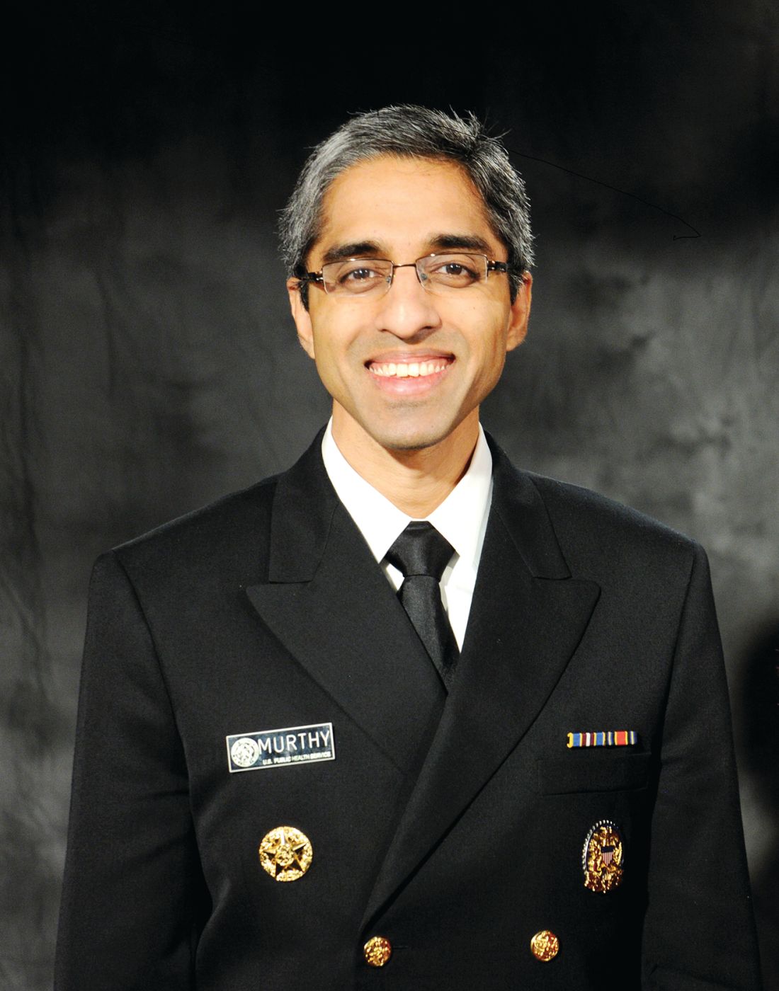
“It’s long past time for us to care for the people who care for all of us and address burnout in our health care workers,” U.S. Surgeon General Vivek H. Murthy, MD, MBA, said during a webinar hosted by the National Institute for Occupational Safety and Health, part of the U.S. Centers for Disease Control and Prevention.
“My hope is that, going forward, we will be able to embark on this journey together to create a health care system, a health care environment, a country where we can not only provide extraordinary care to all those who need it, but where we can take good care of those who have sacrificed so much and make sure that they are well,” Dr. Murthy said.
Burnout is not selective
There are 20 million HCWs in the United States, and no one is immune from burnout, said NIOSH Director John Howard, MD.
![]()
He noted that from June through Sept. of 2020 – the height of the COVID-19 pandemic – 93% of HCWs experienced some degree of stress, with 22% reporting moderate depression and post-traumatic stress disorder.
Looking at subsets of HCWs, a recent survey showed that one in five nurses contemplated leaving the profession because of insufficient staffing, intensity of workload, emotional and physical toll of the job, and lack of support, Dr. Howard noted.
Physician burnout was a significant issue even before the pandemic, with about 79% of physicians reporting burnout. , Dr. Howard said.
Women in health care jobs are especially vulnerable to burnout; 76% of health care jobs are held by women and 64% of physicians that feel burned-out are women, according to federal data.
“We have significant work to do in shoring up the safety and health of women in health care,” Dr. Howard said.
Mental health is also suffering among local and state public health workers. In a recent CDC survey of 26,000 of these workers, 53% reported symptoms of at least one mental health condition in the past 2 weeks.
“That is really an alarming proportion of public health workers who are as vital and essential as nurses and doctors are in our health care system,” Dr. Howard said.
Primary prevention approach
To tackle the burnout crisis, NIOSH plans to:
- Take a deep dive into understanding the personal, social, and economic burdens HCWs face on a daily basis.
- Assimilate the evidence and create a repository of best practices, resources, and interventions.
- Partner with key stakeholders, including the American Hospital Association, the American Nurses Association, National Nurses United, the Joint Commission.
- Identify and adapt tools for the health care workplace that emphasize stress reduction.
NIOSH also plans to “generate awareness through a national, multidimensional social marketing campaign to get the word out about stress so health care workers don’t feel so alone,” Dr. Howard said.
This five-part plan takes a primary prevention approach to identifying and eliminating risk factors for burnout and stress, he added.
Secondary prevention, “when damage has already been done and you’re trying to save a health care worker who is suffering from a mental health issue, that’s a lot harder than taking a good look at what you can do to organizational practices that lead to health care workers’ stress and burnout,” Dr. Howard said.
A version of this article first appeared on Medscape.com.
Federal health officials have outlined a five-part plan to improve and protect the mental health and well-being of America’s health care workers (HCWs) and create sustainable change for the next generation of HCWs.
“It’s long past time for us to care for the people who care for all of us and address burnout in our health care workers,” U.S. Surgeon General Vivek H. Murthy, MD, MBA, said during a webinar hosted by the National Institute for Occupational Safety and Health, part of the U.S. Centers for Disease Control and Prevention.
“My hope is that, going forward, we will be able to embark on this journey together to create a health care system, a health care environment, a country where we can not only provide extraordinary care to all those who need it, but where we can take good care of those who have sacrificed so much and make sure that they are well,” Dr. Murthy said.
Burnout is not selective
There are 20 million HCWs in the United States, and no one is immune from burnout, said NIOSH Director John Howard, MD.
![]()
He noted that from June through Sept. of 2020 – the height of the COVID-19 pandemic – 93% of HCWs experienced some degree of stress, with 22% reporting moderate depression and post-traumatic stress disorder.
Looking at subsets of HCWs, a recent survey showed that one in five nurses contemplated leaving the profession because of insufficient staffing, intensity of workload, emotional and physical toll of the job, and lack of support, Dr. Howard noted.
Physician burnout was a significant issue even before the pandemic, with about 79% of physicians reporting burnout. , Dr. Howard said.
Women in health care jobs are especially vulnerable to burnout; 76% of health care jobs are held by women and 64% of physicians that feel burned-out are women, according to federal data.
“We have significant work to do in shoring up the safety and health of women in health care,” Dr. Howard said.
Mental health is also suffering among local and state public health workers. In a recent CDC survey of 26,000 of these workers, 53% reported symptoms of at least one mental health condition in the past 2 weeks.
“That is really an alarming proportion of public health workers who are as vital and essential as nurses and doctors are in our health care system,” Dr. Howard said.
Primary prevention approach
To tackle the burnout crisis, NIOSH plans to:
- Take a deep dive into understanding the personal, social, and economic burdens HCWs face on a daily basis.
- Assimilate the evidence and create a repository of best practices, resources, and interventions.
- Partner with key stakeholders, including the American Hospital Association, the American Nurses Association, National Nurses United, the Joint Commission.
- Identify and adapt tools for the health care workplace that emphasize stress reduction.
NIOSH also plans to “generate awareness through a national, multidimensional social marketing campaign to get the word out about stress so health care workers don’t feel so alone,” Dr. Howard said.
This five-part plan takes a primary prevention approach to identifying and eliminating risk factors for burnout and stress, he added.
Secondary prevention, “when damage has already been done and you’re trying to save a health care worker who is suffering from a mental health issue, that’s a lot harder than taking a good look at what you can do to organizational practices that lead to health care workers’ stress and burnout,” Dr. Howard said.
A version of this article first appeared on Medscape.com.
Beware of private equity–owned nursing homes: study
When you have to help a parent choose a nursing home or you need nursing home care yourself, you can consult a health care professional, talk to friends, or look at the Nursing Home Compare website of the Centers for Medicare and Medicaid Services (CMS). The CMS website includes star ratings for each nursing home, both overall and on health inspections, staffing and certain quality measures.
But what you might not know is what financial incentives a particular nursing home might have to provide high-quality care, depending on what kind of entity owns the facility.
According to the study, you can expect a somewhat lower level of quality in a PE-owned nursing home than in other for-profit facilities.
The researchers compared CMS data on 302 nursing homes owned by 79 PE firms to data on 9,562 for-profit facilities not owned by such companies from 2013 to 2017. Among fee-for-service Medicare patients in long-term care, private equity acquisitions of nursing homes were associated with an 11.1% increase in ambulatory-care-sensitive (ACS) visits to the emergency department (ED) and an 8.7% increase in ACS hospitalizations per quarter, compared to the changes that occurred in the non-PE-owned facilities, they found.
What’s more, Medicare costs per beneficiary increased 3.9% more – or about $1,000 a year – in the PE-owned nursing homes than they did in the other cohort during the study period.
And when the acquired nursing homes were compared to the nursing homes prior to their acquisition by PE firms, there were no statistically significant differences in unadjusted outcomes, the researchers found. That means the two cohorts were broadly comparable.
The researchers adjusted the numbers in their study for various characteristics of the facilities and their residents. For example, the PE-acquired nursing homes were likely to have a higher percentage of patients covered by Medicare and a lower percentage covered by Medicaid than their non-PE counterparts.
The mean percentages of Black residents, female residents, and residents aged 85 or older were 12.4%, 65.4%, and 36.2%, respectively, for the PE-owned nursing homes and 15.7%, 67.8%, and 39%, respectively, for the non–PE-owned facilities.
Less than optimal outcomes
On average, the residents of non–PE-owned nursing homes had better outcomes than did the patients in the PE-owned facilities. But that doesn’t mean that the average for-profit nursing home had terrific outcomes.
For all the nursing homes in the study, the mean quarterly rate of ACS emergency department visits was 14.1%, and the mean quarterly rate of ACS hospitalizations was 17.3%.
“These events should be largely, although not completely, preventable with appropriate care,” the researchers pointed out.
To date, PE firms have invested about $750 billion in U.S. health care, with nursing homes being a major target of these companies, which currently own 5% of skilled nursing facilities, per the study. PE companies seek annual returns of 20% or more, the paper says, and thus feel pressure to generate high short-term profits. That could lead to reduced staffing, services, supplies, or equipment in their facilities.
Some nursing homes purchased by PE firms may be responsible for the debt incurred in their own leveraged buyouts, the researchers noted. There is also concern that PE firms may focus their properties disproportionately on short-term post-acute care, which is reimbursed at a higher rate than long-term care, the study says.
For all these reasons, some health policy makers are concerned about the long-term impact of private-equity nursing home acquisitions, according to the study.
A version of this article first appeared on WebMD.com.
CDC: All adults should be eligible for Pfizer, Moderna boosters
on its vaccine recommendations.
The Advisory Committee on Immunization Practices, or ACIP, recommended that all adults be eligible for a third dose of a Pfizer or Moderna mRNA vaccine, at least 6 months after their second dose.
They also strengthened a recommendation that everyone over the age of 50 should get a third dose, whether or not they have an underlying health condition that may increase their risk from a COVID-19 infection.
The committee voted 11 to 0 in favor of both policies.
CDC Director Rochelle Walensky, MD, must now sign off on both policies, which she is expected to do.
More than 70 million adults are now eligible for booster shots in the United States, but only about 31 million people have received one. About half of those who have been boosted are over the age of 65.
In a recent survey, the Kaiser Family Foundation found that about 4 in 10 younger adults said they were unsure if they qualified for a booster.
Under the current policy, boosters are recommended for everyone age 65 and older. But people who are younger than age 65 are eligible for boosters if they have an underlying health condition or live or work in a high-risk situation—something individuals have to determine on their own. Experts said that shading of the policy had created confusion that was holding people back.
Nirav Shah, MD, JD, president of the Association of State and Territorial Health Officials, noted that public health officials have been swamped with calls from people who are trying to figure out if they are eligible to get a booster dose.
He said that in a call the evening of Nov. 18 with state health departments, “There was not a single state that voiced opposition to this move,” he told the ACIP.
Dr. Shah said that the current guidelines were well intentioned, but “in pursuit of precision, they create confusion.”
“Our concern is that eligible individuals are not receiving boosters right now as a result of this confusion,” he said.
The committee based its decision on the results of a new study of boosters in Pfizer vaccine recipients, as well as reassuring safety information that’s being collected through the CDC and FDA’s monitoring systems.
Pfizer presented the early results from a study of 10,000 people who had all received two doses of its vaccine. Half of the study participants received a third shot, or booster. The other half got a placebo.
The study is ongoing, but so far, six of the people in the booster group have gotten a COVID-19 infection with symptoms compared to 123 people who got COVID-19 in the placebo group, making boosters 95% effective at keeping people from getting sick. Most people in the study had gotten their original doses about 10 months earlier. They’ve been followed for about 10 weeks since their booster. Importantly, there were no study participants hospitalized for COVID-19 infections in either the placebo or booster group, indicating that the first two doses were still very effective at preventing severe outcomes from infection.
The majority of side effects after a third Pfizer dose were mild and temporary. Side effects like sore arms, swelling, fever, headache, and fatigue were more common in the booster group — affecting about 1 in 4 people who got a third shot. Vaccination side effects were less common after boosters than have been seen after the second dose of the vaccine.
Some cases of myocarditis and pericarditis have been reported after people received vaccine boosters, but the risk for this heart inflammation appears to be extremely low, about two cases for every million doses given. There were 54 cases of myocarditis reported so far to the Vaccine Adverse Event Reporting System, or VAERS. So far, only 12 have met the case definition and are considered related to vaccination. Most of the reported cases are still being studied.
Black young adults: Remember this when facing discrimination
Joel Bervell recalls leaving his hometown of Seattle for the East Coast after being accepted into Yale University.
Still getting accustomed to the big move, Mr. Bervell, who had breezed through high school with straight As, went to see his chemistry professor for advice after getting a low grade on a test.
“He took one look at me and said, ‘Oh, if you’re on the football team, you don’t need to worry about it. So many people from the football team come into the class and end up dropping out, so if you need to drop this class, you can,’ ” Mr. Bervell says.
Mr. Bervell, who is Black, was not on the football team, nor did he receive a sports scholarship of any kind.
“For that professor to make an assumption of me, which to me felt like it was based on my race, made me less likely to want to go into a science field, where I felt like I was being judged before I even had a chance to prove myself,” Mr. Bervell says.
.
Researchers studied health data on 1,834 Americans ages 18-28 over a 10-year span. Findings show that the more instances of discrimination they experienced – including ageism, sexism, and racism – the more likely they were to face mental and behavioral struggles, like mental illness, drug use, severe psychological distress, and poor overall health.
Mr. Bervell, now 26, says he feels lucky that growing up, he was taught healthy ways to process his feelings and emotions.
“Instead of taking that and internalizing it, I said, ‘how can I use this to prove him wrong?’” he says. “Does that mean I need to work harder or does that mean I need to find a different mentor? Surround myself with different people?”
Mr. Bervell is currently a 3rd-year medical student at Washington State University.
When he’s not at the hospital seeing patients, you can find him educating his nearly 340,000 TikTok followers on topics like racial bias in medicine.
Acknowledge the impact
Most Black people don’t tie psychological distress to acts of racism, according to Rheeda Walker, PhD, psychology professor at the University of Houston and author of “The Unapologetic Guide to Black Mental Health” (Oakland, Calif.: New Harbinger Publications, 2020).
Many Black people even normalize it.
“Individuals deal with it [racism] as just another thing, like paying bills, going to work, and studying for class and not as the overwhelming psychological burden that it is,” says Dr. Walker.
And despite what some may say, racial discrimination is not merely “a thing of the past,” Dr. Walker says.
“Instead, discrimination has shifted form from more overt forms of discrimination to less obvious microaggression,” she says.
It’s also critical that young adults are taught how to deal with racism to avoid the risk of “internalizing that they deserve to be mistreated, and/or that they have to work twice as hard to overcome racism,” says Dr. Walker.
“Both scenarios can escalate hopelessness and worry, psychological features of depression and anxiety, respectively,” Dr. Walker says.
Embrace your emotions
Known around the office as “a big teddy bear,” Frederick Herman, a mortgage loan originator based in Charlotte, Va., was coaching a newer employee on how to make sales calls, a common practice in his line of work.
He says a day or 2 days later, his manager let him know that he had made an employee “very uncomfortable” by intimidating them while they were on the phone. Mr. Herman, 29, was told to watch his “aggressive” behavior.
“I’m a bigger Black man. I’m like 6’2, 300 lbs., somewhat muscular. So, if me talking or trying to coach her came off as intimidating, then there’s nothing that I could do or say differently than I was already doing to make her not feel intimidated,” Mr. Herman says.
“If a big teddy bear is now intimidating to you, that just tells me everything I need to know.”
This wasn’t the first time Mr. Herman had been reprimanded for being “too aggressive” or “showing off” when trying to help colleagues at work.
“I’ve had other experiences at work where I may not share my ideas, or I may get super anxious,” says Mr. Herman, a Black man of Haitian descent.
It’s important to allow yourself to feel your emotions after facing acts of discrimination, says Ebony Butler, PhD, a licensed psychologist and creator of My Therapy Cards, a card deck tailored for men, women, and teens of color, with self-care and reflection prompts.
This is a practice called “self-validation” and can reduce the tendency to blame oneself for the mistreatment, says Dr. Butler.
Mr. Herman, 29, says that he recently signed up for therapy to work through his struggles with anxiety.
Relaxation techniques, like grounding and mindfulness, can also be helpful, says Dr. Butler.
“Some example ways to practice grounding are immersing oneself in nature, walking bare feet on the ground, lying on the floor, practicing slow, deep breathing, or engaging the senses,” she says.
“When we are grounded and present, we can better manage our responses and plan our action steps.”
Utilize unique
If you find yourself in a racially charged school or workplace setting, don’t be intimidated, says Wendy Osefo, PhD, education professor at Johns Hopkins University, Baltimore, political commentator, and television personality.
Dr. Osefo made history in 2016 as the first Black woman to earn a PhD in public affairs/community development from Rutgers University.
“Your attitude should be that, no matter how different you might be, you belong, and you earned the right to occupy this space. You’re not less qualified than others who surround you,” she says.
Dr. Ofeso is also CEO of The 1954 Equity Project, an organization that gives minority students tools to succeed in higher education – like mentorships, peer support groups, and other resources and services – all while remaining their authentic selves.
No matter how uncomfortable it might be, staying true to who you are vs. conforming to the masses pays off, says Dr. Osefo.
“Being different is unique and allows you to bring a new and fresh perspective into an environment,” she says.
“Leaning into this uniqueness builds a level of confidence that will aid in your ability to be successful.”
A version of this article first appeared on WebMD.com.
When should psychiatrists retire?
I remember a conversation I had at the end of my training with an older psychiatrist who was closing his practice. I was very excited to finally be a psychiatrist, and therefore a bit shocked that someone would voluntarily end a career I was just beginning. After all, psychiatry is a field where people can practice with flexibility, and a private practice is not an all-or-none endeavor.

“Dinah,” this gentleman said to me, sensing my dismay, “I’m 74. I’m allowed to retire.”
Like many retired psychiatrists, this one continued to come to grand rounds every Monday, dressed in a suit, which was followed by lunch with friends in the dining room. He continued to be involved in professional activities and lived to be 96.
Another dear friend practiced psychiatry until she entered hospice after a 2-year battle with cancer. Others have whittled down their practices, hanging on to a few hours of patient care along with supervision, teaching, and involvement with professional organizations.
In discussing retirement with some of my peers, it’s become immediately clear that each psychiatrist approaches this decision – and how they choose to live after it’s made – with a unique set of concerns and goals.
Fatigued by bureaucracy
Robin Weiss, MD, is in the process of “shrinking” her private practice. She is quick to say she is not retiring, but planning to scale back to 1 day a week starting next summer.
“I want to work less so I have more time for my grandchildren, friends, and travel, and to finally write more.” She also hopes to improve her ping-pong game and exercise habits.
“I’m so tired of prior authorizations, and the one day a week of patients I’ve been committed to feels just about right.”
During the pandemic, Dr. Weiss relinquished her office and she plans to continue with a virtual practice, which allows her more flexibility in terms of where she is physically located.
“The pandemic didn’t influence my decision to scale back, but it did play a role in deciding to give up my office,” she said.
A decision precipitated by medical reasons
Stephen Warres, MD, is a child and adolescent psychiatrist in Maryland who fully retired from practice in June 2021. He started scaling back a few years ago, when he had to give up his office because the building was undergoing renovations.
“I was seeing some patients from my home, but for 2 years I had been working 1 or 2 weekends a month at the Baltimore city jail, and I thought of that as my final act. It was a setting I had never worked in, and I left there 4 months before the pandemic started.”
Dr. Warres noted that his decision to retire was propelled by his diagnosis of Parkinson’s disease at the end of 2019.
“So far I only have a resting tremor, but this is an illness in which cognitive decline is a possibility.”
The emotional roller-coaster that can await
“Why am I leaving when others practice longer? I read about a psychiatrist in California who was still practicing when he died at 102. And the last patient whom I saw when I left practice was a man I started treating just 2 days after I started residency in 1976! When I told him I would be retiring, he found a new psychiatrist who is 82 years old.”
This was followed, he said, by a sense of shame.
“My father was a radiologist and he retired at 76, the same age that I am now, but he volunteered 2 days a week for the state attorney’s office until he was 92, and I’m not doing that.”
What Dr. Warres is choosing to do instead is indulge his many interests, including reading; writing; and practicing on the instrument he’s recently taken up, the harmonium.
This cascade of emotions led to one that was arguably more pleasurable: a sense of immense relief.
“When I got my first request after retirement for a prior authorization, I felt jubilant, like I wanted to throw a party! I felt like I had been walking with a backpack full of weights, and only after the weights were removed did I realize how much lighter it was.
“I loved doing psychotherapy, but more and more psychiatry was not what I had signed up for. I’m relieved that I no longer have to keep up with psychopharmacology. In a way, the Parkinson’s diagnosis sealed the deal. I felt that it gave me license, like a get out of jail card, to retire.”
But even this sense of palpable relief hasn’t closed the cycle of emotions Dr. Warres is experiencing over his retirement.
“You know, the more relieved I am, the more guilt I feel.”
As intellectually adventurous as ever
Marshal Folstein, MD, of Miami retired over a decade ago after a long academic career at Johns Hopkins University, Baltimore, and as chairman of psychiatry at Tufts University, Boston. His Facebook profile states: “Leading the quiet life of a retired professor.”
He said retirement was an easy decision for he and his wife Susan, herself a former academic psychiatrist, which allowed them to immediately change gears.
“At the beginning, we traveled a bit. I wanted to continue with music, so I took flute lessons, and then I played flute in my synagogue, so now I have recently retired from that. I spend my time reading Talmud and the Bible and I keep asking questions. I found a new group of people, some are physicians, and we study and argue. I just turned 80 and I’m intellectually busy and happy.”
The retirement coach
Barbara Fowler, PhD, is a lifespan services consultant at Johns Hopkins who works with faculty and staff getting ready to retire. She said that the university has methods in place to make this decision less jarring.
“The school of medicine has a faculty transition plan that lets people cut back over a set period of time while still keeping benefits. It gives doctors a way to wind up their research and clinical responsibilities, and this is negotiated on an individual level.”
When she’s discussing with someone the possibility of retirement, Dr. Fowler likes to begin by asking them to define what exactly they mean by that word.
“The stereotyped concept is that someone stops what they are doing completely and spends their time playing golf or canasta,” she said. “But the baby boomers are redefining that. Physicians often continue to see some patients or participate in professional organizations. Some people are happy to stop doing the work they have done for years and go do something different, whereas others are interested in scaling back on work activities while adding new ones.”
Timing it right
So, when should psychiatrists retire? The most obvious time to reconsider is when the doctor is no longer able to perform work-related obligations owing to physical or cognitive limitations.
Financial constraints are another factor that comes into play. How necessary is it to work to pay the bills?
“When the kids are out of college and the mortgage is paid off, then there may be the financial means to reconceptualize work life and how you want to rebuild it,” Dr. Fowler said. “Because whether or not people are getting paid, they want to be productive.”
For some, this may come in the form of working in a reduced capacity. Certain practices are more amenable to part-time work or a gradual decrease in hours. A private practice may allow for more control than a position with an institution where an employee may have to continue working full time or not at all.
For others, that productivity might be measured in pursuing their own interests or assisting with family members who need their help. Grandchildren can be an important factor, especially if they live at a distance or childcare is needed. These issues became all the more salient when the pandemic shuttered day care centers and schools, and people limited contact with those outside their households.
Retirement for all physicians is wrapped in issues of identity; for those who have not cultivated other interests, retirement can be a huge loss with no clear path forward. And in an environment where there is a psychiatrist shortage, health care workers are deemed heroes, and human distress is mounting, retirement may come with mixed feelings of guilt, even when the psychiatrist wants a change and is ready for the next chapter. Finally, for those who have launched programs or research projects, there may be the fear that there is no one else who can or will carry on, and that all will be lost.
Yet these considerations focus on the negative, whereas Dr. Fowler said she likes to frame retirement in a positive light. “The key is having more choices; looking for activities that inspire passion; and asking, how can you live your best life?”
Dr. Miller is coauthor of “Committed: The Battle Over Involuntary Psychiatric Care” (Johns Hopkins University Press, 2016). She has a private practice and is assistant professor of psychiatry and behavioral sciences at Johns Hopkins, both in Baltimore. A version of this article first appeared on Medscape.com.
I remember a conversation I had at the end of my training with an older psychiatrist who was closing his practice. I was very excited to finally be a psychiatrist, and therefore a bit shocked that someone would voluntarily end a career I was just beginning. After all, psychiatry is a field where people can practice with flexibility, and a private practice is not an all-or-none endeavor.
“Dinah,” this gentleman said to me, sensing my dismay, “I’m 74. I’m allowed to retire.”
Like many retired psychiatrists, this one continued to come to grand rounds every Monday, dressed in a suit, which was followed by lunch with friends in the dining room. He continued to be involved in professional activities and lived to be 96.
Another dear friend practiced psychiatry until she entered hospice after a 2-year battle with cancer. Others have whittled down their practices, hanging on to a few hours of patient care along with supervision, teaching, and involvement with professional organizations.
In discussing retirement with some of my peers, it’s become immediately clear that each psychiatrist approaches this decision – and how they choose to live after it’s made – with a unique set of concerns and goals.
Fatigued by bureaucracy
Robin Weiss, MD, is in the process of “shrinking” her private practice. She is quick to say she is not retiring, but planning to scale back to 1 day a week starting next summer.
“I want to work less so I have more time for my grandchildren, friends, and travel, and to finally write more.” She also hopes to improve her ping-pong game and exercise habits.
“I’m so tired of prior authorizations, and the one day a week of patients I’ve been committed to feels just about right.”
During the pandemic, Dr. Weiss relinquished her office and she plans to continue with a virtual practice, which allows her more flexibility in terms of where she is physically located.
“The pandemic didn’t influence my decision to scale back, but it did play a role in deciding to give up my office,” she said.
A decision precipitated by medical reasons
Stephen Warres, MD, is a child and adolescent psychiatrist in Maryland who fully retired from practice in June 2021. He started scaling back a few years ago, when he had to give up his office because the building was undergoing renovations.
“I was seeing some patients from my home, but for 2 years I had been working 1 or 2 weekends a month at the Baltimore city jail, and I thought of that as my final act. It was a setting I had never worked in, and I left there 4 months before the pandemic started.”
Dr. Warres noted that his decision to retire was propelled by his diagnosis of Parkinson’s disease at the end of 2019.
“So far I only have a resting tremor, but this is an illness in which cognitive decline is a possibility.”
The emotional roller-coaster that can await
“Why am I leaving when others practice longer? I read about a psychiatrist in California who was still practicing when he died at 102. And the last patient whom I saw when I left practice was a man I started treating just 2 days after I started residency in 1976! When I told him I would be retiring, he found a new psychiatrist who is 82 years old.”
This was followed, he said, by a sense of shame.
“My father was a radiologist and he retired at 76, the same age that I am now, but he volunteered 2 days a week for the state attorney’s office until he was 92, and I’m not doing that.”
What Dr. Warres is choosing to do instead is indulge his many interests, including reading; writing; and practicing on the instrument he’s recently taken up, the harmonium.
This cascade of emotions led to one that was arguably more pleasurable: a sense of immense relief.
“When I got my first request after retirement for a prior authorization, I felt jubilant, like I wanted to throw a party! I felt like I had been walking with a backpack full of weights, and only after the weights were removed did I realize how much lighter it was.
“I loved doing psychotherapy, but more and more psychiatry was not what I had signed up for. I’m relieved that I no longer have to keep up with psychopharmacology. In a way, the Parkinson’s diagnosis sealed the deal. I felt that it gave me license, like a get out of jail card, to retire.”
But even this sense of palpable relief hasn’t closed the cycle of emotions Dr. Warres is experiencing over his retirement.
“You know, the more relieved I am, the more guilt I feel.”
As intellectually adventurous as ever
Marshal Folstein, MD, of Miami retired over a decade ago after a long academic career at Johns Hopkins University, Baltimore, and as chairman of psychiatry at Tufts University, Boston. His Facebook profile states: “Leading the quiet life of a retired professor.”
He said retirement was an easy decision for he and his wife Susan, herself a former academic psychiatrist, which allowed them to immediately change gears.
“At the beginning, we traveled a bit. I wanted to continue with music, so I took flute lessons, and then I played flute in my synagogue, so now I have recently retired from that. I spend my time reading Talmud and the Bible and I keep asking questions. I found a new group of people, some are physicians, and we study and argue. I just turned 80 and I’m intellectually busy and happy.”
The retirement coach
Barbara Fowler, PhD, is a lifespan services consultant at Johns Hopkins who works with faculty and staff getting ready to retire. She said that the university has methods in place to make this decision less jarring.
“The school of medicine has a faculty transition plan that lets people cut back over a set period of time while still keeping benefits. It gives doctors a way to wind up their research and clinical responsibilities, and this is negotiated on an individual level.”
When she’s discussing with someone the possibility of retirement, Dr. Fowler likes to begin by asking them to define what exactly they mean by that word.
“The stereotyped concept is that someone stops what they are doing completely and spends their time playing golf or canasta,” she said. “But the baby boomers are redefining that. Physicians often continue to see some patients or participate in professional organizations. Some people are happy to stop doing the work they have done for years and go do something different, whereas others are interested in scaling back on work activities while adding new ones.”
Timing it right
So, when should psychiatrists retire? The most obvious time to reconsider is when the doctor is no longer able to perform work-related obligations owing to physical or cognitive limitations.
Financial constraints are another factor that comes into play. How necessary is it to work to pay the bills?
“When the kids are out of college and the mortgage is paid off, then there may be the financial means to reconceptualize work life and how you want to rebuild it,” Dr. Fowler said. “Because whether or not people are getting paid, they want to be productive.”
For some, this may come in the form of working in a reduced capacity. Certain practices are more amenable to part-time work or a gradual decrease in hours. A private practice may allow for more control than a position with an institution where an employee may have to continue working full time or not at all.
For others, that productivity might be measured in pursuing their own interests or assisting with family members who need their help. Grandchildren can be an important factor, especially if they live at a distance or childcare is needed. These issues became all the more salient when the pandemic shuttered day care centers and schools, and people limited contact with those outside their households.
Retirement for all physicians is wrapped in issues of identity; for those who have not cultivated other interests, retirement can be a huge loss with no clear path forward. And in an environment where there is a psychiatrist shortage, health care workers are deemed heroes, and human distress is mounting, retirement may come with mixed feelings of guilt, even when the psychiatrist wants a change and is ready for the next chapter. Finally, for those who have launched programs or research projects, there may be the fear that there is no one else who can or will carry on, and that all will be lost.
Yet these considerations focus on the negative, whereas Dr. Fowler said she likes to frame retirement in a positive light. “The key is having more choices; looking for activities that inspire passion; and asking, how can you live your best life?”
Dr. Miller is coauthor of “Committed: The Battle Over Involuntary Psychiatric Care” (Johns Hopkins University Press, 2016). She has a private practice and is assistant professor of psychiatry and behavioral sciences at Johns Hopkins, both in Baltimore. A version of this article first appeared on Medscape.com.
Infectious disease pop quiz: Clinical challenge #2 for the ObGyn
Which major organisms cause urinary tract infections (UTIs) in women?
Continue to the answer...
The most common causative organism is Escherichia coli, which is responsible for approximately 70% of all UTIs. Klebsiella pneumoniae and Proteus species are the 2 other aerobic gram-negative bacilli that are common uropathogens. In addition, 3 gram-positive cocci are important: enterococci, Staphylococcus saprophyticus, and group B streptococcus.
- Duff P. Maternal and perinatal infections: bacterial. In: Landon MB, Galan HL, Jauniaux ERM, et al. Gabbe’s Obstetrics: Normal and Problem Pregnancies. 8th ed. Elsevier; 2021:1124-1146.
- Duff P. Maternal and fetal infections. In: Resnik R, Lockwood CJ, Moore TJ, et al. Creasy & Resnik’s Maternal-Fetal Medicine: Principles and Practice. 8th ed. Elsevier; 2019:862-919.

Dr. Edwards is a Resident in the Department of Medicine, University of Florida College of Medicine, Gainesville.

Dr. Duff is Professor of Maternal-Fetal Medicine, Department of Obstetrics and Gynecology,University of Florida College of Medicine, Gainesville.
The authors report no financial relationships relevant to this article.
Dr. Edwards is a Resident in the Department of Medicine, University of Florida College of Medicine, Gainesville.
Dr. Duff is Professor of Maternal-Fetal Medicine, Department of Obstetrics and Gynecology,University of Florida College of Medicine, Gainesville.
The authors report no financial relationships relevant to this article.
Which major organisms cause urinary tract infections (UTIs) in women?
Continue to the answer...
The most common causative organism is Escherichia coli, which is responsible for approximately 70% of all UTIs. Klebsiella pneumoniae and Proteus species are the 2 other aerobic gram-negative bacilli that are common uropathogens. In addition, 3 gram-positive cocci are important: enterococci, Staphylococcus saprophyticus, and group B streptococcus.
- Duff P. Maternal and perinatal infections: bacterial. In: Landon MB, Galan HL, Jauniaux ERM, et al. Gabbe’s Obstetrics: Normal and Problem Pregnancies. 8th ed. Elsevier; 2021:1124-1146.
- Duff P. Maternal and fetal infections. In: Resnik R, Lockwood CJ, Moore TJ, et al. Creasy & Resnik’s Maternal-Fetal Medicine: Principles and Practice. 8th ed. Elsevier; 2019:862-919.
Infectious disease pop quiz: Clinical challenge #1 for the ObGyn
What are the best tests for the diagnosis of congenital cytomegalovirus (CMV) infection?
Continue to the answer...
When congenital CMV is suspected, if the patient is at least 15 weeks’ gestation, an amniocentesis should be performed to test for CMV DNA in the amniotic fluid using polymerase chain reaction (PCR) methodology. If the initial test is negative, amniocentesis should be repeated in approximately 4 weeks. Coincident with amniocentesis, a detailed ultrasound examination should be performed to search for findings suggestive of fetal injury, such as growth restriction, microcephaly, periventricular calcifications, hepatosplenomegaly, echogenic bowel, and serous effusions in the pleural space or abdomen.
- Duff P. Maternal and perinatal infections: bacterial. In: Landon MB, Galan HL, Jauniaux ERM, et al. Gabbe’s Obstetrics: Normal and Problem Pregnancies. 8th ed. Elsevier; 2021:1124-1146.
- Duff P. Maternal and fetal infections. In: Resnik R, Lockwood CJ, Moore TJ, et al. Creasy & Resnik’s Maternal-Fetal Medicine: Principles and Practice. 8th ed. Elsevier; 2019:862-919.

Dr. Edwards is a Resident in the Department of Medicine, University of Florida College of Medicine, Gainesville.

Dr. Duff is Professor of Maternal-Fetal Medicine, Department of Obstetrics and Gynecology,University of Florida College of Medicine, Gainesville.
The authors report no financial relationships relevant to this article.
Dr. Edwards is a Resident in the Department of Medicine, University of Florida College of Medicine, Gainesville.
Dr. Duff is Professor of Maternal-Fetal Medicine, Department of Obstetrics and Gynecology,University of Florida College of Medicine, Gainesville.
The authors report no financial relationships relevant to this article.
What are the best tests for the diagnosis of congenital cytomegalovirus (CMV) infection?
Continue to the answer...
When congenital CMV is suspected, if the patient is at least 15 weeks’ gestation, an amniocentesis should be performed to test for CMV DNA in the amniotic fluid using polymerase chain reaction (PCR) methodology. If the initial test is negative, amniocentesis should be repeated in approximately 4 weeks. Coincident with amniocentesis, a detailed ultrasound examination should be performed to search for findings suggestive of fetal injury, such as growth restriction, microcephaly, periventricular calcifications, hepatosplenomegaly, echogenic bowel, and serous effusions in the pleural space or abdomen.
- Duff P. Maternal and perinatal infections: bacterial. In: Landon MB, Galan HL, Jauniaux ERM, et al. Gabbe’s Obstetrics: Normal and Problem Pregnancies. 8th ed. Elsevier; 2021:1124-1146.
- Duff P. Maternal and fetal infections. In: Resnik R, Lockwood CJ, Moore TJ, et al. Creasy & Resnik’s Maternal-Fetal Medicine: Principles and Practice. 8th ed. Elsevier; 2019:862-919.