User login
Too Many Migraines, Not Enough Specialists
Key clinical point: Migraine affects millions of patients in Europe. Yet, there aren’t enough specialists adhering to the national and international guidelines on migraines to treat those patients.
Major finding: Patients had seen general practitioners and neurologists for one year prior to seeing specialists. Fifty percent of those patients underwent unnecessary diagnostics and 34.2% had not been treated according to the recommended treatment guidelines.
Study details: Data was collected from 1,935 migraine patients between 2010 and 2018 visiting a headache clinic via standardized questionnaires regarding their prior history and treatment.
Disclosures: None.
Citation: Ziegeler C, et al. J Headache Pain. 2019;20(1):86. doi: 10.1186/s10194-019-1034-8.
Key clinical point: Migraine affects millions of patients in Europe. Yet, there aren’t enough specialists adhering to the national and international guidelines on migraines to treat those patients.
Major finding: Patients had seen general practitioners and neurologists for one year prior to seeing specialists. Fifty percent of those patients underwent unnecessary diagnostics and 34.2% had not been treated according to the recommended treatment guidelines.
Study details: Data was collected from 1,935 migraine patients between 2010 and 2018 visiting a headache clinic via standardized questionnaires regarding their prior history and treatment.
Disclosures: None.
Citation: Ziegeler C, et al. J Headache Pain. 2019;20(1):86. doi: 10.1186/s10194-019-1034-8.
Key clinical point: Migraine affects millions of patients in Europe. Yet, there aren’t enough specialists adhering to the national and international guidelines on migraines to treat those patients.
Major finding: Patients had seen general practitioners and neurologists for one year prior to seeing specialists. Fifty percent of those patients underwent unnecessary diagnostics and 34.2% had not been treated according to the recommended treatment guidelines.
Study details: Data was collected from 1,935 migraine patients between 2010 and 2018 visiting a headache clinic via standardized questionnaires regarding their prior history and treatment.
Disclosures: None.
Citation: Ziegeler C, et al. J Headache Pain. 2019;20(1):86. doi: 10.1186/s10194-019-1034-8.
Grading Dr. Google and treating malaria by the bowlful
Google: A hypochondriac’s dream
Thanks to Google, we have basically free and unlimited access to a huge amount of information right at our fingertips. So when you’re laid up at home with a cold that seems to be taking a turn for the worse, it’s only natural to pull out your phone and ask Google all about your strange new symptoms. After all, the doctor’s office is so far away, and it costs money and time, and who even wants to deal with doctors anyway?

According to a survey commissioned by LetsGetChecked, you’d hardly be alone in turning to Dr. Google. Although 51% of the 2,000 survey respondents consulted a doctor as their first choice for medical help, 65% admitted that they use Google to self-diagnose, 26% have no primary care physician, and about 60% actively avoid doctor’s offices.
So, for the two-thirds of Americans who made an appointment with the good internet doctor, how did things turn out? Not well. About three-quarters reported worrying more afterward, and 43% of all survey respondents managed to convince themselves that they had contracted a serious illness. And the information they looked up? It was wrong more than 60% of the time.
As it turns out, a trained medical professional is actually better than a search engine. Now all we need to do is make accessing health care cheaper, more convenient, and easier to understand. No problem, right?
Better the second time around?
“What’s this stuff?”
“Recycled water. Supposed to taste just like regular water.”
“Did you drink it?”
“I’m not gonna drink it. You drink it.”
“I’m not gonna drink it.”
“Let’s get Mikey!”
“He won’t drink it. He hates everything.”
This time, the kids were right. Mikey did hate the recycled wastewater, or to be more accurate, he was disgusted by it. Like most people, he supports the idea of water conservation but is too disgusted by the source of recycled water to drink it, according to investigators at the University of California, Riverside.
In three separate experiments, volunteers were shown videos about water. One was about water conservation, another was about the urban myth that crocodiles live in New York City sewers, and the third was an educational video demonstrating that recycled wastewater is contaminant free.
In the first experiment, half of the subjects watched the conservation video and half watched the NYC sewer video. Afterward, nearly all participants in each group said no thanks to recycling. In the second experiment, subjects from the two video-watching groups were all shown the third video on recycled water’s purity. That led to a small but insubstantial increase in willingness to use recycled water.
In the third experiment, each of three groups watched one of the videos. Afterward, all subjects were asked to sign a petition supporting conservation and were offered a bottle of water labeled “SMARTdrop – Pure Recycled Water.” About two-thirds of each group signed the petition and took the bottle despite the investigators’ expectation that the group watching the water purity video would have greater acceptance.
Messaging involving water scarcity and conservation alone may not be enough in this case. Instead, the researchers urged “a focus on the more visceral roadblock of disgust.”
Maybe, or maybe not. For now, let’s get back to Mikey and company.
“Don’t tell the kids it’s the recycled wastewater you’ve been trying to get them to drink. You’re the only one who has to know.”
Chicken soup for the malarial soul
A nice hot cup of soup: The refuge of concerned parents who managed to resist looking at Google and decided that their child’s cold wasn’t actually stage IV lung cancer. It’s good, and it’s good for you. But just how good for you? Could the healing powers of soup be harnessed to treat something like, say, malaria?

The research, published in Archives of Disease in Childhood, has perhaps the most adorable setup of any study our cold, stony hearts at LOTME world headquarters have ever seen. The researchers went to a London primary school and asked a group of students to bring in homemade soup for testing and analysis. The students obliged, bringing in dozens of unique soups, of which 56 were tested for 72 hours against the deadliest malaria species, Plasmodium falciparum.
Not every soup was effective, but extracts from five broths were able to halt growth of sexually immature parasites by more than 50%, two of which were about as effective as dihydroartemisinin, a leading antimalarial drug. In addition, four other broths were more than 50% effective at blocking sexual maturation.
The researchers noted that they haven’t analyzed the ingredients of the soups yet and that the utility of soup in combating malaria will depend on a number of factors; regardless, we hope those kids got some serious extra credit. Curing malaria is way more impressive than knowing that mitochondria are the powerhouse of the cell.
Google: A hypochondriac’s dream
Thanks to Google, we have basically free and unlimited access to a huge amount of information right at our fingertips. So when you’re laid up at home with a cold that seems to be taking a turn for the worse, it’s only natural to pull out your phone and ask Google all about your strange new symptoms. After all, the doctor’s office is so far away, and it costs money and time, and who even wants to deal with doctors anyway?
According to a survey commissioned by LetsGetChecked, you’d hardly be alone in turning to Dr. Google. Although 51% of the 2,000 survey respondents consulted a doctor as their first choice for medical help, 65% admitted that they use Google to self-diagnose, 26% have no primary care physician, and about 60% actively avoid doctor’s offices.
So, for the two-thirds of Americans who made an appointment with the good internet doctor, how did things turn out? Not well. About three-quarters reported worrying more afterward, and 43% of all survey respondents managed to convince themselves that they had contracted a serious illness. And the information they looked up? It was wrong more than 60% of the time.
As it turns out, a trained medical professional is actually better than a search engine. Now all we need to do is make accessing health care cheaper, more convenient, and easier to understand. No problem, right?
Better the second time around?
“What’s this stuff?”
“Recycled water. Supposed to taste just like regular water.”
“Did you drink it?”
“I’m not gonna drink it. You drink it.”
“I’m not gonna drink it.”
“Let’s get Mikey!”
“He won’t drink it. He hates everything.”
This time, the kids were right. Mikey did hate the recycled wastewater, or to be more accurate, he was disgusted by it. Like most people, he supports the idea of water conservation but is too disgusted by the source of recycled water to drink it, according to investigators at the University of California, Riverside.
In three separate experiments, volunteers were shown videos about water. One was about water conservation, another was about the urban myth that crocodiles live in New York City sewers, and the third was an educational video demonstrating that recycled wastewater is contaminant free.
In the first experiment, half of the subjects watched the conservation video and half watched the NYC sewer video. Afterward, nearly all participants in each group said no thanks to recycling. In the second experiment, subjects from the two video-watching groups were all shown the third video on recycled water’s purity. That led to a small but insubstantial increase in willingness to use recycled water.
In the third experiment, each of three groups watched one of the videos. Afterward, all subjects were asked to sign a petition supporting conservation and were offered a bottle of water labeled “SMARTdrop – Pure Recycled Water.” About two-thirds of each group signed the petition and took the bottle despite the investigators’ expectation that the group watching the water purity video would have greater acceptance.
Messaging involving water scarcity and conservation alone may not be enough in this case. Instead, the researchers urged “a focus on the more visceral roadblock of disgust.”
Maybe, or maybe not. For now, let’s get back to Mikey and company.
“Don’t tell the kids it’s the recycled wastewater you’ve been trying to get them to drink. You’re the only one who has to know.”
Chicken soup for the malarial soul
A nice hot cup of soup: The refuge of concerned parents who managed to resist looking at Google and decided that their child’s cold wasn’t actually stage IV lung cancer. It’s good, and it’s good for you. But just how good for you? Could the healing powers of soup be harnessed to treat something like, say, malaria?
The research, published in Archives of Disease in Childhood, has perhaps the most adorable setup of any study our cold, stony hearts at LOTME world headquarters have ever seen. The researchers went to a London primary school and asked a group of students to bring in homemade soup for testing and analysis. The students obliged, bringing in dozens of unique soups, of which 56 were tested for 72 hours against the deadliest malaria species, Plasmodium falciparum.
Not every soup was effective, but extracts from five broths were able to halt growth of sexually immature parasites by more than 50%, two of which were about as effective as dihydroartemisinin, a leading antimalarial drug. In addition, four other broths were more than 50% effective at blocking sexual maturation.
The researchers noted that they haven’t analyzed the ingredients of the soups yet and that the utility of soup in combating malaria will depend on a number of factors; regardless, we hope those kids got some serious extra credit. Curing malaria is way more impressive than knowing that mitochondria are the powerhouse of the cell.
Google: A hypochondriac’s dream
Thanks to Google, we have basically free and unlimited access to a huge amount of information right at our fingertips. So when you’re laid up at home with a cold that seems to be taking a turn for the worse, it’s only natural to pull out your phone and ask Google all about your strange new symptoms. After all, the doctor’s office is so far away, and it costs money and time, and who even wants to deal with doctors anyway?
According to a survey commissioned by LetsGetChecked, you’d hardly be alone in turning to Dr. Google. Although 51% of the 2,000 survey respondents consulted a doctor as their first choice for medical help, 65% admitted that they use Google to self-diagnose, 26% have no primary care physician, and about 60% actively avoid doctor’s offices.
So, for the two-thirds of Americans who made an appointment with the good internet doctor, how did things turn out? Not well. About three-quarters reported worrying more afterward, and 43% of all survey respondents managed to convince themselves that they had contracted a serious illness. And the information they looked up? It was wrong more than 60% of the time.
As it turns out, a trained medical professional is actually better than a search engine. Now all we need to do is make accessing health care cheaper, more convenient, and easier to understand. No problem, right?
Better the second time around?
“What’s this stuff?”
“Recycled water. Supposed to taste just like regular water.”
“Did you drink it?”
“I’m not gonna drink it. You drink it.”
“I’m not gonna drink it.”
“Let’s get Mikey!”
“He won’t drink it. He hates everything.”
This time, the kids were right. Mikey did hate the recycled wastewater, or to be more accurate, he was disgusted by it. Like most people, he supports the idea of water conservation but is too disgusted by the source of recycled water to drink it, according to investigators at the University of California, Riverside.
In three separate experiments, volunteers were shown videos about water. One was about water conservation, another was about the urban myth that crocodiles live in New York City sewers, and the third was an educational video demonstrating that recycled wastewater is contaminant free.
In the first experiment, half of the subjects watched the conservation video and half watched the NYC sewer video. Afterward, nearly all participants in each group said no thanks to recycling. In the second experiment, subjects from the two video-watching groups were all shown the third video on recycled water’s purity. That led to a small but insubstantial increase in willingness to use recycled water.
In the third experiment, each of three groups watched one of the videos. Afterward, all subjects were asked to sign a petition supporting conservation and were offered a bottle of water labeled “SMARTdrop – Pure Recycled Water.” About two-thirds of each group signed the petition and took the bottle despite the investigators’ expectation that the group watching the water purity video would have greater acceptance.
Messaging involving water scarcity and conservation alone may not be enough in this case. Instead, the researchers urged “a focus on the more visceral roadblock of disgust.”
Maybe, or maybe not. For now, let’s get back to Mikey and company.
“Don’t tell the kids it’s the recycled wastewater you’ve been trying to get them to drink. You’re the only one who has to know.”
Chicken soup for the malarial soul
A nice hot cup of soup: The refuge of concerned parents who managed to resist looking at Google and decided that their child’s cold wasn’t actually stage IV lung cancer. It’s good, and it’s good for you. But just how good for you? Could the healing powers of soup be harnessed to treat something like, say, malaria?
The research, published in Archives of Disease in Childhood, has perhaps the most adorable setup of any study our cold, stony hearts at LOTME world headquarters have ever seen. The researchers went to a London primary school and asked a group of students to bring in homemade soup for testing and analysis. The students obliged, bringing in dozens of unique soups, of which 56 were tested for 72 hours against the deadliest malaria species, Plasmodium falciparum.
Not every soup was effective, but extracts from five broths were able to halt growth of sexually immature parasites by more than 50%, two of which were about as effective as dihydroartemisinin, a leading antimalarial drug. In addition, four other broths were more than 50% effective at blocking sexual maturation.
The researchers noted that they haven’t analyzed the ingredients of the soups yet and that the utility of soup in combating malaria will depend on a number of factors; regardless, we hope those kids got some serious extra credit. Curing malaria is way more impressive than knowing that mitochondria are the powerhouse of the cell.

Evidence builds for AFib ablation’s efficacy in heart failure
Roughly a third of patients with heart failure also have atrial fibrillation, a comorbid combination notorious for working synergistically to worsen a patient’s quality of life and life expectancy.
During the past year, radiofrequency catheter ablation of atrial fibrillation in patients with both conditions has gathered steam as a way to intervene in at least selected patients, driven by study results that featured attention-grabbing reductions in death and cardiovascular hospitalizations.

The evidence favoring catheter ablation of atrial fibrillation (AFib) in patients with heart failure, particularly patients with heart failure with reduced ejection fraction (HFrEF), ramped up in 2019, spurred largely by a subgroup analysis from the CABANA trial, the largest randomized comparison by far of AFib ablation with antiarrhythmic drug treatment with 2,204 patients.
The past few months also featured release of two meta-analyses that took the CABANA results into account plus findings from about a dozen earlier randomized studies. Both meta-analyses, as well as the heart failure analysis from CABANA, all point in one direction, as stated in the conclusion of one of the meta-analyses: “In patients with AFib, catheter ablation is associated with all-cause mortality benefit, compared with medical therapy, that is driven by patients with AFib and HFrEF. Catheter ablation is safe and reduces cardiovascular hospitalizations and recurrences of atrial arrhythmias” both in patients with paroxysmal and persistent AFib,” wrote Stavros Stavrakis, MD, and his associates in their systematic review of 18 randomized, controlled trials of catheter ablation of AFib in a total of 4,464 patients with or without heart failure (Circ Arrhythm Electrophysiol. 2019 Sep;12[9]: e007414).
Despite these new data and analyses, clinicians seem to have very mixed reactions. Some call for an upgraded recommendation by professional societies that would support more aggressive use of AFib ablation in heart failure patients, and the anecdotal impressions of people who manage these patients are that ablation procedures have recently increased. But others advise caution, and note that in their opinion the efficacy data remain preliminary; the procedure has safety, logistical, and economic concerns; and questions remain about the ability of all active ablation programs to consistently deliver the results seen in published trials.
The meta-analysis led by Dr. Stavrakis showed that catheter ablation of AFib cut all-cause mortality during follow-up by a statistically significant 31%, compared with medical therapy, in all patients regardless of their heart failure status. But in patients with HFrEF, the reduction was 48%, along with a 38% cut in cardiovascular hospitalizations. In contrast, patients without heart failure who underwent AFib ablation showed no significant change in their all-cause mortality, compared with medical management of these patients.
“Based both on our meta-analysis and the CABANA data, patients with AFib most likely to benefit from ablation are patients younger than 65 and those with heart failure,” summed up Dr. Stavrakis, a cardiac electrophysiologist at the Heart Rhythm Institute of the University of Oklahoma in Oklahoma City.
The second meta-analysis, which initially appeared in July, analyzed data from 11 randomized trials of catheter ablations compared with anti-arrhythmic medical therapy for rate or rhythm control with in a total of 3,598 patients who all had heart failure, again including the patients enrolled in the CABANA study. The results showed a significant 49% relative drop in all cause mortality with ablation compared with medical treatment, and a statistically significant 56% cut in hospitalizations, as well as a significant, nearly 7% average, absolute improvement in left ventricular ejection fraction, plus benefits for preventing arrhythmia recurrence and improving quality of life (Eur Heart J. 2019 Jul 11. doi: 10.1093/eurheartj/ehz443).
“The magnitude of the effect seen in the meta-analysis, a 49% reduction in total mortality and a 56% reduction in hospitalizations, is rather staggering, and is larger than typically quoted for other medical interventions or device therapy in heart failure. The treatment effect was uniform among studies, and entirely compatible with the changes in left ventricular function, exercise capacity, and heart failure symptoms. Therefore, although more data are desirable, there are already arguably sufficient data to understand a great deal regarding the impact of a fib ablation,” commented Ross J. Hunter, MRCP, a cardiac electrophysiologist at Barts Heart Centre in London, and his associates in an editorial about this meta-analysis (Eur Heart J. 2019 Oct 22. doi: 10.1093/eurheartj/ehz704).

The heart failure analysis of CABANA (Catheter Ablation vs. Anti-Arrhythmic Drug Therapy for Atrial Fibrillation Trial) itself also showed striking findings when first reported at the annual scientific sessions of the Heart Rhythm Society last May. In presentations he made at this meeting, Douglas L. Packer, MD, CABANA’s lead investigator, reported details of a prespecified subgroup analysis of the 778 patients enrolled in CABANA who had heart failure at baseline, slightly more than a third of the total study enrollment. This was more than double the number of patients identified as specifically having heart failure at entry in the initial publication of CABANA’s findings (JAMA. 2019 Mar 15;321[134]:1261-74). Comparison of the 378 patients with heart failure and randomized to undergo ablation with the 400 with heart failure randomized to medical treatment showed a 36% reduction in the study’s primary, composite endpoint relative to the control group in an intention-to-treat analysis, and a 43% relative cut in all-cause mortality during follow-up, Dr. Packer reported at the May meeting. (As of early November 2019, these results had not yet appeared in a published article.) In contrast, in the 1,422 CABANA patients randomized who did not have heart failure, ablation produced results for these endpoints that were similar to and not statistically different from the outcomes in patients treated medically, said Dr. Packer, a cardiac electrophysiologist and professor of medicine at the Mayo Clinic in Rochester, Minn.
The CABANA results added to what had been previously reported from two other landmark studies that documented incremental efficacy of AFib ablation compared with medical treatment in patients with heart failure: The AATAC (Ablation vs Amiodarone for Treatment of AFib in Patients With Congestive HF and an Implanted Device) study, which randomized 203 patients (Circulation. 2016 Apr 26;133[17]:1637-44), and CASTLE-AF (Catheter Ablation vs. Standard Conventional Therapy in Patients with Left Ventricular Dysfunction and Atrial Fibrillation) trial, which randomized 363 patients (N Engl J Med. 2018 Feb 1;378[5]:417-27). These three studies contributed the most patients and outcomes to the two recent meta-analyses.

“The CASTLE-AF and AATAC trials both showed improved cardiovascular outcomes with ablation in patients with heart failure and AFib. The meta-analysis [by Dr. Stavrakis and his associates] and CABANA subgroup analysis further support use of catheter ablation to improve the outcomes in these patients,” noted Jonathan P. Piccini, MD, a cardiac electrophysiologist at Duke University, Durham, N.C., and a CABANA coinvestigator.
“The CABANA trial was very important because it confirmed the safety of catheter ablation, and more importantly suggested that patients with heart failure may benefit the most [from AFib ablation]. The evidence is very strong to advocate ablation as first-line therapy for selected patients with heart failure. Perhaps the optimal patients are those with [New York Heart Association] class I-III or ambulatory class IV heart failure who are on optimized, guideline-directed medical therapy. We have enough data to make this a class I recommendation. The question that remains is whether this is a cost effective strategy. Because it lowers rehospitalization and death, I suspect it is,” said Luigi Di Biase, MD, lead investigator of AATAC, and director of arrhythmia services at Montefiore Medical Center and professor of medicine at Albert Einstein College of Medicine, both in New York.
Opinions differ on AFib ablation’s role
Despite this expansive assessment of the current status of AFib ablation for patients with heart failure from Dr. Di Biase and shared by others, another camp of cardiologists currently sees ablation as having more limited current utility, as recommended earlier this year by a guideline-update panel representing the American Heart Association, the American College of Cardiology, and the Heart Rhythm Society. The guideline update included this new recommendation for how to use AFib ablation in heart failure patients: “AF catheter ablation may be reasonable in selected patients with symptomatic AFib and heart failure with reduced left ventricular ejection fraction to potentially lower mortality rate and reduce hospitalization for heart failure,” a class IIb recommendation. (J Am Coll Cardiol. 2019 Jul 9;74[1]:104-32). The guideline’s text cited the findings from AATAC and CASTLE-AF, but qualified both studies as “relatively small” and with “highly selected patient populations.” The guideline also incorporated the CABANA results into its considerations (although they may not have had the full analysis in heart failure patients available during their deliberations), but cited the study’s main limitation: CABANA failed to show a statistically significant difference in the primary endpoint in its primary, intention-to-treat analysis, which meant that by the strict statistical criteria that trialists apply to study findings, all other endpoints analyzed using CABANA’s are merely “hypothesis generating” and not definitive.
Questions about the extent of patient selection required to see a clear clinical-endpoint benefit from AFib ablation in heart failure patients, as well as the flawed validity of the CABANA results for making unqualified practice recommendation are the main arguments advanced by experts who caution against broader and more routine ablations.
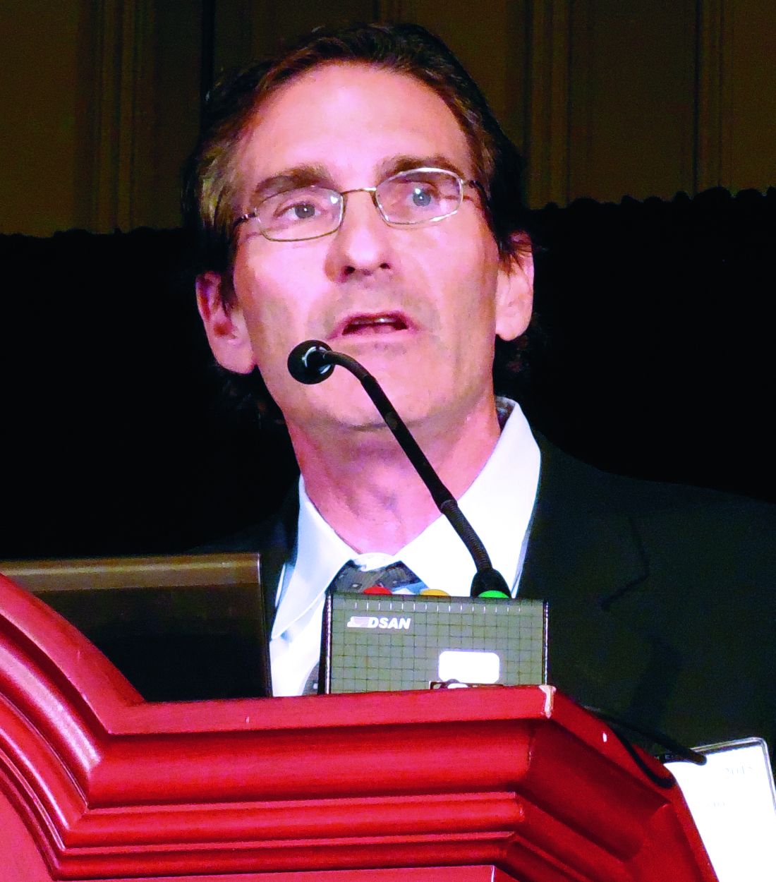
“The findings from the heart failure subgroup of CABANA are hypothesis generating rather than definitive. Even with the recent meta-analysis, uncertainty remains regarding the ability of catheter ablation to improve outcomes beyond reducing AFib-related symptoms,” commented Gregg Fonarow, MD, a heart failure physician and professor of medicine at the University of California, Los Angeles.
“CASTLE-HF had fewer than 100 deaths combined in both arms, which means very unstable results. We don’t know a lot of detail about the heart failure patients in CABANA, and overall we do not have much data from patients with heart failure with preserved ejection fraction [HFpEF],” said Javed Butler, MD, a heart failure physician and professor and chairman of medicine at the University of Mississippi in Jackson. Dr. Butler also voiced his concerns (shared by other heart failure specialists) about the safety of ablation in heart failure patients, noting that “many patients require multiple ablations; many burns result in scarring and can worsen atrial function. In short, ablation of AFib is probably good for selected patients, but to have a class 1 recommendation, we need much larger trials with well-phenotyped heart failure patients,” Dr. Butler said in an interview.
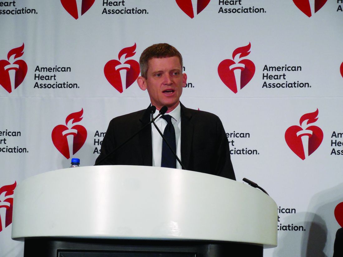
“The totality of data still captures a relatively small number of patients. CASTLE-HF took 8 years to enroll fewer than 400 patients, and the results showed some heterogeneity. Study patients were a decade younger than average HFrEF patients in the community, and thus the effectiveness and safety of catheter ablation in people with more comorbidity and frailty remains in question. Certain HFrEF patients may be less likely to benefit, such as those with amyloid cardiomyopathy. And with the increasing availability of other treatments for HFrEF such as sacubitril/valsartan, dapagliflozin, and MitraClip, it is less clear how catheter ablation would [benefit patients] on top of what is now current best therapy,” said Larry Allen, MD, a heart failure physician and professor of medicine at the University of Colorado in Aurora.
“With these limitations and the fact that catheter ablation is not a simple procedure, a large randomized, controlled trial of ablation, compared with no ablation, in a wide range of HFrEF patients on contemporary therapy would be welcome,” Dr. Allen said. “Given the prevalence of heart failure and AFib and the potential positive and negative implications of catheter ablation running such a trial seems critical for patients and for society.”
“For ablation of AFib in heart failure to become a class I recommendation there will need to be results from larger randomized studies,” summed up Dr. Stavrakis. The meta-analysis that he coauthored noted that “the benefits of catheter ablation for AFib in HFrEF patients have been consistently shown for over a decade now; however, the uptake of this procedure by clinicians in practice has been slow.”
Despite this history of reticence and ongoing caution about ablation, some cardiology experts see the indications for AFib ablation in heart failure steadily creeping forward, buoyed by a safety record that has more benign than ablation’s reputation suggests.

The CABANA results showed that “ablation is remarkably safe in the hands of experienced clinicians, with risks comparable to anti-arrhythmic drugs,” said Peter R. Kowey, MD, a specialist in treating AFib and professor of medicine at Thomas Jefferson University in Philadelphia, who made this assessment during a talk at an AFib meeting in early 2019. Dr. Kowey’s take on what the CABANA safety data showed contrasts with the impression of other cardiologists who are wary of perceived dangers from ablation.
“Ablation comes with a lot of morbidity and mortality. It’s not that the idea of ablation is wrong, but the ability to do it without a lot of adverse effects. ... We’re not quite there yet,” said Douglas L. Mann, MD, a heart failure physician and professor of medicine and chief of the cardiovascular division at Washington University in St. Louis.

“If I had a patient with HFrEF and AFib who was really sick, I’m not so sure I’d send them for ablation, which is not a simple procedure. The patients we tend to send for ablation are selected. Ablation is a big undertaking in patients who are already sick, and it’s expensive. I don’t think the data we have now will change the consensus view, but every heart failure physician is sending some patients for AFib ablation. People are turning to AFib ablation earlier than before. I think the consensus is that ablation is for symptoms or poor rate control, not for better outcomes,” said Mariell Jessup, MD, a heart failure specialist and chief science and medical officer of the American Heart Association in Dallas.

However, this caution about safety and skepticism over efficacy may be dissipating as experience with ablation accumulates.
“CASTLE-AF and other data, including evidence for the apparent isolation of beta-blocker benefit to patients in sinus rhythm, have made me much more proactive about considering catheter ablation in my HFrEF patients. I think many other cardiologists have a similar view,” said Dr. Allen in an interview.
“A lot [of heart failure] patients are [being] referred for ablation, depending on the practice, setting, the local availability of electrophysiologists, and patient interest in ablation,” said Dr. Butler.

“We have no absolutely compelling data, but the data we have all point in the same direction. Like most, I am becoming convinced that AFib ablation in heart failure patients is a very valuable method for managing patients, but I can’t point to one study that was conclusive. Results from lots of studies show that it is likely, and when you add them all together it looks indisputable,” commented A. John Camm, MD, an atrial fibrillation specialist and professor of clinical cardiology at St. George’s University in London. “The findings put a responsibility on cardiologists to assess patients with heart failure for AFib. But there are nothing like enough resources to deal with all the patients who have heart failure who also have AFib.”
A rough estimate of just the U.S. volume of patients with heart failure and AFib is likely in the ball park of 2 million people (a third of the estimated 6 million American currently living with heart failure), and with the prevalence of each of these disorders rising precipitously (more than 5 million Americans have AFib) the confluence of the two should also show a steady increase. “It will take a major change in our concept of heart failure management to really address this. Potentially it would mean a large increase in the number of RF ablations of AFib, but the resources for that are not now present,” Dr. Camm said in an interview.
The attractions of catheter ablation also stand in contrast to the limitations of alternative treatments. Ablation is effective in a majority of patients for reducing AFib burden, both the frequency and duration of AFib episodes, and safety issues are mostly limited to the procedural and immediate postprocedural periods. The drugs available for trying to control AFib are beta-blockers, which provide rate control and can help prevent AFib onset, and rhythm-controlling anti-arrhythmic drugs like amiodarone, which have substantial limitations in both their ability to prevent arrhythmia recurrences as well as for safety.
“Most of the conventional antiarrhythmic drugs are contraindicated, frequently ineffective, or not well tolerated in patients with HFrEF. Catheter ablation of AFib provides an increasingly important option for rhythm control in these patients without using antiarrhythmic drugs,” Dr. Di Biase and his associates wrote in a recent review of AFib ablation in heart failure patients (Eur Heart J. 2019 Feb 21;40[8]:663-71).
“The guidelines that are controversial still make amiodarone a class I drug even though it’s been associated with serious side effects and has been shown in several heart failure trials to increase mortality. I can’t believe that ablation is a class IIb recommendation while a drug like amiodarone is a class I recommendation,” Dr. Di Biase said.
And although beta-blockers are a mainstay of heart failure treatment, once AFib becomes established they are less useful for maintaining sinus rhythm. “Beta-blockers provide effective rate control, but they can’t convert patients to sinus rhythm [once AFib begins], and there is no convincing evidence that patients on beta-blockers stay in sinus rhythm longer. You can’t just say: the patient is on a beta-blocker so I’ve done my best,” noted Dr. Jessup.
CABANA received funding from Biosense Webster, Boston Scientific, Medtronic, and St. Jude. Dr. Stavrakis, Dr, Jessup, and Dr. Di Biase. Dr. Hunter has received research funding, educational grants, and speakers fees from Biosense Webster and Medtronic. Dr. Packer had received honoraria from Biotronik and MediaSphere Medical and research support from several companies. Dr. Piccini has been a consultant to Allergan, Biotronik, Medtronic, Phillips, and Sanofi Aventis, he has received research funding from Abbott, ARCA biopharma, Boston Scientific, Gilead, and Johnson & Johnson, and he had a financial relationship with GlaxoSmithKline. Dr. Fonarow has been a consultant to Abbott, Amgen, Bayer, Janssen, and Novartis. Dr. Butler has been a consultant to several companies. Dr. Allen has been a consultant to Boston Scientific, Janssen, and Novartis. Dr. Kowey has been a consultant to several companies. Dr. Mann has been a consultant to Bristol-Myers Squibb, Corvia, and Novartis, and an adviser to miRagen. Dr. Camm has been a consultant to several companies.
This is part one of a two-part article.
Roughly a third of patients with heart failure also have atrial fibrillation, a comorbid combination notorious for working synergistically to worsen a patient’s quality of life and life expectancy.
During the past year, radiofrequency catheter ablation of atrial fibrillation in patients with both conditions has gathered steam as a way to intervene in at least selected patients, driven by study results that featured attention-grabbing reductions in death and cardiovascular hospitalizations.
The evidence favoring catheter ablation of atrial fibrillation (AFib) in patients with heart failure, particularly patients with heart failure with reduced ejection fraction (HFrEF), ramped up in 2019, spurred largely by a subgroup analysis from the CABANA trial, the largest randomized comparison by far of AFib ablation with antiarrhythmic drug treatment with 2,204 patients.
The past few months also featured release of two meta-analyses that took the CABANA results into account plus findings from about a dozen earlier randomized studies. Both meta-analyses, as well as the heart failure analysis from CABANA, all point in one direction, as stated in the conclusion of one of the meta-analyses: “In patients with AFib, catheter ablation is associated with all-cause mortality benefit, compared with medical therapy, that is driven by patients with AFib and HFrEF. Catheter ablation is safe and reduces cardiovascular hospitalizations and recurrences of atrial arrhythmias” both in patients with paroxysmal and persistent AFib,” wrote Stavros Stavrakis, MD, and his associates in their systematic review of 18 randomized, controlled trials of catheter ablation of AFib in a total of 4,464 patients with or without heart failure (Circ Arrhythm Electrophysiol. 2019 Sep;12[9]: e007414).
Despite these new data and analyses, clinicians seem to have very mixed reactions. Some call for an upgraded recommendation by professional societies that would support more aggressive use of AFib ablation in heart failure patients, and the anecdotal impressions of people who manage these patients are that ablation procedures have recently increased. But others advise caution, and note that in their opinion the efficacy data remain preliminary; the procedure has safety, logistical, and economic concerns; and questions remain about the ability of all active ablation programs to consistently deliver the results seen in published trials.
The meta-analysis led by Dr. Stavrakis showed that catheter ablation of AFib cut all-cause mortality during follow-up by a statistically significant 31%, compared with medical therapy, in all patients regardless of their heart failure status. But in patients with HFrEF, the reduction was 48%, along with a 38% cut in cardiovascular hospitalizations. In contrast, patients without heart failure who underwent AFib ablation showed no significant change in their all-cause mortality, compared with medical management of these patients.
“Based both on our meta-analysis and the CABANA data, patients with AFib most likely to benefit from ablation are patients younger than 65 and those with heart failure,” summed up Dr. Stavrakis, a cardiac electrophysiologist at the Heart Rhythm Institute of the University of Oklahoma in Oklahoma City.
The second meta-analysis, which initially appeared in July, analyzed data from 11 randomized trials of catheter ablations compared with anti-arrhythmic medical therapy for rate or rhythm control with in a total of 3,598 patients who all had heart failure, again including the patients enrolled in the CABANA study. The results showed a significant 49% relative drop in all cause mortality with ablation compared with medical treatment, and a statistically significant 56% cut in hospitalizations, as well as a significant, nearly 7% average, absolute improvement in left ventricular ejection fraction, plus benefits for preventing arrhythmia recurrence and improving quality of life (Eur Heart J. 2019 Jul 11. doi: 10.1093/eurheartj/ehz443).
“The magnitude of the effect seen in the meta-analysis, a 49% reduction in total mortality and a 56% reduction in hospitalizations, is rather staggering, and is larger than typically quoted for other medical interventions or device therapy in heart failure. The treatment effect was uniform among studies, and entirely compatible with the changes in left ventricular function, exercise capacity, and heart failure symptoms. Therefore, although more data are desirable, there are already arguably sufficient data to understand a great deal regarding the impact of a fib ablation,” commented Ross J. Hunter, MRCP, a cardiac electrophysiologist at Barts Heart Centre in London, and his associates in an editorial about this meta-analysis (Eur Heart J. 2019 Oct 22. doi: 10.1093/eurheartj/ehz704).
The heart failure analysis of CABANA (Catheter Ablation vs. Anti-Arrhythmic Drug Therapy for Atrial Fibrillation Trial) itself also showed striking findings when first reported at the annual scientific sessions of the Heart Rhythm Society last May. In presentations he made at this meeting, Douglas L. Packer, MD, CABANA’s lead investigator, reported details of a prespecified subgroup analysis of the 778 patients enrolled in CABANA who had heart failure at baseline, slightly more than a third of the total study enrollment. This was more than double the number of patients identified as specifically having heart failure at entry in the initial publication of CABANA’s findings (JAMA. 2019 Mar 15;321[134]:1261-74). Comparison of the 378 patients with heart failure and randomized to undergo ablation with the 400 with heart failure randomized to medical treatment showed a 36% reduction in the study’s primary, composite endpoint relative to the control group in an intention-to-treat analysis, and a 43% relative cut in all-cause mortality during follow-up, Dr. Packer reported at the May meeting. (As of early November 2019, these results had not yet appeared in a published article.) In contrast, in the 1,422 CABANA patients randomized who did not have heart failure, ablation produced results for these endpoints that were similar to and not statistically different from the outcomes in patients treated medically, said Dr. Packer, a cardiac electrophysiologist and professor of medicine at the Mayo Clinic in Rochester, Minn.
The CABANA results added to what had been previously reported from two other landmark studies that documented incremental efficacy of AFib ablation compared with medical treatment in patients with heart failure: The AATAC (Ablation vs Amiodarone for Treatment of AFib in Patients With Congestive HF and an Implanted Device) study, which randomized 203 patients (Circulation. 2016 Apr 26;133[17]:1637-44), and CASTLE-AF (Catheter Ablation vs. Standard Conventional Therapy in Patients with Left Ventricular Dysfunction and Atrial Fibrillation) trial, which randomized 363 patients (N Engl J Med. 2018 Feb 1;378[5]:417-27). These three studies contributed the most patients and outcomes to the two recent meta-analyses.
“The CASTLE-AF and AATAC trials both showed improved cardiovascular outcomes with ablation in patients with heart failure and AFib. The meta-analysis [by Dr. Stavrakis and his associates] and CABANA subgroup analysis further support use of catheter ablation to improve the outcomes in these patients,” noted Jonathan P. Piccini, MD, a cardiac electrophysiologist at Duke University, Durham, N.C., and a CABANA coinvestigator.
“The CABANA trial was very important because it confirmed the safety of catheter ablation, and more importantly suggested that patients with heart failure may benefit the most [from AFib ablation]. The evidence is very strong to advocate ablation as first-line therapy for selected patients with heart failure. Perhaps the optimal patients are those with [New York Heart Association] class I-III or ambulatory class IV heart failure who are on optimized, guideline-directed medical therapy. We have enough data to make this a class I recommendation. The question that remains is whether this is a cost effective strategy. Because it lowers rehospitalization and death, I suspect it is,” said Luigi Di Biase, MD, lead investigator of AATAC, and director of arrhythmia services at Montefiore Medical Center and professor of medicine at Albert Einstein College of Medicine, both in New York.
Opinions differ on AFib ablation’s role
Despite this expansive assessment of the current status of AFib ablation for patients with heart failure from Dr. Di Biase and shared by others, another camp of cardiologists currently sees ablation as having more limited current utility, as recommended earlier this year by a guideline-update panel representing the American Heart Association, the American College of Cardiology, and the Heart Rhythm Society. The guideline update included this new recommendation for how to use AFib ablation in heart failure patients: “AF catheter ablation may be reasonable in selected patients with symptomatic AFib and heart failure with reduced left ventricular ejection fraction to potentially lower mortality rate and reduce hospitalization for heart failure,” a class IIb recommendation. (J Am Coll Cardiol. 2019 Jul 9;74[1]:104-32). The guideline’s text cited the findings from AATAC and CASTLE-AF, but qualified both studies as “relatively small” and with “highly selected patient populations.” The guideline also incorporated the CABANA results into its considerations (although they may not have had the full analysis in heart failure patients available during their deliberations), but cited the study’s main limitation: CABANA failed to show a statistically significant difference in the primary endpoint in its primary, intention-to-treat analysis, which meant that by the strict statistical criteria that trialists apply to study findings, all other endpoints analyzed using CABANA’s are merely “hypothesis generating” and not definitive.
Questions about the extent of patient selection required to see a clear clinical-endpoint benefit from AFib ablation in heart failure patients, as well as the flawed validity of the CABANA results for making unqualified practice recommendation are the main arguments advanced by experts who caution against broader and more routine ablations.
“The findings from the heart failure subgroup of CABANA are hypothesis generating rather than definitive. Even with the recent meta-analysis, uncertainty remains regarding the ability of catheter ablation to improve outcomes beyond reducing AFib-related symptoms,” commented Gregg Fonarow, MD, a heart failure physician and professor of medicine at the University of California, Los Angeles.
“CASTLE-HF had fewer than 100 deaths combined in both arms, which means very unstable results. We don’t know a lot of detail about the heart failure patients in CABANA, and overall we do not have much data from patients with heart failure with preserved ejection fraction [HFpEF],” said Javed Butler, MD, a heart failure physician and professor and chairman of medicine at the University of Mississippi in Jackson. Dr. Butler also voiced his concerns (shared by other heart failure specialists) about the safety of ablation in heart failure patients, noting that “many patients require multiple ablations; many burns result in scarring and can worsen atrial function. In short, ablation of AFib is probably good for selected patients, but to have a class 1 recommendation, we need much larger trials with well-phenotyped heart failure patients,” Dr. Butler said in an interview.
“The totality of data still captures a relatively small number of patients. CASTLE-HF took 8 years to enroll fewer than 400 patients, and the results showed some heterogeneity. Study patients were a decade younger than average HFrEF patients in the community, and thus the effectiveness and safety of catheter ablation in people with more comorbidity and frailty remains in question. Certain HFrEF patients may be less likely to benefit, such as those with amyloid cardiomyopathy. And with the increasing availability of other treatments for HFrEF such as sacubitril/valsartan, dapagliflozin, and MitraClip, it is less clear how catheter ablation would [benefit patients] on top of what is now current best therapy,” said Larry Allen, MD, a heart failure physician and professor of medicine at the University of Colorado in Aurora.
“With these limitations and the fact that catheter ablation is not a simple procedure, a large randomized, controlled trial of ablation, compared with no ablation, in a wide range of HFrEF patients on contemporary therapy would be welcome,” Dr. Allen said. “Given the prevalence of heart failure and AFib and the potential positive and negative implications of catheter ablation running such a trial seems critical for patients and for society.”
“For ablation of AFib in heart failure to become a class I recommendation there will need to be results from larger randomized studies,” summed up Dr. Stavrakis. The meta-analysis that he coauthored noted that “the benefits of catheter ablation for AFib in HFrEF patients have been consistently shown for over a decade now; however, the uptake of this procedure by clinicians in practice has been slow.”
Despite this history of reticence and ongoing caution about ablation, some cardiology experts see the indications for AFib ablation in heart failure steadily creeping forward, buoyed by a safety record that has more benign than ablation’s reputation suggests.
The CABANA results showed that “ablation is remarkably safe in the hands of experienced clinicians, with risks comparable to anti-arrhythmic drugs,” said Peter R. Kowey, MD, a specialist in treating AFib and professor of medicine at Thomas Jefferson University in Philadelphia, who made this assessment during a talk at an AFib meeting in early 2019. Dr. Kowey’s take on what the CABANA safety data showed contrasts with the impression of other cardiologists who are wary of perceived dangers from ablation.
“Ablation comes with a lot of morbidity and mortality. It’s not that the idea of ablation is wrong, but the ability to do it without a lot of adverse effects. ... We’re not quite there yet,” said Douglas L. Mann, MD, a heart failure physician and professor of medicine and chief of the cardiovascular division at Washington University in St. Louis.
“If I had a patient with HFrEF and AFib who was really sick, I’m not so sure I’d send them for ablation, which is not a simple procedure. The patients we tend to send for ablation are selected. Ablation is a big undertaking in patients who are already sick, and it’s expensive. I don’t think the data we have now will change the consensus view, but every heart failure physician is sending some patients for AFib ablation. People are turning to AFib ablation earlier than before. I think the consensus is that ablation is for symptoms or poor rate control, not for better outcomes,” said Mariell Jessup, MD, a heart failure specialist and chief science and medical officer of the American Heart Association in Dallas.
However, this caution about safety and skepticism over efficacy may be dissipating as experience with ablation accumulates.
“CASTLE-AF and other data, including evidence for the apparent isolation of beta-blocker benefit to patients in sinus rhythm, have made me much more proactive about considering catheter ablation in my HFrEF patients. I think many other cardiologists have a similar view,” said Dr. Allen in an interview.
“A lot [of heart failure] patients are [being] referred for ablation, depending on the practice, setting, the local availability of electrophysiologists, and patient interest in ablation,” said Dr. Butler.
“We have no absolutely compelling data, but the data we have all point in the same direction. Like most, I am becoming convinced that AFib ablation in heart failure patients is a very valuable method for managing patients, but I can’t point to one study that was conclusive. Results from lots of studies show that it is likely, and when you add them all together it looks indisputable,” commented A. John Camm, MD, an atrial fibrillation specialist and professor of clinical cardiology at St. George’s University in London. “The findings put a responsibility on cardiologists to assess patients with heart failure for AFib. But there are nothing like enough resources to deal with all the patients who have heart failure who also have AFib.”
A rough estimate of just the U.S. volume of patients with heart failure and AFib is likely in the ball park of 2 million people (a third of the estimated 6 million American currently living with heart failure), and with the prevalence of each of these disorders rising precipitously (more than 5 million Americans have AFib) the confluence of the two should also show a steady increase. “It will take a major change in our concept of heart failure management to really address this. Potentially it would mean a large increase in the number of RF ablations of AFib, but the resources for that are not now present,” Dr. Camm said in an interview.
The attractions of catheter ablation also stand in contrast to the limitations of alternative treatments. Ablation is effective in a majority of patients for reducing AFib burden, both the frequency and duration of AFib episodes, and safety issues are mostly limited to the procedural and immediate postprocedural periods. The drugs available for trying to control AFib are beta-blockers, which provide rate control and can help prevent AFib onset, and rhythm-controlling anti-arrhythmic drugs like amiodarone, which have substantial limitations in both their ability to prevent arrhythmia recurrences as well as for safety.
“Most of the conventional antiarrhythmic drugs are contraindicated, frequently ineffective, or not well tolerated in patients with HFrEF. Catheter ablation of AFib provides an increasingly important option for rhythm control in these patients without using antiarrhythmic drugs,” Dr. Di Biase and his associates wrote in a recent review of AFib ablation in heart failure patients (Eur Heart J. 2019 Feb 21;40[8]:663-71).
“The guidelines that are controversial still make amiodarone a class I drug even though it’s been associated with serious side effects and has been shown in several heart failure trials to increase mortality. I can’t believe that ablation is a class IIb recommendation while a drug like amiodarone is a class I recommendation,” Dr. Di Biase said.
And although beta-blockers are a mainstay of heart failure treatment, once AFib becomes established they are less useful for maintaining sinus rhythm. “Beta-blockers provide effective rate control, but they can’t convert patients to sinus rhythm [once AFib begins], and there is no convincing evidence that patients on beta-blockers stay in sinus rhythm longer. You can’t just say: the patient is on a beta-blocker so I’ve done my best,” noted Dr. Jessup.
CABANA received funding from Biosense Webster, Boston Scientific, Medtronic, and St. Jude. Dr. Stavrakis, Dr, Jessup, and Dr. Di Biase. Dr. Hunter has received research funding, educational grants, and speakers fees from Biosense Webster and Medtronic. Dr. Packer had received honoraria from Biotronik and MediaSphere Medical and research support from several companies. Dr. Piccini has been a consultant to Allergan, Biotronik, Medtronic, Phillips, and Sanofi Aventis, he has received research funding from Abbott, ARCA biopharma, Boston Scientific, Gilead, and Johnson & Johnson, and he had a financial relationship with GlaxoSmithKline. Dr. Fonarow has been a consultant to Abbott, Amgen, Bayer, Janssen, and Novartis. Dr. Butler has been a consultant to several companies. Dr. Allen has been a consultant to Boston Scientific, Janssen, and Novartis. Dr. Kowey has been a consultant to several companies. Dr. Mann has been a consultant to Bristol-Myers Squibb, Corvia, and Novartis, and an adviser to miRagen. Dr. Camm has been a consultant to several companies.
This is part one of a two-part article.
Roughly a third of patients with heart failure also have atrial fibrillation, a comorbid combination notorious for working synergistically to worsen a patient’s quality of life and life expectancy.
During the past year, radiofrequency catheter ablation of atrial fibrillation in patients with both conditions has gathered steam as a way to intervene in at least selected patients, driven by study results that featured attention-grabbing reductions in death and cardiovascular hospitalizations.
The evidence favoring catheter ablation of atrial fibrillation (AFib) in patients with heart failure, particularly patients with heart failure with reduced ejection fraction (HFrEF), ramped up in 2019, spurred largely by a subgroup analysis from the CABANA trial, the largest randomized comparison by far of AFib ablation with antiarrhythmic drug treatment with 2,204 patients.
The past few months also featured release of two meta-analyses that took the CABANA results into account plus findings from about a dozen earlier randomized studies. Both meta-analyses, as well as the heart failure analysis from CABANA, all point in one direction, as stated in the conclusion of one of the meta-analyses: “In patients with AFib, catheter ablation is associated with all-cause mortality benefit, compared with medical therapy, that is driven by patients with AFib and HFrEF. Catheter ablation is safe and reduces cardiovascular hospitalizations and recurrences of atrial arrhythmias” both in patients with paroxysmal and persistent AFib,” wrote Stavros Stavrakis, MD, and his associates in their systematic review of 18 randomized, controlled trials of catheter ablation of AFib in a total of 4,464 patients with or without heart failure (Circ Arrhythm Electrophysiol. 2019 Sep;12[9]: e007414).
Despite these new data and analyses, clinicians seem to have very mixed reactions. Some call for an upgraded recommendation by professional societies that would support more aggressive use of AFib ablation in heart failure patients, and the anecdotal impressions of people who manage these patients are that ablation procedures have recently increased. But others advise caution, and note that in their opinion the efficacy data remain preliminary; the procedure has safety, logistical, and economic concerns; and questions remain about the ability of all active ablation programs to consistently deliver the results seen in published trials.
The meta-analysis led by Dr. Stavrakis showed that catheter ablation of AFib cut all-cause mortality during follow-up by a statistically significant 31%, compared with medical therapy, in all patients regardless of their heart failure status. But in patients with HFrEF, the reduction was 48%, along with a 38% cut in cardiovascular hospitalizations. In contrast, patients without heart failure who underwent AFib ablation showed no significant change in their all-cause mortality, compared with medical management of these patients.
“Based both on our meta-analysis and the CABANA data, patients with AFib most likely to benefit from ablation are patients younger than 65 and those with heart failure,” summed up Dr. Stavrakis, a cardiac electrophysiologist at the Heart Rhythm Institute of the University of Oklahoma in Oklahoma City.
The second meta-analysis, which initially appeared in July, analyzed data from 11 randomized trials of catheter ablations compared with anti-arrhythmic medical therapy for rate or rhythm control with in a total of 3,598 patients who all had heart failure, again including the patients enrolled in the CABANA study. The results showed a significant 49% relative drop in all cause mortality with ablation compared with medical treatment, and a statistically significant 56% cut in hospitalizations, as well as a significant, nearly 7% average, absolute improvement in left ventricular ejection fraction, plus benefits for preventing arrhythmia recurrence and improving quality of life (Eur Heart J. 2019 Jul 11. doi: 10.1093/eurheartj/ehz443).
“The magnitude of the effect seen in the meta-analysis, a 49% reduction in total mortality and a 56% reduction in hospitalizations, is rather staggering, and is larger than typically quoted for other medical interventions or device therapy in heart failure. The treatment effect was uniform among studies, and entirely compatible with the changes in left ventricular function, exercise capacity, and heart failure symptoms. Therefore, although more data are desirable, there are already arguably sufficient data to understand a great deal regarding the impact of a fib ablation,” commented Ross J. Hunter, MRCP, a cardiac electrophysiologist at Barts Heart Centre in London, and his associates in an editorial about this meta-analysis (Eur Heart J. 2019 Oct 22. doi: 10.1093/eurheartj/ehz704).
The heart failure analysis of CABANA (Catheter Ablation vs. Anti-Arrhythmic Drug Therapy for Atrial Fibrillation Trial) itself also showed striking findings when first reported at the annual scientific sessions of the Heart Rhythm Society last May. In presentations he made at this meeting, Douglas L. Packer, MD, CABANA’s lead investigator, reported details of a prespecified subgroup analysis of the 778 patients enrolled in CABANA who had heart failure at baseline, slightly more than a third of the total study enrollment. This was more than double the number of patients identified as specifically having heart failure at entry in the initial publication of CABANA’s findings (JAMA. 2019 Mar 15;321[134]:1261-74). Comparison of the 378 patients with heart failure and randomized to undergo ablation with the 400 with heart failure randomized to medical treatment showed a 36% reduction in the study’s primary, composite endpoint relative to the control group in an intention-to-treat analysis, and a 43% relative cut in all-cause mortality during follow-up, Dr. Packer reported at the May meeting. (As of early November 2019, these results had not yet appeared in a published article.) In contrast, in the 1,422 CABANA patients randomized who did not have heart failure, ablation produced results for these endpoints that were similar to and not statistically different from the outcomes in patients treated medically, said Dr. Packer, a cardiac electrophysiologist and professor of medicine at the Mayo Clinic in Rochester, Minn.
The CABANA results added to what had been previously reported from two other landmark studies that documented incremental efficacy of AFib ablation compared with medical treatment in patients with heart failure: The AATAC (Ablation vs Amiodarone for Treatment of AFib in Patients With Congestive HF and an Implanted Device) study, which randomized 203 patients (Circulation. 2016 Apr 26;133[17]:1637-44), and CASTLE-AF (Catheter Ablation vs. Standard Conventional Therapy in Patients with Left Ventricular Dysfunction and Atrial Fibrillation) trial, which randomized 363 patients (N Engl J Med. 2018 Feb 1;378[5]:417-27). These three studies contributed the most patients and outcomes to the two recent meta-analyses.
“The CASTLE-AF and AATAC trials both showed improved cardiovascular outcomes with ablation in patients with heart failure and AFib. The meta-analysis [by Dr. Stavrakis and his associates] and CABANA subgroup analysis further support use of catheter ablation to improve the outcomes in these patients,” noted Jonathan P. Piccini, MD, a cardiac electrophysiologist at Duke University, Durham, N.C., and a CABANA coinvestigator.
“The CABANA trial was very important because it confirmed the safety of catheter ablation, and more importantly suggested that patients with heart failure may benefit the most [from AFib ablation]. The evidence is very strong to advocate ablation as first-line therapy for selected patients with heart failure. Perhaps the optimal patients are those with [New York Heart Association] class I-III or ambulatory class IV heart failure who are on optimized, guideline-directed medical therapy. We have enough data to make this a class I recommendation. The question that remains is whether this is a cost effective strategy. Because it lowers rehospitalization and death, I suspect it is,” said Luigi Di Biase, MD, lead investigator of AATAC, and director of arrhythmia services at Montefiore Medical Center and professor of medicine at Albert Einstein College of Medicine, both in New York.
Opinions differ on AFib ablation’s role
Despite this expansive assessment of the current status of AFib ablation for patients with heart failure from Dr. Di Biase and shared by others, another camp of cardiologists currently sees ablation as having more limited current utility, as recommended earlier this year by a guideline-update panel representing the American Heart Association, the American College of Cardiology, and the Heart Rhythm Society. The guideline update included this new recommendation for how to use AFib ablation in heart failure patients: “AF catheter ablation may be reasonable in selected patients with symptomatic AFib and heart failure with reduced left ventricular ejection fraction to potentially lower mortality rate and reduce hospitalization for heart failure,” a class IIb recommendation. (J Am Coll Cardiol. 2019 Jul 9;74[1]:104-32). The guideline’s text cited the findings from AATAC and CASTLE-AF, but qualified both studies as “relatively small” and with “highly selected patient populations.” The guideline also incorporated the CABANA results into its considerations (although they may not have had the full analysis in heart failure patients available during their deliberations), but cited the study’s main limitation: CABANA failed to show a statistically significant difference in the primary endpoint in its primary, intention-to-treat analysis, which meant that by the strict statistical criteria that trialists apply to study findings, all other endpoints analyzed using CABANA’s are merely “hypothesis generating” and not definitive.
Questions about the extent of patient selection required to see a clear clinical-endpoint benefit from AFib ablation in heart failure patients, as well as the flawed validity of the CABANA results for making unqualified practice recommendation are the main arguments advanced by experts who caution against broader and more routine ablations.
“The findings from the heart failure subgroup of CABANA are hypothesis generating rather than definitive. Even with the recent meta-analysis, uncertainty remains regarding the ability of catheter ablation to improve outcomes beyond reducing AFib-related symptoms,” commented Gregg Fonarow, MD, a heart failure physician and professor of medicine at the University of California, Los Angeles.
“CASTLE-HF had fewer than 100 deaths combined in both arms, which means very unstable results. We don’t know a lot of detail about the heart failure patients in CABANA, and overall we do not have much data from patients with heart failure with preserved ejection fraction [HFpEF],” said Javed Butler, MD, a heart failure physician and professor and chairman of medicine at the University of Mississippi in Jackson. Dr. Butler also voiced his concerns (shared by other heart failure specialists) about the safety of ablation in heart failure patients, noting that “many patients require multiple ablations; many burns result in scarring and can worsen atrial function. In short, ablation of AFib is probably good for selected patients, but to have a class 1 recommendation, we need much larger trials with well-phenotyped heart failure patients,” Dr. Butler said in an interview.
“The totality of data still captures a relatively small number of patients. CASTLE-HF took 8 years to enroll fewer than 400 patients, and the results showed some heterogeneity. Study patients were a decade younger than average HFrEF patients in the community, and thus the effectiveness and safety of catheter ablation in people with more comorbidity and frailty remains in question. Certain HFrEF patients may be less likely to benefit, such as those with amyloid cardiomyopathy. And with the increasing availability of other treatments for HFrEF such as sacubitril/valsartan, dapagliflozin, and MitraClip, it is less clear how catheter ablation would [benefit patients] on top of what is now current best therapy,” said Larry Allen, MD, a heart failure physician and professor of medicine at the University of Colorado in Aurora.
“With these limitations and the fact that catheter ablation is not a simple procedure, a large randomized, controlled trial of ablation, compared with no ablation, in a wide range of HFrEF patients on contemporary therapy would be welcome,” Dr. Allen said. “Given the prevalence of heart failure and AFib and the potential positive and negative implications of catheter ablation running such a trial seems critical for patients and for society.”
“For ablation of AFib in heart failure to become a class I recommendation there will need to be results from larger randomized studies,” summed up Dr. Stavrakis. The meta-analysis that he coauthored noted that “the benefits of catheter ablation for AFib in HFrEF patients have been consistently shown for over a decade now; however, the uptake of this procedure by clinicians in practice has been slow.”
Despite this history of reticence and ongoing caution about ablation, some cardiology experts see the indications for AFib ablation in heart failure steadily creeping forward, buoyed by a safety record that has more benign than ablation’s reputation suggests.
The CABANA results showed that “ablation is remarkably safe in the hands of experienced clinicians, with risks comparable to anti-arrhythmic drugs,” said Peter R. Kowey, MD, a specialist in treating AFib and professor of medicine at Thomas Jefferson University in Philadelphia, who made this assessment during a talk at an AFib meeting in early 2019. Dr. Kowey’s take on what the CABANA safety data showed contrasts with the impression of other cardiologists who are wary of perceived dangers from ablation.
“Ablation comes with a lot of morbidity and mortality. It’s not that the idea of ablation is wrong, but the ability to do it without a lot of adverse effects. ... We’re not quite there yet,” said Douglas L. Mann, MD, a heart failure physician and professor of medicine and chief of the cardiovascular division at Washington University in St. Louis.
“If I had a patient with HFrEF and AFib who was really sick, I’m not so sure I’d send them for ablation, which is not a simple procedure. The patients we tend to send for ablation are selected. Ablation is a big undertaking in patients who are already sick, and it’s expensive. I don’t think the data we have now will change the consensus view, but every heart failure physician is sending some patients for AFib ablation. People are turning to AFib ablation earlier than before. I think the consensus is that ablation is for symptoms or poor rate control, not for better outcomes,” said Mariell Jessup, MD, a heart failure specialist and chief science and medical officer of the American Heart Association in Dallas.
However, this caution about safety and skepticism over efficacy may be dissipating as experience with ablation accumulates.
“CASTLE-AF and other data, including evidence for the apparent isolation of beta-blocker benefit to patients in sinus rhythm, have made me much more proactive about considering catheter ablation in my HFrEF patients. I think many other cardiologists have a similar view,” said Dr. Allen in an interview.
“A lot [of heart failure] patients are [being] referred for ablation, depending on the practice, setting, the local availability of electrophysiologists, and patient interest in ablation,” said Dr. Butler.
“We have no absolutely compelling data, but the data we have all point in the same direction. Like most, I am becoming convinced that AFib ablation in heart failure patients is a very valuable method for managing patients, but I can’t point to one study that was conclusive. Results from lots of studies show that it is likely, and when you add them all together it looks indisputable,” commented A. John Camm, MD, an atrial fibrillation specialist and professor of clinical cardiology at St. George’s University in London. “The findings put a responsibility on cardiologists to assess patients with heart failure for AFib. But there are nothing like enough resources to deal with all the patients who have heart failure who also have AFib.”
A rough estimate of just the U.S. volume of patients with heart failure and AFib is likely in the ball park of 2 million people (a third of the estimated 6 million American currently living with heart failure), and with the prevalence of each of these disorders rising precipitously (more than 5 million Americans have AFib) the confluence of the two should also show a steady increase. “It will take a major change in our concept of heart failure management to really address this. Potentially it would mean a large increase in the number of RF ablations of AFib, but the resources for that are not now present,” Dr. Camm said in an interview.
The attractions of catheter ablation also stand in contrast to the limitations of alternative treatments. Ablation is effective in a majority of patients for reducing AFib burden, both the frequency and duration of AFib episodes, and safety issues are mostly limited to the procedural and immediate postprocedural periods. The drugs available for trying to control AFib are beta-blockers, which provide rate control and can help prevent AFib onset, and rhythm-controlling anti-arrhythmic drugs like amiodarone, which have substantial limitations in both their ability to prevent arrhythmia recurrences as well as for safety.
“Most of the conventional antiarrhythmic drugs are contraindicated, frequently ineffective, or not well tolerated in patients with HFrEF. Catheter ablation of AFib provides an increasingly important option for rhythm control in these patients without using antiarrhythmic drugs,” Dr. Di Biase and his associates wrote in a recent review of AFib ablation in heart failure patients (Eur Heart J. 2019 Feb 21;40[8]:663-71).
“The guidelines that are controversial still make amiodarone a class I drug even though it’s been associated with serious side effects and has been shown in several heart failure trials to increase mortality. I can’t believe that ablation is a class IIb recommendation while a drug like amiodarone is a class I recommendation,” Dr. Di Biase said.
And although beta-blockers are a mainstay of heart failure treatment, once AFib becomes established they are less useful for maintaining sinus rhythm. “Beta-blockers provide effective rate control, but they can’t convert patients to sinus rhythm [once AFib begins], and there is no convincing evidence that patients on beta-blockers stay in sinus rhythm longer. You can’t just say: the patient is on a beta-blocker so I’ve done my best,” noted Dr. Jessup.
CABANA received funding from Biosense Webster, Boston Scientific, Medtronic, and St. Jude. Dr. Stavrakis, Dr, Jessup, and Dr. Di Biase. Dr. Hunter has received research funding, educational grants, and speakers fees from Biosense Webster and Medtronic. Dr. Packer had received honoraria from Biotronik and MediaSphere Medical and research support from several companies. Dr. Piccini has been a consultant to Allergan, Biotronik, Medtronic, Phillips, and Sanofi Aventis, he has received research funding from Abbott, ARCA biopharma, Boston Scientific, Gilead, and Johnson & Johnson, and he had a financial relationship with GlaxoSmithKline. Dr. Fonarow has been a consultant to Abbott, Amgen, Bayer, Janssen, and Novartis. Dr. Butler has been a consultant to several companies. Dr. Allen has been a consultant to Boston Scientific, Janssen, and Novartis. Dr. Kowey has been a consultant to several companies. Dr. Mann has been a consultant to Bristol-Myers Squibb, Corvia, and Novartis, and an adviser to miRagen. Dr. Camm has been a consultant to several companies.
This is part one of a two-part article.

Data build on cardiovascular disease risk after GDM, HDP
WASHINGTON – Cardiovascular risk factors may be elevated “as soon as the first postpartum year” in women who have gestational diabetes or hypertensive disorders of pregnancy, recent findings have affirmed, Deborah B. Ehrenthal, MD, MPH, said at the biennial meeting of the Diabetes in Pregnancy Study Group of North America.

Dr. Ehrenthal was one of several researchers who urged innovative strategies and improved care coordination to boost women’s follow-up after gestational diabetes mellitus (GDM) and other adverse pregnancy outcomes and complications. “The metabolic stress of pregnancy can uncover underlying susceptibilities,” she said. “And ”
Evidence that adverse pregnancy outcomes – including GDM and hypertensive disorders of pregnancy (HDP) – can elevate cardiovascular risk comes most recently from the Nulliparous Pregnancy Outcomes Study – Monitoring Mothers to be Heart Health Study (nuMoM2b–HHS study), a prospective observational cohort that followed 4,484 women 2-7 years after their first pregnancy. Women had a follow-up exam, with blood pressure and anthropometric measurements and clinical/biological testing, an average of 3 years post partum.
An analysis published in October 2019 in the Journal of the American Heart Association shows that women with HDP (including preeclampsia and gestational hypertension) had a relative risk of hypertension of 2.5 at follow-up, compared with women without HDP. Women who had preeclampsia specifically were 2.3 times as likely as were women who did not have preeclampsia to have incident hypertension at follow-up, said Dr. Ehrenthal, a coinvestigator of the study.
The analysis focused on incident hypertension as the primary outcome, and adjusted for age, body mass index, and other important cardiovascular disease risk factors, she noted. Researchers utilized the diagnostic threshold for hypertension extant at the time of study design: A systolic blood pressure of 140 mm Hg or greater, or a diastolic BP of 90 mm Hg or greater (J Am Heart Assoc. 2019;8:e013092).
HDP was the most common adverse pregnancy outcome in the nuMoM2b–HHS study (14%). Among all participants, 4% had GDM. Approximately 82% had neither HDP nor GDM. Other adverse pregnancy outcomes included in the analysis were preterm birth, small-for-gestational-age birth, and stillbirth.
Additional preliminary estimates presented by Dr. Ehrenthal show that, based on the new (2017) lower threshold for hypertension – 130 mg Hg systolic or 80 mm Hg diastolic – the disorder afflicted 37% of women who had experienced HDP (relative risk 2.1), and 32% of women who had GDM (RR 1.8). Prediabetes/diabetes (using a fasting blood glucose threshold of 100 mg/dL) at follow-up affected an estimated 21% of women who had HDP (RR 1.4) and 38% of women who had GDM (RR 2.5).
Notably, across the entire study cohort, 20% had hypertension at follow-up, “which is extraordinary” considering the short time frame from pregnancy and the young age of the study population – a mean maternal age of 27 years, said Dr. Ehrenthal, associate professor of population health sciences and obstetrics & gynecology at the University of Wisconsin, Madison.
Also across the cohort, 15% had prediabetes/diabetes at follow-up. “We need to think about women more generally,” she cautioned. “While we recognize the significant elevated risk of HDP and GDM [for the development of subsequent hypertension and cardiovascular risk], we will miss a lot of women [if we focus only on the history of HDP and GDM.]”
The majority of women found to have hypertension or prediabetes/diabetes at follow-up had experienced neither HDP nor GDM, but a good many of them (47% of those who had hypertension and 47% of those found to have prediabetes/diabetes) had a BMI of 30 or above, Dr. Ehrenthal said at the DPSG-NA meeting.
Nurses Health Study, hyperglycemia and adverse pregnancy outcome follow-up data
The new findings from the nuMoM2b–HHS study add to a robust and growing body of evidence that pregnancy is an important window to future health, and that follow up and screening after GDM and HDP are crucial.
Regarding GDM specifically, “there’s quite a bit of literature by now demonstrating that GDM history is a risk factor for hypertension, even 1-2 years post partum, and that the risk is elevated as well for dyslipidemia and vascular dysfunction,” Deirdre K. Tobias, D.Sc., an epidemiologist at Brigham and Women’s Hospital and assistant professor of nutrition at Harvard TH Chan School of Public Health, Boston, said at the DPSG meeting.
An analysis of the Nurses Health Study II (NHS II) cohort published in 2017 found a 40% higher relative risk of cardiovascular disease events (largely myocardial infarction) in women who had GDM, compared with women who did not have GDM over a median follow-up of 26 years. This was after adjustments were made for age, time since pregnancy, menopausal status, family history of MI or stroke, hypertension in pregnancy, white race/ethnicity, prepregnancy BMI, and other factors (JAMA Intern Med. 2017;177[12]:1735-42).
The NHS data also have shown, however, that the elevated risk for cardiovascular disease after a GDM pregnancy “can be mitigated by adopting a healthy lifestyle,” said Dr. Tobias, lead author of the 2017 NHS II analysis. Adjustments for postpregnancy weight gain and lifestyle factors attenuated the relative risk of cardiovascular disease events after a GDM pregnancy to a 30% increased risk.
Dr. Tobias and colleagues currently are looking within the NHS cohort for “metabolomic signatures” or signals – various amino acid and lipid metabolites – to identify the progression of GDM to type 2 diabetes. Metabolomics “may help further refine our understanding of the long-term links between GDM and prevention of type 2 diabetes and of cardiovascular disease in mothers,” she said.
The Hyperglycemia and Adverse Pregnancy Outcome (HAPO) Follow-Up Study, in the meantime, is documenting associations of maternal glucose levels during pregnancy not only with prediabetes or type 2 diabetes 10-14 years later, but also with measures of cardiovascular risk in mothers 10-14 years later.
Just as perinatal outcomes were strongly associated with glucose as a continuous variable in the original HAPO study, “it’s clear there’s a progressive increase in the risk of [later] disorders of glucose metabolism as [fasting blood glucose levels and 1- and-2-hour glucose values] in pregnancy are higher,” said Boyd E. Metzger, MD, the Tom D. Spies emeritus professor of metabolism and nutrition at Northwestern University, Chicago, and principal investigator of the original HAPO study and its follow up.

“Another message is that the more normal you are in pregnancy, the more normal you will be many years later. Good values [during pregnancy] produce good outcomes.”
Currently unpublished data from the HAPO Follow-Up Study are being analyzed, but it appears thus far that GDM is not associated with hypertension (per the old diagnostic threshold) in this cohort after adjustment for maternal age, BMI, smoking, and family history of hypertension. GDM appears to be a significant risk factor for dyslipidemia, however. HDL cholesterol at follow-up was significantly lower for mothers who had GDM compared with those without, whereas LDL cholesterol and triglycerides at follow-up were significantly higher for mothers with GDM, Dr. Metzger said.
Racial/ethnic disparities, postpartum care
Neither long-term study – the NHS II or the HAPO Follow-Up Study – has looked at racial and ethnic differences. The HAPO cohort is racially-ethnically diverse but the NHS II cohort is predominantly white women.
Research suggests that GDM is a heterogeneous condition with some unique phenotypes in subgroups that vary by race and ethnicity. And just as there appear to be racial-ethnic differences in the pathophysiology of GDM, there appear to be racial-ethnic differences in the progression to type 2 diabetes – a known risk factor for cardiovascular disease, said Monique Henderson, PhD, a research scientist at Kaiser Permanente Northern California (KPNC).
On the broadest level, while Asian Americans have the highest prevalence of GDM, African Americans have the highest rates of progressing to type 2 diabetes, Dr. Henderson said. Disparities “may [stem from] metabolic differences in terms of insulin resistance and secretion that are different between pregnancy and the postpartum period, and that might vary [across racial-ethnic subgroups],” she said. Lifestyle differences and variation in postpartum screening rates also may play a role.
At KPNC, where women with GDM receive calls and letters reminding them of the need for postpartum screening, only 48% overall completed an oral glucose tolerance test at 4-12 weeks post partum, as recommended by both the American Diabetes Association and the American College of Obstetricians and Gynecologists. Both before and after adjustment for education, attendance at a postpartum visit, and other variables, Chinese women were most likely to have screening, and black women were least likely, said Dr. Henderson, referring to ongoing research.
A study Dr. Ehrenthal led of women with GDM or HDP recruited from the postpartum service of a large community-based, academic obstetrical hospital in Delaware showed that while nearly all women attended a 6-week postpartum visit with their ob.gyns., 59% of women with GDM had not yet completed diabetes screening when they were interviewed 3 months post partum. Most women with HDP indicated they had follow-up blood pressure testing, and just over half of women with either diagnosis recalled having ever had lipid testing (J Women’s Health 2014;23[9]:760-4).
Women least likely to complete screening tests were those who had no college education, those who had less than a high school level of health literacy, and those who were not privately insured, Dr. Ehrenthal said.
A large national study of privately insured women also found low rates of follow-up testing, however. While the majority of women with GDM had a postpartum visit with an obstetrician or primary care physician within a year after delivery, only a minority of women had a glycemic screening test completed (Obstet Gynecol. 2016;128[1]:159-67).
“We can’t place the blame on women,” Dr. Ehrenthal said. “We need increased attention to screening,” including screening for cardiovascular disease risk factors, and a “deliberate hand-off to primary care.”
For follow-up cardiovascular disease risk factor assessment after HDP, ACOG recommends periodic (perhaps annually) assessment and referral for treatment as needed, and the cardiology professional organizations recommend that pregnancy history be considered when assessing risk in order to decide on lipid treatment, she noted.
Each of the speakers reported that they have no financial or other interests that pose a conflict of interest. The HAPO Follow-Up Study is funded by the National Institute of Diabetes and Digestive and Kidney Diseases, and the nuMoM2b–HHS study has been funded by several National Institutes of Health institutes and other programs and initiatives.
WASHINGTON – Cardiovascular risk factors may be elevated “as soon as the first postpartum year” in women who have gestational diabetes or hypertensive disorders of pregnancy, recent findings have affirmed, Deborah B. Ehrenthal, MD, MPH, said at the biennial meeting of the Diabetes in Pregnancy Study Group of North America.
Dr. Ehrenthal was one of several researchers who urged innovative strategies and improved care coordination to boost women’s follow-up after gestational diabetes mellitus (GDM) and other adverse pregnancy outcomes and complications. “The metabolic stress of pregnancy can uncover underlying susceptibilities,” she said. “And ”
Evidence that adverse pregnancy outcomes – including GDM and hypertensive disorders of pregnancy (HDP) – can elevate cardiovascular risk comes most recently from the Nulliparous Pregnancy Outcomes Study – Monitoring Mothers to be Heart Health Study (nuMoM2b–HHS study), a prospective observational cohort that followed 4,484 women 2-7 years after their first pregnancy. Women had a follow-up exam, with blood pressure and anthropometric measurements and clinical/biological testing, an average of 3 years post partum.
An analysis published in October 2019 in the Journal of the American Heart Association shows that women with HDP (including preeclampsia and gestational hypertension) had a relative risk of hypertension of 2.5 at follow-up, compared with women without HDP. Women who had preeclampsia specifically were 2.3 times as likely as were women who did not have preeclampsia to have incident hypertension at follow-up, said Dr. Ehrenthal, a coinvestigator of the study.
The analysis focused on incident hypertension as the primary outcome, and adjusted for age, body mass index, and other important cardiovascular disease risk factors, she noted. Researchers utilized the diagnostic threshold for hypertension extant at the time of study design: A systolic blood pressure of 140 mm Hg or greater, or a diastolic BP of 90 mm Hg or greater (J Am Heart Assoc. 2019;8:e013092).
HDP was the most common adverse pregnancy outcome in the nuMoM2b–HHS study (14%). Among all participants, 4% had GDM. Approximately 82% had neither HDP nor GDM. Other adverse pregnancy outcomes included in the analysis were preterm birth, small-for-gestational-age birth, and stillbirth.
Additional preliminary estimates presented by Dr. Ehrenthal show that, based on the new (2017) lower threshold for hypertension – 130 mg Hg systolic or 80 mm Hg diastolic – the disorder afflicted 37% of women who had experienced HDP (relative risk 2.1), and 32% of women who had GDM (RR 1.8). Prediabetes/diabetes (using a fasting blood glucose threshold of 100 mg/dL) at follow-up affected an estimated 21% of women who had HDP (RR 1.4) and 38% of women who had GDM (RR 2.5).
Notably, across the entire study cohort, 20% had hypertension at follow-up, “which is extraordinary” considering the short time frame from pregnancy and the young age of the study population – a mean maternal age of 27 years, said Dr. Ehrenthal, associate professor of population health sciences and obstetrics & gynecology at the University of Wisconsin, Madison.
Also across the cohort, 15% had prediabetes/diabetes at follow-up. “We need to think about women more generally,” she cautioned. “While we recognize the significant elevated risk of HDP and GDM [for the development of subsequent hypertension and cardiovascular risk], we will miss a lot of women [if we focus only on the history of HDP and GDM.]”
The majority of women found to have hypertension or prediabetes/diabetes at follow-up had experienced neither HDP nor GDM, but a good many of them (47% of those who had hypertension and 47% of those found to have prediabetes/diabetes) had a BMI of 30 or above, Dr. Ehrenthal said at the DPSG-NA meeting.
Nurses Health Study, hyperglycemia and adverse pregnancy outcome follow-up data
The new findings from the nuMoM2b–HHS study add to a robust and growing body of evidence that pregnancy is an important window to future health, and that follow up and screening after GDM and HDP are crucial.
Regarding GDM specifically, “there’s quite a bit of literature by now demonstrating that GDM history is a risk factor for hypertension, even 1-2 years post partum, and that the risk is elevated as well for dyslipidemia and vascular dysfunction,” Deirdre K. Tobias, D.Sc., an epidemiologist at Brigham and Women’s Hospital and assistant professor of nutrition at Harvard TH Chan School of Public Health, Boston, said at the DPSG meeting.
An analysis of the Nurses Health Study II (NHS II) cohort published in 2017 found a 40% higher relative risk of cardiovascular disease events (largely myocardial infarction) in women who had GDM, compared with women who did not have GDM over a median follow-up of 26 years. This was after adjustments were made for age, time since pregnancy, menopausal status, family history of MI or stroke, hypertension in pregnancy, white race/ethnicity, prepregnancy BMI, and other factors (JAMA Intern Med. 2017;177[12]:1735-42).
The NHS data also have shown, however, that the elevated risk for cardiovascular disease after a GDM pregnancy “can be mitigated by adopting a healthy lifestyle,” said Dr. Tobias, lead author of the 2017 NHS II analysis. Adjustments for postpregnancy weight gain and lifestyle factors attenuated the relative risk of cardiovascular disease events after a GDM pregnancy to a 30% increased risk.
Dr. Tobias and colleagues currently are looking within the NHS cohort for “metabolomic signatures” or signals – various amino acid and lipid metabolites – to identify the progression of GDM to type 2 diabetes. Metabolomics “may help further refine our understanding of the long-term links between GDM and prevention of type 2 diabetes and of cardiovascular disease in mothers,” she said.
The Hyperglycemia and Adverse Pregnancy Outcome (HAPO) Follow-Up Study, in the meantime, is documenting associations of maternal glucose levels during pregnancy not only with prediabetes or type 2 diabetes 10-14 years later, but also with measures of cardiovascular risk in mothers 10-14 years later.
Just as perinatal outcomes were strongly associated with glucose as a continuous variable in the original HAPO study, “it’s clear there’s a progressive increase in the risk of [later] disorders of glucose metabolism as [fasting blood glucose levels and 1- and-2-hour glucose values] in pregnancy are higher,” said Boyd E. Metzger, MD, the Tom D. Spies emeritus professor of metabolism and nutrition at Northwestern University, Chicago, and principal investigator of the original HAPO study and its follow up.
“Another message is that the more normal you are in pregnancy, the more normal you will be many years later. Good values [during pregnancy] produce good outcomes.”
Currently unpublished data from the HAPO Follow-Up Study are being analyzed, but it appears thus far that GDM is not associated with hypertension (per the old diagnostic threshold) in this cohort after adjustment for maternal age, BMI, smoking, and family history of hypertension. GDM appears to be a significant risk factor for dyslipidemia, however. HDL cholesterol at follow-up was significantly lower for mothers who had GDM compared with those without, whereas LDL cholesterol and triglycerides at follow-up were significantly higher for mothers with GDM, Dr. Metzger said.
Racial/ethnic disparities, postpartum care
Neither long-term study – the NHS II or the HAPO Follow-Up Study – has looked at racial and ethnic differences. The HAPO cohort is racially-ethnically diverse but the NHS II cohort is predominantly white women.
Research suggests that GDM is a heterogeneous condition with some unique phenotypes in subgroups that vary by race and ethnicity. And just as there appear to be racial-ethnic differences in the pathophysiology of GDM, there appear to be racial-ethnic differences in the progression to type 2 diabetes – a known risk factor for cardiovascular disease, said Monique Henderson, PhD, a research scientist at Kaiser Permanente Northern California (KPNC).
On the broadest level, while Asian Americans have the highest prevalence of GDM, African Americans have the highest rates of progressing to type 2 diabetes, Dr. Henderson said. Disparities “may [stem from] metabolic differences in terms of insulin resistance and secretion that are different between pregnancy and the postpartum period, and that might vary [across racial-ethnic subgroups],” she said. Lifestyle differences and variation in postpartum screening rates also may play a role.
At KPNC, where women with GDM receive calls and letters reminding them of the need for postpartum screening, only 48% overall completed an oral glucose tolerance test at 4-12 weeks post partum, as recommended by both the American Diabetes Association and the American College of Obstetricians and Gynecologists. Both before and after adjustment for education, attendance at a postpartum visit, and other variables, Chinese women were most likely to have screening, and black women were least likely, said Dr. Henderson, referring to ongoing research.
A study Dr. Ehrenthal led of women with GDM or HDP recruited from the postpartum service of a large community-based, academic obstetrical hospital in Delaware showed that while nearly all women attended a 6-week postpartum visit with their ob.gyns., 59% of women with GDM had not yet completed diabetes screening when they were interviewed 3 months post partum. Most women with HDP indicated they had follow-up blood pressure testing, and just over half of women with either diagnosis recalled having ever had lipid testing (J Women’s Health 2014;23[9]:760-4).
Women least likely to complete screening tests were those who had no college education, those who had less than a high school level of health literacy, and those who were not privately insured, Dr. Ehrenthal said.
A large national study of privately insured women also found low rates of follow-up testing, however. While the majority of women with GDM had a postpartum visit with an obstetrician or primary care physician within a year after delivery, only a minority of women had a glycemic screening test completed (Obstet Gynecol. 2016;128[1]:159-67).
“We can’t place the blame on women,” Dr. Ehrenthal said. “We need increased attention to screening,” including screening for cardiovascular disease risk factors, and a “deliberate hand-off to primary care.”
For follow-up cardiovascular disease risk factor assessment after HDP, ACOG recommends periodic (perhaps annually) assessment and referral for treatment as needed, and the cardiology professional organizations recommend that pregnancy history be considered when assessing risk in order to decide on lipid treatment, she noted.
Each of the speakers reported that they have no financial or other interests that pose a conflict of interest. The HAPO Follow-Up Study is funded by the National Institute of Diabetes and Digestive and Kidney Diseases, and the nuMoM2b–HHS study has been funded by several National Institutes of Health institutes and other programs and initiatives.
WASHINGTON – Cardiovascular risk factors may be elevated “as soon as the first postpartum year” in women who have gestational diabetes or hypertensive disorders of pregnancy, recent findings have affirmed, Deborah B. Ehrenthal, MD, MPH, said at the biennial meeting of the Diabetes in Pregnancy Study Group of North America.
Dr. Ehrenthal was one of several researchers who urged innovative strategies and improved care coordination to boost women’s follow-up after gestational diabetes mellitus (GDM) and other adverse pregnancy outcomes and complications. “The metabolic stress of pregnancy can uncover underlying susceptibilities,” she said. “And ”
Evidence that adverse pregnancy outcomes – including GDM and hypertensive disorders of pregnancy (HDP) – can elevate cardiovascular risk comes most recently from the Nulliparous Pregnancy Outcomes Study – Monitoring Mothers to be Heart Health Study (nuMoM2b–HHS study), a prospective observational cohort that followed 4,484 women 2-7 years after their first pregnancy. Women had a follow-up exam, with blood pressure and anthropometric measurements and clinical/biological testing, an average of 3 years post partum.
An analysis published in October 2019 in the Journal of the American Heart Association shows that women with HDP (including preeclampsia and gestational hypertension) had a relative risk of hypertension of 2.5 at follow-up, compared with women without HDP. Women who had preeclampsia specifically were 2.3 times as likely as were women who did not have preeclampsia to have incident hypertension at follow-up, said Dr. Ehrenthal, a coinvestigator of the study.
The analysis focused on incident hypertension as the primary outcome, and adjusted for age, body mass index, and other important cardiovascular disease risk factors, she noted. Researchers utilized the diagnostic threshold for hypertension extant at the time of study design: A systolic blood pressure of 140 mm Hg or greater, or a diastolic BP of 90 mm Hg or greater (J Am Heart Assoc. 2019;8:e013092).
HDP was the most common adverse pregnancy outcome in the nuMoM2b–HHS study (14%). Among all participants, 4% had GDM. Approximately 82% had neither HDP nor GDM. Other adverse pregnancy outcomes included in the analysis were preterm birth, small-for-gestational-age birth, and stillbirth.
Additional preliminary estimates presented by Dr. Ehrenthal show that, based on the new (2017) lower threshold for hypertension – 130 mg Hg systolic or 80 mm Hg diastolic – the disorder afflicted 37% of women who had experienced HDP (relative risk 2.1), and 32% of women who had GDM (RR 1.8). Prediabetes/diabetes (using a fasting blood glucose threshold of 100 mg/dL) at follow-up affected an estimated 21% of women who had HDP (RR 1.4) and 38% of women who had GDM (RR 2.5).
Notably, across the entire study cohort, 20% had hypertension at follow-up, “which is extraordinary” considering the short time frame from pregnancy and the young age of the study population – a mean maternal age of 27 years, said Dr. Ehrenthal, associate professor of population health sciences and obstetrics & gynecology at the University of Wisconsin, Madison.
Also across the cohort, 15% had prediabetes/diabetes at follow-up. “We need to think about women more generally,” she cautioned. “While we recognize the significant elevated risk of HDP and GDM [for the development of subsequent hypertension and cardiovascular risk], we will miss a lot of women [if we focus only on the history of HDP and GDM.]”
The majority of women found to have hypertension or prediabetes/diabetes at follow-up had experienced neither HDP nor GDM, but a good many of them (47% of those who had hypertension and 47% of those found to have prediabetes/diabetes) had a BMI of 30 or above, Dr. Ehrenthal said at the DPSG-NA meeting.
Nurses Health Study, hyperglycemia and adverse pregnancy outcome follow-up data
The new findings from the nuMoM2b–HHS study add to a robust and growing body of evidence that pregnancy is an important window to future health, and that follow up and screening after GDM and HDP are crucial.
Regarding GDM specifically, “there’s quite a bit of literature by now demonstrating that GDM history is a risk factor for hypertension, even 1-2 years post partum, and that the risk is elevated as well for dyslipidemia and vascular dysfunction,” Deirdre K. Tobias, D.Sc., an epidemiologist at Brigham and Women’s Hospital and assistant professor of nutrition at Harvard TH Chan School of Public Health, Boston, said at the DPSG meeting.
An analysis of the Nurses Health Study II (NHS II) cohort published in 2017 found a 40% higher relative risk of cardiovascular disease events (largely myocardial infarction) in women who had GDM, compared with women who did not have GDM over a median follow-up of 26 years. This was after adjustments were made for age, time since pregnancy, menopausal status, family history of MI or stroke, hypertension in pregnancy, white race/ethnicity, prepregnancy BMI, and other factors (JAMA Intern Med. 2017;177[12]:1735-42).
The NHS data also have shown, however, that the elevated risk for cardiovascular disease after a GDM pregnancy “can be mitigated by adopting a healthy lifestyle,” said Dr. Tobias, lead author of the 2017 NHS II analysis. Adjustments for postpregnancy weight gain and lifestyle factors attenuated the relative risk of cardiovascular disease events after a GDM pregnancy to a 30% increased risk.
Dr. Tobias and colleagues currently are looking within the NHS cohort for “metabolomic signatures” or signals – various amino acid and lipid metabolites – to identify the progression of GDM to type 2 diabetes. Metabolomics “may help further refine our understanding of the long-term links between GDM and prevention of type 2 diabetes and of cardiovascular disease in mothers,” she said.
The Hyperglycemia and Adverse Pregnancy Outcome (HAPO) Follow-Up Study, in the meantime, is documenting associations of maternal glucose levels during pregnancy not only with prediabetes or type 2 diabetes 10-14 years later, but also with measures of cardiovascular risk in mothers 10-14 years later.
Just as perinatal outcomes were strongly associated with glucose as a continuous variable in the original HAPO study, “it’s clear there’s a progressive increase in the risk of [later] disorders of glucose metabolism as [fasting blood glucose levels and 1- and-2-hour glucose values] in pregnancy are higher,” said Boyd E. Metzger, MD, the Tom D. Spies emeritus professor of metabolism and nutrition at Northwestern University, Chicago, and principal investigator of the original HAPO study and its follow up.
“Another message is that the more normal you are in pregnancy, the more normal you will be many years later. Good values [during pregnancy] produce good outcomes.”
Currently unpublished data from the HAPO Follow-Up Study are being analyzed, but it appears thus far that GDM is not associated with hypertension (per the old diagnostic threshold) in this cohort after adjustment for maternal age, BMI, smoking, and family history of hypertension. GDM appears to be a significant risk factor for dyslipidemia, however. HDL cholesterol at follow-up was significantly lower for mothers who had GDM compared with those without, whereas LDL cholesterol and triglycerides at follow-up were significantly higher for mothers with GDM, Dr. Metzger said.
Racial/ethnic disparities, postpartum care
Neither long-term study – the NHS II or the HAPO Follow-Up Study – has looked at racial and ethnic differences. The HAPO cohort is racially-ethnically diverse but the NHS II cohort is predominantly white women.
Research suggests that GDM is a heterogeneous condition with some unique phenotypes in subgroups that vary by race and ethnicity. And just as there appear to be racial-ethnic differences in the pathophysiology of GDM, there appear to be racial-ethnic differences in the progression to type 2 diabetes – a known risk factor for cardiovascular disease, said Monique Henderson, PhD, a research scientist at Kaiser Permanente Northern California (KPNC).
On the broadest level, while Asian Americans have the highest prevalence of GDM, African Americans have the highest rates of progressing to type 2 diabetes, Dr. Henderson said. Disparities “may [stem from] metabolic differences in terms of insulin resistance and secretion that are different between pregnancy and the postpartum period, and that might vary [across racial-ethnic subgroups],” she said. Lifestyle differences and variation in postpartum screening rates also may play a role.
At KPNC, where women with GDM receive calls and letters reminding them of the need for postpartum screening, only 48% overall completed an oral glucose tolerance test at 4-12 weeks post partum, as recommended by both the American Diabetes Association and the American College of Obstetricians and Gynecologists. Both before and after adjustment for education, attendance at a postpartum visit, and other variables, Chinese women were most likely to have screening, and black women were least likely, said Dr. Henderson, referring to ongoing research.
A study Dr. Ehrenthal led of women with GDM or HDP recruited from the postpartum service of a large community-based, academic obstetrical hospital in Delaware showed that while nearly all women attended a 6-week postpartum visit with their ob.gyns., 59% of women with GDM had not yet completed diabetes screening when they were interviewed 3 months post partum. Most women with HDP indicated they had follow-up blood pressure testing, and just over half of women with either diagnosis recalled having ever had lipid testing (J Women’s Health 2014;23[9]:760-4).
Women least likely to complete screening tests were those who had no college education, those who had less than a high school level of health literacy, and those who were not privately insured, Dr. Ehrenthal said.
A large national study of privately insured women also found low rates of follow-up testing, however. While the majority of women with GDM had a postpartum visit with an obstetrician or primary care physician within a year after delivery, only a minority of women had a glycemic screening test completed (Obstet Gynecol. 2016;128[1]:159-67).
“We can’t place the blame on women,” Dr. Ehrenthal said. “We need increased attention to screening,” including screening for cardiovascular disease risk factors, and a “deliberate hand-off to primary care.”
For follow-up cardiovascular disease risk factor assessment after HDP, ACOG recommends periodic (perhaps annually) assessment and referral for treatment as needed, and the cardiology professional organizations recommend that pregnancy history be considered when assessing risk in order to decide on lipid treatment, she noted.
Each of the speakers reported that they have no financial or other interests that pose a conflict of interest. The HAPO Follow-Up Study is funded by the National Institute of Diabetes and Digestive and Kidney Diseases, and the nuMoM2b–HHS study has been funded by several National Institutes of Health institutes and other programs and initiatives.
REPORTING FROM THE DPSG-NA 2019

Ultrasound distinguishes early, late-stage endometriosis
VANCOUVER – and that can help ensure that a patient gets to the right surgeon.
Researchers retrospectively collected data from ultrasounds, using it to create an ASRM stage, and compared the results with the stage seen at surgery. “We’re very good at telling people what they should expect at surgery,” said Mathew Leonardi, MD, who is a gynecologist at the University of Sydney’s Nepean Hospital.
The researchers conducted the study because of perceived mistrust among surgeons when it comes to presurgical imaging. “There is still a lot of cynicism and a lot of hesitancy to adopt this,” Dr. Leonardi said at the meeting sponsored by AAGL. He was unapologetic about the activist nature of the research. “We thought, what better way [to convince surgeons] than to produce an ultrasound-based ASRM scoring system to then match to the surgical findings, because if we can predict the ASRM score preoperatively, there may be more buy-in by the surgeons for the value of imaging.”
He noted that surgeons differ in their training, so getting the patient to the right surgeon is critical. “If you go to a gynecologist who is not minimally invasive trained, you may [end up with] an abandoned surgery, or an incomplete surgical excision leading to residual disease. So being able to predict the severity of the disease preoperatively, you can allow the patient to get to the right surgeon with the right team members.”
The analysis included 204 procedures performed between January 2016 and April 2018. Participants underwent deep endometriosis transvaginal ultrasound at one of two tertiary referral service centers, and laparoscopy by surgeons in the Sydney metropolitan area. Each case was received as a ASRM score of 0-4 at both ultrasound and surgery, and scores of 0-2 and 3-4 were grouped together for analysis.
“We grouped patients that have ASRM 3-4 into one group and those who have less than that [into another group], because clinically that seems to be where the most practical divide is,” said Dr. Leonardi.
It was difficult to differentiate individual ASRM stages from one another using ultrasound, but the technique performed much better in the combined analysis. In assigning a patient to the ASRM stage 0-2 endometriosis group, it had 94.9% sensitivity and 93.8% specificity, and for assigning to ASRM stage 3-4, it had values of 93.8% and 94.9%, respectively.
The success is encouraging, but there is more work to be done. “We are going to have to differentiate those with early-stage endometriosis or stage 1-2, and those that are negative. We are working on being able to identify superficial endometriosis noninvasively, but for now, as a triaging tool ultrasound can get the patient to the right surgeon,” Dr. Leonardi said.
Dr. Leonardi reported no relevant financial disclosures
VANCOUVER – and that can help ensure that a patient gets to the right surgeon.
Researchers retrospectively collected data from ultrasounds, using it to create an ASRM stage, and compared the results with the stage seen at surgery. “We’re very good at telling people what they should expect at surgery,” said Mathew Leonardi, MD, who is a gynecologist at the University of Sydney’s Nepean Hospital.
The researchers conducted the study because of perceived mistrust among surgeons when it comes to presurgical imaging. “There is still a lot of cynicism and a lot of hesitancy to adopt this,” Dr. Leonardi said at the meeting sponsored by AAGL. He was unapologetic about the activist nature of the research. “We thought, what better way [to convince surgeons] than to produce an ultrasound-based ASRM scoring system to then match to the surgical findings, because if we can predict the ASRM score preoperatively, there may be more buy-in by the surgeons for the value of imaging.”
He noted that surgeons differ in their training, so getting the patient to the right surgeon is critical. “If you go to a gynecologist who is not minimally invasive trained, you may [end up with] an abandoned surgery, or an incomplete surgical excision leading to residual disease. So being able to predict the severity of the disease preoperatively, you can allow the patient to get to the right surgeon with the right team members.”
The analysis included 204 procedures performed between January 2016 and April 2018. Participants underwent deep endometriosis transvaginal ultrasound at one of two tertiary referral service centers, and laparoscopy by surgeons in the Sydney metropolitan area. Each case was received as a ASRM score of 0-4 at both ultrasound and surgery, and scores of 0-2 and 3-4 were grouped together for analysis.
“We grouped patients that have ASRM 3-4 into one group and those who have less than that [into another group], because clinically that seems to be where the most practical divide is,” said Dr. Leonardi.
It was difficult to differentiate individual ASRM stages from one another using ultrasound, but the technique performed much better in the combined analysis. In assigning a patient to the ASRM stage 0-2 endometriosis group, it had 94.9% sensitivity and 93.8% specificity, and for assigning to ASRM stage 3-4, it had values of 93.8% and 94.9%, respectively.
The success is encouraging, but there is more work to be done. “We are going to have to differentiate those with early-stage endometriosis or stage 1-2, and those that are negative. We are working on being able to identify superficial endometriosis noninvasively, but for now, as a triaging tool ultrasound can get the patient to the right surgeon,” Dr. Leonardi said.
Dr. Leonardi reported no relevant financial disclosures
VANCOUVER – and that can help ensure that a patient gets to the right surgeon.
Researchers retrospectively collected data from ultrasounds, using it to create an ASRM stage, and compared the results with the stage seen at surgery. “We’re very good at telling people what they should expect at surgery,” said Mathew Leonardi, MD, who is a gynecologist at the University of Sydney’s Nepean Hospital.
The researchers conducted the study because of perceived mistrust among surgeons when it comes to presurgical imaging. “There is still a lot of cynicism and a lot of hesitancy to adopt this,” Dr. Leonardi said at the meeting sponsored by AAGL. He was unapologetic about the activist nature of the research. “We thought, what better way [to convince surgeons] than to produce an ultrasound-based ASRM scoring system to then match to the surgical findings, because if we can predict the ASRM score preoperatively, there may be more buy-in by the surgeons for the value of imaging.”
He noted that surgeons differ in their training, so getting the patient to the right surgeon is critical. “If you go to a gynecologist who is not minimally invasive trained, you may [end up with] an abandoned surgery, or an incomplete surgical excision leading to residual disease. So being able to predict the severity of the disease preoperatively, you can allow the patient to get to the right surgeon with the right team members.”
The analysis included 204 procedures performed between January 2016 and April 2018. Participants underwent deep endometriosis transvaginal ultrasound at one of two tertiary referral service centers, and laparoscopy by surgeons in the Sydney metropolitan area. Each case was received as a ASRM score of 0-4 at both ultrasound and surgery, and scores of 0-2 and 3-4 were grouped together for analysis.
“We grouped patients that have ASRM 3-4 into one group and those who have less than that [into another group], because clinically that seems to be where the most practical divide is,” said Dr. Leonardi.
It was difficult to differentiate individual ASRM stages from one another using ultrasound, but the technique performed much better in the combined analysis. In assigning a patient to the ASRM stage 0-2 endometriosis group, it had 94.9% sensitivity and 93.8% specificity, and for assigning to ASRM stage 3-4, it had values of 93.8% and 94.9%, respectively.
The success is encouraging, but there is more work to be done. “We are going to have to differentiate those with early-stage endometriosis or stage 1-2, and those that are negative. We are working on being able to identify superficial endometriosis noninvasively, but for now, as a triaging tool ultrasound can get the patient to the right surgeon,” Dr. Leonardi said.
Dr. Leonardi reported no relevant financial disclosures
REPORTING FROM THE AAGL GLOBAL CONGRESS
Multimodal therapies almost double survival in anaplastic thyroid cancer
CHICAGO –
Median survival for participants in a specialized program who have been able to benefit from targeted therapy and immunotherapy now stands at 16 months, with 43% of patients surviving 2 years or more, said Anastasios Maniakas, MD, at the annual meeting of the American Thyroid Association.
Median survival was 8 months during 2000-2013, before the program, dubbed FAST (Facilitating Anaplastic Thyroid Cancer Specialized Treatment), was initiated at the University of Texas MD Anderson Cancer Center, Houston.
These increased survival rates were driven primarily by better targeting of mutation-specific therapy and by immunotherapy, said Dr. Maniakas, a fellow in head and neck surgery at the center. This targeting, in turn, was facilitated by timely staging and genetic work-up, as well as appropriate clinical trial enrollment.
As word has spread about the program, referrals went up by 44%, said Dr. Maniakas. Members of the FAST team include representatives from oncologic endocrinology, head and neck surgery, radiation oncology, pathology, and basic science.
Historically, anaplastic thyroid cancer (ATC) has had a 12-month overall survival rate of less than 30% for patients who have advanced disease, said Dr. Maniakas, citing a recent analysis showing that, in 1,567 ATC cases, the median survival was just 4 months, and the 6-month survival rate was 35%.
The FAST team’s engagement starts with rapid intake whereby patients see a physician within 3-5 days of initial contact with the center, explained Dr. Maniakas. A prescheduled work-up is completed within another 3-7 days. It includes basic lab work, cell-free DNA testing, BRAF immunohistochemistry, and molecular testing. Additional consults and appropriate medical imaging for staging are also included in the initial work-up.
With these data in hand, physicians meet again with patients in a treatment-planning clinic to assess eligibility for participation in a clinical trial. Patients will otherwise receive standard-of-care therapy that may include surgery or BRAF-directed therapy. However, said Dr. Maniakas, the FAST approach has resulted in a boost of more than 30% in clinical trial participation by ATC patients. Adjunctive therapies are also tailored to patients under the care of the FAST team, which may include stereotactic body-radiation therapy, surgery, and immunotherapy.
The team is tracking a cohort of patients who received surgery with or without radiation therapy, preceded by neoadjuvant BRAF/MEK inhibitor therapy – an approach used since 2017. Of 20 patients who were positive for BRAF-V600E, 16 are still alive at a median 1.21 years of follow-up since diagnosis, said Dr. Maniakas. The median survival time for those who did not receive surgery is 0.8 years, whereas the median survival has not been reached for those who also had surgery.
Molecular testing and initial screening of ATC patients is an essential component of the cancer center’s precision medicine approach, said Dr. Maniakas. “Genetic profiling has become a key player in ATC management and survival.”
In looking at outcomes at the cancer center, Dr. Maniakas and his collaborators divided the patients into three groups. The first included 227 patients seen during 2000-2013, before the program was initiated. The 100 participants in the second group initiated treatment sometime during 2014-2016, after the program was launched but before the targeted therapy and immunotherapy trial was fully implemented. Since 2017, 152 participants in the third group have had the opportunity to participate in the clinical trial, as well as receiving surgery with or without radiation therapy after neoadjuvant immunotherapy.
Since 2017, 97% of ATC patients have had genetic profiling done. Most patients are receiving rapid determination of BRAF-V600E status with immunohistochemistry, with results available in a few days, followed by liquid biopsy (available in about 2 weeks), and then next-generation sequencing. Results for the latter, considered the gold standard, can take up to 3 weeks.
Patients participating in the program were aged a mean 65 years at diagnosis, and just over half were men. The number of patients receiving targeted therapy has continued to rise, said Dr. Maniakas. From 2000 to 2013, just 9% of patients received targeted therapy; from 2014 to 2016, that figure rose to 43%; and since 2017, 61% of patients have received targeted therapy (P less than .001).
“Landmark changes in the management of ATC patients as a whole have had a direct impact to the significant increase in overall survival,” said Dr. Maniakas.
He added that the cancer center’s experience could inform future ATC guidelines. Patients with this deadliest of thyroid cancers should all have rapid molecular testing, followed by timely, targeted therapy. Clinical trial eligibility should be considered for all patients. Finally, guideline authors should take note of the ongoing favorable survival rates seen for patients receiving surgery after neoadjuvant therapy.
Dr. Maniakas reported no outside sources of funding and that he had no relevant disclosures.
SOURCE: Maniakas A et al. ATA 2019, Short Call Oral Abstract 9.
CHICAGO –
Median survival for participants in a specialized program who have been able to benefit from targeted therapy and immunotherapy now stands at 16 months, with 43% of patients surviving 2 years or more, said Anastasios Maniakas, MD, at the annual meeting of the American Thyroid Association.
Median survival was 8 months during 2000-2013, before the program, dubbed FAST (Facilitating Anaplastic Thyroid Cancer Specialized Treatment), was initiated at the University of Texas MD Anderson Cancer Center, Houston.
These increased survival rates were driven primarily by better targeting of mutation-specific therapy and by immunotherapy, said Dr. Maniakas, a fellow in head and neck surgery at the center. This targeting, in turn, was facilitated by timely staging and genetic work-up, as well as appropriate clinical trial enrollment.
As word has spread about the program, referrals went up by 44%, said Dr. Maniakas. Members of the FAST team include representatives from oncologic endocrinology, head and neck surgery, radiation oncology, pathology, and basic science.
Historically, anaplastic thyroid cancer (ATC) has had a 12-month overall survival rate of less than 30% for patients who have advanced disease, said Dr. Maniakas, citing a recent analysis showing that, in 1,567 ATC cases, the median survival was just 4 months, and the 6-month survival rate was 35%.
The FAST team’s engagement starts with rapid intake whereby patients see a physician within 3-5 days of initial contact with the center, explained Dr. Maniakas. A prescheduled work-up is completed within another 3-7 days. It includes basic lab work, cell-free DNA testing, BRAF immunohistochemistry, and molecular testing. Additional consults and appropriate medical imaging for staging are also included in the initial work-up.
With these data in hand, physicians meet again with patients in a treatment-planning clinic to assess eligibility for participation in a clinical trial. Patients will otherwise receive standard-of-care therapy that may include surgery or BRAF-directed therapy. However, said Dr. Maniakas, the FAST approach has resulted in a boost of more than 30% in clinical trial participation by ATC patients. Adjunctive therapies are also tailored to patients under the care of the FAST team, which may include stereotactic body-radiation therapy, surgery, and immunotherapy.
The team is tracking a cohort of patients who received surgery with or without radiation therapy, preceded by neoadjuvant BRAF/MEK inhibitor therapy – an approach used since 2017. Of 20 patients who were positive for BRAF-V600E, 16 are still alive at a median 1.21 years of follow-up since diagnosis, said Dr. Maniakas. The median survival time for those who did not receive surgery is 0.8 years, whereas the median survival has not been reached for those who also had surgery.
Molecular testing and initial screening of ATC patients is an essential component of the cancer center’s precision medicine approach, said Dr. Maniakas. “Genetic profiling has become a key player in ATC management and survival.”
In looking at outcomes at the cancer center, Dr. Maniakas and his collaborators divided the patients into three groups. The first included 227 patients seen during 2000-2013, before the program was initiated. The 100 participants in the second group initiated treatment sometime during 2014-2016, after the program was launched but before the targeted therapy and immunotherapy trial was fully implemented. Since 2017, 152 participants in the third group have had the opportunity to participate in the clinical trial, as well as receiving surgery with or without radiation therapy after neoadjuvant immunotherapy.
Since 2017, 97% of ATC patients have had genetic profiling done. Most patients are receiving rapid determination of BRAF-V600E status with immunohistochemistry, with results available in a few days, followed by liquid biopsy (available in about 2 weeks), and then next-generation sequencing. Results for the latter, considered the gold standard, can take up to 3 weeks.
Patients participating in the program were aged a mean 65 years at diagnosis, and just over half were men. The number of patients receiving targeted therapy has continued to rise, said Dr. Maniakas. From 2000 to 2013, just 9% of patients received targeted therapy; from 2014 to 2016, that figure rose to 43%; and since 2017, 61% of patients have received targeted therapy (P less than .001).
“Landmark changes in the management of ATC patients as a whole have had a direct impact to the significant increase in overall survival,” said Dr. Maniakas.
He added that the cancer center’s experience could inform future ATC guidelines. Patients with this deadliest of thyroid cancers should all have rapid molecular testing, followed by timely, targeted therapy. Clinical trial eligibility should be considered for all patients. Finally, guideline authors should take note of the ongoing favorable survival rates seen for patients receiving surgery after neoadjuvant therapy.
Dr. Maniakas reported no outside sources of funding and that he had no relevant disclosures.
SOURCE: Maniakas A et al. ATA 2019, Short Call Oral Abstract 9.
CHICAGO –
Median survival for participants in a specialized program who have been able to benefit from targeted therapy and immunotherapy now stands at 16 months, with 43% of patients surviving 2 years or more, said Anastasios Maniakas, MD, at the annual meeting of the American Thyroid Association.
Median survival was 8 months during 2000-2013, before the program, dubbed FAST (Facilitating Anaplastic Thyroid Cancer Specialized Treatment), was initiated at the University of Texas MD Anderson Cancer Center, Houston.
These increased survival rates were driven primarily by better targeting of mutation-specific therapy and by immunotherapy, said Dr. Maniakas, a fellow in head and neck surgery at the center. This targeting, in turn, was facilitated by timely staging and genetic work-up, as well as appropriate clinical trial enrollment.
As word has spread about the program, referrals went up by 44%, said Dr. Maniakas. Members of the FAST team include representatives from oncologic endocrinology, head and neck surgery, radiation oncology, pathology, and basic science.
Historically, anaplastic thyroid cancer (ATC) has had a 12-month overall survival rate of less than 30% for patients who have advanced disease, said Dr. Maniakas, citing a recent analysis showing that, in 1,567 ATC cases, the median survival was just 4 months, and the 6-month survival rate was 35%.
The FAST team’s engagement starts with rapid intake whereby patients see a physician within 3-5 days of initial contact with the center, explained Dr. Maniakas. A prescheduled work-up is completed within another 3-7 days. It includes basic lab work, cell-free DNA testing, BRAF immunohistochemistry, and molecular testing. Additional consults and appropriate medical imaging for staging are also included in the initial work-up.
With these data in hand, physicians meet again with patients in a treatment-planning clinic to assess eligibility for participation in a clinical trial. Patients will otherwise receive standard-of-care therapy that may include surgery or BRAF-directed therapy. However, said Dr. Maniakas, the FAST approach has resulted in a boost of more than 30% in clinical trial participation by ATC patients. Adjunctive therapies are also tailored to patients under the care of the FAST team, which may include stereotactic body-radiation therapy, surgery, and immunotherapy.
The team is tracking a cohort of patients who received surgery with or without radiation therapy, preceded by neoadjuvant BRAF/MEK inhibitor therapy – an approach used since 2017. Of 20 patients who were positive for BRAF-V600E, 16 are still alive at a median 1.21 years of follow-up since diagnosis, said Dr. Maniakas. The median survival time for those who did not receive surgery is 0.8 years, whereas the median survival has not been reached for those who also had surgery.
Molecular testing and initial screening of ATC patients is an essential component of the cancer center’s precision medicine approach, said Dr. Maniakas. “Genetic profiling has become a key player in ATC management and survival.”
In looking at outcomes at the cancer center, Dr. Maniakas and his collaborators divided the patients into three groups. The first included 227 patients seen during 2000-2013, before the program was initiated. The 100 participants in the second group initiated treatment sometime during 2014-2016, after the program was launched but before the targeted therapy and immunotherapy trial was fully implemented. Since 2017, 152 participants in the third group have had the opportunity to participate in the clinical trial, as well as receiving surgery with or without radiation therapy after neoadjuvant immunotherapy.
Since 2017, 97% of ATC patients have had genetic profiling done. Most patients are receiving rapid determination of BRAF-V600E status with immunohistochemistry, with results available in a few days, followed by liquid biopsy (available in about 2 weeks), and then next-generation sequencing. Results for the latter, considered the gold standard, can take up to 3 weeks.
Patients participating in the program were aged a mean 65 years at diagnosis, and just over half were men. The number of patients receiving targeted therapy has continued to rise, said Dr. Maniakas. From 2000 to 2013, just 9% of patients received targeted therapy; from 2014 to 2016, that figure rose to 43%; and since 2017, 61% of patients have received targeted therapy (P less than .001).
“Landmark changes in the management of ATC patients as a whole have had a direct impact to the significant increase in overall survival,” said Dr. Maniakas.
He added that the cancer center’s experience could inform future ATC guidelines. Patients with this deadliest of thyroid cancers should all have rapid molecular testing, followed by timely, targeted therapy. Clinical trial eligibility should be considered for all patients. Finally, guideline authors should take note of the ongoing favorable survival rates seen for patients receiving surgery after neoadjuvant therapy.
Dr. Maniakas reported no outside sources of funding and that he had no relevant disclosures.
SOURCE: Maniakas A et al. ATA 2019, Short Call Oral Abstract 9.
REPORTING FROM ATA 2019
Documentation tips: Acute respiratory failure
It’s always important for everyone to remember why we document things in the chart so that we are on the same page and ultimately do what is best for the patient. We document for insurance companies to prove the need for hospitalization, for legal purposes, and for other clinicians – to clearly communicate the acuity of each patient.
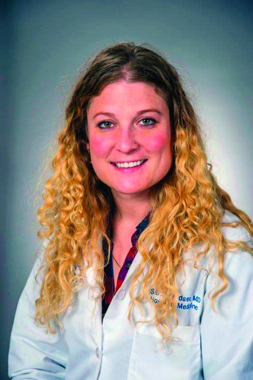
One of the diagnoses that we can often forget to use is acute respiratory failure. Documenting acute respiratory failure matters, regardless if it is, or is not, the primary diagnosis; it increases the estimated Length of Stay (LOS), Severity of Illness (SOI), and Risk of Mortality (ROM). This diagnosis adds an additional degree of specificity to patients with pneumonia, pleural effusions, chronic obstructive pulmonary disease (COPD) exacerbations, etc. While we may be hesitant to document this (perhaps feeling that this applies only to patients who are intubated in the ICU), the reader will hopefully have more confidence using it after reviewing the diagnostic criteria.
Acute respiratory failure can stem from impaired oxygenation or impaired ventilation. The following are some examples that follow these principles:
- Impaired oxygenation. Can be seen in pneumonia, pulmonary edema, and pulmonary embolism, and can present as a low O2 saturation or a low pO2 on an arterial blood gas (ABG) test.
- Impaired ventilation. Can be seen in COPD or asthma where there is increased effort to ventilate the lungs, which can lead to impaired CO2 exchange and subsequent acidosis.
One needs to have two of the following three criteria to make a formal diagnosis of acute respiratory failure:
- pO2 less than 60 mm Hg (hypoxemia).
- pCO2 greater than 50 mm Hg (hypercapnia) with pH less than 7.35.
- Signs and symptoms of acute respiratory distress.
One may think that it would be difficult to meet criteria without an ABG. Although an ABG is the standard, a patient meets criteria 1 without a blood gas if an oxygen saturation less than or equal to 90% is documented. Therefore, in most cases, if you have a documented oxygen saturation less than or equal to 90% on room air with a physical exam showing signs of respiratory distress, your patient will qualify for the diagnosis of acute respiratory failure. This negates the need to always have an ABG.
It is important to document the symptoms and physical exam findings that go along with the diagnosis. Patients should have tachypnea with a respiratory rate (RR) greater than 20 or a decreased rate less than 10. They may have wheezing, difficulty moving air, nasal flaring, and accessory muscle use. All of these findings are extremely helpful to validate the diagnosis and would make it extremely difficult for it to be rejected by a biller or insurance company.
These patients are often given supplemental oxygen (nasal cannula, Venturi mask, non-rebreather) and other treatments including steroids, inhaled bronchodilators, mucolytics, and respiratory therapy. Documenting these interventions in your plans can assist reviewers trying to understand your thought process in the treatment of the patient. If your patient has to be initiated on bilevel positive airway pressure (i.e. – the patient was not on BIPAP at home, but needed to be started because of his/her respiratory status), this almost always means they have acute respiratory failure.
In the two tables accompanying this article, we see some examples of how documenting acute respiratory failure can improve LOS, ROM, SOI, and reimbursement. The number at the top is based off of a specific DRG (Diagnosis Related Group) that is used by coders.
Let’s say we have a 58-year-old male presenting with chest pain, shortness of breath, and concern for unstable angina. Given his symptoms, he is being taken to the cardiac catheterization lab. If we note only that he was hypoxic and required 3L for an O2 saturation of 94%, one can see the ROM, SOI, estimated LOS, and reimbursement in the first column. However, if we write that his oxygen saturation on room air is 87%, he is using intercostal muscles to breathe, and he has marked dyspnea with conversation, we can say that he has acute respiratory failure. Making this distinction increases his expected LOS by almost 4 days and nearly doubles reimbursement.
For the second example, we have an 81-year-old female with diabetes type 2, hypertension, and chronic systolic congestive heart failure who presents with an acute systolic CHF exacerbation. The patient is saturating 85% on room air, has tachypnea (RR 34), and was given large doses of intravenous furosemide in the emergency department. She is stabilized with improvement in her respiratory rate and can go to the floor, but by documenting that this was acute respiratory failure, one can again see the significant improvements in the projected LOS, ROM, and reimbursement as opposed to documenting hypoxia. This has huge implications for our hospitals, and we should continue to strive to document this as clearly as possible.
Key take-home points for hospitalists
- Document accurately, including any comorbid conditions and major comorbid conditions that are applicable.
- Acute respiratory failure comes from impaired oxygenation, impaired ventilation, or both.
- One needs to document two of the three criteria to formally diagnose acute respiratory failure: pO2 less than 60 mm Hg (or room air oxygen saturation less than or equal to 90%), pCO2 greater than 50 mm Hg with pH less than 7.35, and signs/symptoms of respiratory distress.
- Document physical exam findings that correlate with acute respiratory failure (RR greater than 20 or less than 10, wheezing, nasal flaring, accessory muscle use, etc).
- If your patient has to be initiated on BIPAP (i.e. – the patient was not on BIPAP at home, but needed to be started because of his/her respiratory status), they likely have acute respiratory failure.
Dr. DeCaro is a hospitalist and medical director for care coordination at Emory University in Atlanta.
It’s always important for everyone to remember why we document things in the chart so that we are on the same page and ultimately do what is best for the patient. We document for insurance companies to prove the need for hospitalization, for legal purposes, and for other clinicians – to clearly communicate the acuity of each patient.
One of the diagnoses that we can often forget to use is acute respiratory failure. Documenting acute respiratory failure matters, regardless if it is, or is not, the primary diagnosis; it increases the estimated Length of Stay (LOS), Severity of Illness (SOI), and Risk of Mortality (ROM). This diagnosis adds an additional degree of specificity to patients with pneumonia, pleural effusions, chronic obstructive pulmonary disease (COPD) exacerbations, etc. While we may be hesitant to document this (perhaps feeling that this applies only to patients who are intubated in the ICU), the reader will hopefully have more confidence using it after reviewing the diagnostic criteria.
Acute respiratory failure can stem from impaired oxygenation or impaired ventilation. The following are some examples that follow these principles:
- Impaired oxygenation. Can be seen in pneumonia, pulmonary edema, and pulmonary embolism, and can present as a low O2 saturation or a low pO2 on an arterial blood gas (ABG) test.
- Impaired ventilation. Can be seen in COPD or asthma where there is increased effort to ventilate the lungs, which can lead to impaired CO2 exchange and subsequent acidosis.
One needs to have two of the following three criteria to make a formal diagnosis of acute respiratory failure:
- pO2 less than 60 mm Hg (hypoxemia).
- pCO2 greater than 50 mm Hg (hypercapnia) with pH less than 7.35.
- Signs and symptoms of acute respiratory distress.
One may think that it would be difficult to meet criteria without an ABG. Although an ABG is the standard, a patient meets criteria 1 without a blood gas if an oxygen saturation less than or equal to 90% is documented. Therefore, in most cases, if you have a documented oxygen saturation less than or equal to 90% on room air with a physical exam showing signs of respiratory distress, your patient will qualify for the diagnosis of acute respiratory failure. This negates the need to always have an ABG.
It is important to document the symptoms and physical exam findings that go along with the diagnosis. Patients should have tachypnea with a respiratory rate (RR) greater than 20 or a decreased rate less than 10. They may have wheezing, difficulty moving air, nasal flaring, and accessory muscle use. All of these findings are extremely helpful to validate the diagnosis and would make it extremely difficult for it to be rejected by a biller or insurance company.
These patients are often given supplemental oxygen (nasal cannula, Venturi mask, non-rebreather) and other treatments including steroids, inhaled bronchodilators, mucolytics, and respiratory therapy. Documenting these interventions in your plans can assist reviewers trying to understand your thought process in the treatment of the patient. If your patient has to be initiated on bilevel positive airway pressure (i.e. – the patient was not on BIPAP at home, but needed to be started because of his/her respiratory status), this almost always means they have acute respiratory failure.
In the two tables accompanying this article, we see some examples of how documenting acute respiratory failure can improve LOS, ROM, SOI, and reimbursement. The number at the top is based off of a specific DRG (Diagnosis Related Group) that is used by coders.
Let’s say we have a 58-year-old male presenting with chest pain, shortness of breath, and concern for unstable angina. Given his symptoms, he is being taken to the cardiac catheterization lab. If we note only that he was hypoxic and required 3L for an O2 saturation of 94%, one can see the ROM, SOI, estimated LOS, and reimbursement in the first column. However, if we write that his oxygen saturation on room air is 87%, he is using intercostal muscles to breathe, and he has marked dyspnea with conversation, we can say that he has acute respiratory failure. Making this distinction increases his expected LOS by almost 4 days and nearly doubles reimbursement.
For the second example, we have an 81-year-old female with diabetes type 2, hypertension, and chronic systolic congestive heart failure who presents with an acute systolic CHF exacerbation. The patient is saturating 85% on room air, has tachypnea (RR 34), and was given large doses of intravenous furosemide in the emergency department. She is stabilized with improvement in her respiratory rate and can go to the floor, but by documenting that this was acute respiratory failure, one can again see the significant improvements in the projected LOS, ROM, and reimbursement as opposed to documenting hypoxia. This has huge implications for our hospitals, and we should continue to strive to document this as clearly as possible.
Key take-home points for hospitalists
- Document accurately, including any comorbid conditions and major comorbid conditions that are applicable.
- Acute respiratory failure comes from impaired oxygenation, impaired ventilation, or both.
- One needs to document two of the three criteria to formally diagnose acute respiratory failure: pO2 less than 60 mm Hg (or room air oxygen saturation less than or equal to 90%), pCO2 greater than 50 mm Hg with pH less than 7.35, and signs/symptoms of respiratory distress.
- Document physical exam findings that correlate with acute respiratory failure (RR greater than 20 or less than 10, wheezing, nasal flaring, accessory muscle use, etc).
- If your patient has to be initiated on BIPAP (i.e. – the patient was not on BIPAP at home, but needed to be started because of his/her respiratory status), they likely have acute respiratory failure.
Dr. DeCaro is a hospitalist and medical director for care coordination at Emory University in Atlanta.
It’s always important for everyone to remember why we document things in the chart so that we are on the same page and ultimately do what is best for the patient. We document for insurance companies to prove the need for hospitalization, for legal purposes, and for other clinicians – to clearly communicate the acuity of each patient.
One of the diagnoses that we can often forget to use is acute respiratory failure. Documenting acute respiratory failure matters, regardless if it is, or is not, the primary diagnosis; it increases the estimated Length of Stay (LOS), Severity of Illness (SOI), and Risk of Mortality (ROM). This diagnosis adds an additional degree of specificity to patients with pneumonia, pleural effusions, chronic obstructive pulmonary disease (COPD) exacerbations, etc. While we may be hesitant to document this (perhaps feeling that this applies only to patients who are intubated in the ICU), the reader will hopefully have more confidence using it after reviewing the diagnostic criteria.
Acute respiratory failure can stem from impaired oxygenation or impaired ventilation. The following are some examples that follow these principles:
- Impaired oxygenation. Can be seen in pneumonia, pulmonary edema, and pulmonary embolism, and can present as a low O2 saturation or a low pO2 on an arterial blood gas (ABG) test.
- Impaired ventilation. Can be seen in COPD or asthma where there is increased effort to ventilate the lungs, which can lead to impaired CO2 exchange and subsequent acidosis.
One needs to have two of the following three criteria to make a formal diagnosis of acute respiratory failure:
- pO2 less than 60 mm Hg (hypoxemia).
- pCO2 greater than 50 mm Hg (hypercapnia) with pH less than 7.35.
- Signs and symptoms of acute respiratory distress.
One may think that it would be difficult to meet criteria without an ABG. Although an ABG is the standard, a patient meets criteria 1 without a blood gas if an oxygen saturation less than or equal to 90% is documented. Therefore, in most cases, if you have a documented oxygen saturation less than or equal to 90% on room air with a physical exam showing signs of respiratory distress, your patient will qualify for the diagnosis of acute respiratory failure. This negates the need to always have an ABG.
It is important to document the symptoms and physical exam findings that go along with the diagnosis. Patients should have tachypnea with a respiratory rate (RR) greater than 20 or a decreased rate less than 10. They may have wheezing, difficulty moving air, nasal flaring, and accessory muscle use. All of these findings are extremely helpful to validate the diagnosis and would make it extremely difficult for it to be rejected by a biller or insurance company.
These patients are often given supplemental oxygen (nasal cannula, Venturi mask, non-rebreather) and other treatments including steroids, inhaled bronchodilators, mucolytics, and respiratory therapy. Documenting these interventions in your plans can assist reviewers trying to understand your thought process in the treatment of the patient. If your patient has to be initiated on bilevel positive airway pressure (i.e. – the patient was not on BIPAP at home, but needed to be started because of his/her respiratory status), this almost always means they have acute respiratory failure.
In the two tables accompanying this article, we see some examples of how documenting acute respiratory failure can improve LOS, ROM, SOI, and reimbursement. The number at the top is based off of a specific DRG (Diagnosis Related Group) that is used by coders.
Let’s say we have a 58-year-old male presenting with chest pain, shortness of breath, and concern for unstable angina. Given his symptoms, he is being taken to the cardiac catheterization lab. If we note only that he was hypoxic and required 3L for an O2 saturation of 94%, one can see the ROM, SOI, estimated LOS, and reimbursement in the first column. However, if we write that his oxygen saturation on room air is 87%, he is using intercostal muscles to breathe, and he has marked dyspnea with conversation, we can say that he has acute respiratory failure. Making this distinction increases his expected LOS by almost 4 days and nearly doubles reimbursement.
For the second example, we have an 81-year-old female with diabetes type 2, hypertension, and chronic systolic congestive heart failure who presents with an acute systolic CHF exacerbation. The patient is saturating 85% on room air, has tachypnea (RR 34), and was given large doses of intravenous furosemide in the emergency department. She is stabilized with improvement in her respiratory rate and can go to the floor, but by documenting that this was acute respiratory failure, one can again see the significant improvements in the projected LOS, ROM, and reimbursement as opposed to documenting hypoxia. This has huge implications for our hospitals, and we should continue to strive to document this as clearly as possible.
Key take-home points for hospitalists
- Document accurately, including any comorbid conditions and major comorbid conditions that are applicable.
- Acute respiratory failure comes from impaired oxygenation, impaired ventilation, or both.
- One needs to document two of the three criteria to formally diagnose acute respiratory failure: pO2 less than 60 mm Hg (or room air oxygen saturation less than or equal to 90%), pCO2 greater than 50 mm Hg with pH less than 7.35, and signs/symptoms of respiratory distress.
- Document physical exam findings that correlate with acute respiratory failure (RR greater than 20 or less than 10, wheezing, nasal flaring, accessory muscle use, etc).
- If your patient has to be initiated on BIPAP (i.e. – the patient was not on BIPAP at home, but needed to be started because of his/her respiratory status), they likely have acute respiratory failure.
Dr. DeCaro is a hospitalist and medical director for care coordination at Emory University in Atlanta.
Telehealth consults for vascular surgery reimbursed at par with office visits
NEW YORK – Telehealth should be embraced by vascular surgeons for their own self-interest independent of the evidence that it is well accepted and more convenient for patients, according to an update on an evolution that is already underway.

“One of the great advantages of telehealth is the efficacy of time for the clinician,” John W. Hallett, MD, professor of vascular surgery at the Medical University of South Carolina, Charleston, said at a symposium on vascular and endovascular issues sponsored by the Cleveland Clinic Foundation.
This efficiency is purchased with no loss of revenue, he added. He said that many clinicians are unaware of the opportunity this affords.
“Almost every payer reimburses telehealth visit at the same rate as that of an office visit,” Dr. Hallett explained. The only additional step is adding a “GT” modifier when billing Medicare or a “95” modifier when billing private payers.
Telemedicine is not a new concept. Published studies date back decades, but this interaction is increasingly understood to be the future. Along with an increasing array of sensors employing smartphone technology to allow physicians remote access to vital signs and other clinical data, patient attitudes have changed.
“Patients like telemedicine. It is convenient for them,” said Dr. Hallett, who noted that many providers are recognizing telemedicine as a potential marketing tool.
“On my way in from the airport yesterday, there was an advertisement for telemedicine from NYU on the television in the cab,” said Dr. Hallett, referring to the New York University health system.
The data supporting the benefits of telemedicine even include studies undertaken in vascular surgery patients. In one recent retrospective study cited by Dr. Hallett, substantial time and travel costs were saved for every vascular surgery consult conducted by telemedicine rather than in an office visit (Paquette S et al. Ann Vasc Surg. 2019;59:167-172).
“There was no difference in the rate of complications, and 94% of the patients considered the telehealth consultation adequate,” Dr. Hallett said.
He said there is urgency for vascular surgeons to pursue telemedicine. With the number of individuals over the age of 65 growing by thousands in the United States every day, there will be increasing pressure on the relatively fixed pool of vascular surgeons to improve their efficiency.
In addition, telemedicine is coming whether vascular surgeons like it or not.
“Patients are becoming more interested in looking at an app on their smartphone than coming to the office,” said Tony S. Das, MD, an interventional cardiologist who practices in Dallas. Dr. Das also spoke about the value of telemedicine for the vascular and cardiovascular surgeon at the VIETHsymposium.
In his overview, Dr. Das spoke about telehealth in the context of the estimated $12 billion dollars that will be spent on digital health in vascular medicine by 2021. The growth in digital health in vascular medicine is a reflection of a global change in clinical care. According to Dr. Das, there were more than 600 vendors of wearable sensors to monitor disease and health at a recent consumer electronics convention.
“This technology is here to stay,” said Dr. Das, who, appropriately, was not present at the symposium but delivered his presentation remotely.
Both the Centers for Medicare and Medicaid Services and the Food and Drug Administration have digital health action plans, according to Dr. Das. The CMS has already developed reimbursement codes to pay for remote monitoring services and more are expected.
Calling this type of telehealth “untethered vascular care,” Dr. Das agreed with Dr. Hallett that an evolution is coming whether vascular surgeons choose to get on board now or are forced to take action later.
SOURCE: VIETHsymposium
NEW YORK – Telehealth should be embraced by vascular surgeons for their own self-interest independent of the evidence that it is well accepted and more convenient for patients, according to an update on an evolution that is already underway.
“One of the great advantages of telehealth is the efficacy of time for the clinician,” John W. Hallett, MD, professor of vascular surgery at the Medical University of South Carolina, Charleston, said at a symposium on vascular and endovascular issues sponsored by the Cleveland Clinic Foundation.
This efficiency is purchased with no loss of revenue, he added. He said that many clinicians are unaware of the opportunity this affords.
“Almost every payer reimburses telehealth visit at the same rate as that of an office visit,” Dr. Hallett explained. The only additional step is adding a “GT” modifier when billing Medicare or a “95” modifier when billing private payers.
Telemedicine is not a new concept. Published studies date back decades, but this interaction is increasingly understood to be the future. Along with an increasing array of sensors employing smartphone technology to allow physicians remote access to vital signs and other clinical data, patient attitudes have changed.
“Patients like telemedicine. It is convenient for them,” said Dr. Hallett, who noted that many providers are recognizing telemedicine as a potential marketing tool.
“On my way in from the airport yesterday, there was an advertisement for telemedicine from NYU on the television in the cab,” said Dr. Hallett, referring to the New York University health system.
The data supporting the benefits of telemedicine even include studies undertaken in vascular surgery patients. In one recent retrospective study cited by Dr. Hallett, substantial time and travel costs were saved for every vascular surgery consult conducted by telemedicine rather than in an office visit (Paquette S et al. Ann Vasc Surg. 2019;59:167-172).
“There was no difference in the rate of complications, and 94% of the patients considered the telehealth consultation adequate,” Dr. Hallett said.
He said there is urgency for vascular surgeons to pursue telemedicine. With the number of individuals over the age of 65 growing by thousands in the United States every day, there will be increasing pressure on the relatively fixed pool of vascular surgeons to improve their efficiency.
In addition, telemedicine is coming whether vascular surgeons like it or not.
“Patients are becoming more interested in looking at an app on their smartphone than coming to the office,” said Tony S. Das, MD, an interventional cardiologist who practices in Dallas. Dr. Das also spoke about the value of telemedicine for the vascular and cardiovascular surgeon at the VIETHsymposium.
In his overview, Dr. Das spoke about telehealth in the context of the estimated $12 billion dollars that will be spent on digital health in vascular medicine by 2021. The growth in digital health in vascular medicine is a reflection of a global change in clinical care. According to Dr. Das, there were more than 600 vendors of wearable sensors to monitor disease and health at a recent consumer electronics convention.
“This technology is here to stay,” said Dr. Das, who, appropriately, was not present at the symposium but delivered his presentation remotely.
Both the Centers for Medicare and Medicaid Services and the Food and Drug Administration have digital health action plans, according to Dr. Das. The CMS has already developed reimbursement codes to pay for remote monitoring services and more are expected.
Calling this type of telehealth “untethered vascular care,” Dr. Das agreed with Dr. Hallett that an evolution is coming whether vascular surgeons choose to get on board now or are forced to take action later.
SOURCE: VIETHsymposium
NEW YORK – Telehealth should be embraced by vascular surgeons for their own self-interest independent of the evidence that it is well accepted and more convenient for patients, according to an update on an evolution that is already underway.
“One of the great advantages of telehealth is the efficacy of time for the clinician,” John W. Hallett, MD, professor of vascular surgery at the Medical University of South Carolina, Charleston, said at a symposium on vascular and endovascular issues sponsored by the Cleveland Clinic Foundation.
This efficiency is purchased with no loss of revenue, he added. He said that many clinicians are unaware of the opportunity this affords.
“Almost every payer reimburses telehealth visit at the same rate as that of an office visit,” Dr. Hallett explained. The only additional step is adding a “GT” modifier when billing Medicare or a “95” modifier when billing private payers.
Telemedicine is not a new concept. Published studies date back decades, but this interaction is increasingly understood to be the future. Along with an increasing array of sensors employing smartphone technology to allow physicians remote access to vital signs and other clinical data, patient attitudes have changed.
“Patients like telemedicine. It is convenient for them,” said Dr. Hallett, who noted that many providers are recognizing telemedicine as a potential marketing tool.
“On my way in from the airport yesterday, there was an advertisement for telemedicine from NYU on the television in the cab,” said Dr. Hallett, referring to the New York University health system.
The data supporting the benefits of telemedicine even include studies undertaken in vascular surgery patients. In one recent retrospective study cited by Dr. Hallett, substantial time and travel costs were saved for every vascular surgery consult conducted by telemedicine rather than in an office visit (Paquette S et al. Ann Vasc Surg. 2019;59:167-172).
“There was no difference in the rate of complications, and 94% of the patients considered the telehealth consultation adequate,” Dr. Hallett said.
He said there is urgency for vascular surgeons to pursue telemedicine. With the number of individuals over the age of 65 growing by thousands in the United States every day, there will be increasing pressure on the relatively fixed pool of vascular surgeons to improve their efficiency.
In addition, telemedicine is coming whether vascular surgeons like it or not.
“Patients are becoming more interested in looking at an app on their smartphone than coming to the office,” said Tony S. Das, MD, an interventional cardiologist who practices in Dallas. Dr. Das also spoke about the value of telemedicine for the vascular and cardiovascular surgeon at the VIETHsymposium.
In his overview, Dr. Das spoke about telehealth in the context of the estimated $12 billion dollars that will be spent on digital health in vascular medicine by 2021. The growth in digital health in vascular medicine is a reflection of a global change in clinical care. According to Dr. Das, there were more than 600 vendors of wearable sensors to monitor disease and health at a recent consumer electronics convention.
“This technology is here to stay,” said Dr. Das, who, appropriately, was not present at the symposium but delivered his presentation remotely.
Both the Centers for Medicare and Medicaid Services and the Food and Drug Administration have digital health action plans, according to Dr. Das. The CMS has already developed reimbursement codes to pay for remote monitoring services and more are expected.
Calling this type of telehealth “untethered vascular care,” Dr. Das agreed with Dr. Hallett that an evolution is coming whether vascular surgeons choose to get on board now or are forced to take action later.
SOURCE: VIETHsymposium
REPORTING FROM VIETH SYMPOSIUM

Depression linked to persistent opioid use after hysterectomy
VANCOUVER –

Women with depression had an 8% increased risk of perioperative opioid use but a 43% increased risk of persistent use, defined as at least one perioperative prescription followed by at least one prescription 90 days or longer after surgery.
Opioid prescriptions after surgery have been on the rise in recent years, and this has led to a focus on how chronic pain disorders are managed. But studies have shown that patients undergoing general surgery, both minor and major, are at increased risk of persistent opioid use, even after a single surgery, according to Erin Carey, MD, director of the division of minimally invasive gynecologic surgery at the University of North Carolina at Chapel Hill, who presented the research at the meeting sponsored by AAGL.
“We also know that preoperative depression has been linked to adverse outcomes after hysterectomy, both acute postoperative pain in the first 2 days after surgery, and increasing the risk of chronic postoperative pain,” Dr. Carey said.
That prompted her and her team to look at whether preoperative depression might influence the risk of new persistent opioid use after hysterectomy. They analyzed data from the IBM Watson/Truven Health Analytics MarketScan database of claims-based data, which collects information from a variety of sources, including electronic medical records and workplace records such as absences, disability, and long-term disability.
“So it does allow for long-term tracking, which makes it optimal for this type of study,” said Dr. Carey.
The study included 382,078 hysterectomies performed between 2001 and 2015 on women who had continuous prescription plans 180 days before to 180 days after the procedure, excluding anyone who had an opioid prescription in the previous 180 days; 60% of the procedures were minimally invasive. About 20% of women were considered to have depression before the procedure, based on a diagnosis (55%), an antidepressant prescription (22%), or both (23%).
There were some differences at baseline between the two populations: Women with preoperative depression were more likely to have a comorbid pain disorder, compared with patients without depression (20% vs. 14%), another psychiatric disorder (2% vs. less than 1%), and a Charlson comorbidity (12% vs. 9%). They also were less likely to undergo a minimally invasive procedure than women without depression (66% vs. 79%). There was an increase in the prevalence of depression over time, from 16% to 23%.
Overall, 74% of women were prescribed an opioid during the perioperative period; 17% were filled before the hysterectomy was performed. Preoperative fills also increased over time, from 4% in 2001 to 21% in 2015.
Women with preoperative depression were at a slightly greater risk for perioperative opioid use (risk ratio, 1.08), but a greater risk for persistent postoperative opioid use (11% vs. 8%; RR, 1.43). The heightened risk for opioid use was similar whether the surgery was performed on an outpatient or inpatient basis.
The presence of other comorbidities in women with diagnosed depression or prescribed antidepressants complicates the findings, according to Dr. Carey. “There may be additional chronic pain factors that are confounding this data, but it is consistent with other data that de novo postoperative opioid dependence may be a higher risk for these patients, so it’s important for us to look at that critically.”
Dr. Carey has been a consultant for Teleflex Medical and a speaker for Med-IQ.
VANCOUVER –
Women with depression had an 8% increased risk of perioperative opioid use but a 43% increased risk of persistent use, defined as at least one perioperative prescription followed by at least one prescription 90 days or longer after surgery.
Opioid prescriptions after surgery have been on the rise in recent years, and this has led to a focus on how chronic pain disorders are managed. But studies have shown that patients undergoing general surgery, both minor and major, are at increased risk of persistent opioid use, even after a single surgery, according to Erin Carey, MD, director of the division of minimally invasive gynecologic surgery at the University of North Carolina at Chapel Hill, who presented the research at the meeting sponsored by AAGL.
“We also know that preoperative depression has been linked to adverse outcomes after hysterectomy, both acute postoperative pain in the first 2 days after surgery, and increasing the risk of chronic postoperative pain,” Dr. Carey said.
That prompted her and her team to look at whether preoperative depression might influence the risk of new persistent opioid use after hysterectomy. They analyzed data from the IBM Watson/Truven Health Analytics MarketScan database of claims-based data, which collects information from a variety of sources, including electronic medical records and workplace records such as absences, disability, and long-term disability.
“So it does allow for long-term tracking, which makes it optimal for this type of study,” said Dr. Carey.
The study included 382,078 hysterectomies performed between 2001 and 2015 on women who had continuous prescription plans 180 days before to 180 days after the procedure, excluding anyone who had an opioid prescription in the previous 180 days; 60% of the procedures were minimally invasive. About 20% of women were considered to have depression before the procedure, based on a diagnosis (55%), an antidepressant prescription (22%), or both (23%).
There were some differences at baseline between the two populations: Women with preoperative depression were more likely to have a comorbid pain disorder, compared with patients without depression (20% vs. 14%), another psychiatric disorder (2% vs. less than 1%), and a Charlson comorbidity (12% vs. 9%). They also were less likely to undergo a minimally invasive procedure than women without depression (66% vs. 79%). There was an increase in the prevalence of depression over time, from 16% to 23%.
Overall, 74% of women were prescribed an opioid during the perioperative period; 17% were filled before the hysterectomy was performed. Preoperative fills also increased over time, from 4% in 2001 to 21% in 2015.
Women with preoperative depression were at a slightly greater risk for perioperative opioid use (risk ratio, 1.08), but a greater risk for persistent postoperative opioid use (11% vs. 8%; RR, 1.43). The heightened risk for opioid use was similar whether the surgery was performed on an outpatient or inpatient basis.
The presence of other comorbidities in women with diagnosed depression or prescribed antidepressants complicates the findings, according to Dr. Carey. “There may be additional chronic pain factors that are confounding this data, but it is consistent with other data that de novo postoperative opioid dependence may be a higher risk for these patients, so it’s important for us to look at that critically.”
Dr. Carey has been a consultant for Teleflex Medical and a speaker for Med-IQ.
VANCOUVER –
Women with depression had an 8% increased risk of perioperative opioid use but a 43% increased risk of persistent use, defined as at least one perioperative prescription followed by at least one prescription 90 days or longer after surgery.
Opioid prescriptions after surgery have been on the rise in recent years, and this has led to a focus on how chronic pain disorders are managed. But studies have shown that patients undergoing general surgery, both minor and major, are at increased risk of persistent opioid use, even after a single surgery, according to Erin Carey, MD, director of the division of minimally invasive gynecologic surgery at the University of North Carolina at Chapel Hill, who presented the research at the meeting sponsored by AAGL.
“We also know that preoperative depression has been linked to adverse outcomes after hysterectomy, both acute postoperative pain in the first 2 days after surgery, and increasing the risk of chronic postoperative pain,” Dr. Carey said.
That prompted her and her team to look at whether preoperative depression might influence the risk of new persistent opioid use after hysterectomy. They analyzed data from the IBM Watson/Truven Health Analytics MarketScan database of claims-based data, which collects information from a variety of sources, including electronic medical records and workplace records such as absences, disability, and long-term disability.
“So it does allow for long-term tracking, which makes it optimal for this type of study,” said Dr. Carey.
The study included 382,078 hysterectomies performed between 2001 and 2015 on women who had continuous prescription plans 180 days before to 180 days after the procedure, excluding anyone who had an opioid prescription in the previous 180 days; 60% of the procedures were minimally invasive. About 20% of women were considered to have depression before the procedure, based on a diagnosis (55%), an antidepressant prescription (22%), or both (23%).
There were some differences at baseline between the two populations: Women with preoperative depression were more likely to have a comorbid pain disorder, compared with patients without depression (20% vs. 14%), another psychiatric disorder (2% vs. less than 1%), and a Charlson comorbidity (12% vs. 9%). They also were less likely to undergo a minimally invasive procedure than women without depression (66% vs. 79%). There was an increase in the prevalence of depression over time, from 16% to 23%.
Overall, 74% of women were prescribed an opioid during the perioperative period; 17% were filled before the hysterectomy was performed. Preoperative fills also increased over time, from 4% in 2001 to 21% in 2015.
Women with preoperative depression were at a slightly greater risk for perioperative opioid use (risk ratio, 1.08), but a greater risk for persistent postoperative opioid use (11% vs. 8%; RR, 1.43). The heightened risk for opioid use was similar whether the surgery was performed on an outpatient or inpatient basis.
The presence of other comorbidities in women with diagnosed depression or prescribed antidepressants complicates the findings, according to Dr. Carey. “There may be additional chronic pain factors that are confounding this data, but it is consistent with other data that de novo postoperative opioid dependence may be a higher risk for these patients, so it’s important for us to look at that critically.”
Dr. Carey has been a consultant for Teleflex Medical and a speaker for Med-IQ.
REPORTING FROM THE AAGL GLOBAL CONGRESS

An alarming number of bipolar disorder diagnoses or something else?
During a particularly busy day in my inpatient and outpatient practice, I realized that nearly every one of the patients had been given the diagnosis of bipolar disorder at one point or another. The interesting thing is this wasn’t an unusual day.

Nearly all of my patients and their family members have been given the diagnosis of bipolar disorder. Because prevalence of bipolar affective disorders is a little over 2%, this seemed a little odd. Could there be an epidemic of bipolar disorder in the area? Should someone sound the alarm on this unique cluster and get Julia Roberts ready? Unfortunately, the story behind this mystery is a little less sexy but nevertheless interesting.
When I probe more into what symptoms might have led to the diagnosis of bipolar disorder, I most often get some sort of answer about being easily angered (“I’m fine 1 minute and the next minute I’m yelling at my mom”) or mood changing from 1 minute to the next. Rarely do they tell me about sleeping less, increased energy, change in mood (elation, anger, irritability), increase in activity level, and increased pleasurable though dangerous activities all happening around the same time(s). So what is going on?
Beginning in the 1990s, a debate about the phenotypic presentation of pediatric bipolar disorder polarized the field. It was theorized that mania could present with severe nonepisodic irritability with extended periods of very rapid mood cycling within the day as opposed to discrete episodic mood cycles in children and adolescents. With this broader conceptualization in the United States, the rate of bipolar diagnosis increased by over 40 times in less than a decade.1 Similarly, the use of mood stabilizers and atypical antipsychotics in children also rose substantially.2
To help assess if severe nonepisodic irritability belongs in the spectrum of bipolar disorders, the National Institutes of Mental Health proposed a syndrome called “Severe Mood Dysregulation” or SMD, to promote the study of children with this phenotype. In longitudinal studies, Stringaris et al. compared rates of manic episodes in youth with SMD versus bipolar disorder over 2 years and found only one youth (1%) with SMD who presented with manic, hypomanic, or mixed episodes, compared with 58 (62%) with bipolar disorder.3 Leibenluft et al.showed that chronic irritability during early adolescence predicted ADHD at late adolescence and major depressive disorder in early adulthood whereas episodic irritability predicted mania.4 Twenty-year follow-up of the same sample showed chronic irritability in adolescence predicted dysthymia, generalized anxiety disorders, and major depressive disorder.5 Other longitudinal studies essentially have shown the same results.6
At this point, the question of whether chronic irritability is a part of the bipolar spectrum disorder is largely resolved – 7 The diagnosis emphasizes the episodic nature of the illness, and that irritability would wax and wane with other manic symptoms such as changes in energy and sleep. And the ultrarapid mood changes (mood changes within the day) appear to describe mood fluctuations within a manic episode as opposed to each change being a separate episode.

So, most likely, my patients were caught in a time of uncertainty before data were able to clarify their phenotype.
Dr. Chung is a child and adolescent psychiatrist at the University of Vermont Medical Center, Burlington, and practices at Champlain Valley Physician’s Hospital in Plattsburgh, N.Y. Email him at pdnews@mdedge.com.
References
1. Biol Psychiatry. 2007 Jul 15;62(2):107–14.
2. JAMA Psychiatry. 2015 Sep;72(9):859-60.
3. J Am Acad Child Adolesc Psychiatry. 2010 Apr;49(4):397-405.
4. J Child Adolesc Psychopharmacol 2006;16(4):456-66.
5. Am J Psychiatry. 2009 Sep;166(9):1048-54.
6. Biol Psychiatry. 2006 Nov 1;60(9):991-7.
7. Bipolar Disord. 2017 Nov;19(7):524-43.
During a particularly busy day in my inpatient and outpatient practice, I realized that nearly every one of the patients had been given the diagnosis of bipolar disorder at one point or another. The interesting thing is this wasn’t an unusual day.
Nearly all of my patients and their family members have been given the diagnosis of bipolar disorder. Because prevalence of bipolar affective disorders is a little over 2%, this seemed a little odd. Could there be an epidemic of bipolar disorder in the area? Should someone sound the alarm on this unique cluster and get Julia Roberts ready? Unfortunately, the story behind this mystery is a little less sexy but nevertheless interesting.
When I probe more into what symptoms might have led to the diagnosis of bipolar disorder, I most often get some sort of answer about being easily angered (“I’m fine 1 minute and the next minute I’m yelling at my mom”) or mood changing from 1 minute to the next. Rarely do they tell me about sleeping less, increased energy, change in mood (elation, anger, irritability), increase in activity level, and increased pleasurable though dangerous activities all happening around the same time(s). So what is going on?
Beginning in the 1990s, a debate about the phenotypic presentation of pediatric bipolar disorder polarized the field. It was theorized that mania could present with severe nonepisodic irritability with extended periods of very rapid mood cycling within the day as opposed to discrete episodic mood cycles in children and adolescents. With this broader conceptualization in the United States, the rate of bipolar diagnosis increased by over 40 times in less than a decade.1 Similarly, the use of mood stabilizers and atypical antipsychotics in children also rose substantially.2
To help assess if severe nonepisodic irritability belongs in the spectrum of bipolar disorders, the National Institutes of Mental Health proposed a syndrome called “Severe Mood Dysregulation” or SMD, to promote the study of children with this phenotype. In longitudinal studies, Stringaris et al. compared rates of manic episodes in youth with SMD versus bipolar disorder over 2 years and found only one youth (1%) with SMD who presented with manic, hypomanic, or mixed episodes, compared with 58 (62%) with bipolar disorder.3 Leibenluft et al.showed that chronic irritability during early adolescence predicted ADHD at late adolescence and major depressive disorder in early adulthood whereas episodic irritability predicted mania.4 Twenty-year follow-up of the same sample showed chronic irritability in adolescence predicted dysthymia, generalized anxiety disorders, and major depressive disorder.5 Other longitudinal studies essentially have shown the same results.6
At this point, the question of whether chronic irritability is a part of the bipolar spectrum disorder is largely resolved – 7 The diagnosis emphasizes the episodic nature of the illness, and that irritability would wax and wane with other manic symptoms such as changes in energy and sleep. And the ultrarapid mood changes (mood changes within the day) appear to describe mood fluctuations within a manic episode as opposed to each change being a separate episode.
So, most likely, my patients were caught in a time of uncertainty before data were able to clarify their phenotype.
Dr. Chung is a child and adolescent psychiatrist at the University of Vermont Medical Center, Burlington, and practices at Champlain Valley Physician’s Hospital in Plattsburgh, N.Y. Email him at pdnews@mdedge.com.
References
1. Biol Psychiatry. 2007 Jul 15;62(2):107–14.
2. JAMA Psychiatry. 2015 Sep;72(9):859-60.
3. J Am Acad Child Adolesc Psychiatry. 2010 Apr;49(4):397-405.
4. J Child Adolesc Psychopharmacol 2006;16(4):456-66.
5. Am J Psychiatry. 2009 Sep;166(9):1048-54.
6. Biol Psychiatry. 2006 Nov 1;60(9):991-7.
7. Bipolar Disord. 2017 Nov;19(7):524-43.
During a particularly busy day in my inpatient and outpatient practice, I realized that nearly every one of the patients had been given the diagnosis of bipolar disorder at one point or another. The interesting thing is this wasn’t an unusual day.
Nearly all of my patients and their family members have been given the diagnosis of bipolar disorder. Because prevalence of bipolar affective disorders is a little over 2%, this seemed a little odd. Could there be an epidemic of bipolar disorder in the area? Should someone sound the alarm on this unique cluster and get Julia Roberts ready? Unfortunately, the story behind this mystery is a little less sexy but nevertheless interesting.
When I probe more into what symptoms might have led to the diagnosis of bipolar disorder, I most often get some sort of answer about being easily angered (“I’m fine 1 minute and the next minute I’m yelling at my mom”) or mood changing from 1 minute to the next. Rarely do they tell me about sleeping less, increased energy, change in mood (elation, anger, irritability), increase in activity level, and increased pleasurable though dangerous activities all happening around the same time(s). So what is going on?
Beginning in the 1990s, a debate about the phenotypic presentation of pediatric bipolar disorder polarized the field. It was theorized that mania could present with severe nonepisodic irritability with extended periods of very rapid mood cycling within the day as opposed to discrete episodic mood cycles in children and adolescents. With this broader conceptualization in the United States, the rate of bipolar diagnosis increased by over 40 times in less than a decade.1 Similarly, the use of mood stabilizers and atypical antipsychotics in children also rose substantially.2
To help assess if severe nonepisodic irritability belongs in the spectrum of bipolar disorders, the National Institutes of Mental Health proposed a syndrome called “Severe Mood Dysregulation” or SMD, to promote the study of children with this phenotype. In longitudinal studies, Stringaris et al. compared rates of manic episodes in youth with SMD versus bipolar disorder over 2 years and found only one youth (1%) with SMD who presented with manic, hypomanic, or mixed episodes, compared with 58 (62%) with bipolar disorder.3 Leibenluft et al.showed that chronic irritability during early adolescence predicted ADHD at late adolescence and major depressive disorder in early adulthood whereas episodic irritability predicted mania.4 Twenty-year follow-up of the same sample showed chronic irritability in adolescence predicted dysthymia, generalized anxiety disorders, and major depressive disorder.5 Other longitudinal studies essentially have shown the same results.6
At this point, the question of whether chronic irritability is a part of the bipolar spectrum disorder is largely resolved – 7 The diagnosis emphasizes the episodic nature of the illness, and that irritability would wax and wane with other manic symptoms such as changes in energy and sleep. And the ultrarapid mood changes (mood changes within the day) appear to describe mood fluctuations within a manic episode as opposed to each change being a separate episode.
So, most likely, my patients were caught in a time of uncertainty before data were able to clarify their phenotype.
Dr. Chung is a child and adolescent psychiatrist at the University of Vermont Medical Center, Burlington, and practices at Champlain Valley Physician’s Hospital in Plattsburgh, N.Y. Email him at pdnews@mdedge.com.
References
1. Biol Psychiatry. 2007 Jul 15;62(2):107–14.
2. JAMA Psychiatry. 2015 Sep;72(9):859-60.
3. J Am Acad Child Adolesc Psychiatry. 2010 Apr;49(4):397-405.
4. J Child Adolesc Psychopharmacol 2006;16(4):456-66.
5. Am J Psychiatry. 2009 Sep;166(9):1048-54.
6. Biol Psychiatry. 2006 Nov 1;60(9):991-7.
7. Bipolar Disord. 2017 Nov;19(7):524-43.