User login
Closure of women’s health clinics may negatively impact cervical cancer outcomes
.

States with a decreased number of women’s clinics per capita between 2010 and 2013 were found to have less screening for cervical cancer, more advanced stage of cervical cancer at presentation, and higher mortality from cervical cancer than states with no decrease in clinics, reported lead author Amar J. Srivastava, MD, of Washington University in St. Louis, who also noted that these changes occurred within a relatively short time frame.
“We know that women are generally diagnosed through the utilization of Pap smears,” Dr. Srivastava said during a presentation at the annual meeting of the American Society for Radiation Oncology. “These are low-cost tests that are available at multiple low-cost women’s health clinics. Unfortunately ... over the course of the past decade, we’ve seen a significant reduction of these clinics throughout the United States.”
“Between 2010 and 2013, which is the period of interest in this study, we know that about 100 of these women’s health clinics closed,” Dr. Srivastava said. “This was due to a combination of several factors; some of it was due to funding, some of it was due to restructuring of the clinics, and there were also laws passed throughout many states that ultimately led to the closure of many clinics.”
To determine the impact of these closures, the investigators first divided states into those that had women’s clinic closures between 2010 and 2013 and those that did not. Comparisons between these two cohorts involved the use of two databases. The first was the Behavioral Risk Factors Surveillance Study (BRFSS), which provided data from 197,143 cases, enabling assessment of differences between screening availability. The second database was the Surveillance, Epidemiology, and End Results (SEER) registry, which provided data from 10,652 patients, facilitating comparisons of stage at time of diagnosis and mortality rate.
Results were described in terms of relative differences between the two cohorts. For instance, screening rate among women with cervical cancer in states that had a decreased number of clinics was 1.63% lower than in states that did not lose clinics. This disparity was more pronounced in specific demographic subgroups, including Hispanic women (–5.82%), women aged between 21 and 34 years (–5.19%), unmarried women (–4.10%), and uninsured women (–6.88%).
“Historically, these are marginalized, underserved groups, and unfortunately, it comes as no surprise that these were the groups of women who were most dramatically hit by these changes,” Dr. Srivastava said.
Early-stage diagnosis was also significantly less common in states that had a decreased number of clinics, by a margin of 13.2%. Finally, the overall mortality rate among women with cervical cancer was 36% higher in states with clinic closures, a difference that climbed to 40% when comparing only metro residents.
Connecting the dots, Dr. Srivastava suggested that the decreased availability of screening may have led to fewer diagnoses at an early stage, which is more curable than late-stage disease, ultimately translating to a higher mortality rate. After noting that this chain of causality cannot be confirmed, owing to the retrospective nature of the study, Dr. Srivastava finished his presentation with a call to action.
“These findings should really give us some pause,” he said, “as physicians, as people who care about other people, to spend some time, try to figure out what’s going on, and try to address this disparity.”
After the presentation, Geraldine M. Jacobsen, MD, chair of radiation oncology at West Virginia University Cancer Institute, in Morgantown, W.V., echoed Dr. Srivastava’s concern.
“This study really raises broader questions,” Dr. Jacobsen said. “In the United States we’re always engaged in an ongoing dialogue about health care, health care policy, [and] health care costs. But a study like this brings to us the human face of what these dialogues mean. Policy affects people, and if we make changes in health care policy or health care legislation, we’re impacting people’s health and people’s lives.”
The investigators disclosed relationships with Phelps County Regional Medical Center, the Elsa U. Pardee Foundation, the American Society of Clinical Oncology, and ASTRO.
SOURCE: Srivastava AJ et al. ASTRO 2019, Abstract 202.
.
States with a decreased number of women’s clinics per capita between 2010 and 2013 were found to have less screening for cervical cancer, more advanced stage of cervical cancer at presentation, and higher mortality from cervical cancer than states with no decrease in clinics, reported lead author Amar J. Srivastava, MD, of Washington University in St. Louis, who also noted that these changes occurred within a relatively short time frame.
“We know that women are generally diagnosed through the utilization of Pap smears,” Dr. Srivastava said during a presentation at the annual meeting of the American Society for Radiation Oncology. “These are low-cost tests that are available at multiple low-cost women’s health clinics. Unfortunately ... over the course of the past decade, we’ve seen a significant reduction of these clinics throughout the United States.”
“Between 2010 and 2013, which is the period of interest in this study, we know that about 100 of these women’s health clinics closed,” Dr. Srivastava said. “This was due to a combination of several factors; some of it was due to funding, some of it was due to restructuring of the clinics, and there were also laws passed throughout many states that ultimately led to the closure of many clinics.”
To determine the impact of these closures, the investigators first divided states into those that had women’s clinic closures between 2010 and 2013 and those that did not. Comparisons between these two cohorts involved the use of two databases. The first was the Behavioral Risk Factors Surveillance Study (BRFSS), which provided data from 197,143 cases, enabling assessment of differences between screening availability. The second database was the Surveillance, Epidemiology, and End Results (SEER) registry, which provided data from 10,652 patients, facilitating comparisons of stage at time of diagnosis and mortality rate.
Results were described in terms of relative differences between the two cohorts. For instance, screening rate among women with cervical cancer in states that had a decreased number of clinics was 1.63% lower than in states that did not lose clinics. This disparity was more pronounced in specific demographic subgroups, including Hispanic women (–5.82%), women aged between 21 and 34 years (–5.19%), unmarried women (–4.10%), and uninsured women (–6.88%).
“Historically, these are marginalized, underserved groups, and unfortunately, it comes as no surprise that these were the groups of women who were most dramatically hit by these changes,” Dr. Srivastava said.
Early-stage diagnosis was also significantly less common in states that had a decreased number of clinics, by a margin of 13.2%. Finally, the overall mortality rate among women with cervical cancer was 36% higher in states with clinic closures, a difference that climbed to 40% when comparing only metro residents.
Connecting the dots, Dr. Srivastava suggested that the decreased availability of screening may have led to fewer diagnoses at an early stage, which is more curable than late-stage disease, ultimately translating to a higher mortality rate. After noting that this chain of causality cannot be confirmed, owing to the retrospective nature of the study, Dr. Srivastava finished his presentation with a call to action.
“These findings should really give us some pause,” he said, “as physicians, as people who care about other people, to spend some time, try to figure out what’s going on, and try to address this disparity.”
After the presentation, Geraldine M. Jacobsen, MD, chair of radiation oncology at West Virginia University Cancer Institute, in Morgantown, W.V., echoed Dr. Srivastava’s concern.
“This study really raises broader questions,” Dr. Jacobsen said. “In the United States we’re always engaged in an ongoing dialogue about health care, health care policy, [and] health care costs. But a study like this brings to us the human face of what these dialogues mean. Policy affects people, and if we make changes in health care policy or health care legislation, we’re impacting people’s health and people’s lives.”
The investigators disclosed relationships with Phelps County Regional Medical Center, the Elsa U. Pardee Foundation, the American Society of Clinical Oncology, and ASTRO.
SOURCE: Srivastava AJ et al. ASTRO 2019, Abstract 202.
.
States with a decreased number of women’s clinics per capita between 2010 and 2013 were found to have less screening for cervical cancer, more advanced stage of cervical cancer at presentation, and higher mortality from cervical cancer than states with no decrease in clinics, reported lead author Amar J. Srivastava, MD, of Washington University in St. Louis, who also noted that these changes occurred within a relatively short time frame.
“We know that women are generally diagnosed through the utilization of Pap smears,” Dr. Srivastava said during a presentation at the annual meeting of the American Society for Radiation Oncology. “These are low-cost tests that are available at multiple low-cost women’s health clinics. Unfortunately ... over the course of the past decade, we’ve seen a significant reduction of these clinics throughout the United States.”
“Between 2010 and 2013, which is the period of interest in this study, we know that about 100 of these women’s health clinics closed,” Dr. Srivastava said. “This was due to a combination of several factors; some of it was due to funding, some of it was due to restructuring of the clinics, and there were also laws passed throughout many states that ultimately led to the closure of many clinics.”
To determine the impact of these closures, the investigators first divided states into those that had women’s clinic closures between 2010 and 2013 and those that did not. Comparisons between these two cohorts involved the use of two databases. The first was the Behavioral Risk Factors Surveillance Study (BRFSS), which provided data from 197,143 cases, enabling assessment of differences between screening availability. The second database was the Surveillance, Epidemiology, and End Results (SEER) registry, which provided data from 10,652 patients, facilitating comparisons of stage at time of diagnosis and mortality rate.
Results were described in terms of relative differences between the two cohorts. For instance, screening rate among women with cervical cancer in states that had a decreased number of clinics was 1.63% lower than in states that did not lose clinics. This disparity was more pronounced in specific demographic subgroups, including Hispanic women (–5.82%), women aged between 21 and 34 years (–5.19%), unmarried women (–4.10%), and uninsured women (–6.88%).
“Historically, these are marginalized, underserved groups, and unfortunately, it comes as no surprise that these were the groups of women who were most dramatically hit by these changes,” Dr. Srivastava said.
Early-stage diagnosis was also significantly less common in states that had a decreased number of clinics, by a margin of 13.2%. Finally, the overall mortality rate among women with cervical cancer was 36% higher in states with clinic closures, a difference that climbed to 40% when comparing only metro residents.
Connecting the dots, Dr. Srivastava suggested that the decreased availability of screening may have led to fewer diagnoses at an early stage, which is more curable than late-stage disease, ultimately translating to a higher mortality rate. After noting that this chain of causality cannot be confirmed, owing to the retrospective nature of the study, Dr. Srivastava finished his presentation with a call to action.
“These findings should really give us some pause,” he said, “as physicians, as people who care about other people, to spend some time, try to figure out what’s going on, and try to address this disparity.”
After the presentation, Geraldine M. Jacobsen, MD, chair of radiation oncology at West Virginia University Cancer Institute, in Morgantown, W.V., echoed Dr. Srivastava’s concern.
“This study really raises broader questions,” Dr. Jacobsen said. “In the United States we’re always engaged in an ongoing dialogue about health care, health care policy, [and] health care costs. But a study like this brings to us the human face of what these dialogues mean. Policy affects people, and if we make changes in health care policy or health care legislation, we’re impacting people’s health and people’s lives.”
The investigators disclosed relationships with Phelps County Regional Medical Center, the Elsa U. Pardee Foundation, the American Society of Clinical Oncology, and ASTRO.
SOURCE: Srivastava AJ et al. ASTRO 2019, Abstract 202.
REPORTING FROM ASTRO 2019

Business case for interoperability remains elusive

In a recent survey issued by the Center for Connected Medicine that asked, “What is needed most to push interoperability forward in health care?” 53% of the 100 IT and business leader participants at hospitals and health systems answered “senior leadership commitment to interoperability as a top strategic priority.”
That interoperability continues to be an elusive target despite being a regulatory emphasis for more than a decade comes as no surprise.
“You have to put interoperability into the context of the challenges of being a leader in health care delivery organization or a hospital,” Robert Bart, MD, chief medical information officer of the Health Services Division at the University of Pittsburgh Medical Center, said in an interview. UPMC operates the Center for Connected Medicine in partnership with GE Healthcare and Nokia.
“For many health care delivery systems, the operational margin is extremely small,” he continued. “From a strategic perspective, they may be prioritizing things or opportunities that directly contribute to the bottom line and the financial success or even financial viability of the organization. “Interoperability certainly helps health care overall in the U.S., but whether it contributes directly to the financial bottom line of any given specific organization might vary significantly.”
But Dr. Bart said it really is not the financial incentives, a staple in the early days of the meaningful use program to spur adoption, that will get electronic health records and other health IT into a more interoperable space.
“I am not sure that financial incentives – unless they are significantly different in amounts than they currently are – are going to be the sole single reason why organizations prioritize interoperability higher,” he said.
Rather, he sees two key components that will drive interoperability.
First, he cited efforts by the Centers for Medicare & Medicaid Services to bring ownership of health data to the individual as an important driver.
“I think that is a better direction to push as opposed to financial incentives because I think that is really where we philosophically and then therefore operationally need to get to,” Dr. Bart said.
The second would take a lot more involvement from government that likely is more difficult to achieve: getting a more clear definition of health IT standards.
“We have accomplished some forward movement in interoperability, but we probably have far more road in front of us than we have left behind us as it relates to interoperability,” he said. “A fair amount of that is related to standards which, unfortunately, oftentimes are left up to interpretation. So when it comes to operationalizing these standards between different vendors or different health care systems, they sometimes don’t match up and add even more challenge to the exchange of information.”
And with industry not taking an active lead in solving some of the problems related to the standardization of data needed to drive interoperability, he suggested government should play a bigger role.
“I would like the government to take a stronger hand in helping health care define the standards so the standards are actually much more executable and there is a lot less interpretation, which I think would ease the ability for interoperability to flourish a lot more,” he said.
The move to value-based care could also provide added incentive to drive interoperability, Dr. Bart said.
Value-based care is “certainly not going to raise more barriers toward interoperability and potentially could lower some as organizations recognize that it may be a path to decreasing the costs of repeating tests that will not improve the quality of care delivered,” he said.
But overall, it would appear that getting to an interoperable point is going to be a challenge.
The report notes that “beyond tasks required by regulation or related to basic functioning (such as sharing data within their own system), interoperability still presents a challenge to organizations. Fewer than 4 in 10 report success with sharing data with other health systems, effectiveness in tapping into unstructured data, and effectiveness in reducing the cost of care.”
SOURCE: A report issued by the Center for Connected Medicine called “Improving Health Care Interoperability: Are We Making Progress?”
In a recent survey issued by the Center for Connected Medicine that asked, “What is needed most to push interoperability forward in health care?” 53% of the 100 IT and business leader participants at hospitals and health systems answered “senior leadership commitment to interoperability as a top strategic priority.”
That interoperability continues to be an elusive target despite being a regulatory emphasis for more than a decade comes as no surprise.
“You have to put interoperability into the context of the challenges of being a leader in health care delivery organization or a hospital,” Robert Bart, MD, chief medical information officer of the Health Services Division at the University of Pittsburgh Medical Center, said in an interview. UPMC operates the Center for Connected Medicine in partnership with GE Healthcare and Nokia.
“For many health care delivery systems, the operational margin is extremely small,” he continued. “From a strategic perspective, they may be prioritizing things or opportunities that directly contribute to the bottom line and the financial success or even financial viability of the organization. “Interoperability certainly helps health care overall in the U.S., but whether it contributes directly to the financial bottom line of any given specific organization might vary significantly.”
But Dr. Bart said it really is not the financial incentives, a staple in the early days of the meaningful use program to spur adoption, that will get electronic health records and other health IT into a more interoperable space.
“I am not sure that financial incentives – unless they are significantly different in amounts than they currently are – are going to be the sole single reason why organizations prioritize interoperability higher,” he said.
Rather, he sees two key components that will drive interoperability.
First, he cited efforts by the Centers for Medicare & Medicaid Services to bring ownership of health data to the individual as an important driver.
“I think that is a better direction to push as opposed to financial incentives because I think that is really where we philosophically and then therefore operationally need to get to,” Dr. Bart said.
The second would take a lot more involvement from government that likely is more difficult to achieve: getting a more clear definition of health IT standards.
“We have accomplished some forward movement in interoperability, but we probably have far more road in front of us than we have left behind us as it relates to interoperability,” he said. “A fair amount of that is related to standards which, unfortunately, oftentimes are left up to interpretation. So when it comes to operationalizing these standards between different vendors or different health care systems, they sometimes don’t match up and add even more challenge to the exchange of information.”
And with industry not taking an active lead in solving some of the problems related to the standardization of data needed to drive interoperability, he suggested government should play a bigger role.
“I would like the government to take a stronger hand in helping health care define the standards so the standards are actually much more executable and there is a lot less interpretation, which I think would ease the ability for interoperability to flourish a lot more,” he said.
The move to value-based care could also provide added incentive to drive interoperability, Dr. Bart said.
Value-based care is “certainly not going to raise more barriers toward interoperability and potentially could lower some as organizations recognize that it may be a path to decreasing the costs of repeating tests that will not improve the quality of care delivered,” he said.
But overall, it would appear that getting to an interoperable point is going to be a challenge.
The report notes that “beyond tasks required by regulation or related to basic functioning (such as sharing data within their own system), interoperability still presents a challenge to organizations. Fewer than 4 in 10 report success with sharing data with other health systems, effectiveness in tapping into unstructured data, and effectiveness in reducing the cost of care.”
SOURCE: A report issued by the Center for Connected Medicine called “Improving Health Care Interoperability: Are We Making Progress?”
In a recent survey issued by the Center for Connected Medicine that asked, “What is needed most to push interoperability forward in health care?” 53% of the 100 IT and business leader participants at hospitals and health systems answered “senior leadership commitment to interoperability as a top strategic priority.”
That interoperability continues to be an elusive target despite being a regulatory emphasis for more than a decade comes as no surprise.
“You have to put interoperability into the context of the challenges of being a leader in health care delivery organization or a hospital,” Robert Bart, MD, chief medical information officer of the Health Services Division at the University of Pittsburgh Medical Center, said in an interview. UPMC operates the Center for Connected Medicine in partnership with GE Healthcare and Nokia.
“For many health care delivery systems, the operational margin is extremely small,” he continued. “From a strategic perspective, they may be prioritizing things or opportunities that directly contribute to the bottom line and the financial success or even financial viability of the organization. “Interoperability certainly helps health care overall in the U.S., but whether it contributes directly to the financial bottom line of any given specific organization might vary significantly.”
But Dr. Bart said it really is not the financial incentives, a staple in the early days of the meaningful use program to spur adoption, that will get electronic health records and other health IT into a more interoperable space.
“I am not sure that financial incentives – unless they are significantly different in amounts than they currently are – are going to be the sole single reason why organizations prioritize interoperability higher,” he said.
Rather, he sees two key components that will drive interoperability.
First, he cited efforts by the Centers for Medicare & Medicaid Services to bring ownership of health data to the individual as an important driver.
“I think that is a better direction to push as opposed to financial incentives because I think that is really where we philosophically and then therefore operationally need to get to,” Dr. Bart said.
The second would take a lot more involvement from government that likely is more difficult to achieve: getting a more clear definition of health IT standards.
“We have accomplished some forward movement in interoperability, but we probably have far more road in front of us than we have left behind us as it relates to interoperability,” he said. “A fair amount of that is related to standards which, unfortunately, oftentimes are left up to interpretation. So when it comes to operationalizing these standards between different vendors or different health care systems, they sometimes don’t match up and add even more challenge to the exchange of information.”
And with industry not taking an active lead in solving some of the problems related to the standardization of data needed to drive interoperability, he suggested government should play a bigger role.
“I would like the government to take a stronger hand in helping health care define the standards so the standards are actually much more executable and there is a lot less interpretation, which I think would ease the ability for interoperability to flourish a lot more,” he said.
The move to value-based care could also provide added incentive to drive interoperability, Dr. Bart said.
Value-based care is “certainly not going to raise more barriers toward interoperability and potentially could lower some as organizations recognize that it may be a path to decreasing the costs of repeating tests that will not improve the quality of care delivered,” he said.
But overall, it would appear that getting to an interoperable point is going to be a challenge.
The report notes that “beyond tasks required by regulation or related to basic functioning (such as sharing data within their own system), interoperability still presents a challenge to organizations. Fewer than 4 in 10 report success with sharing data with other health systems, effectiveness in tapping into unstructured data, and effectiveness in reducing the cost of care.”
SOURCE: A report issued by the Center for Connected Medicine called “Improving Health Care Interoperability: Are We Making Progress?”
Judicious EEG use identifies pseudosyncope during tilt-table testing
NEW ORLEANS – , according to an investigation from Vanderbilt University, Nashville.
Pseudosyncope, also known as nonsyncopal fainting (NSF), is a conversion disorder where people appear to faint, but don’t lose consciousness. It’s generally thought to be a physical manifestation of traumatic stress. Patients “aren’t faking it; they believe they are fainting,” said senior investigator Italo Biaggioni, MD, a professor of medicine and pharmacology at the university and director of the Vanderbilt Autonomic Dysfunction Center.
It’s “not benign. These patients stop working, stop driving, and need to depend on other people. They can have very dramatic episodes and hurt themselves” when they fall. “They are very disabled,” he said.
NSF is often misdiagnosed and mistreated, and sometimes unrecognized for years. Almost a third of the 39 NSF cases in Dr. Biaggioni’s series, for instance, were on anticonvulsants, and several had undergone cardiac catheterization. Often, NSF is treated as vasovagal syncope, but patients don’t respond to medications. A better way to identify it is needed. “By the time we get them, they’ve been through a lot.” Dr. Biaggioni said at the joint scientific sessions of the American Heart Association Council on Hypertension, AHA Council on Kidney in Cardiovascular Disease, and American Society of Hypertension.
He and his team had a hunch that judicious use of EEG would help, so they limited EEG to suspected NSF cases in a series of 107 refractory syncope patients referred to Vanderbilt for head-up tilt-table testing; 39 (36%) had normal EEGs during an apparent loss of consciousness, as opposed to the slow-wave pattern of true loss of consciousness, and were diagnosed with NSF.
The 36% identified was a marked increase in incidence over more common approaches. Among 64 patients who had tilt-table testing without EEG at Vanderbilt, for instance, three (5%) were diagnosed with NSF. Historically, tilt table plus EEG in all comers has a diagnostic yield of around 18% for the condition. In short, “elective EEG monitoring during tilt-table testing” better “distinguishes between syncope” and NSF, the team concluded.
NSF is suggested by a history of more than 20 episodes of apparent fainting; episodes once a week or more; or losing consciousness for more than 5 minutes. Fainting with eyes closed, or while supine, is also suggestive.
The elective approach prevents inappropriate treatment but is also therapeutic in itself. “When we document with EEG that patients are not really fainting, and explain that to them, it automatically reduces the number of episodes,” Dr. Biaggioni said.
It also saves NSF patients from a nitroglycerin challenge and repeat tilt testing, which is the default in many places when the first round of testing doesn’t trigger an episode. Challenge testing provokes vasovagal syncope in around 10% of even healthy people, so it puts NSF patients at risk for a false positive. As a rule, “we don’t use provocative agents when we [suspect NSF],” he said.
In addition to the 39 NSF cases, 11 patients in the series were diagnosed with vasovagal syncope, and testing didn’t provoke an event in 57 (53%), which isn’t uncommon.
Baseline blood pressure and heart rates were similar across the three groups, and there were more women than men in each. Subjects were in their early 40s, on average.
NSF patients were more likely to be taking anxiety and depression medications. One NSF patient had posttraumatic stress disorder, and two had sexual abuse histories, compared with none in the nondiagnostic and vasovagal groups. The NSF group had a shorter time to an event: 9 minutes versus 19 minutes among vasovagal patients.
Tilt-table testing was done after 6 hours of fasting, and the team used standard 22-channel EEG. Cognitive-behavioral therapy is the go-to treatment for NSF, Dr. Biaggioni said.
There was no industry funding, and the authors didn’t have any disclosures.
SOURCE: Muldowney JA et al. Joint Hypertension 2019, Abstract P3061.
NEW ORLEANS – , according to an investigation from Vanderbilt University, Nashville.
Pseudosyncope, also known as nonsyncopal fainting (NSF), is a conversion disorder where people appear to faint, but don’t lose consciousness. It’s generally thought to be a physical manifestation of traumatic stress. Patients “aren’t faking it; they believe they are fainting,” said senior investigator Italo Biaggioni, MD, a professor of medicine and pharmacology at the university and director of the Vanderbilt Autonomic Dysfunction Center.
It’s “not benign. These patients stop working, stop driving, and need to depend on other people. They can have very dramatic episodes and hurt themselves” when they fall. “They are very disabled,” he said.
NSF is often misdiagnosed and mistreated, and sometimes unrecognized for years. Almost a third of the 39 NSF cases in Dr. Biaggioni’s series, for instance, were on anticonvulsants, and several had undergone cardiac catheterization. Often, NSF is treated as vasovagal syncope, but patients don’t respond to medications. A better way to identify it is needed. “By the time we get them, they’ve been through a lot.” Dr. Biaggioni said at the joint scientific sessions of the American Heart Association Council on Hypertension, AHA Council on Kidney in Cardiovascular Disease, and American Society of Hypertension.
He and his team had a hunch that judicious use of EEG would help, so they limited EEG to suspected NSF cases in a series of 107 refractory syncope patients referred to Vanderbilt for head-up tilt-table testing; 39 (36%) had normal EEGs during an apparent loss of consciousness, as opposed to the slow-wave pattern of true loss of consciousness, and were diagnosed with NSF.
The 36% identified was a marked increase in incidence over more common approaches. Among 64 patients who had tilt-table testing without EEG at Vanderbilt, for instance, three (5%) were diagnosed with NSF. Historically, tilt table plus EEG in all comers has a diagnostic yield of around 18% for the condition. In short, “elective EEG monitoring during tilt-table testing” better “distinguishes between syncope” and NSF, the team concluded.
NSF is suggested by a history of more than 20 episodes of apparent fainting; episodes once a week or more; or losing consciousness for more than 5 minutes. Fainting with eyes closed, or while supine, is also suggestive.
The elective approach prevents inappropriate treatment but is also therapeutic in itself. “When we document with EEG that patients are not really fainting, and explain that to them, it automatically reduces the number of episodes,” Dr. Biaggioni said.
It also saves NSF patients from a nitroglycerin challenge and repeat tilt testing, which is the default in many places when the first round of testing doesn’t trigger an episode. Challenge testing provokes vasovagal syncope in around 10% of even healthy people, so it puts NSF patients at risk for a false positive. As a rule, “we don’t use provocative agents when we [suspect NSF],” he said.
In addition to the 39 NSF cases, 11 patients in the series were diagnosed with vasovagal syncope, and testing didn’t provoke an event in 57 (53%), which isn’t uncommon.
Baseline blood pressure and heart rates were similar across the three groups, and there were more women than men in each. Subjects were in their early 40s, on average.
NSF patients were more likely to be taking anxiety and depression medications. One NSF patient had posttraumatic stress disorder, and two had sexual abuse histories, compared with none in the nondiagnostic and vasovagal groups. The NSF group had a shorter time to an event: 9 minutes versus 19 minutes among vasovagal patients.
Tilt-table testing was done after 6 hours of fasting, and the team used standard 22-channel EEG. Cognitive-behavioral therapy is the go-to treatment for NSF, Dr. Biaggioni said.
There was no industry funding, and the authors didn’t have any disclosures.
SOURCE: Muldowney JA et al. Joint Hypertension 2019, Abstract P3061.
NEW ORLEANS – , according to an investigation from Vanderbilt University, Nashville.
Pseudosyncope, also known as nonsyncopal fainting (NSF), is a conversion disorder where people appear to faint, but don’t lose consciousness. It’s generally thought to be a physical manifestation of traumatic stress. Patients “aren’t faking it; they believe they are fainting,” said senior investigator Italo Biaggioni, MD, a professor of medicine and pharmacology at the university and director of the Vanderbilt Autonomic Dysfunction Center.
It’s “not benign. These patients stop working, stop driving, and need to depend on other people. They can have very dramatic episodes and hurt themselves” when they fall. “They are very disabled,” he said.
NSF is often misdiagnosed and mistreated, and sometimes unrecognized for years. Almost a third of the 39 NSF cases in Dr. Biaggioni’s series, for instance, were on anticonvulsants, and several had undergone cardiac catheterization. Often, NSF is treated as vasovagal syncope, but patients don’t respond to medications. A better way to identify it is needed. “By the time we get them, they’ve been through a lot.” Dr. Biaggioni said at the joint scientific sessions of the American Heart Association Council on Hypertension, AHA Council on Kidney in Cardiovascular Disease, and American Society of Hypertension.
He and his team had a hunch that judicious use of EEG would help, so they limited EEG to suspected NSF cases in a series of 107 refractory syncope patients referred to Vanderbilt for head-up tilt-table testing; 39 (36%) had normal EEGs during an apparent loss of consciousness, as opposed to the slow-wave pattern of true loss of consciousness, and were diagnosed with NSF.
The 36% identified was a marked increase in incidence over more common approaches. Among 64 patients who had tilt-table testing without EEG at Vanderbilt, for instance, three (5%) were diagnosed with NSF. Historically, tilt table plus EEG in all comers has a diagnostic yield of around 18% for the condition. In short, “elective EEG monitoring during tilt-table testing” better “distinguishes between syncope” and NSF, the team concluded.
NSF is suggested by a history of more than 20 episodes of apparent fainting; episodes once a week or more; or losing consciousness for more than 5 minutes. Fainting with eyes closed, or while supine, is also suggestive.
The elective approach prevents inappropriate treatment but is also therapeutic in itself. “When we document with EEG that patients are not really fainting, and explain that to them, it automatically reduces the number of episodes,” Dr. Biaggioni said.
It also saves NSF patients from a nitroglycerin challenge and repeat tilt testing, which is the default in many places when the first round of testing doesn’t trigger an episode. Challenge testing provokes vasovagal syncope in around 10% of even healthy people, so it puts NSF patients at risk for a false positive. As a rule, “we don’t use provocative agents when we [suspect NSF],” he said.
In addition to the 39 NSF cases, 11 patients in the series were diagnosed with vasovagal syncope, and testing didn’t provoke an event in 57 (53%), which isn’t uncommon.
Baseline blood pressure and heart rates were similar across the three groups, and there were more women than men in each. Subjects were in their early 40s, on average.
NSF patients were more likely to be taking anxiety and depression medications. One NSF patient had posttraumatic stress disorder, and two had sexual abuse histories, compared with none in the nondiagnostic and vasovagal groups. The NSF group had a shorter time to an event: 9 minutes versus 19 minutes among vasovagal patients.
Tilt-table testing was done after 6 hours of fasting, and the team used standard 22-channel EEG. Cognitive-behavioral therapy is the go-to treatment for NSF, Dr. Biaggioni said.
There was no industry funding, and the authors didn’t have any disclosures.
SOURCE: Muldowney JA et al. Joint Hypertension 2019, Abstract P3061.
REPORTING FROM JOINT HYPERTENSION 2019

Machine learning–derived risk score predicts heart failure risk in diabetes patients
PHILADELPHIA – For patients with high-risk diabetes, a novel, machine learning–derived risk score based on 10 common clinical variables can identify those facing a heart failure risk of up to nearly 20% over the ensuing 5 years, an investigator said at the annual meeting of the Heart Failure Society of America.
The risk score, dubbed WATCH-DM, has greater accuracy in predicting incident heart failure than traditional risk-based models, and requires no specific cardiovascular biomarkers or imaging, according to Muthiah Vaduganathan, MD, MPH, a cardiologist at Brigham and Women’s Hospital and faculty at Harvard Medical School in Boston.
The tool may help inform risk-based monitoring and introduction of sodium-glucose transporter 2 (SGLT2) inhibitors, which have been shown in multiple clinical trials to prevent heart failure in at-risk patients with type 2 diabetes mellitus (T2DM), Dr. Vaduganathan said.
“Patients identified at high risk based on WATCH-DM should be strongly considered for initiation of SGLT2 inhibitors in clinical practice,” Dr. Vaduganathan said in an interview.
WATCH-DM is available online at cvriskscores.com. Work is underway to integrate the tool into electronic health record systems at Brigham and Women’s Hospital and at the University of Texas Southwestern Medical Center in Dallas. “I expect that to be launched in the next year,” he said.
The WATCH-DM score was developed based on data from the ACCORD (Action to Control Cardiovascular Risk in Diabetes) trial, including 8,756 T2DM patients with inadequate glycemic control at high cardiovascular risk and no heart failure at baseline.
Starting with 147 variables, the investigators used a decision-tree machine learning approach to identify predictors of heart failure.
“What machine learning does is automate the variable selection process, as a form of artificial intelligence,” Dr. Vaduganathan said.
The WATCH-DM risk score was based on the 10 best-performing predictors as selected by machine learning, including body mass index, age, systolic blood pressure, diastolic blood pressure, fasting plasma glucose, serum creatinine, high-density lipoprotein cholesterol, QRS duration, prior myocardial infarction, and prior coronary artery bypass grafting.
The 5-year risk of heart failure was just 1.1% for patients with WATCH-DM scores in the lowest quintile, increasing in a graded fashion to nearly 20% (17.4%) in the highest quintile, study results show.
Findings of the study were simultaneously published in the journal Diabetes Care.
Dr. Vaduganathan said he is supported by an award from Harvard Catalyst. He provided disclosures related to Amgen, AstraZeneca, Baxter Healthcare, Bayer AG, Boehringer Ingelheim (advisory boards), and with Novartis and the National Institutes of Health (participation on clinical endpoint committees).
SOURCE: HFSA 2019; Segar MW, Vaduganathan M et al. Diabetes Care. doi: 10.2337/dc19-0587.
PHILADELPHIA – For patients with high-risk diabetes, a novel, machine learning–derived risk score based on 10 common clinical variables can identify those facing a heart failure risk of up to nearly 20% over the ensuing 5 years, an investigator said at the annual meeting of the Heart Failure Society of America.
The risk score, dubbed WATCH-DM, has greater accuracy in predicting incident heart failure than traditional risk-based models, and requires no specific cardiovascular biomarkers or imaging, according to Muthiah Vaduganathan, MD, MPH, a cardiologist at Brigham and Women’s Hospital and faculty at Harvard Medical School in Boston.
The tool may help inform risk-based monitoring and introduction of sodium-glucose transporter 2 (SGLT2) inhibitors, which have been shown in multiple clinical trials to prevent heart failure in at-risk patients with type 2 diabetes mellitus (T2DM), Dr. Vaduganathan said.
“Patients identified at high risk based on WATCH-DM should be strongly considered for initiation of SGLT2 inhibitors in clinical practice,” Dr. Vaduganathan said in an interview.
WATCH-DM is available online at cvriskscores.com. Work is underway to integrate the tool into electronic health record systems at Brigham and Women’s Hospital and at the University of Texas Southwestern Medical Center in Dallas. “I expect that to be launched in the next year,” he said.
The WATCH-DM score was developed based on data from the ACCORD (Action to Control Cardiovascular Risk in Diabetes) trial, including 8,756 T2DM patients with inadequate glycemic control at high cardiovascular risk and no heart failure at baseline.
Starting with 147 variables, the investigators used a decision-tree machine learning approach to identify predictors of heart failure.
“What machine learning does is automate the variable selection process, as a form of artificial intelligence,” Dr. Vaduganathan said.
The WATCH-DM risk score was based on the 10 best-performing predictors as selected by machine learning, including body mass index, age, systolic blood pressure, diastolic blood pressure, fasting plasma glucose, serum creatinine, high-density lipoprotein cholesterol, QRS duration, prior myocardial infarction, and prior coronary artery bypass grafting.
The 5-year risk of heart failure was just 1.1% for patients with WATCH-DM scores in the lowest quintile, increasing in a graded fashion to nearly 20% (17.4%) in the highest quintile, study results show.
Findings of the study were simultaneously published in the journal Diabetes Care.
Dr. Vaduganathan said he is supported by an award from Harvard Catalyst. He provided disclosures related to Amgen, AstraZeneca, Baxter Healthcare, Bayer AG, Boehringer Ingelheim (advisory boards), and with Novartis and the National Institutes of Health (participation on clinical endpoint committees).
SOURCE: HFSA 2019; Segar MW, Vaduganathan M et al. Diabetes Care. doi: 10.2337/dc19-0587.
PHILADELPHIA – For patients with high-risk diabetes, a novel, machine learning–derived risk score based on 10 common clinical variables can identify those facing a heart failure risk of up to nearly 20% over the ensuing 5 years, an investigator said at the annual meeting of the Heart Failure Society of America.
The risk score, dubbed WATCH-DM, has greater accuracy in predicting incident heart failure than traditional risk-based models, and requires no specific cardiovascular biomarkers or imaging, according to Muthiah Vaduganathan, MD, MPH, a cardiologist at Brigham and Women’s Hospital and faculty at Harvard Medical School in Boston.
The tool may help inform risk-based monitoring and introduction of sodium-glucose transporter 2 (SGLT2) inhibitors, which have been shown in multiple clinical trials to prevent heart failure in at-risk patients with type 2 diabetes mellitus (T2DM), Dr. Vaduganathan said.
“Patients identified at high risk based on WATCH-DM should be strongly considered for initiation of SGLT2 inhibitors in clinical practice,” Dr. Vaduganathan said in an interview.
WATCH-DM is available online at cvriskscores.com. Work is underway to integrate the tool into electronic health record systems at Brigham and Women’s Hospital and at the University of Texas Southwestern Medical Center in Dallas. “I expect that to be launched in the next year,” he said.
The WATCH-DM score was developed based on data from the ACCORD (Action to Control Cardiovascular Risk in Diabetes) trial, including 8,756 T2DM patients with inadequate glycemic control at high cardiovascular risk and no heart failure at baseline.
Starting with 147 variables, the investigators used a decision-tree machine learning approach to identify predictors of heart failure.
“What machine learning does is automate the variable selection process, as a form of artificial intelligence,” Dr. Vaduganathan said.
The WATCH-DM risk score was based on the 10 best-performing predictors as selected by machine learning, including body mass index, age, systolic blood pressure, diastolic blood pressure, fasting plasma glucose, serum creatinine, high-density lipoprotein cholesterol, QRS duration, prior myocardial infarction, and prior coronary artery bypass grafting.
The 5-year risk of heart failure was just 1.1% for patients with WATCH-DM scores in the lowest quintile, increasing in a graded fashion to nearly 20% (17.4%) in the highest quintile, study results show.
Findings of the study were simultaneously published in the journal Diabetes Care.
Dr. Vaduganathan said he is supported by an award from Harvard Catalyst. He provided disclosures related to Amgen, AstraZeneca, Baxter Healthcare, Bayer AG, Boehringer Ingelheim (advisory boards), and with Novartis and the National Institutes of Health (participation on clinical endpoint committees).
SOURCE: HFSA 2019; Segar MW, Vaduganathan M et al. Diabetes Care. doi: 10.2337/dc19-0587.
REPORTING FROM HFSA 2019
Hyponatremia almost as common with spironolactone as chlorthalidone
NEW ORLEANS – .
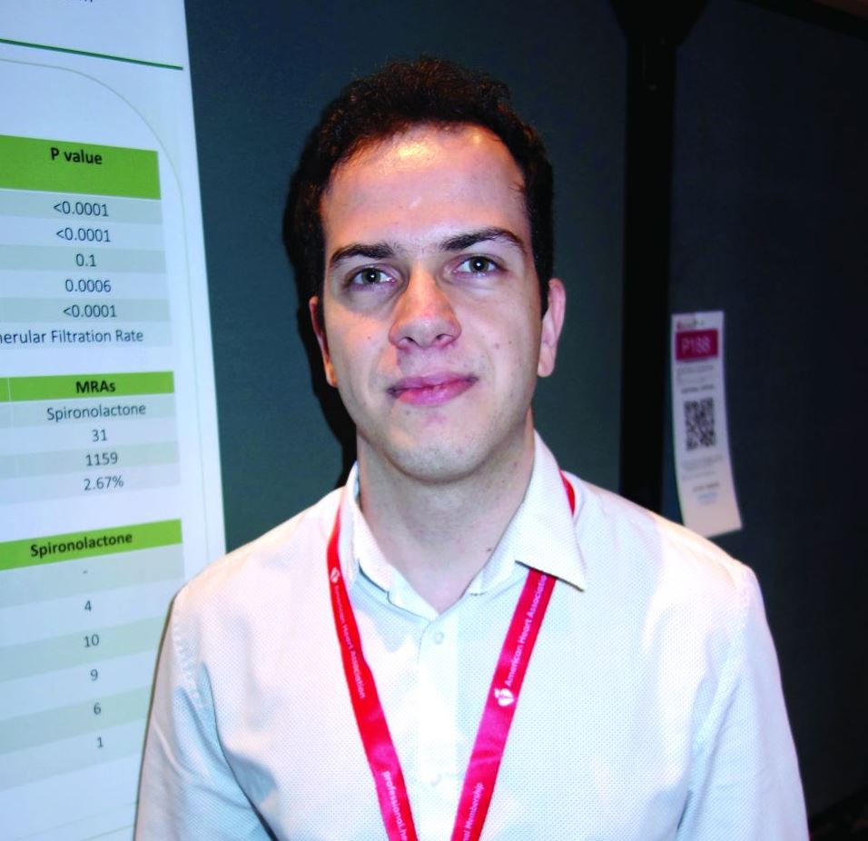
The investigators reviewed hypertension patients whose treatment regimens included one diuretic. Forty on chlorthalidone developed hyponatremia – defined as a serum sodium below 133 mEq/L – across 1,322 prescriptions, for an incidence of 3.03%. There were 31 cases across 1,159 spironolactone prescriptions, an incidence of 2.67%.
Among 14 patients in a substudy who discontinued chlorthalidone after developing hyponatremia at a mean of about 2 weeks, six (43%) subsequently developed hyponatremia on spironolactone, also at an average of about 2 weeks.
The findings suggest that spironolactone is more likely than generally thought to cause hyponatremia, a potentially severe complication of diuretics, and that hyponatremia on chlorthalidone increases the risk, said lead investigator Faris Matanes, MD, a hypertension researcher at the university.
“We used to think” that hyponatremia on spironolactone was “very unlikely, but actually it’s not; the incidence is really close to chlorthalidone,” a well-known cause, and “if a patient develops hyponatremia on chlorthalidone, we should be more careful about giving them spironolactone,” he said.
Almost half the spironolactone cases were on 25 mg/day or less, and over a quarter of the chlorthalidone cases were on 12.5 mg/day. Of the 154 hyponatremia cases across 10,660 hydrochlorothiazide prescriptions (1.44%), over a third were taking 12.5 mg/day or less.
Overall, hyponatremia was diagnosed at a mean of 40.4 days, but sometimes after 2 or more months of treatment.
The findings “mean that even if we start patients on a low dose, we can’t stop checking after one or two normal sodium levels.” Measurements need to be ongoing, Dr. Matanes said at the joint scientific sessions of the American Heart Association Council on Hypertension, AHA Council on Kidney in Cardiovascular Disease, and American Society of Hypertension.
He and his team wanted to get around the limitations of previous diuretic hyponatremia studies, including use of more than one diuretic, markedly poor kidney function, and other confounders. To that end, the study was limited to outpatients on a single diuretic who had normal sodium levels both before and after their hyponatremic episode, and estimated glomerular filtration rates (eGFR) of at least 30 mL/min/1.73 m2. Exclusion criteria included heart failure, cirrhosis, and adrenal insufficiency.
Older white people with lower baseline sodium and eGFR values were most at risk. Contrary to previous reports, hyponatremia wasn’t more likely in men.
The mean sodium level during an episode was 130.2 mEq/L; the majority of patients eventually normalized and continued treatment.
Subjects in the main study were a mean of 66 years old, about two-thirds were white, and about 60% were women. The baseline eGFR was 77.2 mL/min/1.73 m2, and baseline sodium level 135.8 mEq/L.
All but one of the 14 substudy patients were women. Those who became hyponatremic when switched to spironolactone were older (mean 74.2 versus 65.8 years), had lower baseline eGFRs (63.7 versus 69.7 mL/min/1.73 m2), and were more likely to be white, but the differences were not statistically significant.
There was no external funding, and the investigators didn’t have any industry disclosures.
SOURCE: Matanes F et al. Joint Hypertension 2019, Abstracts 187 and 174.
NEW ORLEANS – .
The investigators reviewed hypertension patients whose treatment regimens included one diuretic. Forty on chlorthalidone developed hyponatremia – defined as a serum sodium below 133 mEq/L – across 1,322 prescriptions, for an incidence of 3.03%. There were 31 cases across 1,159 spironolactone prescriptions, an incidence of 2.67%.
Among 14 patients in a substudy who discontinued chlorthalidone after developing hyponatremia at a mean of about 2 weeks, six (43%) subsequently developed hyponatremia on spironolactone, also at an average of about 2 weeks.
The findings suggest that spironolactone is more likely than generally thought to cause hyponatremia, a potentially severe complication of diuretics, and that hyponatremia on chlorthalidone increases the risk, said lead investigator Faris Matanes, MD, a hypertension researcher at the university.
“We used to think” that hyponatremia on spironolactone was “very unlikely, but actually it’s not; the incidence is really close to chlorthalidone,” a well-known cause, and “if a patient develops hyponatremia on chlorthalidone, we should be more careful about giving them spironolactone,” he said.
Almost half the spironolactone cases were on 25 mg/day or less, and over a quarter of the chlorthalidone cases were on 12.5 mg/day. Of the 154 hyponatremia cases across 10,660 hydrochlorothiazide prescriptions (1.44%), over a third were taking 12.5 mg/day or less.
Overall, hyponatremia was diagnosed at a mean of 40.4 days, but sometimes after 2 or more months of treatment.
The findings “mean that even if we start patients on a low dose, we can’t stop checking after one or two normal sodium levels.” Measurements need to be ongoing, Dr. Matanes said at the joint scientific sessions of the American Heart Association Council on Hypertension, AHA Council on Kidney in Cardiovascular Disease, and American Society of Hypertension.
He and his team wanted to get around the limitations of previous diuretic hyponatremia studies, including use of more than one diuretic, markedly poor kidney function, and other confounders. To that end, the study was limited to outpatients on a single diuretic who had normal sodium levels both before and after their hyponatremic episode, and estimated glomerular filtration rates (eGFR) of at least 30 mL/min/1.73 m2. Exclusion criteria included heart failure, cirrhosis, and adrenal insufficiency.
Older white people with lower baseline sodium and eGFR values were most at risk. Contrary to previous reports, hyponatremia wasn’t more likely in men.
The mean sodium level during an episode was 130.2 mEq/L; the majority of patients eventually normalized and continued treatment.
Subjects in the main study were a mean of 66 years old, about two-thirds were white, and about 60% were women. The baseline eGFR was 77.2 mL/min/1.73 m2, and baseline sodium level 135.8 mEq/L.
All but one of the 14 substudy patients were women. Those who became hyponatremic when switched to spironolactone were older (mean 74.2 versus 65.8 years), had lower baseline eGFRs (63.7 versus 69.7 mL/min/1.73 m2), and were more likely to be white, but the differences were not statistically significant.
There was no external funding, and the investigators didn’t have any industry disclosures.
SOURCE: Matanes F et al. Joint Hypertension 2019, Abstracts 187 and 174.
NEW ORLEANS – .
The investigators reviewed hypertension patients whose treatment regimens included one diuretic. Forty on chlorthalidone developed hyponatremia – defined as a serum sodium below 133 mEq/L – across 1,322 prescriptions, for an incidence of 3.03%. There were 31 cases across 1,159 spironolactone prescriptions, an incidence of 2.67%.
Among 14 patients in a substudy who discontinued chlorthalidone after developing hyponatremia at a mean of about 2 weeks, six (43%) subsequently developed hyponatremia on spironolactone, also at an average of about 2 weeks.
The findings suggest that spironolactone is more likely than generally thought to cause hyponatremia, a potentially severe complication of diuretics, and that hyponatremia on chlorthalidone increases the risk, said lead investigator Faris Matanes, MD, a hypertension researcher at the university.
“We used to think” that hyponatremia on spironolactone was “very unlikely, but actually it’s not; the incidence is really close to chlorthalidone,” a well-known cause, and “if a patient develops hyponatremia on chlorthalidone, we should be more careful about giving them spironolactone,” he said.
Almost half the spironolactone cases were on 25 mg/day or less, and over a quarter of the chlorthalidone cases were on 12.5 mg/day. Of the 154 hyponatremia cases across 10,660 hydrochlorothiazide prescriptions (1.44%), over a third were taking 12.5 mg/day or less.
Overall, hyponatremia was diagnosed at a mean of 40.4 days, but sometimes after 2 or more months of treatment.
The findings “mean that even if we start patients on a low dose, we can’t stop checking after one or two normal sodium levels.” Measurements need to be ongoing, Dr. Matanes said at the joint scientific sessions of the American Heart Association Council on Hypertension, AHA Council on Kidney in Cardiovascular Disease, and American Society of Hypertension.
He and his team wanted to get around the limitations of previous diuretic hyponatremia studies, including use of more than one diuretic, markedly poor kidney function, and other confounders. To that end, the study was limited to outpatients on a single diuretic who had normal sodium levels both before and after their hyponatremic episode, and estimated glomerular filtration rates (eGFR) of at least 30 mL/min/1.73 m2. Exclusion criteria included heart failure, cirrhosis, and adrenal insufficiency.
Older white people with lower baseline sodium and eGFR values were most at risk. Contrary to previous reports, hyponatremia wasn’t more likely in men.
The mean sodium level during an episode was 130.2 mEq/L; the majority of patients eventually normalized and continued treatment.
Subjects in the main study were a mean of 66 years old, about two-thirds were white, and about 60% were women. The baseline eGFR was 77.2 mL/min/1.73 m2, and baseline sodium level 135.8 mEq/L.
All but one of the 14 substudy patients were women. Those who became hyponatremic when switched to spironolactone were older (mean 74.2 versus 65.8 years), had lower baseline eGFRs (63.7 versus 69.7 mL/min/1.73 m2), and were more likely to be white, but the differences were not statistically significant.
There was no external funding, and the investigators didn’t have any industry disclosures.
SOURCE: Matanes F et al. Joint Hypertension 2019, Abstracts 187 and 174.
REPORTING FROM JOINT HYPERTENSION 2019
Oral triplet shows promise for relapsed/refractory myeloma
BOSTON – The all-oral combination of selinexor, lenalidomide, and dexamethasone has demonstrated efficacy in patients with relapsed/refractory multiple myeloma, particularly those who are lenalidomide naive.
In the phase 1/2 STOMP trial (NCT02343042), selinexor plus lenalidomide and dexamethasone produced an overall response rate (ORR) of 60% in all evaluable patients and an ORR of 92% in lenalidomide-naive patients.
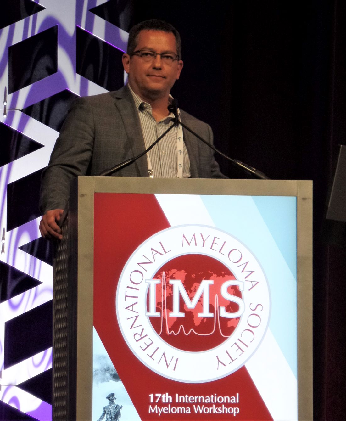
Darrell White, MD, of Dalhousie University in Halifax, N.S., presented these results at the International Myeloma Workshop held by the International Myeloma Society.
Dr. White discussed results in myeloma patients who had received at least one prior line of therapy. This arm of the STOMP trial has enrolled 24 patients. Their median age at baseline was 67 years (range, 49-84 years), and their median time from diagnosis was 4.5 years (range, less than 1-22 years).
The patients had received a median of 1 (range, 1-8) prior therapies. Half of patients had received a transplant. All patients had received a proteasome inhibitor (65% refractory), and 38% had received prior lenalidomide (21% refractory).
Dosing
Patients received selinexor at 60 mg once or twice weekly or at 80 mg once weekly on a 28-day cycle. They received dexamethasone at 20 mg twice weekly or 40 mg once weekly and lenalidomide at 25 mg once daily every 21 days.
The recommended phase 2 dosing schedule was selinexor at 60 mg once weekly plus lenalidomide at 25 mg daily and dexamethasone at 40 mg once weekly. Half of patients (n = 12) received this dosing schedule.
There were no dose-limiting toxicities (DLTs) at the recommended phase 2 dose. When selinexor was given at 60 mg twice weekly, DLTs included grade 3 fatigue, grade 3 anorexia and weight loss, and grade 4 thrombocytopenia (n = 2). When selinexor was given at 80 mg once weekly, the DLTs were grade 4 thrombocytopenia (n = 2).
Safety
“The side-effect profile was as expected, with mainly grade 3/4 toxicity being hematologic and primarily thrombocytopenia and neutropenia,” Dr. White said. “Frequent gastrointestinal side effects [were] expected. Investigators were able to manage that with appropriate supportive care and antiemetics in particular.”
Among patients who received the recommended phase 2 dose, the grade 4 treatment-related adverse events (AEs) were thrombocytopenia (n = 4) and neutropenia (n = 4). Grade 3 treatment-related AEs were thrombocytopenia (n = 3), neutropenia (n = 4), anemia (n = 1), nausea (n = 1), asthenia (n = 1), fatigue (n = 2), and dehydration (n = 1). Common grade 1/2 treatment-related AEs in patients who received the recommended phase 2 dose were nausea (n = 6), anorexia (n = 5), weight loss (n = 5), vomiting (n = 4), diarrhea (n = 4), and fatigue (n = 4).
Efficacy
In the 20 patients evaluable for response, the ORR was 60% (n = 12). One patient achieved a stringent complete response, three had a very good partial response, and eight had a partial response. The median time to response was 1 month.
The ORR was 92% (11/12) in lenalidomide-naive patients and 13% (1/8) in lenalidomide-exposed patients. The single responder in the lenalidomide-exposed group achieved a partial response.
Among lenalidomide-naive patients who received the recommended phase 2 dose, the median time on treatment was 12 months. The patient who achieved a stringent complete response remained on treatment at last follow-up, as did one partial responder and one patient with a very good partial response.
These results suggest the combination of selinexor, lenalidomide, and dexamethasone “is active, relatively well tolerated, and could warrant further investigation,” Dr. White said.
The STOMP trial is sponsored by Karyopharm Therapeutics. Dr. White disclosed relationships with Karyopharm, Amgen, Celgene, Janssen, Sanofi, and Takeda.
SOURCE: White D et al. IMW 2019, Abstract OAB-083.
BOSTON – The all-oral combination of selinexor, lenalidomide, and dexamethasone has demonstrated efficacy in patients with relapsed/refractory multiple myeloma, particularly those who are lenalidomide naive.
In the phase 1/2 STOMP trial (NCT02343042), selinexor plus lenalidomide and dexamethasone produced an overall response rate (ORR) of 60% in all evaluable patients and an ORR of 92% in lenalidomide-naive patients.
Darrell White, MD, of Dalhousie University in Halifax, N.S., presented these results at the International Myeloma Workshop held by the International Myeloma Society.
Dr. White discussed results in myeloma patients who had received at least one prior line of therapy. This arm of the STOMP trial has enrolled 24 patients. Their median age at baseline was 67 years (range, 49-84 years), and their median time from diagnosis was 4.5 years (range, less than 1-22 years).
The patients had received a median of 1 (range, 1-8) prior therapies. Half of patients had received a transplant. All patients had received a proteasome inhibitor (65% refractory), and 38% had received prior lenalidomide (21% refractory).
Dosing
Patients received selinexor at 60 mg once or twice weekly or at 80 mg once weekly on a 28-day cycle. They received dexamethasone at 20 mg twice weekly or 40 mg once weekly and lenalidomide at 25 mg once daily every 21 days.
The recommended phase 2 dosing schedule was selinexor at 60 mg once weekly plus lenalidomide at 25 mg daily and dexamethasone at 40 mg once weekly. Half of patients (n = 12) received this dosing schedule.
There were no dose-limiting toxicities (DLTs) at the recommended phase 2 dose. When selinexor was given at 60 mg twice weekly, DLTs included grade 3 fatigue, grade 3 anorexia and weight loss, and grade 4 thrombocytopenia (n = 2). When selinexor was given at 80 mg once weekly, the DLTs were grade 4 thrombocytopenia (n = 2).
Safety
“The side-effect profile was as expected, with mainly grade 3/4 toxicity being hematologic and primarily thrombocytopenia and neutropenia,” Dr. White said. “Frequent gastrointestinal side effects [were] expected. Investigators were able to manage that with appropriate supportive care and antiemetics in particular.”
Among patients who received the recommended phase 2 dose, the grade 4 treatment-related adverse events (AEs) were thrombocytopenia (n = 4) and neutropenia (n = 4). Grade 3 treatment-related AEs were thrombocytopenia (n = 3), neutropenia (n = 4), anemia (n = 1), nausea (n = 1), asthenia (n = 1), fatigue (n = 2), and dehydration (n = 1). Common grade 1/2 treatment-related AEs in patients who received the recommended phase 2 dose were nausea (n = 6), anorexia (n = 5), weight loss (n = 5), vomiting (n = 4), diarrhea (n = 4), and fatigue (n = 4).
Efficacy
In the 20 patients evaluable for response, the ORR was 60% (n = 12). One patient achieved a stringent complete response, three had a very good partial response, and eight had a partial response. The median time to response was 1 month.
The ORR was 92% (11/12) in lenalidomide-naive patients and 13% (1/8) in lenalidomide-exposed patients. The single responder in the lenalidomide-exposed group achieved a partial response.
Among lenalidomide-naive patients who received the recommended phase 2 dose, the median time on treatment was 12 months. The patient who achieved a stringent complete response remained on treatment at last follow-up, as did one partial responder and one patient with a very good partial response.
These results suggest the combination of selinexor, lenalidomide, and dexamethasone “is active, relatively well tolerated, and could warrant further investigation,” Dr. White said.
The STOMP trial is sponsored by Karyopharm Therapeutics. Dr. White disclosed relationships with Karyopharm, Amgen, Celgene, Janssen, Sanofi, and Takeda.
SOURCE: White D et al. IMW 2019, Abstract OAB-083.
BOSTON – The all-oral combination of selinexor, lenalidomide, and dexamethasone has demonstrated efficacy in patients with relapsed/refractory multiple myeloma, particularly those who are lenalidomide naive.
In the phase 1/2 STOMP trial (NCT02343042), selinexor plus lenalidomide and dexamethasone produced an overall response rate (ORR) of 60% in all evaluable patients and an ORR of 92% in lenalidomide-naive patients.
Darrell White, MD, of Dalhousie University in Halifax, N.S., presented these results at the International Myeloma Workshop held by the International Myeloma Society.
Dr. White discussed results in myeloma patients who had received at least one prior line of therapy. This arm of the STOMP trial has enrolled 24 patients. Their median age at baseline was 67 years (range, 49-84 years), and their median time from diagnosis was 4.5 years (range, less than 1-22 years).
The patients had received a median of 1 (range, 1-8) prior therapies. Half of patients had received a transplant. All patients had received a proteasome inhibitor (65% refractory), and 38% had received prior lenalidomide (21% refractory).
Dosing
Patients received selinexor at 60 mg once or twice weekly or at 80 mg once weekly on a 28-day cycle. They received dexamethasone at 20 mg twice weekly or 40 mg once weekly and lenalidomide at 25 mg once daily every 21 days.
The recommended phase 2 dosing schedule was selinexor at 60 mg once weekly plus lenalidomide at 25 mg daily and dexamethasone at 40 mg once weekly. Half of patients (n = 12) received this dosing schedule.
There were no dose-limiting toxicities (DLTs) at the recommended phase 2 dose. When selinexor was given at 60 mg twice weekly, DLTs included grade 3 fatigue, grade 3 anorexia and weight loss, and grade 4 thrombocytopenia (n = 2). When selinexor was given at 80 mg once weekly, the DLTs were grade 4 thrombocytopenia (n = 2).
Safety
“The side-effect profile was as expected, with mainly grade 3/4 toxicity being hematologic and primarily thrombocytopenia and neutropenia,” Dr. White said. “Frequent gastrointestinal side effects [were] expected. Investigators were able to manage that with appropriate supportive care and antiemetics in particular.”
Among patients who received the recommended phase 2 dose, the grade 4 treatment-related adverse events (AEs) were thrombocytopenia (n = 4) and neutropenia (n = 4). Grade 3 treatment-related AEs were thrombocytopenia (n = 3), neutropenia (n = 4), anemia (n = 1), nausea (n = 1), asthenia (n = 1), fatigue (n = 2), and dehydration (n = 1). Common grade 1/2 treatment-related AEs in patients who received the recommended phase 2 dose were nausea (n = 6), anorexia (n = 5), weight loss (n = 5), vomiting (n = 4), diarrhea (n = 4), and fatigue (n = 4).
Efficacy
In the 20 patients evaluable for response, the ORR was 60% (n = 12). One patient achieved a stringent complete response, three had a very good partial response, and eight had a partial response. The median time to response was 1 month.
The ORR was 92% (11/12) in lenalidomide-naive patients and 13% (1/8) in lenalidomide-exposed patients. The single responder in the lenalidomide-exposed group achieved a partial response.
Among lenalidomide-naive patients who received the recommended phase 2 dose, the median time on treatment was 12 months. The patient who achieved a stringent complete response remained on treatment at last follow-up, as did one partial responder and one patient with a very good partial response.
These results suggest the combination of selinexor, lenalidomide, and dexamethasone “is active, relatively well tolerated, and could warrant further investigation,” Dr. White said.
The STOMP trial is sponsored by Karyopharm Therapeutics. Dr. White disclosed relationships with Karyopharm, Amgen, Celgene, Janssen, Sanofi, and Takeda.
SOURCE: White D et al. IMW 2019, Abstract OAB-083.
REPORTING FROM IMW 2019
Is EVAR for ruptured AAA worth revisiting?
CHICAGO – Numerous studies have shown conflicting results for endovascular repair in ruptured abdominal aortic aneurysms (AAA), but an analysis of 4,000-plus cases from a national registry has found a 41% reduction in mortality with endovascular repair vs. open repair, according to a presentation at the annual meeting of the Midwestern Vascular Surgery Society.
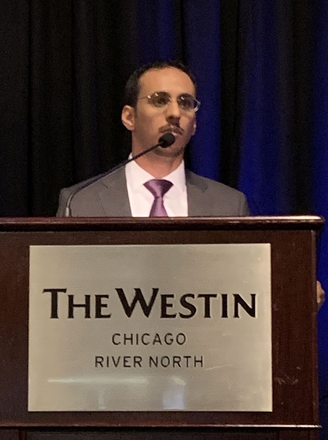
“EVAR is becoming an increasingly popular strategy for treatment of AAA,” said Samer Alharthi, MD, MPH, of the University of Toledo in Ohio. “As surgeon experience and endovascular technology have improved, a greater percentage of ruptured AAA are being treated by EVAR.”
Dr. Alharthi reported on a retrospective analysis of 4,133 patients who had repair for ruptured AAA in the American College of Surgeons National Surgical Quality Improvement Program database from 2010 to 2016. Notably, the number of EVAR repairs continue to increase and peaked in 2015, with 53% of ruptured AAA treated by EVAR.
Over the term of the study, the overall mortality rate was 22.6% for EVAR and 33.2% for open repair (P less than .001), Dr. Alharthi said. “After adjusting for cofounders, there was a 41% reduction in the mortality rate with the EVAR approach,” he said.
The only appreciable significant difference in demographics between the two groups was a higher percentage of smokers with chronic obstructive pulmonary disease having open repair – 942 (49.2%) vs. 701 (36.2%) – and a higher percentage of patients with end-stage renal disease having EVAR, Dr. Alharthi said. Other comorbidities had no statistically significant difference.
“Complications – pneumonia, reintubation, and acute renal failure – were higher in the open than the EVAR group,” he said. For example, rates of acute renal failure were 15.4% and 8.2% (P less than.001), respectively. Rates of myocardial infarction were similar between the two groups: 6.3% and 6% (P = .74), respectively.
Dr. Alharthi had no financial relationships to disclose.
SOURCE: Alharthi S et al. Midwestern Vascular 2019, Abstract 13.
CHICAGO – Numerous studies have shown conflicting results for endovascular repair in ruptured abdominal aortic aneurysms (AAA), but an analysis of 4,000-plus cases from a national registry has found a 41% reduction in mortality with endovascular repair vs. open repair, according to a presentation at the annual meeting of the Midwestern Vascular Surgery Society.
“EVAR is becoming an increasingly popular strategy for treatment of AAA,” said Samer Alharthi, MD, MPH, of the University of Toledo in Ohio. “As surgeon experience and endovascular technology have improved, a greater percentage of ruptured AAA are being treated by EVAR.”
Dr. Alharthi reported on a retrospective analysis of 4,133 patients who had repair for ruptured AAA in the American College of Surgeons National Surgical Quality Improvement Program database from 2010 to 2016. Notably, the number of EVAR repairs continue to increase and peaked in 2015, with 53% of ruptured AAA treated by EVAR.
Over the term of the study, the overall mortality rate was 22.6% for EVAR and 33.2% for open repair (P less than .001), Dr. Alharthi said. “After adjusting for cofounders, there was a 41% reduction in the mortality rate with the EVAR approach,” he said.
The only appreciable significant difference in demographics between the two groups was a higher percentage of smokers with chronic obstructive pulmonary disease having open repair – 942 (49.2%) vs. 701 (36.2%) – and a higher percentage of patients with end-stage renal disease having EVAR, Dr. Alharthi said. Other comorbidities had no statistically significant difference.
“Complications – pneumonia, reintubation, and acute renal failure – were higher in the open than the EVAR group,” he said. For example, rates of acute renal failure were 15.4% and 8.2% (P less than.001), respectively. Rates of myocardial infarction were similar between the two groups: 6.3% and 6% (P = .74), respectively.
Dr. Alharthi had no financial relationships to disclose.
SOURCE: Alharthi S et al. Midwestern Vascular 2019, Abstract 13.
CHICAGO – Numerous studies have shown conflicting results for endovascular repair in ruptured abdominal aortic aneurysms (AAA), but an analysis of 4,000-plus cases from a national registry has found a 41% reduction in mortality with endovascular repair vs. open repair, according to a presentation at the annual meeting of the Midwestern Vascular Surgery Society.
“EVAR is becoming an increasingly popular strategy for treatment of AAA,” said Samer Alharthi, MD, MPH, of the University of Toledo in Ohio. “As surgeon experience and endovascular technology have improved, a greater percentage of ruptured AAA are being treated by EVAR.”
Dr. Alharthi reported on a retrospective analysis of 4,133 patients who had repair for ruptured AAA in the American College of Surgeons National Surgical Quality Improvement Program database from 2010 to 2016. Notably, the number of EVAR repairs continue to increase and peaked in 2015, with 53% of ruptured AAA treated by EVAR.
Over the term of the study, the overall mortality rate was 22.6% for EVAR and 33.2% for open repair (P less than .001), Dr. Alharthi said. “After adjusting for cofounders, there was a 41% reduction in the mortality rate with the EVAR approach,” he said.
The only appreciable significant difference in demographics between the two groups was a higher percentage of smokers with chronic obstructive pulmonary disease having open repair – 942 (49.2%) vs. 701 (36.2%) – and a higher percentage of patients with end-stage renal disease having EVAR, Dr. Alharthi said. Other comorbidities had no statistically significant difference.
“Complications – pneumonia, reintubation, and acute renal failure – were higher in the open than the EVAR group,” he said. For example, rates of acute renal failure were 15.4% and 8.2% (P less than.001), respectively. Rates of myocardial infarction were similar between the two groups: 6.3% and 6% (P = .74), respectively.
Dr. Alharthi had no financial relationships to disclose.
SOURCE: Alharthi S et al. Midwestern Vascular 2019, Abstract 13.
REPORTING FROM MIDWESTERN VASCULAR 2019
ABPM rarely used for hypertension management in United States
NEW ORLEANS – , according to a University of Florida, Gainesville, analysis of claims data for almost 4 million people.

“With each iteration, evidence-based guidelines have more strongly recommended out-of-office blood pressure measurement, but it’s basically had no impact. If we are going to continue to recommend this aggressively, we need to put some pressure on both payers and providers,” said lead investigator Steven M. Smith, PharmD, of the department of pharmacotherapy & translational research, associate director of the Center for Integrative Cardiovascular and Metabolic Diseases at the university.
“A number of studies show that ambulatory blood pressure monitoring [ABPM] is more strongly predictive of outcomes than office pressure.” It’s “considered the gold standard for hypertension,” he said at the joint scientific sessions of the American Heart Association Council on Hypertension, AHA Council on Kidney in Cardiovascular Disease, and American Society of Hypertension
The reason is that ABPM gives a continual reading of blood pressure over 24 hours, not just an office snapshot, and can do things that office measurements cannot, including ruling out white coat hypertension, identifying masked hypertension, and checking nocturnal dipping and morning surge, both of which are related to cardiovascular risk.
Although common in Canada and Europe, it’s no secret that ABPM hasn’t caught on in the United States. The goal of Dr. Smith’s work was to help quantify the situation.
Using Truven Health Analytics commercial insurance claims data, he and his team identified 3,378,645 adults starting their first hypertension medication and 335,200 starting their fourth from 2008 to 2017. They looked for ABPM claims in the previous 6 months as well as the month after patients started their new medication. The idea was to assess ABPM use in both new and resistant hypertensive patients.
ABPM claims were submitted for 0.15% of patients starting their first drug in 2008, rising to 0.3% in 2017. ABPM was used mostly before treatment initiation.
ABPM use actually declined among resistant patients from about 0.27% in 2008 to about 0.12% in 2017. Use was split about evenly before and after they started their fourth medication.
About 80% of claims – generally for interpreting ABPM results, not the upfront cost of the machine – were paid. Claims submitted tended to come from more high-end plans. Reimbursement rates were similar for more bargain plans, but there were many fewer claims submitted, Dr. Smith said.
He thought plans would at least follow Medicare’s reimbursement policy, which, at the time of the study, covered ABPM to rule out white coat hypertension, “but they didn’t seem to,” he said. Medicare recently added coverage for suspected masked hypertension.
The study doesn’t address why uptake is so low in the United States, but outside the world of hypertension specialists, “physicians don’t see a value in it. They don’t recognize what they would get from ABPM and how that would change what they do,” in part because treatment is currently based on office measurements. There’s also probably uncertainty about how to interpret the results, Dr. Smith said.
Standardization across payers about what they’ll cover and for whom would probably help, he added.
Findings in the study were similar for home blood pressure monitoring, but probably not an accurate gauge of use. Patients mostly buy their own devices and report the results to their physician, without getting insurance involved, he said.
There was no industry funding, and the investigators didn’t have any disclosures.
SOURCE: Smith SM et al. Joint Hypertension 2019, Abstract P2067.
NEW ORLEANS – , according to a University of Florida, Gainesville, analysis of claims data for almost 4 million people.
“With each iteration, evidence-based guidelines have more strongly recommended out-of-office blood pressure measurement, but it’s basically had no impact. If we are going to continue to recommend this aggressively, we need to put some pressure on both payers and providers,” said lead investigator Steven M. Smith, PharmD, of the department of pharmacotherapy & translational research, associate director of the Center for Integrative Cardiovascular and Metabolic Diseases at the university.
“A number of studies show that ambulatory blood pressure monitoring [ABPM] is more strongly predictive of outcomes than office pressure.” It’s “considered the gold standard for hypertension,” he said at the joint scientific sessions of the American Heart Association Council on Hypertension, AHA Council on Kidney in Cardiovascular Disease, and American Society of Hypertension
The reason is that ABPM gives a continual reading of blood pressure over 24 hours, not just an office snapshot, and can do things that office measurements cannot, including ruling out white coat hypertension, identifying masked hypertension, and checking nocturnal dipping and morning surge, both of which are related to cardiovascular risk.
Although common in Canada and Europe, it’s no secret that ABPM hasn’t caught on in the United States. The goal of Dr. Smith’s work was to help quantify the situation.
Using Truven Health Analytics commercial insurance claims data, he and his team identified 3,378,645 adults starting their first hypertension medication and 335,200 starting their fourth from 2008 to 2017. They looked for ABPM claims in the previous 6 months as well as the month after patients started their new medication. The idea was to assess ABPM use in both new and resistant hypertensive patients.
ABPM claims were submitted for 0.15% of patients starting their first drug in 2008, rising to 0.3% in 2017. ABPM was used mostly before treatment initiation.
ABPM use actually declined among resistant patients from about 0.27% in 2008 to about 0.12% in 2017. Use was split about evenly before and after they started their fourth medication.
About 80% of claims – generally for interpreting ABPM results, not the upfront cost of the machine – were paid. Claims submitted tended to come from more high-end plans. Reimbursement rates were similar for more bargain plans, but there were many fewer claims submitted, Dr. Smith said.
He thought plans would at least follow Medicare’s reimbursement policy, which, at the time of the study, covered ABPM to rule out white coat hypertension, “but they didn’t seem to,” he said. Medicare recently added coverage for suspected masked hypertension.
The study doesn’t address why uptake is so low in the United States, but outside the world of hypertension specialists, “physicians don’t see a value in it. They don’t recognize what they would get from ABPM and how that would change what they do,” in part because treatment is currently based on office measurements. There’s also probably uncertainty about how to interpret the results, Dr. Smith said.
Standardization across payers about what they’ll cover and for whom would probably help, he added.
Findings in the study were similar for home blood pressure monitoring, but probably not an accurate gauge of use. Patients mostly buy their own devices and report the results to their physician, without getting insurance involved, he said.
There was no industry funding, and the investigators didn’t have any disclosures.
SOURCE: Smith SM et al. Joint Hypertension 2019, Abstract P2067.
NEW ORLEANS – , according to a University of Florida, Gainesville, analysis of claims data for almost 4 million people.
“With each iteration, evidence-based guidelines have more strongly recommended out-of-office blood pressure measurement, but it’s basically had no impact. If we are going to continue to recommend this aggressively, we need to put some pressure on both payers and providers,” said lead investigator Steven M. Smith, PharmD, of the department of pharmacotherapy & translational research, associate director of the Center for Integrative Cardiovascular and Metabolic Diseases at the university.
“A number of studies show that ambulatory blood pressure monitoring [ABPM] is more strongly predictive of outcomes than office pressure.” It’s “considered the gold standard for hypertension,” he said at the joint scientific sessions of the American Heart Association Council on Hypertension, AHA Council on Kidney in Cardiovascular Disease, and American Society of Hypertension
The reason is that ABPM gives a continual reading of blood pressure over 24 hours, not just an office snapshot, and can do things that office measurements cannot, including ruling out white coat hypertension, identifying masked hypertension, and checking nocturnal dipping and morning surge, both of which are related to cardiovascular risk.
Although common in Canada and Europe, it’s no secret that ABPM hasn’t caught on in the United States. The goal of Dr. Smith’s work was to help quantify the situation.
Using Truven Health Analytics commercial insurance claims data, he and his team identified 3,378,645 adults starting their first hypertension medication and 335,200 starting their fourth from 2008 to 2017. They looked for ABPM claims in the previous 6 months as well as the month after patients started their new medication. The idea was to assess ABPM use in both new and resistant hypertensive patients.
ABPM claims were submitted for 0.15% of patients starting their first drug in 2008, rising to 0.3% in 2017. ABPM was used mostly before treatment initiation.
ABPM use actually declined among resistant patients from about 0.27% in 2008 to about 0.12% in 2017. Use was split about evenly before and after they started their fourth medication.
About 80% of claims – generally for interpreting ABPM results, not the upfront cost of the machine – were paid. Claims submitted tended to come from more high-end plans. Reimbursement rates were similar for more bargain plans, but there were many fewer claims submitted, Dr. Smith said.
He thought plans would at least follow Medicare’s reimbursement policy, which, at the time of the study, covered ABPM to rule out white coat hypertension, “but they didn’t seem to,” he said. Medicare recently added coverage for suspected masked hypertension.
The study doesn’t address why uptake is so low in the United States, but outside the world of hypertension specialists, “physicians don’t see a value in it. They don’t recognize what they would get from ABPM and how that would change what they do,” in part because treatment is currently based on office measurements. There’s also probably uncertainty about how to interpret the results, Dr. Smith said.
Standardization across payers about what they’ll cover and for whom would probably help, he added.
Findings in the study were similar for home blood pressure monitoring, but probably not an accurate gauge of use. Patients mostly buy their own devices and report the results to their physician, without getting insurance involved, he said.
There was no industry funding, and the investigators didn’t have any disclosures.
SOURCE: Smith SM et al. Joint Hypertension 2019, Abstract P2067.
REPORTING FROM JOINT HYPERTENSION 2019

Project ECHO helps osteoporosis specialists connect with PCPs
ORLANDO – The use of a teleconferencing program to share knowledge about osteoporosis has helped health care professionals learn about the disease and may potentially reduce the osteoporosis treatment gap in underserved communities, according to a speaker at the annual meeting of the American Society for Bone and Mineral Research.
The concept, called “technology-enabled collaborative learning,” is intended to address the problem of there being not enough specialists to see patients who need treatment, and the ineffectiveness of educating primary care providers in how to treat complex medical conditions, E. Michael Lewiecki, MD, the director of the New Mexico Clinical Research & Osteoporosis Center said in his presentation.
“Primary care doctors are busy,” said Dr. Lewiecki. “They have limited time taking care of patients. They don’t have the time or often the skills to manage patients who have any questions or concerns about osteoporosis and treatments for osteoporosis.”
One solution, he said, is to find health care professionals in underserved communities who are already interested in and motivated to learn more about osteoporosis, turn them into near-experts on osteoporosis for their patients as well as in their own community.
Dr. Lewiecki proposed the Extension for Community Healthcare Outcomes (ECHO), or Project ECHO, an initiative out of the University of New Mexico School of Medicine, as a potential answer. Project ECHO uses videoconferencing to connect experts in a therapeutic area, with Bone Health TeleECHO focusing on raising knowledge of osteoporosis for its participants. “The idea of ECHO is to be a force multiplier to educate health care professionals, each of whom takes care of many patients, and to have many ECHO programs around the world in convenient time zones and convenient languages for people who are interested in participating,” said Dr. Lewiecki.
The idea began when a gastroenterologist at Dr. Lewiecki’s own center was frustrated that patients were not seeking treatment for hepatitis C because of time or travel issues. In response, a pilot program for Project ECHO was developed through a collaboration between the University of New Mexico Health Sciences Center and the Osteoporosis Foundation of New Mexico where gastroenterologists at University of New Mexico connected with primary care providers across the state, sharing information about hepatitis C and discussing case studies. The results of the pilot program were published in the New England Journal of Medicine and showed a similar rate of sustained viral response between patients treated at the University of New Mexico clinic (84 of 146 patients; 57.5%) and at 21 ECHO clinics (152 of 261 patients; 58.2%) (Arora S et al. N Eng J Med. 2011. doi: 10.1056/NEJMoa1009370).
“ECHO expands the capacity to deliver best practice medical care through collegial, interactive, case-based discussions with minimal disruption to the office routine,” said Dr. Lewiecki. “Patients benefit from better care, closer to home, with greater convenience and lower cost than referral to a medical center. And the potential is to reduce the osteoporosis treatment gap by having many ECHOs starting up in many places in the world.”
Today, the ECHO program is in 37 countries, with 322 ECHO hubs and 677 ECHO programs. The top three specialties are endocrinology, orthopedics, and rheumatology; 51% of ECHO participants are primary care providers, 24% are advanced care providers, and 19% are health care providers such as nutritionists, physical therapists, and other providers that have an interest in bone health.
In survey results adapted from a 2017 study from his own group, Dr. Lewiecki showed that 263 health care professionals who participated in Bone Health TeleECHO rated themselves as more confident in 20 different domains of osteoporosis treatment, such as secondary osteoporosis and anabolic therapy, after 21 months of using the ECHO program (Lewiecki EM et al. J Endocr Soc. 2017. doi: 10.1210/js.2017-00361). However, he admitted that showing fracture prevention outcomes at these ECHO centers has proven more difficult.
“Of course, we’re all interested in outcomes. The ultimate outcome here is preventing fractures, but it is extraordinarily difficult to design a study to actually show that we’re reducing fractures, but certainly self-confidence in managing osteoporosis has improved,” he said.
There have also been some misconceptions of the Project ECHO. The program is not only for beginners or primary care providers, said Dr. Lewiecki. It is also not limited to providers in rural areas, as the program has many participants at urban centers, he added.
“We are a virtual community of practice. It’s a collegial relationship,” he said. “It’s really recapitulating the way that we learned during our postgraduate training: When we see a patient, we present the case to our attending, the attending pontificates a little bit, we bounce things off of one another, and we go back and then we do some different things with our patients. And that’s exactly what we do with Echo. It makes learning fun again.”
Dr. Lewiecki challenged the attendees in the room who are already experts in osteoporosis to help share their knowledge of the disease to help other health care professionals learn more about how to better care for their patients. “If you have a passion for teaching, if you want to share knowledge and you’re willing to devote a little bit of your time to doing that and reaching out to more people, this is the way that you can do it.”
Dr. Lewiecki reports research grant support from Amgen, consulting fees from Alexion, Amgen, Radius, Shire, and Ultragenyx, speaking fees from Alexion, Radius, and Shire, and is an advisory board member with the National Osteoporosis Foundation, International Society for Clinical Densitometry, and the Osteoporosis Foundation of New Mexico.
SOURCE: Lewiecki ME. ASBMR 2019. Symposia: Cutting Edge Concepts: Novel Approaches to Reducing Fractures. Bone Health TeleECHO.
ORLANDO – The use of a teleconferencing program to share knowledge about osteoporosis has helped health care professionals learn about the disease and may potentially reduce the osteoporosis treatment gap in underserved communities, according to a speaker at the annual meeting of the American Society for Bone and Mineral Research.
The concept, called “technology-enabled collaborative learning,” is intended to address the problem of there being not enough specialists to see patients who need treatment, and the ineffectiveness of educating primary care providers in how to treat complex medical conditions, E. Michael Lewiecki, MD, the director of the New Mexico Clinical Research & Osteoporosis Center said in his presentation.
“Primary care doctors are busy,” said Dr. Lewiecki. “They have limited time taking care of patients. They don’t have the time or often the skills to manage patients who have any questions or concerns about osteoporosis and treatments for osteoporosis.”
One solution, he said, is to find health care professionals in underserved communities who are already interested in and motivated to learn more about osteoporosis, turn them into near-experts on osteoporosis for their patients as well as in their own community.
Dr. Lewiecki proposed the Extension for Community Healthcare Outcomes (ECHO), or Project ECHO, an initiative out of the University of New Mexico School of Medicine, as a potential answer. Project ECHO uses videoconferencing to connect experts in a therapeutic area, with Bone Health TeleECHO focusing on raising knowledge of osteoporosis for its participants. “The idea of ECHO is to be a force multiplier to educate health care professionals, each of whom takes care of many patients, and to have many ECHO programs around the world in convenient time zones and convenient languages for people who are interested in participating,” said Dr. Lewiecki.
The idea began when a gastroenterologist at Dr. Lewiecki’s own center was frustrated that patients were not seeking treatment for hepatitis C because of time or travel issues. In response, a pilot program for Project ECHO was developed through a collaboration between the University of New Mexico Health Sciences Center and the Osteoporosis Foundation of New Mexico where gastroenterologists at University of New Mexico connected with primary care providers across the state, sharing information about hepatitis C and discussing case studies. The results of the pilot program were published in the New England Journal of Medicine and showed a similar rate of sustained viral response between patients treated at the University of New Mexico clinic (84 of 146 patients; 57.5%) and at 21 ECHO clinics (152 of 261 patients; 58.2%) (Arora S et al. N Eng J Med. 2011. doi: 10.1056/NEJMoa1009370).
“ECHO expands the capacity to deliver best practice medical care through collegial, interactive, case-based discussions with minimal disruption to the office routine,” said Dr. Lewiecki. “Patients benefit from better care, closer to home, with greater convenience and lower cost than referral to a medical center. And the potential is to reduce the osteoporosis treatment gap by having many ECHOs starting up in many places in the world.”
Today, the ECHO program is in 37 countries, with 322 ECHO hubs and 677 ECHO programs. The top three specialties are endocrinology, orthopedics, and rheumatology; 51% of ECHO participants are primary care providers, 24% are advanced care providers, and 19% are health care providers such as nutritionists, physical therapists, and other providers that have an interest in bone health.
In survey results adapted from a 2017 study from his own group, Dr. Lewiecki showed that 263 health care professionals who participated in Bone Health TeleECHO rated themselves as more confident in 20 different domains of osteoporosis treatment, such as secondary osteoporosis and anabolic therapy, after 21 months of using the ECHO program (Lewiecki EM et al. J Endocr Soc. 2017. doi: 10.1210/js.2017-00361). However, he admitted that showing fracture prevention outcomes at these ECHO centers has proven more difficult.
“Of course, we’re all interested in outcomes. The ultimate outcome here is preventing fractures, but it is extraordinarily difficult to design a study to actually show that we’re reducing fractures, but certainly self-confidence in managing osteoporosis has improved,” he said.
There have also been some misconceptions of the Project ECHO. The program is not only for beginners or primary care providers, said Dr. Lewiecki. It is also not limited to providers in rural areas, as the program has many participants at urban centers, he added.
“We are a virtual community of practice. It’s a collegial relationship,” he said. “It’s really recapitulating the way that we learned during our postgraduate training: When we see a patient, we present the case to our attending, the attending pontificates a little bit, we bounce things off of one another, and we go back and then we do some different things with our patients. And that’s exactly what we do with Echo. It makes learning fun again.”
Dr. Lewiecki challenged the attendees in the room who are already experts in osteoporosis to help share their knowledge of the disease to help other health care professionals learn more about how to better care for their patients. “If you have a passion for teaching, if you want to share knowledge and you’re willing to devote a little bit of your time to doing that and reaching out to more people, this is the way that you can do it.”
Dr. Lewiecki reports research grant support from Amgen, consulting fees from Alexion, Amgen, Radius, Shire, and Ultragenyx, speaking fees from Alexion, Radius, and Shire, and is an advisory board member with the National Osteoporosis Foundation, International Society for Clinical Densitometry, and the Osteoporosis Foundation of New Mexico.
SOURCE: Lewiecki ME. ASBMR 2019. Symposia: Cutting Edge Concepts: Novel Approaches to Reducing Fractures. Bone Health TeleECHO.
ORLANDO – The use of a teleconferencing program to share knowledge about osteoporosis has helped health care professionals learn about the disease and may potentially reduce the osteoporosis treatment gap in underserved communities, according to a speaker at the annual meeting of the American Society for Bone and Mineral Research.
The concept, called “technology-enabled collaborative learning,” is intended to address the problem of there being not enough specialists to see patients who need treatment, and the ineffectiveness of educating primary care providers in how to treat complex medical conditions, E. Michael Lewiecki, MD, the director of the New Mexico Clinical Research & Osteoporosis Center said in his presentation.
“Primary care doctors are busy,” said Dr. Lewiecki. “They have limited time taking care of patients. They don’t have the time or often the skills to manage patients who have any questions or concerns about osteoporosis and treatments for osteoporosis.”
One solution, he said, is to find health care professionals in underserved communities who are already interested in and motivated to learn more about osteoporosis, turn them into near-experts on osteoporosis for their patients as well as in their own community.
Dr. Lewiecki proposed the Extension for Community Healthcare Outcomes (ECHO), or Project ECHO, an initiative out of the University of New Mexico School of Medicine, as a potential answer. Project ECHO uses videoconferencing to connect experts in a therapeutic area, with Bone Health TeleECHO focusing on raising knowledge of osteoporosis for its participants. “The idea of ECHO is to be a force multiplier to educate health care professionals, each of whom takes care of many patients, and to have many ECHO programs around the world in convenient time zones and convenient languages for people who are interested in participating,” said Dr. Lewiecki.
The idea began when a gastroenterologist at Dr. Lewiecki’s own center was frustrated that patients were not seeking treatment for hepatitis C because of time or travel issues. In response, a pilot program for Project ECHO was developed through a collaboration between the University of New Mexico Health Sciences Center and the Osteoporosis Foundation of New Mexico where gastroenterologists at University of New Mexico connected with primary care providers across the state, sharing information about hepatitis C and discussing case studies. The results of the pilot program were published in the New England Journal of Medicine and showed a similar rate of sustained viral response between patients treated at the University of New Mexico clinic (84 of 146 patients; 57.5%) and at 21 ECHO clinics (152 of 261 patients; 58.2%) (Arora S et al. N Eng J Med. 2011. doi: 10.1056/NEJMoa1009370).
“ECHO expands the capacity to deliver best practice medical care through collegial, interactive, case-based discussions with minimal disruption to the office routine,” said Dr. Lewiecki. “Patients benefit from better care, closer to home, with greater convenience and lower cost than referral to a medical center. And the potential is to reduce the osteoporosis treatment gap by having many ECHOs starting up in many places in the world.”
Today, the ECHO program is in 37 countries, with 322 ECHO hubs and 677 ECHO programs. The top three specialties are endocrinology, orthopedics, and rheumatology; 51% of ECHO participants are primary care providers, 24% are advanced care providers, and 19% are health care providers such as nutritionists, physical therapists, and other providers that have an interest in bone health.
In survey results adapted from a 2017 study from his own group, Dr. Lewiecki showed that 263 health care professionals who participated in Bone Health TeleECHO rated themselves as more confident in 20 different domains of osteoporosis treatment, such as secondary osteoporosis and anabolic therapy, after 21 months of using the ECHO program (Lewiecki EM et al. J Endocr Soc. 2017. doi: 10.1210/js.2017-00361). However, he admitted that showing fracture prevention outcomes at these ECHO centers has proven more difficult.
“Of course, we’re all interested in outcomes. The ultimate outcome here is preventing fractures, but it is extraordinarily difficult to design a study to actually show that we’re reducing fractures, but certainly self-confidence in managing osteoporosis has improved,” he said.
There have also been some misconceptions of the Project ECHO. The program is not only for beginners or primary care providers, said Dr. Lewiecki. It is also not limited to providers in rural areas, as the program has many participants at urban centers, he added.
“We are a virtual community of practice. It’s a collegial relationship,” he said. “It’s really recapitulating the way that we learned during our postgraduate training: When we see a patient, we present the case to our attending, the attending pontificates a little bit, we bounce things off of one another, and we go back and then we do some different things with our patients. And that’s exactly what we do with Echo. It makes learning fun again.”
Dr. Lewiecki challenged the attendees in the room who are already experts in osteoporosis to help share their knowledge of the disease to help other health care professionals learn more about how to better care for their patients. “If you have a passion for teaching, if you want to share knowledge and you’re willing to devote a little bit of your time to doing that and reaching out to more people, this is the way that you can do it.”
Dr. Lewiecki reports research grant support from Amgen, consulting fees from Alexion, Amgen, Radius, Shire, and Ultragenyx, speaking fees from Alexion, Radius, and Shire, and is an advisory board member with the National Osteoporosis Foundation, International Society for Clinical Densitometry, and the Osteoporosis Foundation of New Mexico.
SOURCE: Lewiecki ME. ASBMR 2019. Symposia: Cutting Edge Concepts: Novel Approaches to Reducing Fractures. Bone Health TeleECHO.
FROM ASBMR 2019
Study aims to define symptoms of Sjögren’s syndrome secondary to SLE
Sjögren’s syndrome secondary to systemic lupus erythematosus rises in frequency with age, affects nearly one-quarter of all people with SLE, and is marked by a systemic inflammatory state with high levels of proinflammatory cytokines.
Those are key findings from a Swedish study that set out to evaluate the subjective and objective symptoms of secondary Sjögren’s syndrome (sSS) from a large cohort of SLE patients and matched controls.
“The diagnosis SS is a clinical entity, based on dryness of eyes and mouth due to destructive inflammation in the exocrine glands, especially tear and salivary glands,” researchers led by Guillermo Ruacho, DMD, and Marika Kvarnström, MD, PhD, of the Karolinska Institute, wrote in a study published in the Journal of Rheumatology (doi: 10.3899/jrheum.190250). “SS can exist [as] isolated, primary SS (pSS) or together with other rheumatic diseases, referred to as secondary SS (sSS). A major difference according to the 2002 Revised American-European Consensus Criteria (AECC) is the classification where the serologic item (SSA/SSB antibodies) is included for pSS, but not for sSS (Ann Rheum Dis. 2002;61:554-8). In SLE, these autoantibodies are common, usually stable over time, and they appear early, even several years before disease onset.”
The researchers evaluated 504 consecutive SLE patients and 319 controls from the general population, who were matched for age and gender to the first 319 SLE patients. They used AECC to define SLE-sSS and conducted a thorough clinical investigation of all patients, including analysis of autoantibodies and 20 selected cytokines.
The researchers found that SLE-sSS occurred in 23% of the SLE patients. In comparison with SLE patients who did not have sSS, those in the SLE-sSS group were an average of 9 years older, more likely to be female (96% vs. 84%, respectively), and more likely to have leukopenia (57% vs. 45%), yet less likely to have nephritis (32% vs. 43%). Of 20 proinflammatory cytokines investigated, 6 were higher in the SLE-sSS group: TNF-alpha, IL-6, MCP-4, MIP-1beta, IL-12/IL-23p40, and IP-10. Other clinical measures higher in the SLE-sSS group were total IgG, anti-SSA/Ro52, anti-SSA/Ro60, anti-SSB/La antibodies, and rheumatoid factor (IgM and IgA; P less than .05 for all comparisons).
“To our knowledge this is the first study to investigate if systemic inflammation, as measured by cytokine levels, differs between SLE-sSS and SLE-nonsSS,” the researchers wrote. “In clinical practice, it is often difficult to delineate pSS from SLE-sSS. Organ manifestations commonly reported in pSS are fever, lymphadenopathy, parotid gland enlargement, Raynaud’s phenomenon, interstitial lung disease, peripheral neuropathy, and vasculitis. All these clinical features, except parotid gland enlargement, were investigated in the present study, but only peripheral neuropathy differed and was more frequent in SLE-sSS than in SLE-nonsSS.”
They acknowledged certain limitations of the study, including the fact that they did not measure saliva and tear production in controls without sicca symptoms.
The study was supported by funds from Swedish local and national governments, medical societies, foundations, and patient advocacy groups, One author is an employee at AstraZeneca, which provided reagents for the cytokine analyses but had no impact on the analyses, the authors said.
SOURCE: Ruacho G et al. J Rheumatol. 2019 Sep 1. doi: 10.3899/jrheum.190250.
Sjögren’s syndrome secondary to systemic lupus erythematosus rises in frequency with age, affects nearly one-quarter of all people with SLE, and is marked by a systemic inflammatory state with high levels of proinflammatory cytokines.
Those are key findings from a Swedish study that set out to evaluate the subjective and objective symptoms of secondary Sjögren’s syndrome (sSS) from a large cohort of SLE patients and matched controls.
“The diagnosis SS is a clinical entity, based on dryness of eyes and mouth due to destructive inflammation in the exocrine glands, especially tear and salivary glands,” researchers led by Guillermo Ruacho, DMD, and Marika Kvarnström, MD, PhD, of the Karolinska Institute, wrote in a study published in the Journal of Rheumatology (doi: 10.3899/jrheum.190250). “SS can exist [as] isolated, primary SS (pSS) or together with other rheumatic diseases, referred to as secondary SS (sSS). A major difference according to the 2002 Revised American-European Consensus Criteria (AECC) is the classification where the serologic item (SSA/SSB antibodies) is included for pSS, but not for sSS (Ann Rheum Dis. 2002;61:554-8). In SLE, these autoantibodies are common, usually stable over time, and they appear early, even several years before disease onset.”
The researchers evaluated 504 consecutive SLE patients and 319 controls from the general population, who were matched for age and gender to the first 319 SLE patients. They used AECC to define SLE-sSS and conducted a thorough clinical investigation of all patients, including analysis of autoantibodies and 20 selected cytokines.
The researchers found that SLE-sSS occurred in 23% of the SLE patients. In comparison with SLE patients who did not have sSS, those in the SLE-sSS group were an average of 9 years older, more likely to be female (96% vs. 84%, respectively), and more likely to have leukopenia (57% vs. 45%), yet less likely to have nephritis (32% vs. 43%). Of 20 proinflammatory cytokines investigated, 6 were higher in the SLE-sSS group: TNF-alpha, IL-6, MCP-4, MIP-1beta, IL-12/IL-23p40, and IP-10. Other clinical measures higher in the SLE-sSS group were total IgG, anti-SSA/Ro52, anti-SSA/Ro60, anti-SSB/La antibodies, and rheumatoid factor (IgM and IgA; P less than .05 for all comparisons).
“To our knowledge this is the first study to investigate if systemic inflammation, as measured by cytokine levels, differs between SLE-sSS and SLE-nonsSS,” the researchers wrote. “In clinical practice, it is often difficult to delineate pSS from SLE-sSS. Organ manifestations commonly reported in pSS are fever, lymphadenopathy, parotid gland enlargement, Raynaud’s phenomenon, interstitial lung disease, peripheral neuropathy, and vasculitis. All these clinical features, except parotid gland enlargement, were investigated in the present study, but only peripheral neuropathy differed and was more frequent in SLE-sSS than in SLE-nonsSS.”
They acknowledged certain limitations of the study, including the fact that they did not measure saliva and tear production in controls without sicca symptoms.
The study was supported by funds from Swedish local and national governments, medical societies, foundations, and patient advocacy groups, One author is an employee at AstraZeneca, which provided reagents for the cytokine analyses but had no impact on the analyses, the authors said.
SOURCE: Ruacho G et al. J Rheumatol. 2019 Sep 1. doi: 10.3899/jrheum.190250.
Sjögren’s syndrome secondary to systemic lupus erythematosus rises in frequency with age, affects nearly one-quarter of all people with SLE, and is marked by a systemic inflammatory state with high levels of proinflammatory cytokines.
Those are key findings from a Swedish study that set out to evaluate the subjective and objective symptoms of secondary Sjögren’s syndrome (sSS) from a large cohort of SLE patients and matched controls.
“The diagnosis SS is a clinical entity, based on dryness of eyes and mouth due to destructive inflammation in the exocrine glands, especially tear and salivary glands,” researchers led by Guillermo Ruacho, DMD, and Marika Kvarnström, MD, PhD, of the Karolinska Institute, wrote in a study published in the Journal of Rheumatology (doi: 10.3899/jrheum.190250). “SS can exist [as] isolated, primary SS (pSS) or together with other rheumatic diseases, referred to as secondary SS (sSS). A major difference according to the 2002 Revised American-European Consensus Criteria (AECC) is the classification where the serologic item (SSA/SSB antibodies) is included for pSS, but not for sSS (Ann Rheum Dis. 2002;61:554-8). In SLE, these autoantibodies are common, usually stable over time, and they appear early, even several years before disease onset.”
The researchers evaluated 504 consecutive SLE patients and 319 controls from the general population, who were matched for age and gender to the first 319 SLE patients. They used AECC to define SLE-sSS and conducted a thorough clinical investigation of all patients, including analysis of autoantibodies and 20 selected cytokines.
The researchers found that SLE-sSS occurred in 23% of the SLE patients. In comparison with SLE patients who did not have sSS, those in the SLE-sSS group were an average of 9 years older, more likely to be female (96% vs. 84%, respectively), and more likely to have leukopenia (57% vs. 45%), yet less likely to have nephritis (32% vs. 43%). Of 20 proinflammatory cytokines investigated, 6 were higher in the SLE-sSS group: TNF-alpha, IL-6, MCP-4, MIP-1beta, IL-12/IL-23p40, and IP-10. Other clinical measures higher in the SLE-sSS group were total IgG, anti-SSA/Ro52, anti-SSA/Ro60, anti-SSB/La antibodies, and rheumatoid factor (IgM and IgA; P less than .05 for all comparisons).
“To our knowledge this is the first study to investigate if systemic inflammation, as measured by cytokine levels, differs between SLE-sSS and SLE-nonsSS,” the researchers wrote. “In clinical practice, it is often difficult to delineate pSS from SLE-sSS. Organ manifestations commonly reported in pSS are fever, lymphadenopathy, parotid gland enlargement, Raynaud’s phenomenon, interstitial lung disease, peripheral neuropathy, and vasculitis. All these clinical features, except parotid gland enlargement, were investigated in the present study, but only peripheral neuropathy differed and was more frequent in SLE-sSS than in SLE-nonsSS.”
They acknowledged certain limitations of the study, including the fact that they did not measure saliva and tear production in controls without sicca symptoms.
The study was supported by funds from Swedish local and national governments, medical societies, foundations, and patient advocacy groups, One author is an employee at AstraZeneca, which provided reagents for the cytokine analyses but had no impact on the analyses, the authors said.
SOURCE: Ruacho G et al. J Rheumatol. 2019 Sep 1. doi: 10.3899/jrheum.190250.
FROM THE JOURNAL OF RHEUMATOLOGY