User login
A Neurotoxin, an Antidepressant, and More Emerging Options for Treating Rosacea
ORLANDO, FLORIDA — At the same time, there is new recognition that systemic inflammation can occur with rosacea, and targeting treatment to the phenotype continues to gain steam as a way to help people with this difficult-to-manage condition.
“Anyone here think they’ve got rosacea under control? No, I wish — not yet,” Diane Dr. Thiboutot, MD, said at the annual ODAC Dermatology, Aesthetic & Surgical Conference.
Botulinum Toxin Benefits
With that in mind, Dr. Thiboutot highlighted emerging therapies for treating rosacea. “Last year, there were a couple of reports … looking at the use of botulinum toxin injections for patients with rosacea,” said Dr. Thiboutot, professor of dermatology and vice chair for research in the Department of Dermatology at Penn State College of Medicine, Hershey, Pennsylvania.
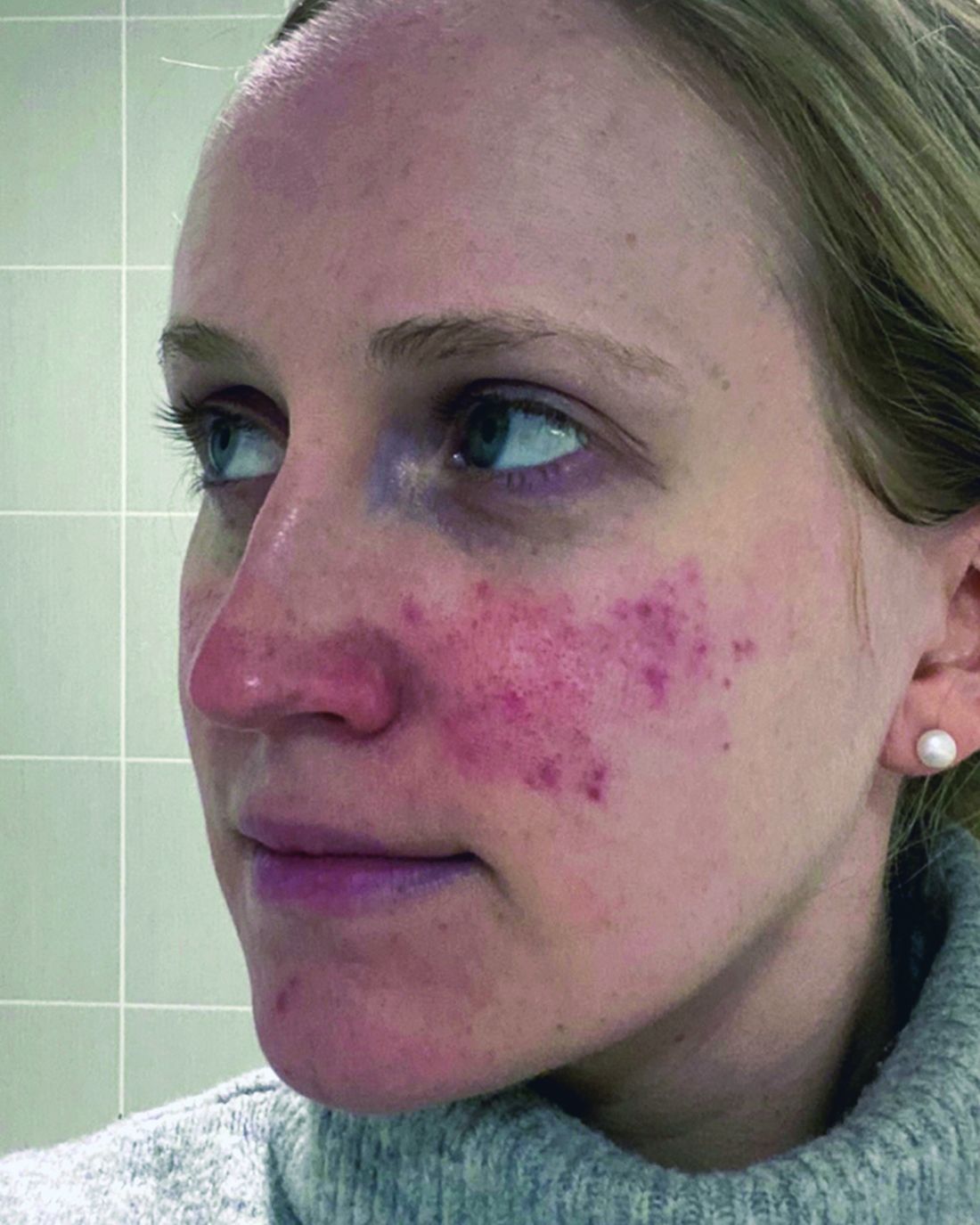
One report describes the case of a woman with rosacea who had severe recurrent episodes of erythema and flushing. She also experienced occasional papules and pustules and had been recalcitrant to multiple treatments for rosacea, according to the report published in the Journal of Drugs in Dermatology in June 2023. The patient was treated with a total of 150-180 units of botulinum toxin administered as 3-6 units spaced 1 cm apart every 2-4 months. She was “eventually maintained every 6 months with excellent improvement,” Dr. Thiboutot said.
In another case, a man with refractory vascular and papulopustular rosacea was treated with half of a unit of botulinum toxin spaced every 0.5 cm. Images taken at baseline, 1 month, and 3 months after treatment demonstrated improvements, as reported in June 2023.
Regarding botulinum toxin for rosacea, Dr. Thiboutot said, “it’s a very interesting thing to think about.”
Susan Weinkle, MD, ODAC conference cochair, session moderator, and collaborative associate professor of dermatology at the University of South Florida, Tampa, Florida, agreed. “I do think it holds some interesting potential,” she said. “How good are your hands? Because administering 0.5-unit injections evenly is a little bit challenging.”
However, one approach that might help is “if we could be a little more innovative like they are in Europe.” Physicians in Europe can use a metered syringe, one where they dial in the exact amount per injection, which allows them to be consistent, she added.
With rosacea erythema, Dr. Thiboutot noted, a spotted effect can result if injections are not administered uniformly.
Potential Role for Paroxetine
The antidepressant paroxetine, a potent selective serotonin reuptake inhibitor, could be an effective treatment for refractory erythema of rosacea, Dr. Thiboutot said. It is approved for treating depression, obsessive-compulsive disorder, and social phobia. The agent has also shown effectiveness in alleviating hot flashes associated with vascular dysregulation in menopause.
Uptake in serotonin and changes in receptors are closely related to vascular dilation and constriction, Dr. Thiboutot added, so paroxetine “may be beneficial in treating vascular dysfunction” including in people with rosacea. Evidence to support this potential approach comes from the primary results of a randomized controlled trial published in June 2023. Based on the results, the researchers concluded that paroxetine “appears to be an efficacious and well-tolerated treatment for refractory erythema in rosacea.”
In the trial, almost 43% of people treated with paroxetine met the primary endpoint for improving recalcitrant erythema at week 12 compared with almost 21% who took a placebo, a statistically significant difference.
Heparan Sulfate Analog in a Cream
Evidence suggests that a low-molecular-weight heparan sulfate analog is another agent that holds potential for treating rosacea. For example, a 2023 randomized controlled trial evaluated the immune response in rosacea, focusing on a specific cathelicidin peptide called LL-37 that activates an inflammasome in rosacea. Low-molecular-weight heparan sulfate holds the potential to inhibit LL-37 activity, as LL-37 is inhibited by binding to heparan sulfate, a cell surface glycosaminoglycan.
The study of 16 people assessed the ability of the analog to modulate this response; they were also treated with the pulsed dye laser. Participants who applied a dermal repair cream that contained this ingredient experienced a one-grade reduction in erythema at weeks 4 and 8 compared with a control group applying a moisturizer.
A Growing Case for Systemic Inflammation
In the meantime, treating rosacea with more traditional therapies remains challenging.
But there’s hope. Success has been reported in the few years since an expert panel recommended treating based on phenotype — a treat-what-you-see approach, Dr. Thiboutot said.
“We don’t have a single treatment that is one-size-fits-all. We have to individualize our treatment [based] more on what we are seeing and what the patient is experiencing.”
Eventually, therapies to treat systemic inflammation could provide benefits as well. As with hidradenitis suppurativa and psoriasis, “there’s evidence of systemic inflammation in some of our rosacea patients,” Dr. Thiboutot said.
For example, researchers compared blood taken from people with and without rosacea and found increased levels of some inflammatory markers among participants with the condition.
The retrospective study published in June 2023 in Scientific Reports included 100 patients with rosacea and 58 controls. The investigators found significantly higher elevations in the SII index, erythrocyte sedimentation rate (ESR), and C-reactive protein (CRP) levels in the patients with rosacea.
“There was no significant link between the severity of rosacea and the ESR, CRP, or SII index values, Dr. Thiboutot added. “This study suggests inflammation beyond the skin in rosacea patients.”
For more guidance on treating rosacea through standard management options, including how to tailor therapy to each individual, she recommended the 2019 Update by the National Rosacea Society Expert Committee. “It’s a nice quick way to see, based on expert opinion, the most effective treatments and what the evidence base is,” said Dr. Thiboutot, lead author of the paper, published in the Journal of the American Academy of Dermatology in February 2020.
Dr. Thiboutot reported no relevant financial relationships.
ORLANDO, FLORIDA — At the same time, there is new recognition that systemic inflammation can occur with rosacea, and targeting treatment to the phenotype continues to gain steam as a way to help people with this difficult-to-manage condition.
“Anyone here think they’ve got rosacea under control? No, I wish — not yet,” Diane Dr. Thiboutot, MD, said at the annual ODAC Dermatology, Aesthetic & Surgical Conference.
Botulinum Toxin Benefits
With that in mind, Dr. Thiboutot highlighted emerging therapies for treating rosacea. “Last year, there were a couple of reports … looking at the use of botulinum toxin injections for patients with rosacea,” said Dr. Thiboutot, professor of dermatology and vice chair for research in the Department of Dermatology at Penn State College of Medicine, Hershey, Pennsylvania.
One report describes the case of a woman with rosacea who had severe recurrent episodes of erythema and flushing. She also experienced occasional papules and pustules and had been recalcitrant to multiple treatments for rosacea, according to the report published in the Journal of Drugs in Dermatology in June 2023. The patient was treated with a total of 150-180 units of botulinum toxin administered as 3-6 units spaced 1 cm apart every 2-4 months. She was “eventually maintained every 6 months with excellent improvement,” Dr. Thiboutot said.
In another case, a man with refractory vascular and papulopustular rosacea was treated with half of a unit of botulinum toxin spaced every 0.5 cm. Images taken at baseline, 1 month, and 3 months after treatment demonstrated improvements, as reported in June 2023.
Regarding botulinum toxin for rosacea, Dr. Thiboutot said, “it’s a very interesting thing to think about.”
Susan Weinkle, MD, ODAC conference cochair, session moderator, and collaborative associate professor of dermatology at the University of South Florida, Tampa, Florida, agreed. “I do think it holds some interesting potential,” she said. “How good are your hands? Because administering 0.5-unit injections evenly is a little bit challenging.”
However, one approach that might help is “if we could be a little more innovative like they are in Europe.” Physicians in Europe can use a metered syringe, one where they dial in the exact amount per injection, which allows them to be consistent, she added.
With rosacea erythema, Dr. Thiboutot noted, a spotted effect can result if injections are not administered uniformly.
Potential Role for Paroxetine
The antidepressant paroxetine, a potent selective serotonin reuptake inhibitor, could be an effective treatment for refractory erythema of rosacea, Dr. Thiboutot said. It is approved for treating depression, obsessive-compulsive disorder, and social phobia. The agent has also shown effectiveness in alleviating hot flashes associated with vascular dysregulation in menopause.
Uptake in serotonin and changes in receptors are closely related to vascular dilation and constriction, Dr. Thiboutot added, so paroxetine “may be beneficial in treating vascular dysfunction” including in people with rosacea. Evidence to support this potential approach comes from the primary results of a randomized controlled trial published in June 2023. Based on the results, the researchers concluded that paroxetine “appears to be an efficacious and well-tolerated treatment for refractory erythema in rosacea.”
In the trial, almost 43% of people treated with paroxetine met the primary endpoint for improving recalcitrant erythema at week 12 compared with almost 21% who took a placebo, a statistically significant difference.
Heparan Sulfate Analog in a Cream
Evidence suggests that a low-molecular-weight heparan sulfate analog is another agent that holds potential for treating rosacea. For example, a 2023 randomized controlled trial evaluated the immune response in rosacea, focusing on a specific cathelicidin peptide called LL-37 that activates an inflammasome in rosacea. Low-molecular-weight heparan sulfate holds the potential to inhibit LL-37 activity, as LL-37 is inhibited by binding to heparan sulfate, a cell surface glycosaminoglycan.
The study of 16 people assessed the ability of the analog to modulate this response; they were also treated with the pulsed dye laser. Participants who applied a dermal repair cream that contained this ingredient experienced a one-grade reduction in erythema at weeks 4 and 8 compared with a control group applying a moisturizer.
A Growing Case for Systemic Inflammation
In the meantime, treating rosacea with more traditional therapies remains challenging.
But there’s hope. Success has been reported in the few years since an expert panel recommended treating based on phenotype — a treat-what-you-see approach, Dr. Thiboutot said.
“We don’t have a single treatment that is one-size-fits-all. We have to individualize our treatment [based] more on what we are seeing and what the patient is experiencing.”
Eventually, therapies to treat systemic inflammation could provide benefits as well. As with hidradenitis suppurativa and psoriasis, “there’s evidence of systemic inflammation in some of our rosacea patients,” Dr. Thiboutot said.
For example, researchers compared blood taken from people with and without rosacea and found increased levels of some inflammatory markers among participants with the condition.
The retrospective study published in June 2023 in Scientific Reports included 100 patients with rosacea and 58 controls. The investigators found significantly higher elevations in the SII index, erythrocyte sedimentation rate (ESR), and C-reactive protein (CRP) levels in the patients with rosacea.
“There was no significant link between the severity of rosacea and the ESR, CRP, or SII index values, Dr. Thiboutot added. “This study suggests inflammation beyond the skin in rosacea patients.”
For more guidance on treating rosacea through standard management options, including how to tailor therapy to each individual, she recommended the 2019 Update by the National Rosacea Society Expert Committee. “It’s a nice quick way to see, based on expert opinion, the most effective treatments and what the evidence base is,” said Dr. Thiboutot, lead author of the paper, published in the Journal of the American Academy of Dermatology in February 2020.
Dr. Thiboutot reported no relevant financial relationships.
FROM ODAC 2024
RNA Vaccines: Risk for Heavy Menstrual Bleeding Clarified
Cases of menstrual disorders, particularly unusually heavy menstrual bleeding, have been reported following RNA vaccination against COVID-19.
In France, this safety signal has been confirmed and added to the product characteristics summaries and vaccine leaflets for mRNA vaccines in October 2022. However, few studies have accurately measured this risk to date.
To address this gap in research, the French scientific interest group in the epidemiology of health products, ANSM-Cnam EPI-PHARE, conducted a study to assess the risk for heavy menstrual bleeding requiring hospitalization after COVID-19 vaccination in France.
“This study provides new evidence supporting the existence of an increased risk for heavy menstrual bleeding following COVID-19 vaccination with mRNA vaccines,” wrote the authors.
Study Details
The study included all women aged 15-50 years who were diagnosed with heavy menstrual bleeding in the hospital between May 12, 2021, and August 31, 2022. Participants were identified in the National Health Data System, and the study population totaled 4610 women.
Each participant was randomly matched with as many as 30 women who had not been hospitalized for abnormal genital bleeding and had similar characteristics in terms of age, department of residence, social deprivation index of the commune of residence, and contraceptive method.
Women who had a recent pregnancy, hysterectomy, or coagulation disorder within the specified time frames were excluded.
At the time of the study, 71% of cases and 70% of controls had received at least one dose of the COVID-19 vaccine. Among vaccinated participants, 68% and 66%, respectively, received a vaccination dose (first or second dose). An mRNA vaccine (Comirnaty or Spikevax) was the last vaccine for 99.8% of the population.
Increased Risk
Compared with control women, those hospitalized for heavy menstrual bleeding were more likely to have received their last dose of mRNA vaccine (Comirnaty or Spikevax) in the previous 1-3 months. This association was observed for vaccination doses (odds ratio [OR], 1.20), indicating a 20% increased risk, but it was not found for booster doses (OR, 1.07).
This association was particularly notable for women residing in socially disadvantaged communities (OR, 1.28) and women not using hormonal contraception (OR, 1.28).
The risk did not appear to be increased beyond 3 months after vaccination. Researchers noted that the increased risk may have occurred earlier, considering the likely interval between initial symptoms and hospitalization.
Assuming a causal relationship, the estimated number of cases attributable to vaccination was 8 cases per million vaccinated women, totaling 103 cases among all women aged 15-50 years who were vaccinated in France between May 12, 2021, and August 31, 2022.
As of the study date and in the 3 years before the study, none of the authors had any conflicts of interest with pharmaceutical companies.
This article was translated from the Medscape French edition. A version of this article appeared on Medscape.com.
Automated ADR Software Shows Promise
, according to investigators.
The new software, which automatically integrates endoscopy and pathology reports across a variety of practice settings, delivered an ADR on par with manual review, supporting its accuracy and feasibility for real-world usage, reported Todd A. Brenner, MD, of Johns Hopkins Hospital, Baltimore, and colleagues.
“ADR calculation is resource-intensive, often requiring manual collation of endoscopy and pathology data across multiple reporting modalities, making it an impractical tool for frequent quality audits at many centers,” the investigators wrote in Techniques and Innovations in Gastrointestinal Endoscopy.
Although others have tried to streamline ADR calculation, most efforts have relied upon manual entry of pathology data, while approaches using artificial intelligence tend to be costly and clumsy to implement across different databases, according to the investigators.
“Thus, there is a substantial demand for a novel tool to extract and analyze colonoscopy indicators from text-based reports that provides accurate data extraction in a package that is easily implemented and modified by clinicians,” they wrote.
Dr. Brenner and colleagues developed a web-based platform to meet these goals.
Following colonoscopy, the system gathers procedural and histopathology results, extracts and classifies relevant data, then outputs ADR, along with cecal intubation rate, Boston Bowel Preparation Score (BBPS), and withdrawal time.
The software was evaluated using endoscopy and pathology reports from 3,809 colonoscopies performed at six centers over 3 months. Six months later, the investigators manually reviewed data from a validation cohort of 1384 colonoscopies conducted over a 1-month period.
Comparing the automated versus manual approach revealed high congruity, with an ADR of 45.1% for the automated system vs 44.3% for manual review. The software also correctly identified most ADR-qualifying screening colonoscopies (sensitivity, 0.918; specificity, 1.0).
“The discrepancy between manual and automated ADR calculations was exclusively attributable to missed (i.e., false negative) identification of ADR-qualifying procedures,” the investigators wrote.
Of these 43 mislabeled cases, about half involved pending pathology results or erroneous pathology sample entries, while the remainder were due to spelling and/or syntax issues that stumped the system.
Still, Dr. Brenner and colleagues suggested that additional programming can overcome these kinds of issues and allow for generalizability across institutions. They noted that search terms can be edited to match local practice patterns, while the web-based reporting platform can be customized to deliver desired quality metrics.
The publication includes a screenshot of one such dashboard, including a readout of ADR, a comparison of ADR across sexes, a pie chart of BBPS score distribution, and gauge charts for cecal intubation rate and mean withdrawal time.
“Further development of this Internet-based colonoscopy quality reporting platform will focus on integrating additional metrics, such as adenomas per colonoscopy, as well as novel metrics, such as a size-stratified ADR, location-stratified ADR, or ADR stratified by polyp histology,” the investigators wrote.
They predicted that automating data collection in this way could help determine which metrics provide clinically meaningful insights, potentially expanding the roster of standard performance benchmarks.
“We further intend to study the integration of this platform into colonoscopy quality improvement and transparency programs to better characterize the impact of frequent, on-demand ADR feedback on colonoscopy performance,” Dr. Brenner and colleagues concluded.The investigators disclosed relationships with Olympus, Medtronic, Apollo Endosurgery, and others.
Adenoma detection rate (ADR) has proven to be a useful metric for the evaluation of quality in screening colonoscopies. Outside of its proven inverse associations with interval colon cancer, ADR also can facilitate quality improvement interventions aimed at improving colonoscopy quality among low performing endoscopists. By focusing on this metric, healthcare providers can identify areas for improvement, ensuring a higher standard of care and ensuring maximum benefit of screening colonoscopies for patients.

Brenner and colleagues describe an automated system importing smart-phrase–based pathology reports into the endoscopy reporting software allowing for the subsequent calculation of an endoscopist-specific ADR. The automated reporting system provided a high level of agreement against manual review and correlated with average withdrawal time. Additional available quality metrics included cecal intubation rate and individual endoscopist procedural volumes.
The added methodology for developing endoscopist and site-specific ADR is an exciting and potentially more generalizable tool that will allow for widespread adoption of this quality metric. Site-specific data limitations and the use of smart-phrase–based reporting systems may limit the utility of this methodology, but it can also encourage more uniform reporting in pathologic and endoscopic reports. Regular service intervals may be required to inspect the quality of the reporting when initially implementing systems at a variety of practice settings.
Vijaya L. Rao, MD, is Assistant Professor of Medicine in the Division of Digestive Diseases & Nutrition at Rush University Medical Center, Chicago. She reports no conflicts of interest.
Adenoma detection rate (ADR) has proven to be a useful metric for the evaluation of quality in screening colonoscopies. Outside of its proven inverse associations with interval colon cancer, ADR also can facilitate quality improvement interventions aimed at improving colonoscopy quality among low performing endoscopists. By focusing on this metric, healthcare providers can identify areas for improvement, ensuring a higher standard of care and ensuring maximum benefit of screening colonoscopies for patients.
Brenner and colleagues describe an automated system importing smart-phrase–based pathology reports into the endoscopy reporting software allowing for the subsequent calculation of an endoscopist-specific ADR. The automated reporting system provided a high level of agreement against manual review and correlated with average withdrawal time. Additional available quality metrics included cecal intubation rate and individual endoscopist procedural volumes.
The added methodology for developing endoscopist and site-specific ADR is an exciting and potentially more generalizable tool that will allow for widespread adoption of this quality metric. Site-specific data limitations and the use of smart-phrase–based reporting systems may limit the utility of this methodology, but it can also encourage more uniform reporting in pathologic and endoscopic reports. Regular service intervals may be required to inspect the quality of the reporting when initially implementing systems at a variety of practice settings.
Vijaya L. Rao, MD, is Assistant Professor of Medicine in the Division of Digestive Diseases & Nutrition at Rush University Medical Center, Chicago. She reports no conflicts of interest.
, according to investigators.
The new software, which automatically integrates endoscopy and pathology reports across a variety of practice settings, delivered an ADR on par with manual review, supporting its accuracy and feasibility for real-world usage, reported Todd A. Brenner, MD, of Johns Hopkins Hospital, Baltimore, and colleagues.
“ADR calculation is resource-intensive, often requiring manual collation of endoscopy and pathology data across multiple reporting modalities, making it an impractical tool for frequent quality audits at many centers,” the investigators wrote in Techniques and Innovations in Gastrointestinal Endoscopy.
Although others have tried to streamline ADR calculation, most efforts have relied upon manual entry of pathology data, while approaches using artificial intelligence tend to be costly and clumsy to implement across different databases, according to the investigators.
“Thus, there is a substantial demand for a novel tool to extract and analyze colonoscopy indicators from text-based reports that provides accurate data extraction in a package that is easily implemented and modified by clinicians,” they wrote.
Dr. Brenner and colleagues developed a web-based platform to meet these goals.
Following colonoscopy, the system gathers procedural and histopathology results, extracts and classifies relevant data, then outputs ADR, along with cecal intubation rate, Boston Bowel Preparation Score (BBPS), and withdrawal time.
The software was evaluated using endoscopy and pathology reports from 3,809 colonoscopies performed at six centers over 3 months. Six months later, the investigators manually reviewed data from a validation cohort of 1384 colonoscopies conducted over a 1-month period.
Comparing the automated versus manual approach revealed high congruity, with an ADR of 45.1% for the automated system vs 44.3% for manual review. The software also correctly identified most ADR-qualifying screening colonoscopies (sensitivity, 0.918; specificity, 1.0).
“The discrepancy between manual and automated ADR calculations was exclusively attributable to missed (i.e., false negative) identification of ADR-qualifying procedures,” the investigators wrote.
Of these 43 mislabeled cases, about half involved pending pathology results or erroneous pathology sample entries, while the remainder were due to spelling and/or syntax issues that stumped the system.
Still, Dr. Brenner and colleagues suggested that additional programming can overcome these kinds of issues and allow for generalizability across institutions. They noted that search terms can be edited to match local practice patterns, while the web-based reporting platform can be customized to deliver desired quality metrics.
The publication includes a screenshot of one such dashboard, including a readout of ADR, a comparison of ADR across sexes, a pie chart of BBPS score distribution, and gauge charts for cecal intubation rate and mean withdrawal time.
“Further development of this Internet-based colonoscopy quality reporting platform will focus on integrating additional metrics, such as adenomas per colonoscopy, as well as novel metrics, such as a size-stratified ADR, location-stratified ADR, or ADR stratified by polyp histology,” the investigators wrote.
They predicted that automating data collection in this way could help determine which metrics provide clinically meaningful insights, potentially expanding the roster of standard performance benchmarks.
“We further intend to study the integration of this platform into colonoscopy quality improvement and transparency programs to better characterize the impact of frequent, on-demand ADR feedback on colonoscopy performance,” Dr. Brenner and colleagues concluded.The investigators disclosed relationships with Olympus, Medtronic, Apollo Endosurgery, and others.
FROM TECHNIQUES AND INNOVATIONS IN GASTROINTESTINAL ENDOSCOPY
Expert Shares Tips for Diagnosing, Managing Spitz Nevi
SAN DIEGO —
“For a pigmented Spitz nevus, we were taught to look for a starburst pattern, a central area of homogeneous pigment, and peripheral symmetrical streaks or pseudopods,” Dr. Piggott, an adult and pediatric dermatologist at Scripps Clinic, San Diego, said at the annual Cutaneous Malignancy Update. “For Spitz nevi without pigment, we were taught to look for symmetric dotted vessels.”
However, results from a retrospective study published in 2015 gave her pause in relying on dermoscopy alone for assessing Spitz nevi. Researchers from Italy, Japan, and Brazil studied excision specimens of 384 Spitzoid-looking lesions in patients 12 years and older. On histology, 86.7% were diagnosed as benign Spitz nevi and 13.3% were diagnosed as melanoma.
When the researchers looked at the dermoscopic images, many cases of atypical Spitz nevi were indistinguishable from the benign Spitz nevi. Now, Dr. Piggott said, “I respect the dermoscopy criteria, but I don’t rely solely on it.”
If a child presents with Spitzoid-looking lesion, biopsy is generally preferred to observation. “The traditional belief was that punch biopsy was preferable, followed by shave biopsy,” she said. “This is always on a case-by-case basis.”
However, results from a retrospective study of the records of 123 cases of biopsy-proven Spitz nevi with incomplete removal on biopsy suggests that the method of biopsy matters. The researchers found that the presence of residual lesion in the re-excision specimen was significantly higher when the initial biopsy was done by punch biopsy (90.9%) when compared with shave biopsy (48.9%) and formal excision (62.5%; P < .05).
“This suggests that shave may better than punch for the initial biopsy, but the study was limited by its retrospective design,” Dr. Piggott said at the meeting, which was hosted by Scripps Cancer Center. “Even today it remains controversial whether you should do a shave or punch biopsy.”
Parameters for diagnosing Spitzoid tumors that pathologists look for under the microscope are asymmetry, Clark’s level IV/V, lack of maturation, solid growth, nuclear pleomorphism, high nuclear-cytoplasmic ratio, and mitoses that are atypical, deep, or that exceed 6 mm2 in size.
In terms of treatment recommendations for children with biopsy-proven Spitz nevi, Dr. Piggott said that there is no consensus among pediatric dermatologists. If the biopsy comes back as a benign Spitz nevus, the most reasonable approach is observation, “especially if there is no clinical residue — no pigment on exam, no papule left over in the scar,” she said. “You also want to educate the family about the rare potential for transformation down the line. Monitor for recurrence and consider re-excision if recurrence occurs.”
If the initial Spitz nevus biopsy reveals any degree of atypia, excision is preferred. “In young children, you have to weigh the risks of anesthesia for removal,” she said. “If you’re unable to excise the lesion, close observation is recommended at 6 months or 1 year.”
Treatment for borderline atypical Spitz tumor is excision, she continued, but no outcomes data exist that document a survival benefit with sentinel lymph node (SLN) biopsy. “The decision on whether to do a SLN biopsy is usually made on a case-by-case basis,” Dr. Piggott said. “Nodal metastases from atypical Spitzoid tumors are not uncommon, but death from widespread disease is rare. If the SLN biopsy is positive, complete lymphadenectomy is associated with increased risk of morbidity and no evidence of increased survival. If lymph node disease is found, we in pediatric dermatology would consider referral to a pediatric oncologist for consideration of systemic therapy such as interferon or a newer immunotherapy.”
Dr. Piggott reported having no relevant disclosures.
FROM MELANOMA 2024
Disparities Seen in Weight Loss Drug Prescriptions, Fills
Socioeconomic factors and insurance type greatly influence the odds of a person with obesity receiving a prescription for a weight loss medication and subsequently filling it, new research finds.
The results come from a retrospective study of Florida and Ohio electronic health records of more than 50,000 adults with a body mass index (BMI) of ≥ 30 kg/m2 who sought care for obesity from 2015 through June 2023.
The fill rate increased to 26% in 2022-2023 after the newer glucagon-like peptide 1 (GLP-1) agonists became available, but the identified disparities persisted throughout, study author Hamlet Gasoyan, PhD, told this news organization. “Things are changing, but this study provides a very good picture of who’s getting prescriptions and the implications for policy decisions.”
Dr. Gasoyan, of the Center for Value-Based Care Research at the Cleveland Clinic, Cleveland, Ohio, noted that Medicare doesn’t currently cover antiobesity medications nor do most Medicaid programs (neither Florida’s nor Ohio’s do), but there is now at least one bill in Congress to change that. “Medicare and other government payers are currently facing important policy decisions about antiobesity medication coverage. I think they should consider how their policies could impact existing inequalities in obesity care.”
Another noteworthy finding, Dr. Gasoyan said, is that “despite all the recent hype, the real data shows these medications are underutilized and probably will remain so.”
Asked to comment, David B. Sarwer, PhD, Director of the Center for Obesity Research and Education at Temple University, Philadelphia, Pennsylvania, told this news organization, “there’s a tremendous amount of enthusiasm in the obesity treatment community that these newer medications have the potential to be game-changers. I think what this study shows us, as does other work from this group and others, is that we still have some significant issues around access to care and long-term engagement with these medications that we need to address for them to realize their full potential.”
Dr. Sarwer acknowledged, as did Dr. Gasoyan, that the study timing is a limitation and more data will need to be collected prospectively with the new incretin drugs. As of now, though, “These medications are very expensive. While there are some insurance plans that are offering payment for them, many are not. Until we wrestle that to the ground there are always going to be questions about whether these medications are getting to the people who need them the most. I think one of the highlights of this paper is it reminds us that obesity is a disease that differentially impacts persons from underserved groups.”
Moreover, Dr. Sarwer noted, “In this day and age, many physicians don’t have a lot of time to spend with individual patients. Conversations around weight can be challenging and often very emotional for patients. I’m not sure we’ve trained physicians how to have productive, targeted conversations that lead to effective use of a weight-loss intervention. Maybe in some ways that’s what we’re seeing here.”
Disparities Seen in Both Prescriptions and Fills
The 50,678 study subjects all not only met BMI criteria (≥ 30 kg/m2) but also attended at least one weight management program (n = 48,711) and/or received a weight-loss medication prescription (n = 4047). “We know BMI isn’t a perfect measure of obesity, so we specifically looked at where the patient or provider had identified excess weight as an issue and wanted to do something about it…You would expect that in this group the use of antiobesity medications would be high, but it wasn’t, unfortunately,” Dr. Gasoyan commented.
Participants had a mean BMI of 38 kg/m2 and mean age 50 years. Slightly more than half (54%) were women, 66% were White individuals, 24% Black individuals, and 5.3% Hispanic individuals. A majority (56%) had private insurance, and 41% had diabetes. Mean follow-up time was 4.7 years.
The main measures were prescriptions for naltrexone-bupropion, orlistat, phentermine-topiramate, 3.0 mg liraglutide, 2.4 mg semaglutide, and a fill for one of those during the study follow-up.
Overall, 8.0% had a new anti-obesity medication prescription, and of those, 55% had at least one documented fill of the prescription. Among the fills, 39% were for naltrexone-buproprion, 29% for phentermine-topiramate, 19% for semaglutide, 11% for liraglutide, and 1.2% for orlistat.
In the multivariable model, receipt of an antiobesity medication prescription was significantly less likely among Black patients (adjusted odds ratio, 0.68), Hispanic individuals (0.72), and those from other racial or ethnic backgrounds (0.70) than among White patients. Men had lower odds than women (0.38).
Compared with privately insured patients, significantly lower odds of receiving prescriptions were seen in those with Medicaid (0.44), traditional Medicare (0.35), Medicare Advantage (0.36), and self-paying (0.65) and other insurance types (0.53). Those in the highest quartile of economic disadvantage also had lower antiobesity medication prescription odds (0.81).
Also associated with lower prescription odds were younger age, higher age-adjusted Charlson comorbidity score, presence of diabetes diagnosis, and a history of myocardial infarction or heart failure.
Factors associated with lower odds of filling antiobesity medication prescriptions included Hispanic ethnicity vs White ethnicity (0.51) but not Black race. Compared with private insurance, lower odds of filling the prescriptions were seen among those with Medicaid (0.41), traditional Medicare (0.38), and Medicare Advantage (0.37).
Over the study period, compared with naltrexone-buproprion, phentermine-topiramate had higher odds of being filled (1.27), while liraglutide (0.61) and orlistat (0.11) had lower odds, and semaglutide didn’t differ significantly (0.90).
Older age, female sex, and the presence of diabetes diagnosis were associated with higher odds of prescription fills, while deprivation quartile, history of myocardial infarction, history of heart failure, and age-adjusted Charlson comorbidity score were not significantly associated with medication fill.
Dr. Gasoyan told this news organization, “This study is unique in that we were able to look at patterns of use and barriers at several stages…We just recently published another study where we found patients weren’t often taking these medications long-term. So, patients are facing challenges on receiving obesity pharmacotherapy at several stages. …Hopefully these data will highlight the issues and inform future decisions. We see clear areas where we could obviously do better.”
Dr. Gasoyan had no disclosures. Dr. Sarwer received grant funding from the National Institutes of Health and declared having consulting relationships with NovoNordisk and Twenty30 Health.
A version of this article appeared on Medscape.com.
GLP-1s for Obesity: Your Questions Answered
The arrival of GLP-1 receptor agonists has revolutionized treatment options for people with obesity and medical practice.
This news organization recently hosted a panel of experts across specialties — including endocrinology, gastroenterology, and obesity medicine — to discuss these potentially life-changing medications and to answer questions from the audience.
Because of the flood of queries from our audience, we asked our panelists to address some of the questions that didn’t make the recording. Their answers are below.
Beverly Tchang, MD, endocrinologist, Weill Cornell Medicine, New York City
Audience member: Can you initiate glucagon-like peptide-1 agonists (GLP-1 RAs) as a primary drug in a patient with obesity and newly diagnosed type 2 diabetes?
BT: We often prescribe GLP-1 RAs to individuals with type 2 diabetes as a first-line medication. Guidelines from the American Diabetes Association are really emphasizing a patient-centered approach, and metformin may not be the best first-line medication anymore.
Audience member:
BT: GLP-1 RAs do not need to be renally dosed, but I still recommend conferring with the patient’s nephrologist because the glomerular filtration rate might decrease in the setting of dehydration. Because GLP1s suppress the thirst, not just appetite, patients can go all day without drinking water and not feel thirsty.
Michael Camilleri, MD, gastroenterologist, Mayo Clinic, Rochester, Minnesota
Audience member: Should GLP-1 RAs be held for 1 week or 4 weeks prior to surgery to reduce the patient’s risk for aspiration? And is tapering required?
MC: For a patient taking liraglutide, I would hold the drug for 1 week prior to surgery. For patients taking other GLP-1 RAs, including extended exenatide, I advise holding for between 2 and 3 weeks before the procedure. It’s also important to make sure the patient’s diabetes is well-controlled with other medications — not GLP-1 RAs — during this period.
After surgery, you can restart GLP-1 RA therapy once there is recovery of oral food intake and normal bowel function.
Audience member: Is treatment with GLP-1 RAs appropriate for a patient with a family history of colon cancer but an otherwise unremarkable medical and family history?
MC: I have not seen a contraindication to receiving GLP-1 RAs based on a family history of colorectal cancer or other malignancies. An analysis of the French national healthcare insurance system database has suggested 1-3 years use of GLP-1 RAs (exenatide, liraglutide, and dulaglutide) may be linked with increased occurrence of thyroid cancer. Data from 37 randomized controlled trials and 19 real-world studies having 16,839 patients in placebo control group, 16,550 patients in active control group, and 13,330 patients in real-world studies were analyzed in a 2023 systematic review and meta-analysis. Compared to placebo or active control treatments, occurrence of pancreatic cancer, thyroid cancer, and all neoplasms — benign, malignant, and otherwise unspecified — were similar in the semaglutide group.
Toshi Iroku-Malize, MD, MPH, MBA, FAAFP, family physician, Zucker School of Medicine, Hempstead, New York
Audience member: What do you do about elevated liver functions after starting treatment with GLP-1 RAs, and what do you do when a patient has reached their weight loss goal?
TI-M: I recommend monitoring the liver function tests, evaluating for underlying causes, such as viral hepatitis, alcohol-related damage, or problems with other medications, and consulting a gastroenterologist or liver specialist if necessary. It’s also important to discuss the risk-benefit of continuing on the GP-1 RA for that particular patient.
Audience member: What effects will GLP-1 RAs have on sleep-disordered breathing/obstructive sleep apnea (OSA)? Are you aware of any ongoing trials addressing this subject?
TI-M: GLP-1 RAs may have beneficial effects on sleep-disordered breathing and OSA through weight loss, which can lead to a reduction in excess adipose tissue, and improvements in metabolic parameters. In terms of studies, a 2023 paper addressed this question, but more research is needed.
Audience member: Is it within a psychiatric provider’s scope of practice to prescribe GLP-1 agents for the reduction of weight gain associated with psychiatric medications?
TI-M: Obesity medicine is an interdisciplinary process. Numerous medications prescribed for mental health can contribute to obesity, and psychiatrists can play a role in collaborating with a patient’s primary care provider and/or obesity medicine specialist to determine which medications can be adjusted or replaced. It is important to remember that obesity management is not just about medications. It requires managing nutrition and activity in addition to behavioral health issues and social determinants of health. If the clinician has had the training to manage these pillars and is comfortable managing this chronic illness — similar to diabetes, hypertension, and other conditions — then this is a possibility. Otherwise, team-based care is appropriate.
Holly Lofton, MD, obesity medicine, NYU Langone Health, New York City
Audience member: Can we safely use them on patients who have had bariatric surgery and regularly develop dumping syndrome?
HL: These medications can be used after bariatric surgery in patients who meet the criteria for pharmacologic treatment. If a patient is having postoperative symptoms of dumping syndrome or excessive gastrointestinal losses from vomiting or diarrhea, dietary adjustments and other methods of managing the dumping syndrome in gastric bypass patients should be initiated before considering GLP-1 RAs because these patients do not have a functioning pylorus in their alimentary tract and these drugs are not indicated to treat dumping syndrome. The first-line approach typically involves reducing the patient’s intake of simple carbohydrates but can also include medications or surgical intervention when appropriate.
Audience member: Would teaching a patient to fast intermittently while they’re on GLP-1 RAs help them preserve weight loss if they choose to wean off the medication?
HL: Personally, I feel it is best to use the titration period and the time in which the patient is actively losing weight when on GLP-1 RAs. These are the best periods to help develop an individualized treatment plan, one that includes nutrition, activity, behavior modification, and resistance training. The patient’s lifestyle plan will likely change based on their environment and other factors. Intermittent fasting can be a part of such a plan. There is no consensus as to exactly which eating pattern will help patients maintain weight once they lose the physiologic benefit of the weight loss medications. However, studies have been published that demonstrate an average weight regain of 66% or greater when patients go from taking the maximum dose of a GLP-1 RA to taking none at all. Thus, patients should still be followed closely for weight regain when they discontinue a GLP-1 RA.
A version of this article appeared on Medscape.com.
Is Your Patient With PCOS at Risk for Suicide?
Women with polycystic ovary syndrome (PCOS) may be as much as eight times more likely to attempt suicide than are those without the disorder, according to a new study published in the Annals of Internal Medicine on February 5.
The results point to the importance of mental health screening for all patients who may have syndrome, the researchers concluded.
“If we can know such conditions earlier in our clinical practice, we may reduce the subsequence risk and bad consequences,” said Mu-Hong Chen, MD, PhD, an attending psychiatrist at the Department of Psychiatry at Taipei Veterans General Hospital in Taiwan, a coauthor of the study.
PCOS affects as many as 15% of reproductive-age women in the United States, or approximately six million people. The condition is associated with an increased risk for metabolic disorders, like diabetes and metabolic syndrome, and cardiovascular problems, like hypertension and stroke. The disorder is associated with infertility, weight gain, hirsutism, and skin changes. Evidence also shows that these changes can lead to poorer self-image and mental health conditions like depression and anxiety.
Dr. Chen and his coauthors compared the records of nearly 19,000 women between ages 12 and 64 years who had a PCOS diagnosis with a matched control group of 189,600 women and girls without PCOS using data from 1997 to 2012 in the Taiwan National Health Insurance Research Database. Cohorts were matched by age, income, urbanization level, and mental health conditions.
Older women with PCOS had slightly lower risk compared with younger women, but the risk was higher compared with older women without PCOS. Studies in other countries have shown similar results.
Adolescents with PCOS had more than five times the risk for attempted suicide than did the control group (hazard ratio [HR], 5.38; 95% CI, 3.93-7.3). Those between ages 20 and 40 years had more than nine times the risk for attempted suicide (HR, 9.15; 95% CI, 8.03-10.42), and those older than 40 years had the lowest risk (HR, 3.75; 95% CI, 2.23-6.28).
The number of women with PCOS in the study was likely underreported, and those who were included likely had more serious cases, according to Ricardo Azziz, MD, MPH, MBA, professor in the Department of Obstetrics & Gynecology and the Department of Medicine at the University of Alabama at Birmingham.
The findings, “speak to the fact that women with PCOS do have a greater incidence of mental health disorders and do require clinicians and patients themselves and their families to be aware of these risks,” said Dr. Azziz, former CEO of the American Society for Reproductive Medicine.
Clinicians should ask their patients with PCOS about suicide risk and mental health, according to Dr. Azziz.
“It’s not infrequent that those of us in clinical practice see patients who are significantly depressed, and we need to ask the right questions,” he said.
Though he was only aware of a few patients with PCOS who have attempted suicide, he said that clinicians should be prepared to refer these patients to another professional who can address mental health concerns if they express any signs of distress.
“Simply asking and inviting patients to speak about this will allow physicians to identify patients who may need to be referred,” Dr. Azziz said.
The study was funded by grants from the Taipei Veterans General Hospital, Yen Tjing Ling Medical Foundation, and the Ministry of Science and Technology of Taiwan.
The study authors report no relevant financial relationships.
A version of this article appeared on Medscape.com.
New Findings on Vitamin D, Omega-3 Supplements for Preventing Autoimmune Diseases
Two years after the end of a randomized trial that showed a benefit of daily vitamin D and omega-3 fatty acid (n-3 FA) supplementation for reducing risk for autoimmune diseases, the salubrious effects of daily vitamin D appear to have waned after the supplement was discontinued, while the protection from n-3 lived on for at least 2 additional years.
As previously reported, the randomized VITAL, which was designed primarily to study the effects of vitamin D and n-3 supplementation on incident cancer and cardiovascular disease, also showed that 5 years of vitamin D supplementation was associated with a 22% reduction in risk for confirmed autoimmune diseases, and 5 years of n-3 FA supplementation was associated with an 18% reduction in confirmed and probable incident autoimmune diseases.
Now, investigators Karen H. Costenbader, MD, MPH, of Brigham & Women’s Hospital in Boston, Massachusetts, and colleagues reported that among 21,592 participants in VITAL who agreed to be followed for an additional 2 years after discontinuation, the protection against autoimmune diseases from daily vitamin D (cholecalciferol; 2000 IU/d) was no longer statistically significant, but the benefits of daily marine n-3 FAs (1 g/d as a fish-oil capsule containing 460 mg of eicosapentaenoic acid and 380 mg of docosahexaenoic acid) remained significant.
“VITAL observational extension results suggest that vitamin D supplementation should be given on a continuous basis for long-term prevention of [autoimmune diseases]. The beneficial effects of n-3 fatty acids, however, may be prolonged for at least 2 years after discontinuation,” they wrote in an article published in Arthritis & Rheumatology.
Dr. Costenbader told this news organization that the results of the observational extension study suggest that the benefits of vitamin D “wear off more quickly, and it should be continued for a longer period of time or indefinitely, rather than only for 5 years.”
In addition to the disparity in the duration of the protective effect, the investigators also saw differences in the effects across different autoimmune diseases.
“The protective effect of vitamin D seemed strongest for psoriasis, while for omega-3 fatty acids, the protective effects were strongest for rheumatoid arthritis and inflammatory bowel disease,” she said.
Mixed Effects
In an interview with this news organization, Janet Funk, MD, MS, vice chair of research in the Department of Medicine and professor in the School of Nutritional Science and Wellness at the University of Arizona, Tucson, Arizona, who was not involved in the study, saidthat the results suggest that while each supplement may offer protection against autoimmune diseases, the effects are inconsistent and may not apply to all patients.
“I think the VITAL extension results suggest that either supplement (or both together) may have benefits in reducing risk of autoimmune diseases, including possible persistent effects posttreatment, but that these effects are nuanced (ie, only in normal weight post-vitamin D treatment) and possibly not uniform across all autoimmune diseases (including possible adverse effects for some — eg, inverse association between prior omega-3 and psoriasis and tendency for increased autoimmune thyroid disease for vitamin D), although the study was not powered sufficiently to draw disease-specific conclusions,” she said.
In an editorial accompanying the study, rheumatologist Joel M. Kremer, MD, of Albany Medical College and the Corrona Research Foundation in Delray Beach, Florida, wrote that “[T]he studies by Dr. Costenbader, et al. have shed new light on the possibility that dietary supplements of n-3 FA [fatty acid] may prevent the onset of [autoimmune disease]. The sustained benefits they describe for as long as 2 years after the supplements are discontinued are consistent with the chronicity of FA species in cellular plasma membranes where they serve as substrates for a diverse array of salient metabolic and inflammatory pathways.”
VITAL Then
To test whether vitamin D or marine-derived long-chain n-3 FA supplementation could protect against autoimmune disease over time, Dr. Costenbader and colleagues piggybacked an ancillary study onto the VITAL trial, which had primary outcomes of cancer and cardiovascular disease incidence.
A total of 25,871 participants were enrolled, including 12,786 men aged 50 and older and 13,085 women aged 55 and older. The study had a 2 × 2 factorial design, with patients randomly assigned to vitamin D 2000 IU/d or placebo and then further randomized to either 1 g/d n-3 FAs or placebo in both the vitamin D and placebo primary randomization arms.
In multivariate analysis adjusted for age, sex, race, and other supplement arm, vitamin D alone was associated with a hazard ratio (HR) of 0.68 (P = .02) for incident autoimmune disease, n-3 alone was associated with a nonsignificant HR of 0.74, and the combination was associated with an HR of 0.69 (P = .03). However, when probable incident autoimmune disease cases were included, the effect of n-3 became significant, with an HR of 0.82.
VITAL Now
In the current analysis, Dr. Costenbader and colleagues reported observational data on 21,592 VITAL participants, a sample representing 83.5% of those who were initially randomized, and 87.9% of those who were alive and could be contacted at the end of the study.
As in the initial trial, the investigators used annual questionnaires to assess incident autoimmune diseases during the randomized follow-up. Participants were asked about new-onset, doctor-diagnosed rheumatoid arthritis, polymyalgia rheumatica, psoriasis, autoimmune thyroid disease, and inflammatory bowel disease. Participants could also write in any other new autoimmune disease diagnoses.
There were 236 new cases of confirmed autoimmune disease that occurred since the initial publication of the trial results, as well as 65 probable cases identified during the median 5.3 years of the randomized portion, and 42 probable cases diagnosed during the 2-year observational phase.
The investigators found that after the 2-year observation period, 255 participants initially randomized to receive vitamin D had a newly developed confirmed autoimmune disease, compared with 259 of those initially randomized to a vitamin D placebo. This translated into a nonsignificant HR of 0.98.
Adding probable autoimmune cases to the confirmed cases made little difference, resulting in a nonsignificant adjusted HR of 0.95.
In contrast, there were 234 confirmed autoimmune disease cases among patients initially assigned to n-3, compared with 280 among patients randomized to the n-3 placebo, translating into a statistically significant HR of 0.83 for new-onset autoimmune disease with n-3.
Dr. Costenbader and colleagues acknowledged that the study was limited by the use of doses intended to prevent cancer or cardiovascular disease and that higher doses intended for high-risk or nutritionally deficient populations might reveal larger effects of supplementation. In addition, they noted the difficulty of identifying the timing and onset of incident disease, and that the small number of cases that occurred during the 2-year observational period precluded detailed analyses of individual autoimmune diseases.
The study was funded by grants from the National Institutes of Health. Dr. Costenbader, Dr. Funk, and Dr. Kremer reported no relevant financial relationships.
A version of this article appeared on Medscape.com.
FROM ARTHRITIS & RHEUMATOLOGY
SGLT2 Inhibitors Reduce Kidney Stone Risk in Type 2 Diabetes
People with type 2 diabetes treated with sodium-glucose cotransporter 2 inhibitors (SGLT2) inhibitors show a significantly reduced risk of developing kidney stones compared with those treated with other commonly used diabetes drugs.
“To our knowledge, this study is the first and largest to assess the association between SGLT2 inhibitors use and risk of nephrolithiasis [kidney stones] in patients with type 2 diabetes in routine US clinical practice,” said the authors of the study, published in JAMA Internal Medicine.
they wrote.
The prevalence of kidney stones has been on the rise, and the problem is especially relevant to those with type 2 diabetes, which is known to have an increased risk of kidney stones, potentially causing severe pain and leading to kidney function decline.
With SGLT2 inhibitors showing renoprotective, in addition to cardiovascular benefits, first author Julie Paik, MD, MPH, an associate professor of medicine in the Division of Pharmacoepidemiology and Pharmacoeconomics and the Division of Renal (Kidney) Medicine at Brigham and Women’s Hospital in Boston, Massachusetts, and colleagues conducted an active comparator cohort study using data from three nationwide databases on patients with type 2 diabetes in routine clinical practice.
In the study’s two arms of propensity score-matched patients, 358,203 pairs of patients with type 2 diabetes were matched 1:1 to either those who were new users of SGLT2 inhibitors or glucagon-like peptide-1 (GLP-1) receptor agonists (RAs), with patients in those groups having a mean age of 61 and being about 51% female.
In addition, 331,028 pairs matched new SGLT2 inhibitor users 1:1 with didpeptidyl peptidase-4 (DPP4) inhibitor users, who also had a mean age of about 61.5 years and were about 47% female.
Over a median follow-up of 192 days, those treated with SGLT2 inhibitors had about a 31% significantly lower risk of kidney stones than GLP-1RA users (14.9 vs 21.3 events per 1000 person-years; hazard ratio [HR], 0.69).
And the SGLT2 group also had a 26% lower kidney stone risk vs DPP4 inhibitor users (14.6 vs 19.9 events per 1000 person-years; HR, 0.74).
There were no differences in the results with either groups of pairs based on sex, race, ethnicity, a history of chronic kidney disease, or obesity.
Of note, the magnitude of the risk reduction observed with SGLT2 inhibitors was greater in adults aged < 70 years than in those aged ≥ 70 years (HR, 0.85; P for interaction < .001).
The age-related difference could possibly be due to changes in stone composition that occurs with aging, which may influence SGLT2 inhibitor response, Dr Paik told this news organization.
“However, we did not have information on stone composition in our study.”
In the study, patients were taking, on average, more than two antidiabetic medications upon entrance to the study, with 13% taking thiazides and 12% taking loop diuretics. In addition, approximately half of patients discontinued SGLT2 inhibitors (52.6%) and DPP4 inhibitors (53.2%).
However, the results remained consistent after adjusting for those factors, Dr. Paik noted.
Mechanisms: Urinary Citrate Excretion?
Among key possible explanations for the lower risk of kidney stones with SGLT2 inhibitors is that the drugs have increased urinary citrate excretion, with one study showing a nearly 50% increase in urinary citrate excretion among patients treated with empagliflozin vs placebo over 4 weeks and other studies also showing similar increases.
“This increased urinary citrate excretion may play a pivotal role in decreasing stone risk by inhibiting supersaturation and crystallization of calcium crystals,” the authors explained.
In addition, the urinary citrate excretion could further play a role by “forming complexes with calcium and thus lowering urinary calcium concentration, and raising urinary pH, thereby reducing the risk of uric acid stones,” they added.
SGLT inhibitors’ anti-inflammatory effects could also reduce stone formation by “suppressing the expression of a stone core matrix protein, osteopontin, and markers of kidney injury, inflammation, and macrophages that promote stone formation,” the authors noted.
Ultimately, however, “while we found a lower risk of kidney stones in our study, we don’t fully understand how they lower the risk,” Dr. Paik said. The potential explanations “remain to be studied further.”
Either way, “the risk of kidney stones in a patient might be one additional consideration for a clinician to take into account when choosing among the different glucose-lowering agents for patients with type 2 diabetes,” Dr. Paik said.
The study was funded by the Division of Pharmacoepidemiology and Pharmacoeconomics, Department of Medicine, Brigham and Women’s Hospital, the National Institute of Aging the Patient-Centered Outcomes Research Institute, the US Food and Drug Administration, and the National Institute of Arthritis and Musculoskeletal and Skin Diseases.
A version of this article appeared on Medscape.com .
FROM JAMA INTERNAL MEDICINE
Near-Death Experiences During CPR: An Impetus for Better Care
If someone has been in cardiac arrest for 10 minutes, the brain is permanently damaged and there’s nothing to do, right?
Not so according to emerging evidence that suggests that the brain shows signs of electrical recovery for as long as an hour into ongoing cardiopulmonary resuscitation (CPR). This time between cardiac arrest and awakening can be a period of vivid experiences for the dying patient before they return to life — a phenomenon known as “recalled death.”
This should be an impetus to increase the use of devices that measure the quality of CPR and to find new treatments to restart the heart or prevent brain injury, experts advised. Cardiologists and critical care clinicians are among those who will need to manage patients in the aftermath.
said Jasmeet Soar, MD, consultant in Anesthetics & Intensive Care Medicine, North Bristol NHS Trust, Bristol, England, and an editor of the journal Resuscitation.
“We know that because if chest compressions are stopped, the person becomes unconscious again,” he said. “This CPR-induced consciousness has become more common when professionals do the CPR because resuscitation guidelines now place a much bigger focus on high-quality CPR — ‘push hard, push fast.’ ”
“People are giving up too soon on trying to revive individuals, and they should be trying more modern strategies, such as extracorporeal membrane oxygenation,” said Sam Parnia, MD, PhD, associate professor in the Department of Medicine at NYU Langone Health and director of critical care and resuscitation research at NYU Langone, New York City.
Brain Activity, Heightened Experiences
Two types of brain activity may occur when CPR works. The first, called CPR-induced consciousness, is when an individual recovers consciousness while in cardiac arrest. Signs of consciousness include combativeness, groaning, and eye-opening, Soar explained.
The second type is a perception of lucidity with recall of events, he said. “Patients who experience this may form memories that they can recall. We’re not sure whether that happens during CPR or while the patient is waking up during intensive care, or how the brain creates these memories, or if they’re real memories or coincidental, but it’s clear the brain does form them during the dying and recovery process.”
This latter phenomenon was explored in detail in a recent study led by Dr. Parnia.
In that study of 567 in-hospital patients with cardiac arrest from 25 centers in the United States and United Kingdom, 53 survived, 28 of those survivors were interviewed, and 11 reported memories or perceptions suggestive of consciousness.
Four types of experiences occurred:
- Recalled experiences of death: “I thought I heard my grandma [who had passed] saying ‘you need to go back.’”
- Emergence from coma during CPR/CPR-induced consciousness: “I remember when I came back and they were putting those two electrodes to my chest, and I remember the shock.”
- Emergence from coma in the post-resuscitation period: “I heard my partner saying [patient’s name] and my son saying ‘mom.’”
- Dreams and dream-like experiences: “[I] felt as though someone was holding my hand. It was very black; I couldn’t see anything.”
In a complementary cross-sectional study, 126 community cardiac arrest survivors reported similar experiences plus a fifth type, “delusions,” or “misattribution of medical events,” for example, “I heard my name, over and over again. All around me were things like demons and monsters. It felt like they were trying to tear off my body parts.”
“Many people label recalled experiences of death as ‘near-death’ experiences, but they’re not,” Dr. Parnia said. “Medically speaking, being near to death means your heart is about to stop. But the whole point is that these people are not near death. They actually died and came back from it.”
One of the big implications of the study, he said, is that “a lot of physicians are taught that somehow after, say, 3-5 minutes of oxygen deprivation, the brain dies. Our study showed this is not true. It showed that the brain may not be functioning, which is why they flatline. But if you’re able to resuscitate them appropriately, you can restore activity up to an hour later.”
Because some clinicians questioned or dismissed previous work in this area by Dr. Parnia and others, the latest study used EEG monitoring in a subset of 53 patients. Among those with evaluable EEG data, brain activity returned to normal or near-normal after flatlining in about 40% of images; spikes were seen in the delta (22%), theta (12%), alpha (6%), and beta (1%) waves associated with higher mental function.
“The team recorded what was happening in the brain during real-time CPR using various tests of consciousness, including EEG measurements and tests of visual and auditory awareness using a tablet with a special app and a Bluetooth headphone.”
“Incredibly, we found that even though the brain flatlines, which is what we expect when the heart stops, with professionally given CPR even up to about an hour after this, the brainwaves changed into normal to near-normal patterns,” Dr. Parnia said. “We were able to identify these brain waves in patients while they were being resuscitated, which confirms the fact that people can have lucid consciousness even though they appear to be unconscious.”
Asked what implications, if any, his work has for current definitions of brain death and cardiac death, Dr. Parnia said that the problem is that these are based on the concept of “a permanent irreversible loss of function,” but “that’s only relative to what medical treatments are developed at a given time.”
Potential Mechanism
Dr. Parnia and his team proposed a potential mechanism for recalled experiences of death. Essentially, when the brain flatlines, the dying brain removes natural inhibitory (braking) systems that are needed to support daily functioning. This disinhibition may open access to “new dimensions of reality, including lucid recall of stored memories from early childhood to death,” he said.
From a clinical perspective, he noted, “although the brain stops working when it flatlines, it does not die within 5 or 10 minutes of oxygen deprivation.”
This is contrary to what many doctors believe, and because of that, he said, “nobody has tried to find treatments or new ways to restart the heart or prevent brain injury. They think it’s futile. So, with this work, we’ve opened up the window to developing cocktails of drugs that could be given to patients who have technically gone through death to bring them back to life again.”
Probe Patients or Leave Well Enough Alone?
The findings have ramifications for clinicians who may be caring for patients who survive cardiac arrest, said Lance B. Becker, MD, professor and chair, Department of Emergency Medicine, Donald & Barbara Zucker School of Medicine at Hofstra/Northwell, Manhasset, New York, and chair, Department of Emergency Medicine at North Shore University Hospital, Manhasset, and Long Island Jewish Medical Center, Queens, New York.
“I’ve talked with a lot of patients who have had some kind of recalled experience around cardiac arrest and some who have had zero recall, as well, like in the paper,” he told this news organization. “The ones who do have an experience are sometimes mystified by it and have questions. And very often, clinicians don’t want to listen, don’t think it’s important, and downplay it.”
“I think it is important, and when people have important things happen to them, it’s really imperative that doctors listen, learn, and respond,” he said. “When I started in this field a long time ago, there were so few survivors that there wasn’t even a concept of survivorship,” he said.
Dr. Becker noted that it’s not uncommon for cardiac arrest survivors to have depression, problems with executive function, or a small brain injury they need to recover from. “Now survivorship organizations are springing up that these people can turn to, but clinicians still need to become more aware and sensitive to this.”
Not all are. “I had a number of patients who said I was the only doctor who ever asked them about what they experienced,” he recalled. “I was a young doctor at the time and didn’t exactly know what to say to them, but they were just happy to have a doctor who would listen to them and not be afraid to hear what they had to say.”
Recognizing that support is an issue, the American Heart Association released a scientific statement in 2020 on sudden cardiac arrest survivorship, which “expands the cardiac arrest resuscitation system of care to include patients, caregivers, and rehabilitative healthcare partnerships, which are central to cardiac survivorship.”
Soar has a more nuanced view of survivorship support, however. “I suspect some people are very glad to be alive, and that trying to dig deep and bring things out may actually be harmful,” he said. “It’s not as clear cut as everybody thinks.”
He noted that follow-up and rehabilitation should be an option for people who specifically need it who would need to be identified. “But human beings are resilient, and while some people will require help, not everybody will,” he said.
Better CPR, New Treatments
Experts in emergency and intensive care medicine studying survival after cardiac arrest hope to find ways to save patients before too much damage is done to the brain and other organs from loss of oxygen, Dr. Parnia said. He is the lead author in a recent multidisciplinary consensus statement on guidelines and standards for the study of death and recalled experiences of death.
“One of my bugbears is that our survival outcomes from cardiac arrest resuscitation have not changed very much for 60 years because we haven’t developed new treatments and innovative methods,” he said. “Unlike the rest of medicine, we’re living in the past.”
Currently, his team is developing cocktails of treatments. These include hypothermic circulatory arrest — cooling the body to stop blood circulation and brain function for up to 40 minutes — and giving magnesium, a brain-protective treatment, to people whose hearts stop.
Dr. Becker would like to see optimal care of patients with cardiac arrest. “The first step is to increase blood flow with good CPR and then measure whether CPR is working,” he said. Adding that despite the availability of devices that provide feedback on the quality of CPR, they’re rarely used. He cited ultrasound devices that measure the blood flow generated during CPR, compression meter devices that go between the patient’s chest and the rescuer’s hands that gauge the rate and depth of compression, and invasive devices that measure blood pressure during CPR.
His group is trying to design even better devices, he said. “An example would be a little probe that you could pop on the neck that would study blood flow to the brain with ultrasound, so that while you were pumping on the person, you could see if you’re making them better or not.”
“We also have some preliminary data showing that the American Heart Association recommended position on the chest for doing CPR is not the perfect place for everybody,” he said. The 2020 AHA guidelines recommended the center of the lower half of the sternum. At the 2023 American College of Emergency Physicians meeting, Dr. Becker›s team at Hofstra/Northwell presented data on 175 video-recorded adult cardiac arrests in their emergency department over more than 2 years, 22 of which involved at least one change of compression location (for a total of 29 location changes). They found that 41% of compression location changes were associated with return of spontaneous circulation.
For about a third of people, the hands need to be repositioned slightly. “This is not anything that is taught to the public because you can only figure it out if you have some kind of sensor that will let you know how you’re doing. That’s very achievable. We could have that in the future on every ambulance and even in people’s homes.”
When the person arrives at the hospital, he said, “we can make it easier and more likely that they can be put on extracorporeal membrane oxygenation (ECMO). We do that on selected patients in our hospital, even though it’s very difficult to do, because we know that when it’s done properly, it can change survival rates dramatically, from maybe 10%-50%.”
Dr. Dr. Becker, like Dr. Parnia, also favors the development of drug cocktails, and his team has been experimenting with various combinations in animal models. “We think those two things together — ECMO and a drug cocktail — would be a very powerful one to two knock out for cardiac arrest,” he said. “We have a long way to go — 10 or 20 years. But most people around the world working in this area believe that will be the future.”
Dr. Parnia’s study on recalled death was supported by The John Templeton Foundation, Resuscitation Council (UK), and New York University Grossman School of Medicine, with research support staff provided by the UK’s National Institutes for Health Research. Soar is the editor of the journal Resuscitation and receives payment from the publisher Elsevier. Dr. Becker’s institute has received grants from Philips Medical Systems, NIH, Zoll Medical Corp, Nihon Kohden, PCORI, BrainCool, and United Therapeutics. He has received advisory/consultancy honoraria from NIH, Nihon Kohden, HP, and Philips, and he holds several patents in hypothermia induction and reperfusion therapies and several pending patents involving the use of medical slurries as human coolant devices to create reperfusion cocktails and measurement of respiratory quotient.
A version of this article appeared on Medscape.com.