User login
Tazemetostat receives fast track designation for DLBCL

The US Food and Drug Administration (FDA) has granted fast track designation for tazemetostat as a treatment for patients with relapsed or refractory diffuse large B-cell lymphoma (DLBCL) with EZH2 activating mutations.
Tazemetostat inhibits EZH2, a histone methyltransferase that appears to play a role in the growth and proliferation of a number of cancers, including DLBCL.
Tazemetostat is being developed by Epizyme, Inc.
The FDA’s fast track program is designed to facilitate the development and expedite the review of products intended to treat or prevent serious or life-threatening conditions and address unmet medical need.
Through the FDA’s fast track program, a product may be eligible for priority review. In addition, the company developing the product may be allowed to submit sections of the biologic license application or new drug application on a rolling basis as data become available.
Fast track designation also provides the company with opportunities for more frequent meetings and written communications with the FDA.
Tazemetostat trials
Tazemetostat is under investigation as monotherapy and in combination with other agents as a treatment for multiple cancers.
Results from a phase 1 study suggested tazemetostat monotherapy can produce durable responses in patients with advanced non-Hodgkin lymphomas, including DLBCL. The study was presented at the 2015 ASH Annual Meeting.
Now, Epizyme is conducting a phase 2 study of tazemetostat monotherapy in adults with relapsed or refractory DLBCL or follicular lymphoma.
Tazemetostat is also being evaluated in 2 combination studies in patients with DLBCL.
In a phase 1b/2 trial, researchers are investigating tazemetostat in combination with R-CHOP (rituximab, cyclophosphamide, doxorubicin, vincristine, and prednisone) as a front-line treatment for patients with DLBCL.
In a phase 1b study, researchers are evaluating tazemetostat in combination with atezolizumab, an anti-PD-L1 immunotherapy, in patients with relapsed and refractory DLBCL. ![]()
Increased death rate with platelets for aspirin/clopidogrel GI bleed
Patients with normal platelet counts who have a GI bleed while on antiplatelets were almost six times more likely to die in the hospital if they had a platelet transfusion in a retrospective cohort study from the Yale University in New Haven, Conn.
Ten of the 14 deaths in the 204 transfused patients – versus none of the 3 deaths in the 204 nontransfused patients - were due to bleeding, so it’s possible that the mortality difference was simply because patients with worse bleeding were more likely to get transfused. “On the other hand, the adjusted [odds ratios] for mortality (4.5-6.8 with different sensitivity analyses) [were] large, increasing the likelihood of a cause-and-effect relationship,” said investigators led by gastroenterologist Liam Zakko, MD, now at the Mayo Clinic in Rochester, Minn. (Clin Gastroenterol Hepatol. 2016 Jul 25. doi: 10.1016/j.cgh.2016.07.017).
Current guidelines suggest platelet transfusions are an option for antiplatelet patients with serious GI bleeds, but the Yale team found that they did not reduce rebleeding. “The observation of increased mortality without documentation of clinical benefit suggests a very cautious approach to the use of platelet transfusion. ... We do not support the use of platelet transfusions in patients with GI [bleeds] who are taking antiplatelet agents,” the investigators wrote.
Subjects in the two groups were matched for sex, age, and GI bleed location, and all had platelet counts above 100 × 109/L. Almost everyone was on aspirin for cardiovascular protection, and 30% were on also on clopidogrel.
Just over half in both groups had upper GI bleeds, and about 40% in each group had colonic bleeds. Transfused patients had more-severe bleeding, with overall lower blood pressure and lower hemoglobin; a larger proportion was admitted to the ICU.
On univariate analyses, platelet patients had more cardiovascular events (23% vs. 13%) while in the hospital. They were also more likely to stay in the hospital for more than 4 days (47% vs. 33%) and more likely to die while there (7% vs. 1%). On multivariable analysis, only the greater risk for death during admission remained statistically significant (odds ratio, 5.57; 95% confidence interval, 1.52-27.1). The adjusted odds ratio for recurrent bleeding was not significant.
Four patients in the platelet group died from cardiovascular causes. One patient in the control group had a fatal cardiovascular event.
Although counterintuitive, the authors said that it’s possible that platelet transfusions might actually increase the risk of severe and fatal GI bleeding. “Mechanisms by which platelet transfusion would increase mortality or [GI bleeding]–related mortality are not clear,” but “platelet transfusions are reported to be proinflammatory and alter recipient immunity,” they said.
At least for now, “the most prudent way to manage patients on antiplatelet agents with [GI bleeding] is to follow current evidence-based recommendations,” including early endoscopy, endoscopic hemostatic therapy for high-risk lesions, and intensive proton pump inhibitor therapy in patients with ulcers and high-risk endoscopic features.
“Although not based on high-quality evidence, we believe that hemostatic techniques that do not cause significant tissue damage (e.g., clips rather than thermal devices or sclerosants) should be used in patients on antiplatelet agents, especially if patients are expected to remain on these agents in the future,” they said.
The mean age in the study was 74 years, and about two-thirds of the subjects were men.
The authors had no disclosures.
The management of patients with gastrointestinal bleeding on antithrombotic drugs is a major challenge for gastroenterologists. Unfortunately, the use of aspirin alone has been shown to increase the risk of GI bleed twofold, and the addition of a thienopyridine additionally increases the risk of bleeding twofold. Furthermore, there is no available agent to reverse antiplatelet affects of these drugs, which irreversibly block platelet function for the life of the platelet (8-10 days). Current recommendations for the management of severe GI bleeding in patients receiving antithrombotic therapy include platelet transfusion, including those with a normal platelet count. However, this comes with a price as reversal of platelet function may increase the rate of cardiovascular events.
Zakko et al. performed a retrospective case-control study evaluating the role of platelet transfusion in patients presenting with GI bleeding. Patients were matched by age, sex, and the location of the GI bleed. Most patients included in the study were on low-dose aspirin and almost a third of the patients were taking both aspirin and a thienopyridine. Patients receiving platelet transfusions appeared to have more severe GI bleeding compared with matched controls, as patients receiving transfusion were more likely to have been hypotensive, tachycardic, have a low hemoglobin level, and require treatment in the intensive care unit (72% vs. 28%, P less than .0001). Patients receiving platelet transfusions were also more likely than matched controls to have recurrent GI bleeding as well as major cardiovascular adverse events, including myocardial infarction and inpatient death. After adjusting for patient characteristics, patients receiving platelet transfusions were more likely to have an increased risk of death (adjusted OR, 5.57; 95% CI, 1.52-27.1). The authors conclude that “the use of platelet transfusions in patients with GI bleeding who are taking antiplatelet agents without thrombocytopenia did not reduce rebleeding but was associated with higher mortality.”
Currently, there is no convincing evidence to support platelet transfusion in patients with bleeding on aspirin and/or a thienopyridine. Because the majority of the deaths were due to GI bleeding and not cardiovascular events, the observed increase in adverse events in patients receiving platelet transfusions likely reflects more severe GI bleeding in patients receiving platelet transfusions than in controls. We should avoid platelet transfusions and focus our management on achieving adequate resuscitation, use of proton pump inhibitors for patients with high-risk ulcers, and early endoscopy with endoscopic therapy for high-risk lesions.
John R. Saltzman, MD, AGAF, is director of endoscopy, Brigham and Women’s Hospital, professor of medicine, Harvard Medical School, Boston. He has no conflicts of interest.
Patients with normal platelet counts who have a GI bleed while on antiplatelets were almost six times more likely to die in the hospital if they had a platelet transfusion in a retrospective cohort study from the Yale University in New Haven, Conn.
Ten of the 14 deaths in the 204 transfused patients – versus none of the 3 deaths in the 204 nontransfused patients - were due to bleeding, so it’s possible that the mortality difference was simply because patients with worse bleeding were more likely to get transfused. “On the other hand, the adjusted [odds ratios] for mortality (4.5-6.8 with different sensitivity analyses) [were] large, increasing the likelihood of a cause-and-effect relationship,” said investigators led by gastroenterologist Liam Zakko, MD, now at the Mayo Clinic in Rochester, Minn. (Clin Gastroenterol Hepatol. 2016 Jul 25. doi: 10.1016/j.cgh.2016.07.017).
Current guidelines suggest platelet transfusions are an option for antiplatelet patients with serious GI bleeds, but the Yale team found that they did not reduce rebleeding. “The observation of increased mortality without documentation of clinical benefit suggests a very cautious approach to the use of platelet transfusion. ... We do not support the use of platelet transfusions in patients with GI [bleeds] who are taking antiplatelet agents,” the investigators wrote.
Subjects in the two groups were matched for sex, age, and GI bleed location, and all had platelet counts above 100 × 109/L. Almost everyone was on aspirin for cardiovascular protection, and 30% were on also on clopidogrel.
Just over half in both groups had upper GI bleeds, and about 40% in each group had colonic bleeds. Transfused patients had more-severe bleeding, with overall lower blood pressure and lower hemoglobin; a larger proportion was admitted to the ICU.
On univariate analyses, platelet patients had more cardiovascular events (23% vs. 13%) while in the hospital. They were also more likely to stay in the hospital for more than 4 days (47% vs. 33%) and more likely to die while there (7% vs. 1%). On multivariable analysis, only the greater risk for death during admission remained statistically significant (odds ratio, 5.57; 95% confidence interval, 1.52-27.1). The adjusted odds ratio for recurrent bleeding was not significant.
Four patients in the platelet group died from cardiovascular causes. One patient in the control group had a fatal cardiovascular event.
Although counterintuitive, the authors said that it’s possible that platelet transfusions might actually increase the risk of severe and fatal GI bleeding. “Mechanisms by which platelet transfusion would increase mortality or [GI bleeding]–related mortality are not clear,” but “platelet transfusions are reported to be proinflammatory and alter recipient immunity,” they said.
At least for now, “the most prudent way to manage patients on antiplatelet agents with [GI bleeding] is to follow current evidence-based recommendations,” including early endoscopy, endoscopic hemostatic therapy for high-risk lesions, and intensive proton pump inhibitor therapy in patients with ulcers and high-risk endoscopic features.
“Although not based on high-quality evidence, we believe that hemostatic techniques that do not cause significant tissue damage (e.g., clips rather than thermal devices or sclerosants) should be used in patients on antiplatelet agents, especially if patients are expected to remain on these agents in the future,” they said.
The mean age in the study was 74 years, and about two-thirds of the subjects were men.
The authors had no disclosures.
FROM CLINICAL GASTROENTEROLOGY AND HEPATOLOGY
Key clinical point:
Major finding: Compared with those not transfused, the risk for death during admission remained statistically significant on multivariate analysis (OR, 5.57; 95% CI, 1.52-27.1).
Data source: Retrospective cohort study of 408 GI bleed patients
Disclosures: The authors had no disclosures.
SPG Stimulation May Enhance Delivery of Drugs to the Brain
BALTIMORE—Stimulation of the sphenopalatine ganglion (SPG) may be a safe and effective method of temporarily disrupting the blood–brain barrier to deliver therapeutics to the brain. In an animal model of stroke, SPG stimulation enhances the delivery of mesenchymal stem cells and improves functional outcomes, according to research presented at the 141st Annual Meeting of the American Neurological Association. The technique does not entail unwanted systemic effects and potentially could be applied in the treatment of other neurologic disorders.
Although it would be undesirable to deliver bone-marrow stem cells to the human brain, SPG stimulation could aid the delivery of neural stem cells, viral vectors, antibody infusions, and gene therapies, said Lorraine Iacovitti, PhD, Director of the Jefferson Stem Cell and Regenerative Neuroscience Center at Thomas Jefferson University in Philadelphia. She and her colleagues plan to investigate the mechanisms responsible for the response to SPG stimulation. In addition, they will examine various stimulation frequencies and determine the size of therapies that can be delivered to the brain. 
Disruption of the Blood–Brain Barrier
Modifying the blood–brain barrier has been a longstanding goal of medicine. Achieving this goal would “improve treatments for many neurologic diseases and disorders, particularly if you could combine it with a focused endovascular delivery system so that these reagents get to the appropriate regions,” said Dr. Iacovitti. In 2004, Yarnitsky et al found that stimulating the SPG caused a transient, reversible increase in blood–brain barrier permeability in animals. The technique enabled Evans blue to penetrate nearly the entire side of the brain that received stimulation.
Michael Lang, MD, a fifth-year neurosurgical resident, led Dr. Iacovitti’s group in a study of SPG stimulation in rats with middle cerebral artery (MCA) occlusion. The researchers previously had found that injection of exogenous bone-marrow mesenchymal stem cells reduced infarct size, improved behavioral deficits, and decreased proinflammatory factors in this model of stroke. Although some stem cells reached the brain, most collected in the lungs, the kidneys, and the liver. Dr. Iacovitti’s group hypothesized that SPG stimulation would increase mesenchymal stem cell engraftment following intra-arterial delivery.
SPG Stimulation in a Stroke Model
The investigators studied three groups of rats. One group received MCA occlusion. The second group received MCA occlusion and an intra-arterial infusion of mesenchymal stem cells at one day post stroke. The third group underwent MCA occlusion, intra-arterial infusion of mesenchymal stem cells, and SPG stimulation at one day post stroke. The stimulation frequency was 10 Hz, and the potential was 5 V. Stimulation continuously alternated between 90-s on and 60-s off for a total of 20 minutes.
In the absence of SPG stimulation, few, if any, stem cells reached the parenchyma. The cells did reach the parenchyma, however, in rats that received SPG stimulation. In addition, SPG stimulation was associated with an improvement in functional outcome. At day 7 and at day 14, the researchers observed a difference in function between animals that received mesenchymal stem cells alone and those that received mesenchymal stem cells plus SPG stimulation. At day 14, the Modified Neurologic Severity score was approximately 50% lower in rats that received stem cells and SPG stimulation, compared with untreated rats.
Electron microscopy revealed that most tight junctions in the rats’ brains appeared normal after SPG stimulation, although tight junction discontinuity was common. The effect was similar to that of a mannitol infusion, said Dr. Iacovitti. “It is possible that stem cells are moving out of circulation into the brain in a fashion similar to what you would see after tumor-necrosis-factor-alpha-stimulated inflammation, where you would get immune cells to move out of the blood vessels and into the damaged brain area through a process of diapedesis.” Unlike mannitol administration, which causes dangerous systemic side effects, SPG stimulation has no observed adverse side effects.
“The combination of endovascular selectivity with SPG stimulation is potentially an extremely powerful tool to deliver [therapies] across the blood–brain barrier into the brain,” she continued. “We have just started to look at getting viruses across…. This work has really just begun.”
Dr. Iacovitti’s research was funded by grants awarded by the NIH, the Joseph and Marie Field Family Foundation, and the Mary E. Groff Charitable Trust.
—Erik Greb
BALTIMORE—Stimulation of the sphenopalatine ganglion (SPG) may be a safe and effective method of temporarily disrupting the blood–brain barrier to deliver therapeutics to the brain. In an animal model of stroke, SPG stimulation enhances the delivery of mesenchymal stem cells and improves functional outcomes, according to research presented at the 141st Annual Meeting of the American Neurological Association. The technique does not entail unwanted systemic effects and potentially could be applied in the treatment of other neurologic disorders.
Although it would be undesirable to deliver bone-marrow stem cells to the human brain, SPG stimulation could aid the delivery of neural stem cells, viral vectors, antibody infusions, and gene therapies, said Lorraine Iacovitti, PhD, Director of the Jefferson Stem Cell and Regenerative Neuroscience Center at Thomas Jefferson University in Philadelphia. She and her colleagues plan to investigate the mechanisms responsible for the response to SPG stimulation. In addition, they will examine various stimulation frequencies and determine the size of therapies that can be delivered to the brain.
Disruption of the Blood–Brain Barrier
Modifying the blood–brain barrier has been a longstanding goal of medicine. Achieving this goal would “improve treatments for many neurologic diseases and disorders, particularly if you could combine it with a focused endovascular delivery system so that these reagents get to the appropriate regions,” said Dr. Iacovitti. In 2004, Yarnitsky et al found that stimulating the SPG caused a transient, reversible increase in blood–brain barrier permeability in animals. The technique enabled Evans blue to penetrate nearly the entire side of the brain that received stimulation.
Michael Lang, MD, a fifth-year neurosurgical resident, led Dr. Iacovitti’s group in a study of SPG stimulation in rats with middle cerebral artery (MCA) occlusion. The researchers previously had found that injection of exogenous bone-marrow mesenchymal stem cells reduced infarct size, improved behavioral deficits, and decreased proinflammatory factors in this model of stroke. Although some stem cells reached the brain, most collected in the lungs, the kidneys, and the liver. Dr. Iacovitti’s group hypothesized that SPG stimulation would increase mesenchymal stem cell engraftment following intra-arterial delivery.
SPG Stimulation in a Stroke Model
The investigators studied three groups of rats. One group received MCA occlusion. The second group received MCA occlusion and an intra-arterial infusion of mesenchymal stem cells at one day post stroke. The third group underwent MCA occlusion, intra-arterial infusion of mesenchymal stem cells, and SPG stimulation at one day post stroke. The stimulation frequency was 10 Hz, and the potential was 5 V. Stimulation continuously alternated between 90-s on and 60-s off for a total of 20 minutes.
In the absence of SPG stimulation, few, if any, stem cells reached the parenchyma. The cells did reach the parenchyma, however, in rats that received SPG stimulation. In addition, SPG stimulation was associated with an improvement in functional outcome. At day 7 and at day 14, the researchers observed a difference in function between animals that received mesenchymal stem cells alone and those that received mesenchymal stem cells plus SPG stimulation. At day 14, the Modified Neurologic Severity score was approximately 50% lower in rats that received stem cells and SPG stimulation, compared with untreated rats.
Electron microscopy revealed that most tight junctions in the rats’ brains appeared normal after SPG stimulation, although tight junction discontinuity was common. The effect was similar to that of a mannitol infusion, said Dr. Iacovitti. “It is possible that stem cells are moving out of circulation into the brain in a fashion similar to what you would see after tumor-necrosis-factor-alpha-stimulated inflammation, where you would get immune cells to move out of the blood vessels and into the damaged brain area through a process of diapedesis.” Unlike mannitol administration, which causes dangerous systemic side effects, SPG stimulation has no observed adverse side effects.
“The combination of endovascular selectivity with SPG stimulation is potentially an extremely powerful tool to deliver [therapies] across the blood–brain barrier into the brain,” she continued. “We have just started to look at getting viruses across…. This work has really just begun.”
Dr. Iacovitti’s research was funded by grants awarded by the NIH, the Joseph and Marie Field Family Foundation, and the Mary E. Groff Charitable Trust.
—Erik Greb

Metabolomics of liquid biopsies offer a comprehensive look at NAFLD
BOSTON – Metabolomics of liquid biopsies noninvasively identified nonalcoholic fatty liver disease (NAFLD) with and without steatosis, and assessed the severity of both steatosis and fibrosis, Puneet Puri, MD, reported at the annual meeting of the American Association for the Study of Liver Diseases.
“These data provide proof of concept that liquid biopsy metabolomics can be used to resolve diagnostic questions in NAFLD management,” said Dr. Puri of Virginia Commonwealth University Medical Center in Richmond.
The researchers first developed a model that distinguished NAFLD patients from controls based on body mass index and the relative plasma concentrations of 11 triglycerides. This model correctly classified patients and controls 90% of the time (area under the receiver operating characteristic curve [AUROC], 0.90; standard deviation, 0.02) in the discovery cohort, and 93% of the time in the validation cohort (AUROC, 0.93; SD, 0.03). The sensitivity of the model was 98% in the discovery cohort and 97% in the validation cohort, and its specificity was 78% in the discovery cohort and 82% in the validation cohort.
The investigators then developed a lipodomic signature to assess the severity of steatosis in NAFLD patients, using magnetic resonance (MR) hepatic fat fraction data as the standard. This lipodomic signature correlated with MR with an r value of 0.81 (P less than .0001).
Next, they evaluated metabolomics for diagnosing nonalcoholic steatohepatitis (NASH). A model that accounted for body mass index (BMI) and the relative concentrations of 20 triglycerides distinguished biopsy-confirmed nonalcoholic fatty liver without steatosis from NASH with an AUROC of 0.95, a sensitivity of 0.83, and a specificity of 0.94 in the discovery cohort. In the validation cohort, the AUROC was 0.84, sensitivity was 79%, and specificity was 92%.
Finally, the researchers developed a way to use metabolomics to evaluate the severity of fibrosis. An algorithm that incorporated 16 variables for phospholipids, triacylglycerols, and nonesterified fatty acids distinguished F0 from F1 through F4 fibrosis with an AUROC of 0.92. Its sensitivity was 90%, and its specificity was 77%. A separate algorithm that incorporated five variables for phospholipids, triacylglycerols, acylcarnitines, sphingolipids, and sterols distinguished F1/F2 fibrosis from F3/F4 fibrosis with an AUROC of 0.89. Its sensitivity was only 62%, but its specificity was 93%.
This proof-of-concept study supports the idea that NAFLD and NASH cause metabolic changes, which in turn alter the circulating metabolome and can be noninvasively measured for diagnostic purposes, Dr. Puri concluded.
Dr. Puri did not list funding sources. He reported having no relevant financial conflicts of interest.
AT THE LIVER MEETING 2016
Key clinical point: Metabolomics of liquid biopsies identified and characterized nonalcoholic fatty liver disease in a proof-of-concept study.
Major finding: Four distinct models diagnosed NAFLD, diagnosed NASH, and characterized the severity of steatosis and fibrosis.
Data source: A multicenter study of 817 patients with biopsy-confirmed NAFLD and 130 biopsy-confirmed controls.
Disclosures: Dr. Puri did not list funding sources. He reported having no relevant financial conflicts of interest.
VIDEO: Denosumab trumps risedronate in bone building for glucocorticoid-induced osteoporosis
WASHINGTON – Denosumab built significantly more bone at the hip and lumbar spine than did risedronate when given for 1 year to patients with glucocorticoid-induced osteoporosis in an ongoing 2-year, head-to-head, randomized trial.
Denosumab (Prolia) is currently approved for the treatment of postmenopausal osteoporosis, and it performed so well in the trial that it could be put forward for the indication of glucocorticoid-induced osteoporosis as well, Kenneth Saag, MD, said at the annual meeting of the American College of Rheumatology.
“I would say there is definitely potential for this as a new therapeutic option for these patients,” he said in a video interview about the trial’s primary outcome of denosumab’s noninferiority to risedronate in percentage change in bone mineral density (BMD) at the lumbar spine after 1 year and secondary outcomes of the superiority of denosumab over risedronate in total hip and lumbar spine BMD at 1 year.
Denosumab is a particularly intriguing treatment option for patients with glucocorticoid-induced osteoporosis. They experience a double hit on bone health: increased RANKL, a protein that stimulates osteoclast development, and decreased osteoprotegerin, a protein that inhibits osteoclasts. Denosumab is a RANKL-inhibitor and, as such, tamps down on osteoclastic bone remodeling, said Dr. Saag, vice chair of the department of medicine and director of the Center for Education and Research on Therapeutics at the University of Alabama at Birmingham.
The phase III trial comprised 795 patients who were taking corticosteroids for a variety of rheumatic diseases, including rheumatoid arthritis, polymyalgia rheumatica, and systemic lupus erythematosus, and randomized them to denosumab or risedronate, which is already FDA approved for glucocorticoid-induced bone loss. Patients were randomized to 24 months of subcutaneous denosumab 60 mg given every 6 months or oral risedronate 5-mg daily. The study is still ongoing to test secondary outcomes at 24 months.
The patients were split into those who were continuing glucocorticoid therapy (505) and those who were just initiating it (290). Patients’ mean age ranged from 61 to 67 years, with the glucocorticoid-initiating group (GC-I) being somewhat older. The mean daily prednisone-equivalent dose was 16 mg in that group and 12 mg in the glucocorticoid-continuing group (GC-C). The mean BMD T-scores in the GC-C group were –1.96 at the lumbar spine and –1.56 at the total hip. In the GC-I group, BMD T-scores were –1.06 at the lumbar spine and –0.98 at the total hip.
In the GC-C group, denosumab increased BMD significantly more than risedronate at both spine and hip. At the lumbar spine, denosumab was associated with a mean increase of 4.4% over baseline, compared with a 2.3% increase with risedronate. Total hip BMD increased 2.1% with denosumab and 0.6% with risedronate.
The results were similar in the GC-I group. Denosumab increased lumbar spine BMD by 3.8% over baseline, compared with an increase of 0.8% with risedronate. Total hip BMD increased 1.7% with denosumab and 0.2% with risedronate.
Denosumab was also associated with significantly greater increases in femoral neck BMD in both groups, Dr. Saag noted. There were no significant differences in markers of bone turnover between the treatment groups. Adverse events, including pneumonia, diverticulitis, and bronchitis, were similar.
Amgen, manufacturer of denosumab, is sponsoring the 24-month study. Dr. Saag has been a consultant for Amgen. One coauthor is an employee of Amgen, and others disclosed financial relationships with Amgen and other pharmaceutical companies.
The video associated with this article is no longer available on this site. Please view all of our videos on the MDedge YouTube channel
msullivan@frontlinemedcom.com
On Twitter @alz_gal
AT THE ACR ANNUAL MEETING
Key clinical point:
Major finding: In patients on continuous glucocorticoid therapy, denosumab increased BMD by 4.4% at the lumbar spine and 2.1% at the total hip, compared with increases of 2.3% and 0.6% with risedronate.
Data source: 12-month results of the 24-month, phase III study of 795 patients.
Disclosures: Amgen sponsored the study. Dr. Saag has been a consultant for the company. One coauthor is an employee of Amgen, and others disclosed financial relationships with Amgen and other pharmaceutical companies.

Study shows NJ tube and PEG-J on par for enteral nutrition, but each has complications
CORONADO, CALIF. – Percutaneous gastrostomy with jejunal extension (PEG-J) is an appealing and effective method for delivery of enteral nutrition in necrotizing pancreatitis patients, without the mechanical issues and discomfort associated with nasojejunal (NJ) tube, results from a single-center retrospective study showed.
“The advantages of PEG-J route for enteral nutrition in necrotizing pancreatitis patients must be weighed carefully against the potentially severe complication profile,” study author Alexandra M. Roch, MD, said at the annual meeting of the Western Surgical Association.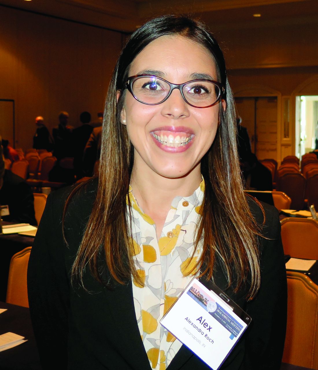
Historically, the preferred way to manage patients with necrotizing pancreatitis was via parenteral nutrition with a lack of pancreatic stimulation, said Dr. Roch, of the department of surgery at Indiana University, Indianapolis. However, parenteral nutrition is associated with increased permeability, a lack of peristaltic stimulation, changes in intestinal flora, and an increased risk of infection.
“More recently, enteral nutrition has been used, despite a potential for pancreatic stimulation,” she said. “From 16 randomized, controlled trials with 847 patients, it was associated with decreased mortality, decreased infectious complications, decreased length of hospital stay, and a trend toward decreased rate of organ failure. Based on those findings, enteral nutrition has become the standard of care in acute pancreatitis. The optimal enteral nutrition route, however, is still debated. The traditional route is the nasojejunal [NJ] tube. Its placement is noninvasive, but it is associated with discomfort for the patient, dislodgement in 16%-63% of cases, and potentially sinusitis. Conversely, percutaneous gastrostomy with jejunal extension [PEG-J] is beneficial for patient comfort but has the drawbacks of being an invasive procedure with the risk of cellulitis and more severe complications.”
The aim of the current study was to compare the safety and efficacy of NJ tube and PEG-J enteral nutrition delivery before surgical debridement in patients with necrotizing pancreatitis. Dr. Roch and her associates hypothesized that NJ tube and PEG-J would have a similar complication profile. They retrospectively reviewed the medical records of all patients who underwent surgical debridement for necrotizing pancreatitis at Indiana University Medical Center between 2005 and 2015. Patients with exclusive total parenteral nutrition were excluded from the study, as were those who had incomplete data.
Dr. Roch reported results from 242 patients with a mean age of 54 years. More than half (64%) were men and the main etiology was biliary (47%), followed by alcohol (16%). The median duration of preoperative enteral nutrition was 29 days. Of the 242 patients, 187 had an NJ tube only, 25 had PEG-J only, and 30 patients had an NJ tube followed by PEG-J. More than half of PEG-Js were placed under fluoroscopic guidance, while the remaining 41% were placed endoscopically.
In terms of safety, patients in the NJ tube group had a significantly higher rate of all complications, compared with those in the PEG-J group (52% vs. 27%, respectively; P = .0015). Conversely, there was a significantly higher rate of serious complications among patients in the PEG-J group, compared with the NJ group (11% vs. 0%; P less than .0001). The researchers also found that compared with patients in the PEG-J group, those in the NJ group were more prone to mechanical complications such as difficulty to place (5% vs. 0%, respectively), replacement (30% vs. 5.5%), and repositioning (30% vs. 2%), while PEG-J patients were more prone to infectious complications such as skin infections/cellulitis (4% vs. 0%) and perforation/leakage/peritonitis (11% vs. 0%). When they limited the analysis to grade III or IV complications, the mechanism was always the same: early dislodgement from the GI tract. “The presentation ranged from asymptomatic patients to severe peritonitis,” Dr. Roch said. “Two patients out of the six with severe complications required emergent laparotomy.”
In terms of efficacy, the NJ and PEG-J groups were equivalent in achieving enteral nutrition (67% vs. 68%, respectively). There were also no differences between the two groups in nutritional status when assessed by an increase of serum albumin (38% vs. 36%; P = .87), normalization of serum albumin (9% vs. 16%; P = .14), or in the prevalence of infected necrosis (53% vs. 49%; P = .64).
Dr. Roch acknowledged certain limitations of the study, including its single-center, retrospective design. “Furthermore, we are a tertiary care center, and most patients are referred to us late in the course of their disease,” she said. “Finally, no PEG-Js were placed outside of our institution, raising the question of a selection bias. She reported having no financial disclosures.
AT WSA 2016
Key clinical point:
Major finding: In terms of efficacy, the NJ and PEG-J groups were equivalent in achieving enteral nutrition (67% vs. 68%, respectively).
Data source: A retrospective review of 242 patients who underwent surgical debridement for necrotizing pancreatitis at Indiana University Medical Center between 2005 and 2015.
Disclosures: Dr. Roch reported having no financial disclosures.
DBS and Optimal Therapy May Have Long-Term Benefit in Early Parkinson’s Disease
BALTIMORE—Deep brain stimulation (DBS) of the subthalamic nucleus, along with optimal drug therapy (ODT), produces clinically meaningful improvements in clinician-assessed motor control for at least five years in patients with early-stage Parkinson’s disease, according to a prospective pilot study. If the findings are confirmed in a phase III trial that has been approved, DBS may become a valuable method to achieve long-term improvement in motor skills in patients with Parkinson’s disease.
Mallory Hacker, PhD, Research Assistant Professor of Neurology at Vanderbilt University in Nashville, presented the latest results of a subanalysis of a randomized, controlled, single-blind clinical trial at the 141st Annual Meeting of the American Neurological Association. 
The pilot trial included 30 patients with Parkinson’s disease between ages 50 and 75 and demonstrated the safety and benefit of DBS in conjunction with ODT—which includes drugs such as carbidopa–levodopa, pramipexole, ropinirole, and selegiline—compared with ODT alone in improving motor scores of the patients through two years. The latest subanalysis of the trial data examined the effect of DBS for at least five years.
The subanalysis included 28 patients: 14 in the DBS and ODT group and 14 in the ODT-only group. Both groups were predominantly male and were similar in age (about 61) at baseline. Motor control was assessed using the Unified Parkinson’s Disease Rating Scale (UPDRS) Part III (clinician assessed) and the Hoehn and Yahr scale.
Over the five-year period, mean UPDRS motor scores progressively worsened for the ODT group, but improved for patients who received DBS. Compared with patients who received ODT only, patients who received DBS plus ODT had UPDRS improvements of 4.6 points at 1.5 years, 5.8 points at two years, 8.9 points at four years, and 10.1 points at five years.
“These results demonstrate that subthalamic nucleus DBS applied in early-stage Parkinson’s disease may provide long-term, clinically meaningful improvement in motor function over standard clinical therapy,” Dr. Hacker said.
The exact mechanism of DBS is still unknown, but evidence suggests that synaptic plasticity is involved. Results from the pilot trial led to FDA approval of a large-scale, multicenter clinical trial. Participants will be implanted with a DBS device and will receive ODT. Some subjects will be randomized to receive DBS during the first two years, along with ODT, and the remainder will receive ODT only. In the following two years, all subjects will receive DBS and ODT.
—Brian Hoyle
BALTIMORE—Deep brain stimulation (DBS) of the subthalamic nucleus, along with optimal drug therapy (ODT), produces clinically meaningful improvements in clinician-assessed motor control for at least five years in patients with early-stage Parkinson’s disease, according to a prospective pilot study. If the findings are confirmed in a phase III trial that has been approved, DBS may become a valuable method to achieve long-term improvement in motor skills in patients with Parkinson’s disease.
Mallory Hacker, PhD, Research Assistant Professor of Neurology at Vanderbilt University in Nashville, presented the latest results of a subanalysis of a randomized, controlled, single-blind clinical trial at the 141st Annual Meeting of the American Neurological Association.
The pilot trial included 30 patients with Parkinson’s disease between ages 50 and 75 and demonstrated the safety and benefit of DBS in conjunction with ODT—which includes drugs such as carbidopa–levodopa, pramipexole, ropinirole, and selegiline—compared with ODT alone in improving motor scores of the patients through two years. The latest subanalysis of the trial data examined the effect of DBS for at least five years.
The subanalysis included 28 patients: 14 in the DBS and ODT group and 14 in the ODT-only group. Both groups were predominantly male and were similar in age (about 61) at baseline. Motor control was assessed using the Unified Parkinson’s Disease Rating Scale (UPDRS) Part III (clinician assessed) and the Hoehn and Yahr scale.
Over the five-year period, mean UPDRS motor scores progressively worsened for the ODT group, but improved for patients who received DBS. Compared with patients who received ODT only, patients who received DBS plus ODT had UPDRS improvements of 4.6 points at 1.5 years, 5.8 points at two years, 8.9 points at four years, and 10.1 points at five years.
“These results demonstrate that subthalamic nucleus DBS applied in early-stage Parkinson’s disease may provide long-term, clinically meaningful improvement in motor function over standard clinical therapy,” Dr. Hacker said.
The exact mechanism of DBS is still unknown, but evidence suggests that synaptic plasticity is involved. Results from the pilot trial led to FDA approval of a large-scale, multicenter clinical trial. Participants will be implanted with a DBS device and will receive ODT. Some subjects will be randomized to receive DBS during the first two years, along with ODT, and the remainder will receive ODT only. In the following two years, all subjects will receive DBS and ODT.
—Brian Hoyle
PINK1 Mutation Raises Risk of Early-Onset Parkinson’s Disease
The rare p.G411S mutation of PINK1 greatly increases the risk of early-onset Parkinson’s disease, even if present in only one allele, according to a genetic association study published online ahead of print November 2 in Brain. By reducing kinase activity towards ubiquitin, the mutation impairs the elimination of damaged mitochondria from cells.
Previous data had indicated that mutations in both PINK1 alleles confer a risk of early-onset Parkinson’s disease (age younger than 45). Genetic studies had suggested that a single mutated PINK1 allele also might increase the risk of developing the disease.
Wolfdieter Springer, PhD, Associate Professor of Neuroscience at Mayo Clinic in Jacksonville, Florida, and colleagues examined DNA samples from 2,560 patients with Parkinson’s disease and 2,145 controls from the United States, Poland, Norway, Ireland, and Sweden. Patients were included in the study irrespective of age at disease onset. The controls included the patients’ spouses and caregivers, as well as unrelated individuals. Dr. Springer and colleagues also searched the literature for previous studies that identified or excluded PINK1 p.G411S mutations in cases of Parkinson’s disease and controls. 
The investigators found PINK1 p.G411S substitution in 19 cases (0.74%) and five control subjects (0.23%), indicating a significant association with an intermediate effect size between heterozygous PINK1 p.G411S carrier status and Parkinson’s disease (odds ratio [OR], 2.92). The median age at disease onset was significantly lower in the 19 cases carrying PINK1 p.G411S than in noncarrier cases (59 vs 64). When the researchers combined their data with those of six studies that they had identified in the literature review, the increased risk of Parkinson’s disease associated with the p.G411S mutation remained evident (OR, 2.89).
Levels of PINK1 protein, which helps selectively eliminate damaged mitochondria from cells, were similar in p.G411S heterozygous cells and in wild-type controls. Levels of p-Ser65-Ub, a marker of mitochondrial stress, were persistently lower, however, and were significantly reduced at later time points. In a HeLa cell model, the researchers observed that p.G411S overexpressing cells had reduced p-Ser65-Ub levels, compared with PINK1 wild-type cells. Furthermore, p.G411S showed strongly reduced kinase activity towards ubiquitin, compared with PINK1 wild-type. Coexpression of p.G411S along with PINK1 wild-type significantly impaired ubiquitin phosphorylation.
“Genetic analyses, as well as functional, cell-based and structural, computational characterization, for the first time provided evidence for a partial dominant-negative function of the heterozygous PINK1 p.G411S mutation that confers a markedly increased risk for Parkinson’s disease,” said Dr. Springer. “The low frequency of homozygote p.G411S carriers may be due to the rarity of the mutation or could indicate [that] it is particularly damaging or clinically manifests with an alternate phenotypic presentation. Replication of our genetic association in other case–control series and further clinical studies is now warranted.”
—Erik Greb
Suggested Reading
Puschmann A, Fiesel FC, Caulfield TR, et al. Heterozygous PINK1 p.G411S increases risk of Parkinson’s disease via a dominant-negative mechanism. Brain. 2016 Nov 2 [Epub ahead of print].
The rare p.G411S mutation of PINK1 greatly increases the risk of early-onset Parkinson’s disease, even if present in only one allele, according to a genetic association study published online ahead of print November 2 in Brain. By reducing kinase activity towards ubiquitin, the mutation impairs the elimination of damaged mitochondria from cells.
Previous data had indicated that mutations in both PINK1 alleles confer a risk of early-onset Parkinson’s disease (age younger than 45). Genetic studies had suggested that a single mutated PINK1 allele also might increase the risk of developing the disease.
Wolfdieter Springer, PhD, Associate Professor of Neuroscience at Mayo Clinic in Jacksonville, Florida, and colleagues examined DNA samples from 2,560 patients with Parkinson’s disease and 2,145 controls from the United States, Poland, Norway, Ireland, and Sweden. Patients were included in the study irrespective of age at disease onset. The controls included the patients’ spouses and caregivers, as well as unrelated individuals. Dr. Springer and colleagues also searched the literature for previous studies that identified or excluded PINK1 p.G411S mutations in cases of Parkinson’s disease and controls.
The investigators found PINK1 p.G411S substitution in 19 cases (0.74%) and five control subjects (0.23%), indicating a significant association with an intermediate effect size between heterozygous PINK1 p.G411S carrier status and Parkinson’s disease (odds ratio [OR], 2.92). The median age at disease onset was significantly lower in the 19 cases carrying PINK1 p.G411S than in noncarrier cases (59 vs 64). When the researchers combined their data with those of six studies that they had identified in the literature review, the increased risk of Parkinson’s disease associated with the p.G411S mutation remained evident (OR, 2.89).
Levels of PINK1 protein, which helps selectively eliminate damaged mitochondria from cells, were similar in p.G411S heterozygous cells and in wild-type controls. Levels of p-Ser65-Ub, a marker of mitochondrial stress, were persistently lower, however, and were significantly reduced at later time points. In a HeLa cell model, the researchers observed that p.G411S overexpressing cells had reduced p-Ser65-Ub levels, compared with PINK1 wild-type cells. Furthermore, p.G411S showed strongly reduced kinase activity towards ubiquitin, compared with PINK1 wild-type. Coexpression of p.G411S along with PINK1 wild-type significantly impaired ubiquitin phosphorylation.
“Genetic analyses, as well as functional, cell-based and structural, computational characterization, for the first time provided evidence for a partial dominant-negative function of the heterozygous PINK1 p.G411S mutation that confers a markedly increased risk for Parkinson’s disease,” said Dr. Springer. “The low frequency of homozygote p.G411S carriers may be due to the rarity of the mutation or could indicate [that] it is particularly damaging or clinically manifests with an alternate phenotypic presentation. Replication of our genetic association in other case–control series and further clinical studies is now warranted.”
—Erik Greb
Suggested Reading
Puschmann A, Fiesel FC, Caulfield TR, et al. Heterozygous PINK1 p.G411S increases risk of Parkinson’s disease via a dominant-negative mechanism. Brain. 2016 Nov 2 [Epub ahead of print].

Bone marrow cells prove limb-saving for some
NEW ORLEANS – Treatment with autologous bone marrow cells can avert amputation in selected patients who have critical limb ischemia and are not candidates for revascularization surgery, based on results from the randomized phase III MOBILE trial.
Critical limb ischemia, the end stage of peripheral arterial disease, accounts for more than 53,000 major limb amputations in the United States annually, noted principal investigator Dr. Michael P. Murphy, director of the Vascular and Cardiac Center for Adult Stem Cell Therapy at Indiana University, Indianapolis.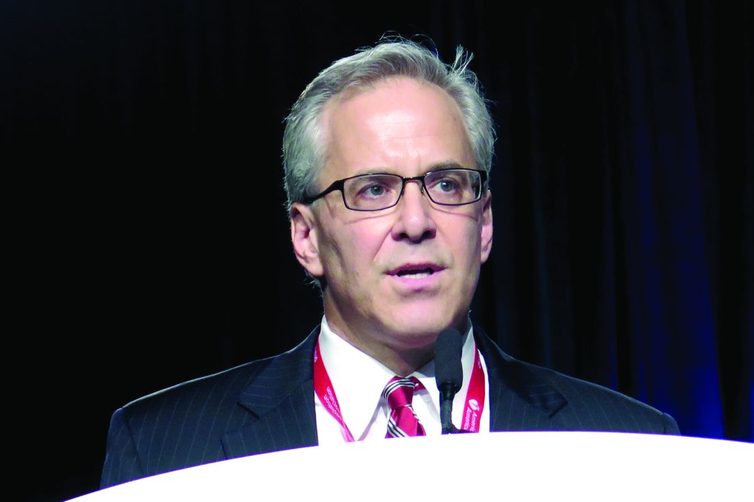
“About 30% of our patient population with critical limb ischemia have no options for the standard of care, such as surgical bypass, due to absence of a surgical target or chronic total occlusion, which mitigates an endovascular approach,” he said at the American Heart Association scientific sessions. “Thus, these no-option critical limb ischemia patients represent an unmet medical need for a novel agent that may promote limb salvage and prevent amputation and its associated disabilities.”
In the trial, investigators randomized 155 affected limbs (in 152 patients) 3:1 to receive double-blinded treatment with injections of concentrated autologous bone marrow aspirate or placebo.
Amputation-free survival 1 year after treatment, the trial’s primary endpoint, was not significantly better in the aspirate group than in the placebo group, according to the researchers. However, the aspirate reduced the risk of major amputation by 73% in the subgroup with less severe (Rutherford class 4) disease and by 67% in the subgroup of patients who did not have diabetes.
“Personally, I would recommend cell therapy for my Rutherford 4 patients and my nondiabetic Rutherford 5 patients,” Dr. Murphy said. “And one would say even for the Rutherford 5 diabetics, there were no safety concerns and there is hope for improvement rather than amputation – it might be worth the roll of the dice.”
The investigators are preparing a manuscript based on the full data and working with Biomet, the trial sponsor, to apply for Food and Drug Administration approval of the product for the treatment of critical limb ischemia, he said.
Lessons learned
A session attendee asked whether stronger demonstration of benefit in a larger trial is needed to justify the use and cost of such new cellular therapies.
“If we could go back and do it all over again, we would do things differently knowing what we know now,” Dr. Murphy replied, noting that the investigators had to stick to a trial design created more than a decade ago.
“We discovered that the event rate in patients with critical limb ischemia in the control group is actually 30% and not 40% because medical management has changed,” he said. “Secondly, we would do a 1:1 randomization and most likely would increase our sample size to 200, and we probably would have seen a difference overall. That difference overall would have been provided by, of course, increasing the Rutherford 4 and Rutherford 5 nondiabetics, which would overshadow the increased amputation rate in the diabetic group.”
“I think from this [experience], we can launch a much larger study, if there were funding for it, and look at autologous versus allogeneic cells in this domain,” Dr. Murphy concluded.
Identifying patients who benefit
Session panelist Roberto Bolli, MD, chief of the division of cardiovascular medicine at the University of Louisville (Ky.), commended the MOBILE trial for its study design and prespecified subgroup analyses.
“The whole problem we are coping with, not just in this trial, but in other studies, is that patients are different and some patients may respond better than others, or some patients may not respond at all. And really, we still don’t understand why some patients respond or not – it may have to do with their immune system, their regenerative capacity, as well as the type of cells that are being used,” he said.
“Even though it was in its entirety a negative study, it’s very important because it identified a possible target for future trials, which is patients without diabetes in Rutherford class 4, which may benefit from therapy,” Dr. Bolli concluded.
Trial details
Patients were enrolled in MOBILE from 24 U.S. centers. All had critical limb ischemia, were ineligible for revascularization, had an ankle-brachial index of less than 0.60 or a toe-brachial index of less than 0.40, and had Rutherford 4 disease (rest pain) or Rutherford 5 disease (tissue loss).
They received concentrated autologous bone marrow aspirate or placebo by intramuscular injection at 35-40 sites in the affected limb.
Results showed that the groups did not differ significantly with respect to the rates of adverse events or serious adverse events overall, Dr. Murphy reported. The aspirate group had lower rates of respiratory failure and fever; they also had a higher rate of anemia (68.9% vs. 36.1%, P less than .001) as expected, from the aspiration procedure, but with no associated complications.
The 1-year rate of amputation-free survival events, reflecting both major amputations and death, was lower with the aspirate, at 20.2%, than with placebo, at 30.5%, but not significantly so (P = .224). Findings were similar for major amputation in the entire trial population (16.0% vs. 22.2%, P = .392). However, this outcome was less common with the aspirate among patients with Rutherford 4 disease (7.7% vs. 26.3%, P = .041) and nondiabetic patients (10.0% vs. 27.7%, P = .046).
“Looking at these data, it became apparent to us that the Rutherford 5 diabetic was the outlying group,” said Dr. Murphy, who disclosed that he had no relevant conflicts of interest.
Considering all other patients – Rutherford 4 regardless of diabetes status and nondiabetic Rutherford 5 – the aspirate was associated with a dramatically lower rate of amputation compared with the placebo (9.6% vs. 26.7%, P = .021; hazard ratio, 0.33). In this subset, the number needed to treat with bone marrow aspirate to prevent a single amputation was just 6.
The aspirate and placebo groups did not differ with respect to ankle-brachial index, toe-brachial index, or 6-minute walk test distance in the entire trial population. Transcutaneous oxygen pressure (TcP02), an indicator of microvascular perfusion in the subcutaneous tissue of the ischemic limb, increased by 59% in the aspirate group but by only 7% in the placebo group.
AT THE AHA SCIENTIFIC SESSIONS
Key clinical point:
Major finding: The 52-week rate of amputation-free survival events was 20.2% with the aspirate and 30.5% with placebo (P = .224).
Data source: A randomized phase III trial among 152 patients with critical limb ischemia who were not candidates for revascularization surgery (MOBILE trial).
Disclosures: Dr. Murphy disclosed that he had no relevant conflicts of interest. The trial was sponsored by Biomet Biologics, LLC.
VIDEO: HeartMate 3 LVAD solves pump thrombosis
NEW ORLEANS – HeartMate 3, the latest left ventricular assist device in the HeartMate line, appears to have solved the problem of pump thrombosis, a complication that has dogged ventricular pumps since the issue leapt into medical awareness about 3 years ago (New Engl J Med. 2014 Jan 2;370:33-40).
During 6 months of follow-up, none of 152 heart failure patients assigned to receive a HeartMate 3 left ventricular assist device (LVAD) developed suspected or confirmed pump thrombosis, compared with 14 patients (10%) having pump thrombosis out of 138 recipients of the prior-generation HeartMate II LVAD who served as the control group for the study.
“Three years ago, when the issue of pump thrombosis was first revealed, there was a lot of consternation and some drop in LVAD use, especially as destination therapy. We think that seeing no pump thrombosis whatsoever will give people renewed confidence in this technology,” said Dr. Mehra, professor of medicine at Harvard Medical School and medical director of the Heart and Vascular Center of Brigham and Women’s Hospital, both in Boston.
Pump thrombosis has also been a problem for the patients who have received a competitor LVAD, the HeartWare HVAD device (Circulation. 2015 Nov 10;132[suppl 3]:A19675), approved for U.S. use as bridge to transplant. HeartMate II is approved for both bridge to transplant and for destination therapy.
In addition to apparently eliminating pump thrombosis, HeartMate 3’s size and potential implantation approach should make its placement during routine use as quick and minimally invasive as the HeartWare device, features that should further help broader use of HeartMate 3, commented Mark Slaughter, MD, professor and chairman of cardiovascular and thoracic surgery at the University of Louisville (Ky.). But Dr. Slaughter and others were also quick to highlight the shortcomings that remain with both devices that will continue to hamper a broader role for LVAD treatment of patients with advanced heart failure.
“We thought that if there was less pump thrombosis we’d see less stroke, but that is not what the data suggest. It’s the big puzzle we need to figure out before we see widespread acceptance of this treatment,” Dr. Sweitzer said.
“This will not shift LVAD use substantially,” commented Christopher B. Granger, MD, a professor of medicine and a heart failure specialist at Duke University, Durham, N.C. “Reducing the need for reoperation is good for the field, and is an incremental advance, but it is not transformational,” he said in an interview.
The MOMENTUM 3 (Multicenter Study of MagLev Technology in Patients Undergoing Mechanical Circulatory Support Therapy with HeartMate 3) trial randomized 294 patients at 69 U.S. centers. The study’s primary endpoint of 6-month survival free from disabling stroke or reoperation to repair or replace the LVAD occurred in 86% of 152 patients who received a HeartMate 3 and 77% of 142 patients randomized to HeartMate II, a statistical difference that met the prespecified criteria for both noninferiority and superiority. Concurrently with Dr. Mehra’s report at the meeting, a journal article appeared online (New Engl J Med. 2016 Nov 16. doi: 10.1056/NEJMoa1610426). He stated that as far as he understood, St. Jude would submit the 6-month data he reported to the Food and Drug Administration in an application for marketing approval for HeartMate 3.
“I agree that there are still morbid evens [with HeartMate 3] that need to be surmounted, but this is a confidence-building step in the right direction,” Dr. Mehra said.
mzoler@frontlinemedcom.com
On Twitter @mitchelzoler
By eliminating all episodes of pump thrombosis during 6-month follow-up, the HeartMate 3 appeared to resolve one of the major issues that has stood in the way of patients and physicians feeling comfortable with left ventricular assist devices. The smaller size of the HeartMate 3 pump and its ability to be placed with minimally invasive and fairly rapid surgery is another big advance, putting this device on par with the rival pump, the HeartWare HVAD.
But the performance of the HeartMate 3 left ventricular assist device (LVAD) in MOMENTUM 3 also highlighted the shortcomings that still remain for these devices: the unchanged rates of stroke, gastrointestinal bleeds, and infections with HeartMate 3, compared with HeartMate II in this trial, and similar 6-month survival rates in the two arms of the study.
The HeartMate 3 can be implanted without sternotomy, using an 8 cm incision on the lateral chest wall, resulting in a shorter postoperative stay and fewer perisurgical adverse events. Despite the less invasive surgery and absence of pump thrombosis, some patients and physicians will remain hesitant to use an LVAD unless it is unavoidable because of concern about strokes. Until further design and procedural refinements change the rate of serious strokes and other adverse events, LVADs will not be fully competitive with heart transplantation.
The competition between HeartMate and the HeartWare devices will help drive this field forward, leading to further improvements in outcomes and expanded LVAD use.
Mark Slaughter, MD, is professor of surgery and chairman of cardiovascular and thoracic surgery at the University of Louisville (Ky.). He was an investigator in MOMENTUM 3, he has been a consultant to EvaHeart and Oregon Heart, and he has received research support from Carmat and HeartWare. He made these comments as designated discussant for the report and in a video interview.
The video associated with this article is no longer available on this site. Please view all of our videos on the MDedge YouTube channel
NEW ORLEANS – HeartMate 3, the latest left ventricular assist device in the HeartMate line, appears to have solved the problem of pump thrombosis, a complication that has dogged ventricular pumps since the issue leapt into medical awareness about 3 years ago (New Engl J Med. 2014 Jan 2;370:33-40).
During 6 months of follow-up, none of 152 heart failure patients assigned to receive a HeartMate 3 left ventricular assist device (LVAD) developed suspected or confirmed pump thrombosis, compared with 14 patients (10%) having pump thrombosis out of 138 recipients of the prior-generation HeartMate II LVAD who served as the control group for the study.
“Three years ago, when the issue of pump thrombosis was first revealed, there was a lot of consternation and some drop in LVAD use, especially as destination therapy. We think that seeing no pump thrombosis whatsoever will give people renewed confidence in this technology,” said Dr. Mehra, professor of medicine at Harvard Medical School and medical director of the Heart and Vascular Center of Brigham and Women’s Hospital, both in Boston.
Pump thrombosis has also been a problem for the patients who have received a competitor LVAD, the HeartWare HVAD device (Circulation. 2015 Nov 10;132[suppl 3]:A19675), approved for U.S. use as bridge to transplant. HeartMate II is approved for both bridge to transplant and for destination therapy.
In addition to apparently eliminating pump thrombosis, HeartMate 3’s size and potential implantation approach should make its placement during routine use as quick and minimally invasive as the HeartWare device, features that should further help broader use of HeartMate 3, commented Mark Slaughter, MD, professor and chairman of cardiovascular and thoracic surgery at the University of Louisville (Ky.). But Dr. Slaughter and others were also quick to highlight the shortcomings that remain with both devices that will continue to hamper a broader role for LVAD treatment of patients with advanced heart failure.
“We thought that if there was less pump thrombosis we’d see less stroke, but that is not what the data suggest. It’s the big puzzle we need to figure out before we see widespread acceptance of this treatment,” Dr. Sweitzer said.
“This will not shift LVAD use substantially,” commented Christopher B. Granger, MD, a professor of medicine and a heart failure specialist at Duke University, Durham, N.C. “Reducing the need for reoperation is good for the field, and is an incremental advance, but it is not transformational,” he said in an interview.
The MOMENTUM 3 (Multicenter Study of MagLev Technology in Patients Undergoing Mechanical Circulatory Support Therapy with HeartMate 3) trial randomized 294 patients at 69 U.S. centers. The study’s primary endpoint of 6-month survival free from disabling stroke or reoperation to repair or replace the LVAD occurred in 86% of 152 patients who received a HeartMate 3 and 77% of 142 patients randomized to HeartMate II, a statistical difference that met the prespecified criteria for both noninferiority and superiority. Concurrently with Dr. Mehra’s report at the meeting, a journal article appeared online (New Engl J Med. 2016 Nov 16. doi: 10.1056/NEJMoa1610426). He stated that as far as he understood, St. Jude would submit the 6-month data he reported to the Food and Drug Administration in an application for marketing approval for HeartMate 3.
“I agree that there are still morbid evens [with HeartMate 3] that need to be surmounted, but this is a confidence-building step in the right direction,” Dr. Mehra said.
mzoler@frontlinemedcom.com
On Twitter @mitchelzoler
AT THE AHA SCIENTIFIC SESSIONS
Key clinical point:
Major finding: During 6 months, suspected or confirmed pump thrombosis occurred in no HeartMate 3 patients and in 10% of HeartMate II recipients.
Data source: The MOMENTUM 3 trial, which randomized 294 patients at 69 U.S. centers.
Disclosures: MOMENTUM 3 was sponsored by St. Jude, the company developing the HeartMate 3 LVAD. Dr. Mehra has received travel reimbursements from St. Jude and has been a consultant to Medtronic, Stealth, and Teva. Dr. Sweitzer was an investigator in MOMENTUM 3 and has been a consultant to Acorda and Medtronic and received research support from Bayer, Corvia, and Novartis. Dr. Granger has been a consultant to Boehringer Ingelheim, and received research support from Medtronic and several other drug and device companies. Dr. Slaughter was an investigator in MOMENTUM 3, has been a consultant to EvaHeart and Oregon Heart, and has received research support from Carmat and HeartWare.