User login
The Right Choice? Kindness and Surgical Ethics: Reflections on a Friend and Mentor
As I sit down to write this column, I reflect on the news that my mentor and friend, Norman W. Thompson, M.D, FACS, passed away yesterday. I had the good fortune to spend 1 year as an endocrine surgery fellow with Dr. Thompson at the University of Michigan in 1995-96. That year was certainly the most significant of my training in terms of defining my professional life as an endocrine surgeon. However, as I think back on my time with Dr. Thompson, I am struck by how much more I learned from him than how to take out a thyroid or a parathyroid or manage multiple endocrine neoplasia.
Dr. Thompson was an excellent technical surgeon, and he would have had a tremendous career helping thousands of patients if that was all that he had done. However, he was much more than an excellent technician. He was also a great doctor. In order for a surgeon to be a great doctor, it is necessary to be technically excellent, but that alone is not sufficient. I believe that what makes a surgeon a great doctor is the combination of technical mastery with outstanding interpersonal skills and ethically sound clinical judgment. Dr. Thompson had all of that, and he was exceptionally kind.
Kindness is not a word that we commonly use in describing surgeons today. In an era of surgeons being pressured to see more patients and generate more RVUs [relative value units], it is unusual to hear kindness mentioned as an essential attribute of a great surgeon. However, Dr. Thompson’s kindness was immediately apparent to all who spent time with him. He treated each patient as a unique individual. In addition, he treated his trainees and his colleagues in Ann Arbor and around the world with respect and incredible humility. He was generous with his time and was always approachable no matter how inexperienced the surgeon asking him a question. Dr. Thompson was kind to all of us and made us feel that he valued spending time with us.
What does kindness have to do with a column that traditionally focuses on ethical issues in the practice of surgery? Although acting with kindness is not the same as acting in an ethical manner, I believe that there is more overlap of the terms than we often imagine. The kind surgeon is the one who treats people – whether they are patients or colleagues – as though they matter. The ethical surgeon respects the patient’s wishes and acts to benefit the patient as much as possible in all circumstances. I am certain that I have met ethical surgeons who were not kind, but I have met very few kind surgeons who are not ethical.
As someone who has spent significant time and energy in the last 19 years as a surgery faculty member trying to teach ethics, I am also struck by a clear truth. Actions always speak louder than words. It may be valuable to talk about the ethical principles that may come to play in a particularly difficult surgical case. Defining the competing interests and assessing the patient’s wishes are important components of the ethical practice of surgery. However, no amount of discussion of these issues can substitute for the value of behavior. Treating patients and colleagues with kindness and respect is modeling the behaviors of an ethical surgeon – perhaps learned from a wise and thoughtful mentor.
Dr. Thompson was an excellent role model for me and so many others in how he treated patients and everyone around him. As I see patients and perform surgery, I still hear myself saying many of the same things that he said many years ago. His genuine expressions of optimism before difficult operations, honesty in communicating, and sadness when things did not go well were tremendous examples to me of how a great doctor treats those around him. These lessons that I learned from Dr. Thompson have influenced my practice significantly, and I am grateful for the opportunity to try to model them on a daily basis.
Although I remain convinced that formal curricula in ethics and professionalism remain important in the education of today’s surgeons, it is valuable to remember the impact that the behaviors of those we respect have on us. Perhaps we surgeons more than other physicians are molded by the people who train us, but there is no question that the ethical behaviors of our teachers and mentors will have a greater impact than any lecture or manuscript. I want to acknowledge and commemorate the kindness and ethical behaviors that Dr. Thompson modeled daily for all who were fortunate enough to work with him.
Dr. Angelos is an ACS Fellow; the Linda Kohler Anderson Professor of Surgery and Surgical Ethics; chief, endocrine surgery; and associate director of the MacLean Center for Clinical Medical Ethics at the University of Chicago.

T-VEC: Advancing the Fight Against Melanoma

Following a phase III, open-label trial conducted by Andtbacka et al (J Clin Oncol. 2015;33:2780-2788), the US Food and Drug Administration recently approved the first oncolytic immunotherapy talimogene laherparepvec (T-VEC) for the treatment of unresectable cutaneous, subcutaneous, and nodal lesions in patients with advanced melanoma (stage IIIB/C–stage IV) following initial surgery.
A group of 436 patients with injectable melanomas (melanomas that are accessible via a percutaneous injection) that were not surgically resectable were randomly assigned (2:1) to treatment with intralesional T-VEC or subcutaneous granulocyte macrophage colony-stimulating factor (GM-CSF). The primary endpoint of the study was durable response rate (DRR), defined as objective response lasting continuously for 6 months or longer. Secondary endpoints included overall survival (OS) and overall response rate.
Talimogene laherparepvec was shown to extend DRRs compared to GM-CSF. Durable response rates were significantly higher with T-VEC (16.3%; 95% confidence interval [CI], 12.1%–20.5%) versus GM-CSF (2.1%; 95% CI, 0%–4.5%)(odds ratio, 8.9; P<.001).
In the OS analysis, a 4.4-month extension with T-VEC was observed; however, this was not deemed to be statistically significant (P=.051). The median OS was 23.3 months (95% CI, 19.5–29.6 months) with T-VEC and 18.9 months (95% CI, 16.0–23.7 months) with GM-CSF (hazard ratio, 0.79; 95% CI, 0.62–1.00; P=.051). Overall response rate also was higher in the T-VEC arm (26.4%; 95% CI, 21.4%–31.5%) versus GM-CSF (5.7%; 95% CI, 1.9%–9.5%).
Talimogene laherparepvec is a herpes simplex virus type 1–derived oncolytic immunotherapy designed to replicate within tumors and produce GM-CSF, which enhances systemic antitumor immune responses and induces tumor lysis.
In this study, T-VEC efficacy was greatest in patients with stage IIIB, IIIC, or IVM1a melanomas and in patients with treatment-naive disease. Differences in DRRs in patients with stage IIIB/C melanomas were 33% in the T-VEC group versus 0% for patients treated with GM-CSF alone. In the stage IVM1a group, DRR was 16% with T-VEC versus 2% with GM-CSF. The difference between both treatments was smaller in more advanced melanomas (IVM1b group, 3% vs 4%; IVM1c, 7% vs 3%). In the first-line treatment, the DRR with T-VEC was 24% versus 0% with GM-CSF. In the second-line and beyond, the DRR with T-VEC was 10% compared to 4% for GM-CSF.
The main adverse events seen in this study were fatigue, chills, and pyrexia. Serious adverse events occurred in 25.7% and 13.4% of participants in the T-VEC and GM-CSF arms, respectively, with disease progression (3.1% vs 1.6%) and cellulitis (2.4% vs 0.8%) being the most common. Six immune-mediated events occurred in the T-VEC group compared to 3 in the GM-CSF group.
Twelve patient deaths occurred within 30 days of the last dose of T-VEC; 9 were associated with progressive disease and the other 3 were associated with myocardial infarction, cardiac arrest, and sepsis, respectively. Four patient deaths were reported in the GM-CSF arm within the same 30 days.
What’s the Issue?
Immunotherapy represents a promising treatment option for metastatic melanoma. These promising results along with the US Food and Drug Administration’s approval of T-VEC will lead to further studies of the uses of T-VEC in combination with other therapies, including a phase I/II study to assess T-VEC in combination with ipilimumab for unresected melanomas (NCT01740297) and a phase III study of T-VEC in combination with pembrolizumab for unresected melanomas (NCT02263508). It is important for dermatologists to be familiar with the new frontier of melanoma treatments. How will these new immunotherapies affect your treatment of melanoma?

Video etiquette
FaceTime with my mother would be better described as ForeheadTime. She loves to use video for our Sunday calls, yet when she does, she always talks into her iPhone as if it’s a speakerphone. As a result, all I see is the top of her head. “Mom. Lower the phone. Mom, I can’t see you,” I must repeat weekly.
Video provides a richer experience compared with telephone. It allows for a deeper, emotional connection. That’s why moms like mine prefer it to telephone conversations. In medicine, video visits are uncommon, but that’s changing as payers are now reimbursing and patients are demanding the service. For many, they offer a far more convenient and still effective method to receive medical care. Psychiatry is an obvious example. Less obvious, but still effective examples, include endocrinology, pediatrics, primary care, surgery (post operatively), and dermatology.
Like the example with my mom, quality of the experience matters, and issues often arise not from the technology, but from the technique. Making eye contact is more difficult on video, and not looking patients in the eye can harm doctor-patient bonding. Here are a few basic tips when using video with your patients:
• Be sure the light source is in front of you. Having windows behind you often puts you in shadow.
• The best place for the camera is at the top of your screen. It’s nearly impossible to look into the camera and see the patient if the camera is next to the screen instead of above.
• Remember, to look directly at the patient, you have to look into the camera. This is tricky and easy to forget.
• Be sure your entire head and upper torso are in the frame. Talking heads can be intimidating.
• When possible, use a headset with a microphone. Headsets help both you and your patient hear better and give the patient an increased sense of privacy.
• Generally speaking, video visits take as long or longer than in-person visits. Remember to be patient as some of your patients may experience technical difficulties. Our IT colleagues have a word for it: “picnic,” which stands for “Problem In Chair Not In Computer.” You should also train your staff to aid you and the patients. For instance, if a patient is struggling with the computer, you might have your assistant help him or her while you move on to the next patient.
• Although the patient can be home, it is best for you to be in your office. It’s possible to do video consults from home, but it is more difficult because you have to ensure that both your technology and your environment are secure and private. Otherwise, you risk violating HIPAA or other compliance requirements.
• Be sure to get the appropriate consent before conducting a virtual visit. In California, it requires only verbal consent, but your state’s requirements might be different.
• As for your appearance, there’s a reason why Kennedy won the Kennedy-Nixon debates. Video does reveal details that you might not want emphasized. A two-day beard might appear hip in person but unkempt and uncaring online. Bold stripes or checks on your shirt sometimes appear distorted, so opt for solids in soft shades. Scrubs are okay, but be sure to check your neckline, particularly as you move about. Whether it’s clothing or accessories, avoid anything overly distracting.
Video visits have had a long, slow ramp-up, but they seem to be gaining momentum. You may not use them in your practice now, but it’s likely we all will someday. Soon.
Dr. Benabio is a partner physician in the department of dermatology of the Southern California Permanente Group in San Diego, and a volunteer clinical assistant professor at the University of California, San Diego. He is @dermdoc on Twitter.

Neurosurgery at the End of Life
The juxtaposition between my first 2 days of neurosurgery could not have been more profound. On my first day as a third-year medical student, the attending and chief resident let me take the lead on the first case: a straightforward brain biopsy. I got to make the incision, drill the burr hole, and perform the needle biopsy. I still remember the thrill of the technical challenge, the controlled violence of drilling into the skull, and the finesse of accessing the tumor core.
The buzz was so strong that I barely registered the diagnosis that was called back from the pathologist: glioblastoma. It was not until I saw the face of the disease the next morning that I understood the reality of a GBM diagnosis. That face belonged to a 47-year-old man who hadn’t slept all night, wide eyed with apprehension at what news I might bring. He beseeched me with questions, and though his aphasia left him stammering to get the words out, I knew exactly what he was asking: Would he live or die? It was a question I was in no position to answer. Instead, I reassured him that we were waiting on the final pathology, all the while trying to forget the fact that the frozen section suggested an aggressive subtype, surely heralding a poor prognosis.
In his poignant memoir, “Do No Harm: Stories of Life, Death and Brain Surgery” (New York: Thomas Dunne Book, 2015), Dr. Henry Marsh writes beautifully about how difficult it can be to find the balance between optimism and realism. In one memorable passage, Dr. Marsh shows a house officer a scan of a highly malignant brain tumor and asks him what he would say to the patient. The trainee reflexively hides behind jargon, skirting around what he knew to be the truth: This tumor would kill her. Marsh presses him to admit that he’s lying, before lamenting at how hard it is to improve these critical communication skills: “When I have had to break bad news I never know whether I have done it well or not. The patients aren’t going to ring me up afterward and say, ‘Mr. Marsh, I really liked the way you told me that I was going to die,’ or ‘Mr. Marsh, you were crap.’ You can only hope that you haven’t made too much of a mess of it.”
I could certainly relate to Dr. Marsh’s house officer as I walked away from my own patient. I felt almost deceitful withholding diagnostic information from him, even if I did the “right” thing. It made me wonder, why did I want to become a neurosurgeon? Surely to help people through some of the most difficult moments of their lives. But is it possible to be a source of comfort when you are required so often to be a harbinger of death? The answer depends on whether one can envision a role for the neurosurgeon beyond the mandate of “life at all costs.”
While the field has become known for its life-saving procedures, neurosurgeons are called just as often to preside over the end of their patient’s lives – work that requires just as much skill as any technical procedure. Dr. Marsh recognized the tremendous human cost of neglecting that work. For cases that appear “hopeless,” he writes, “We often end up operating because it’s easier than being honest, and it means that we can avoid a painful conversation.”
We are only beginning to understand the many issues that neurosurgical patients face at the end of life, but so far it is clear that neurosurgical trainees require substantive training in prognostication, communication, and palliation (Crit Care Med. 2015 Sep;43[9]:1964-77 1,2; J Neurooncol. 2009 Jan;91[1]:39-43). Is there room in the current training paradigm for more formal education in these domains? As we move further into the 21st century, we must embrace the need for masterful clinicians outside of the operating room if we are to ever challenge the axiom set forth by the renowned French surgeon, René Leriche, some 65 years ago: “Every surgeon carries within himself a small cemetery, where from time to time he goes to pray – a place of bitterness and regret, where he must look for an explanation for his failures.” Let us look forward to the day when this is no longer the case.
Stephen Miranda is a medical student from the University of Rochester, who is now working as a research fellow at Ariadne Labs, a joint center for health systems innovation at Brigham & Women’s Hospital and Harvard T.H. Chan School of Public Health, both in Boston.

Immediate treatment yields best outcomes in NSTEMI MI
Immediate, rather than delayed, angiography reduced by 62% the chance of both recurrent heart attack and death in patients with non–ST-segment myocardial infarction.
The significant advantage of early treatment was apparent at both 30 days and 1 year after myocardial infarction, Dr. Aleksandra Milosevic and colleagues wrote (JACC Cardiovasc Interv. 2016;10.1016/j.jcin.2015.11.018).
RIDDLE-NSTEMI (Randomized Study of Immediate vs. Delayed Invasive Intervention in Patients with Non–ST-Segment Elevation MI) is not the first to examine immediate vs. delayed outcomes in coronary angiography. But it is the first to look at a pure cohort of NSTEMI patients, and the first to use purely clinical, rather than biochemical, outcomes, said Dr. Milosevic of the Clinical Center of Serbia, Belgrade.
The study group comprised 323 patients with a confirmed NSTEMI MI to either immediate or delayed angiography. Those in the immediate treatment group went to catheterization as soon as possible (median, 1.4 hours); those in the delayed-treatment group underwent the procedure within 72 hours of randomization (median, 61 hours).
All patients received a loading dose of dual-antiplatelet therapy. For immediate-treatment patients, this consisted of 300 mg aspirin and 600 mg clopidogrel. Those in the delayed-treatment group received 300 mg aspirin and 300 mg clopidogrel. Everyone then received daily treatment of aspirin 100 mg and clopidogrel 75 mg.
Patients who had already been taking dual-antiplatelet therapy continued their maintenance dose. Low-molecular-weight heparin, nitrates, beta-blockers, and ACE inhibitors were given according to clinical guidelines; glycoprotein IIb/IIIa inhibitors were allowed at the treating physician’s discretion.
The time from symptom onset to randomization was 5 hours in the immediate- and 6.5 hours in the delayed-treatment groups. Manual thrombectomy was more common in the immediate-treatment group (3.9% vs. 1%), as was percutaneous coronary intervention (78% vs. 65%). Coronary artery bypass grafting was more common in the delayed-treatment group (23.8% vs. 12%).
At 30 days, the composite primary endpoint of mortality and new MI was significantly less common in the immediate-treatment group (4.3% vs. 13.0%; hazard ratio, 0.32). Mortality was the same in both groups (5 patients; 3%). The difference was driven by the significantly larger number of nonfatal MIs in the delayed-treatment group (16 vs. 2).
The advantage of early treatment held at 1 year, with significantly lower rates of death and new MI in the immediate-treatment group (16.8% vs. 18.8%). This was entirely driven by early events; there was no significant difference after the first 30 days.
A composite secondary endpoint of death, new MI or recurrent ischemia, was also significantly lower in the immediate-intervention group at 30 days (6.8% vs. 26.7%) and 1 year (15.4% vs. 33%).
A multivariate regression analysis controlled for demographics, past medical history, prior coronary procedures, and electrocardiogram changes. After considering these variables, immediate treatment was associated with a 62% reduction in the chance of a new MI within 30 days (HR, 0.42; P = .052).
There was no between-group difference in major bleeding, which was low both at 30 days and 1 year. One patient in the immediate-treatment group and two in the delayed-treatment group needed a transfusion. One intracranial bleed occurred in an immediately treated patient. Four patients in the delayed treatment group were treated for gastrointestinal bleeding.
The authors had no financial declarations.
FROM JACC CARDIOVASCULAR INTERVENTIONS
Key clinical point: Immediate angiography improved both short- and long-term outcomes in non–ST-segment myocardial infarction.
Major finding: Compared with delayed treatment, an immediate cardiac angiography reduced the risk of recurrent MI and death by more than 60% at both 30 days and 1 year.
Data source: The randomized study comprised 323 patients.
Disclosures: The authors made no financial disclosures.
Nick Fitterman, MD, SFHM, Discusses Population Health and Hospital Medicine's Role

Nick Fitterman, MD, SFHM, vice chair of hospital medicine for the Hofstra North Shore-LIJ School of Medicine in Hempstead, N.Y., and North Shore-Long Island Jewish Health System in New Hyde Park, N.Y., discusses how hospital medicine factors into population health—where is the intersection and what is the hospitalist’s role?
Nick Fitterman, MD, SFHM, vice chair of hospital medicine for the Hofstra North Shore-LIJ School of Medicine in Hempstead, N.Y., and North Shore-Long Island Jewish Health System in New Hyde Park, N.Y., discusses how hospital medicine factors into population health—where is the intersection and what is the hospitalist’s role?
Nick Fitterman, MD, SFHM, vice chair of hospital medicine for the Hofstra North Shore-LIJ School of Medicine in Hempstead, N.Y., and North Shore-Long Island Jewish Health System in New Hyde Park, N.Y., discusses how hospital medicine factors into population health—where is the intersection and what is the hospitalist’s role?
Dr. Jaime Upegui, MD, Chats about Motorcycles, Skydiving, and Zen

Movement is key to hospitalist Jaime Upegui, MD, who says riding a motorcycle is a lot like yoga, "you have to focus on the moment, the task at hand." Listen to more of our interview:
Movement is key to hospitalist Jaime Upegui, MD, who says riding a motorcycle is a lot like yoga, "you have to focus on the moment, the task at hand." Listen to more of our interview:
Movement is key to hospitalist Jaime Upegui, MD, who says riding a motorcycle is a lot like yoga, "you have to focus on the moment, the task at hand." Listen to more of our interview:
Harvard Professor Robert Blendon, ScD, Discusses the Republican Presidential Candidates
Listen to more of our interview on the GOP candidates with Robert Blendon, ScD, professor of health policy and political analysis at the Harvard School of Public Health and Harvard Kennedy School of Government in Cambridge, Mass.
Listen to more of our interview on the GOP candidates with Robert Blendon, ScD, professor of health policy and political analysis at the Harvard School of Public Health and Harvard Kennedy School of Government in Cambridge, Mass.
Listen to more of our interview on the GOP candidates with Robert Blendon, ScD, professor of health policy and political analysis at the Harvard School of Public Health and Harvard Kennedy School of Government in Cambridge, Mass.
QUIZ: What Is Your Risk from a Needlestick?
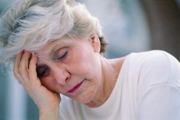
Later menopause lowers risk of later depression
The longer a woman’s reproductive years last, the less she may be prone to postmenopausal depression, a large meta-analysis has determined.
The risk of depression declined by 2% for every 2 premenopausal years after age 40. Women who entered menopause after age 40 experienced a 50% decrease in the risk of depression, compared with women who experienced premature menopause, Dr. Marios K. Georgakis and colleagues reported Jan. 6 in JAMA Psychiatry (2016. doi:10.1001/jamapsychiatry.2015.2653).

The findings suggest that longer exposure to endogenous estrogens mediates the pathophysiology of late-life depression, wrote Dr. Georgakis of the National and Kapodistrian University of Athens and coauthors.
“If confirmed in prospective and culturally diverse studies … these findings could have a significant clinical effect by allowing for the identification of a group of women at higher risk for depression who may benefit from psychiatric monitoring or estrogen-based therapies.”
The meta-analysis comprised 14 studies that included 67,714 women. They controlled for numerous factors, including age, body mass index, obesity, smoking, and hormone therapy. However, only two controlled for past depression – one of the biggest risk factors for recurring depression.
In addition to the 2% decline per 2 premenopausal years after 40, a subanalysis of three studies examining severe depression found a 5% decreased risk for the same time measure. Another analysis of women with premature menopause found a doubling in the risk of depression for those who experienced menopause before age 40.
Estrogen is known to have neuroprotective and antidepressive properties, and the brain is richly endowed with estrogen receptors, the authors said. The exact pathway of protection against depression, however, remains unknown. Potentiation of neurotransmitters and moderation of atherosclerosis might play protective roles.
“Given the results of our study, it remains to be investigated whether women with menopause at younger ages could benefit by preventive use of hormone therapy against late-life depression, provided that adverse effects associated with long-term use are considered,” the authors said. “In this context, the development of estrogen receptor subtype–specific ligands could decrease the proportion of estrogen therapy adverse effects.”
Neither Dr. Georgakis nor any of the coauthors declared any financial conflicts.
The study is a “commendable effort” to examine the role of reproductive hormones in postmenopausal depression, but several important caveats should temper enthusiasm for its conclusions, Dr. Hadine Joffe and Joyce T. Bromberger, Ph.D., wrote in an accompanying editorial.
In most of the studies, women were aged 55-60 years – considerably beyond the average menopausal age of 52. Additionally, most were at least 5 years past their menopause, reflecting a group that might have passed the period of highest risk for hormone-mediated depression.
“This meta-analysis does not address depression associated with the gonadal steroid fluctuations of the perimenopause or recent estradiol withdrawal of the immediate postmenopause,” the colleagues wrote. “Rather, the analysis applies to depression in older women whose brains have not recently been exposed to estradiol or other reproductive hormones and for whom hormonal risk factors have previously been considered less relevant.”
However, the study is one of the few to investigate the psychotropic effects of estrogen on aging women. “In contrast to the acute effects of reproductive hormones on mood in cycling women, the article highlights a potential neuroprotective effect of gonadal steroids on mood that is delayed and extends into the stable hypoestrogenic and hypoprogestinemic environment of the postmenopause.”
Its conclusions are strengthened by studies of nonpsychiatric diseases associated with earlier menopause, including cardiovascular disease, cognitive decline, and dementia. Nevertheless, it’s too early to recommend prophylactic hormone therapy, the authors concluded.
“Given the small effect size and limitations of the studies used in this analysis, more direct evidence supporting a sustained and delayed neuroprotective effect of extended exposure to estradiol, cyclic progestins, and their neurosteroid derivatives is required to support use of hormonal therapy as a therapeutic approach to protecting against postmenopausal depression.”
Dr. Joffe is director of the Women’s Hormone and Aging Research Program at Brigham and Women’s Hospital, Boston. Dr. Bromberger is a professor of epidemiology and psychiatry at the University of Pittsburgh.
The longer a woman’s reproductive years last, the less she may be prone to postmenopausal depression, a large meta-analysis has determined.
The risk of depression declined by 2% for every 2 premenopausal years after age 40. Women who entered menopause after age 40 experienced a 50% decrease in the risk of depression, compared with women who experienced premature menopause, Dr. Marios K. Georgakis and colleagues reported Jan. 6 in JAMA Psychiatry (2016. doi:10.1001/jamapsychiatry.2015.2653).
The findings suggest that longer exposure to endogenous estrogens mediates the pathophysiology of late-life depression, wrote Dr. Georgakis of the National and Kapodistrian University of Athens and coauthors.
“If confirmed in prospective and culturally diverse studies … these findings could have a significant clinical effect by allowing for the identification of a group of women at higher risk for depression who may benefit from psychiatric monitoring or estrogen-based therapies.”
The meta-analysis comprised 14 studies that included 67,714 women. They controlled for numerous factors, including age, body mass index, obesity, smoking, and hormone therapy. However, only two controlled for past depression – one of the biggest risk factors for recurring depression.
In addition to the 2% decline per 2 premenopausal years after 40, a subanalysis of three studies examining severe depression found a 5% decreased risk for the same time measure. Another analysis of women with premature menopause found a doubling in the risk of depression for those who experienced menopause before age 40.
Estrogen is known to have neuroprotective and antidepressive properties, and the brain is richly endowed with estrogen receptors, the authors said. The exact pathway of protection against depression, however, remains unknown. Potentiation of neurotransmitters and moderation of atherosclerosis might play protective roles.
“Given the results of our study, it remains to be investigated whether women with menopause at younger ages could benefit by preventive use of hormone therapy against late-life depression, provided that adverse effects associated with long-term use are considered,” the authors said. “In this context, the development of estrogen receptor subtype–specific ligands could decrease the proportion of estrogen therapy adverse effects.”
Neither Dr. Georgakis nor any of the coauthors declared any financial conflicts.
FROM JAMA PSYCHIATRY
Key clinical point: Later menopause, with its longer estrogen exposure, appears tied to a lower risk of postmenopausal depression.
Major finding: The risk of depression decreased by 2% for each 2 premenopausal years after age 40.
Data source: The meta-analysis comprised 14 studies with more than 67,700 women.
Disclosures: Neither Dr. Georgakis nor any of the coauthors declared any financial conflicts.