User login
Joint Commission Resource Educates Patients, Hospitalists about Antibiotics
The Joint Commission has launched a new online resource for patients and hospitalists to help change mindsets and start conversations about proper antibiotic usage.
The SpeakUp: Antibiotics campaign is a package of free materials, including an infographic illustrating which illnesses may require an antibiotic, a list of questions for patients to ask when prescribed an antibiotic, a podcast, and a video reminding patients that antibiotics are not needed for colds or the flu.
“The new SpeakUp campaign provides a variety of resources to help patients and caregivers understand that how they use antibiotics today can affect how well the drugs work for them tomorrow,” says Lisa Waldowski, MS, APRN, CIC, infection control specialist at The Joint Commission.
The primary audience for these materials is the consumer, but hospitalists and healthcare workers are the crucial secondary audience. “This is a partnership; the knowledge needs to go both ways,” Waldowski says. “Sometimes there’s an expectation that when you see a physician, you are somehow shortchanged if you don’t leave with a prescription for an antibiotic.
There’s an education that needs to go on in the mindset of the physician, [in terms of] looking at whether this situation warrants an antibiotic and educating the patient if it does not. It takes time to have that conversation.”
The campaign can also provide a starting point for hospitalists to make changes in the workplace. “The information needs to be digested by everyone individually, but collectively in the organization where you work, this can lead to an antibiotic stewardship program, a coordinated intervention,” she says. She recommends a multidisciplinary approach. “Sometimes successful programs are led by a physician, and they have a strong pharmacy component, working together and supporting one another to use antibiotics appropriately.”
Visit our website for more information on antibiotic overuse.
Medicare Tests New Quality Measure: Readmission Rates for Heart Failure
The Centers for Medicare & Medicaid Services (CMS) has wrapped up a test run of a new measure for readmission of heart failure patients as the federal agency tries to educate hospitals and hospitalists before formally including it as a quality metric in fiscal year 2018.
The trial concludes October 7, 2015, for the new claims-based measurement, excess days in acute care (EDAC) after hospitalization for heart failure. It captures the number of days within the 30-day postdischarge period that a patient spends in acute care following an index admission for heart failure. The measure will be included in CMS’ Hospital Inpatient Quality Reporting Program in fiscal year 2018, but the agency plans to publicly report hospitals’ results on Hospital Compare next year.
CMS hopes the new measure will help educate hospitals and hospitalists about 30-day postdischarge outcomes for patients with heart failure and provide a better understanding of what services are utilized, which could translate to better interventions. CMS spokesperson Alper Ozinal says that hospitalists currently get “little feedback about what happens to their patients after discharge.”
“CMS found substantial variation in utilization across hospitals, which suggests an opportunity for improvement in transitional care practices,” Ozinal says. “CMS anticipates that the measure will support hospital efforts to further optimize quality of care, particularly the quality of transitional care, by providing a more comprehensive picture of postdischarge events.”
The measure’s trial run, which began September 8, 2015, measured Medicare fee-for-service patients age 65 and older who were hospitalized with a principal discharge diagnosis of heart failure. The outcomes are risk-adjusted, taking into account age, sex, and comorbidities.
Once the results are calculated, CMS will post a report on the QualityNet website. Comments are welcome as CMS is expected to discuss whether tweaks are needed in the measure’s methodology.
Visit our website for more information on hospital readmissions.
Billing, Coding Documentation to Support Services, Minimize Risks

The electronic health record (EHR) has many benefits:
- Improved patient care;
- Improved care coordination;
- Improved diagnostics and patient outcomes;
- Increased patient participation; and
- Increased practice efficiencies and cost savings.1
EHRs also introduce risks, however. Heightened concern about EHR misuse and vulnerability elevates the level of scrutiny placed on provider documentation as it relates to billing and coding. Without clear guidelines from the Centers for Medicare and Medicaid Services (CMS) or other payers, the potential for unintentional misapplication exists. Auditor misinterpretation is also possible. Providers should utilize simple defensive documentation principles to support their services and minimize their risks.
Reason for Encounter
Under section 1862 (a)(1)(A) of the Social Security Act, the Medicare Program may only pay for items and services that are “reasonable and necessary for the diagnosis or treatment of illness or injury or to improve the functioning of a malformed body member,” unless there is another statutory authorization for payment (e.g. colorectal cancer screening).2
A payer can determine if a service is “reasonable and necessary” based on the service indication. The reason for the patient encounter, otherwise known as the chief complaint, must be evident. This can be a symptom, problem, condition, diagnosis, physician-recommended return, or another factor that necessitates the encounter.1 It cannot be inferred and must be clearly stated in the documentation. Without it, a payer may question the medical necessity of the service, especially if it involves hospital-based services in the course of which multiple specialists will see the patient on any given date. Payers are likely to deny services that cannot be easily differentiated (e.g. “no c/o”). Furthermore, payers can deny concurrent care services for the following reasons:3
- Services exceed normal frequency or duration for a given condition without documented circumstances requiring additional care; or
- Services by one physician duplicate/overlap those of the other provider without any recognizable distinction.
Providers should be specific in identifying the encounter reason, as in the following examples: “Patient seen for shortness of breath” or “Patient with COPD, feeling improved with 3L O2 NC.”
Assessment and Plan
Accurately representing patient complexity for every visit throughout the hospitalization presents its challenges. Although the problem list may not dramatically change day to day, providers must formulate an assessment of the patient’s condition with a corresponding plan of care for each encounter. Documenting problems without a corresponding plan of care does not substantiate physician participation in the management of that problem. Providing a brief, generalized comment (e.g. “DM, CKD, CHF: Continue current treatment plan”) minimizes the complexity and effort put forth in the encounter and could result in auditor downgrading upon documentation review.
Developing shortcuts might falsely minimize the provider’s documentation burden. An electronic documentation system might make it possible to copy previous progress notes into the current encounter to save time; however, the previously entered information could include elements that do not require reassessment during a subsequent encounter or contain information about conditions that are being managed concurrently by another specialist (e.g. CKD being managed by the nephrologist). Leaving the copied information unmodified may not accurately reflect the patient’s current condition or the care provided by the hospitalist during the current encounter. Information that is pulled forward or copied and pasted from a previous entry should be modified to demonstrate updated content and nonoverlapping care relevant to that date.
According to the Office of Inspector General (OIG), “inappropriate copy-pasting could facilitate attempts to inflate claims and duplicate or create fraudulent claims.”4
An equally problematic EHR function involves “overdocumentation,” the practice of inserting false or irrelevant documentation to create the appearance of support for billing higher level services.4 EHR technology has the ability to auto-populate fields using templates built into the system or generate extensive documentation on the basis of a single click. The OIG cautions providers to use these features carefully, because they can produce information suggesting the practitioner performed more comprehensive services than were actually rendered.4
An example is the inclusion of the same lab results more than once. Although clinicians include this information as a reference to avoid having to “find it somewhere in the chart” when it is needed—as a basis for comparison, for example—auditors mistake this as an attempt to gain credit for the daily review of the same “old” information. Including only relevant data will mitigate this concern.
Authorship
Dates and signatures are essential to each encounter. Medicare requires services provided/ordered to be authenticated by the author.5 A reviewer must be able to identify each individual who performs, documents, and bills for a service on a given date. Progress notes that fail to identify the service date or service provider will likely result in denial.
Additionally, a service is questioned when two different sets of handwriting appear on a note, yet only one signature is provided. Since the reviewer cannot confirm the credentials of the unidentified individual and cannot be sure which portion belongs to the identified individual, the entire note is disregarded.
Notes that contain an illegible signature are equally problematic. If the legibility of the signature prevents the reviewer from correctly identifying the rendering provider, the service may be denied.
CMS has instructed Medicare contractors to request a signed provider attestation before issuing a denial.5 The provider should print his/her name beside the signature or include a separate signature sheet with the requested documentation to assist the reviewer in provider identification. Stamped signatures are not acceptable under any circumstance. Medicare accepts only handwritten or electronic signatures.5
Carol Pohlig is a billing and coding expert with the University of Pennsylvania Medical Center, Philadelphia. She is also on the faculty of SHM’s inpatient coding course.
References
- HealthIT.gov. Benefits of electronic health records (EHRs). Accessed August 1, 2015.
- Social Security Administration. Exclusions from coverage and Medicare as secondary payer. Accessed August 1, 2015.
- Centers for Medicare and Medicaid Services. Medicare Benefit Policy Manual: Chapter 15—Covered medical and other health services. Chapter 15, Section 30.E. Concurrent care. Accessed August 1, 2015.
- Department of Health and Human Services. Office of Inspector General. CMS and its contractors have adopted few program integrity practices to address vulnerabilities in EHRs. Accessed August 1, 2015.
- Centers for Medicare and Medicaid Services. Signature guidelines for medical review purposes. Accessed August 1, 2015.
- Centers for Medicare and Medicaid Services. 1995 documentation guidelines for evaluation and management services. Accessed August 1, 2015.
Do we need lower extremity physiologic studies in the age of duplex scanners?
Lower-Extremity Physiologic Studies Are No Longer Required – Wrong!
Okay Gene, consider an example from the cardiology world.
You walk up a flight of stairs and develop chest pain. Is it angina? If subsequent coronary catheterization reveals a 60% stenosis in your proximal right coronary artery, then you have an anatomic coronary lesion. But is this lesion causing angina? Maybe. If the catheter is pushed across the stenosis and a 12mm Hg resting pressure gradient is identified, the lesion also has hemodynamic significance. But is it causing angina? Possibly.

To clarify things, a functional study is necessary; specifically, you need to stress the heart (using treadmill exercise, biking, infusion of a catecholamine, etc.) and determine the subsequent cardiac response (pain, ECG changes, wall motion abnormalities, sestamibi/thallium uptake, etc.). If stress produces an abnormal cardiac response, does that mean you have angina? Probably.
Yes, it’s so simple a cardiologist can get it. Yet many vascular specialists seem to struggle with the concept that anatomic, hemodynamic, and functional tests provide different types of information about the arterial system. These tests are complimentary; when used properly they combine to yield information that no single test can provide. But the siren song of an image is hard to resist – after all, everyone loves pictures, and Duplex provides nice ones. With imaging, you can scan for aneurysms. You can see what plaque looks like. In some instances you can even perform operations or procedures solely on the basis of Duplex scanning. What more do you need? (Insert my sarcastic sneer here, Gene.)
Duplex even provides basic hemodynamic information about specific lesions (based on blood flow velocity changes across the lesion). However, it’s a mistake to think that hemodynamic changes across a particular lesion tell us much about the overall “hemodynamics” in a limb. Major arteries may be narrowed (or even completely occluded), but if the collateral vessels are good, the resulting hemodynamic compromise may be minimal. Unfortunately, Duplex scanning is relatively poor at identifying and assessing collateral flow. Tests like segmental pressures (and the ankle-brachial index, or ABI) often provide a better assessment of overall arterial status because the adequacy of collateral flow influences (and is reflected in) distal blood pressure measurements. It’s also naive to think that Duplex is the only means for noninvasively obtaining anatomic information about arterial disease; nonimaging tests using segmental pressures, multilevel pulse-volume recordings (PVRs), continuous-wave (CW) Doppler, etc. may not produce pretty pictures, but they can still localize arterial lesions to a particular anatomic level (aortoiliac vs. femoral-popliteal vs. infrapopliteal, etc.).
The combination of hemodynamic and functional testing is typically referred to as “physiologic” testing, but there’s much more to this than just CW Dopplerand pneumatic cuffs. Specific tests using Laser-Doppler to measure skin blood flow or microvascular perfusion pressure make it possible to (physiologically) assess the microcirculation. Transcutaneous Oxygen (TcPO2) measurement is available in many labs; this test provides valuable physiological information about skin viability and healing potential.
Where Duplex scanning meets its biggest challenge is in the area of functional testing. Yes, it’s possible to exercise the legs and identify subsequent flow (velocity) changes across certain lesions or vessels, but any seasoned sonographer knows that these studies are extremely difficult and time consuming. And because they typically can’t assess/account for collateral flow, they are often of questionable value for determining the adverse impact of arterial disease on the patient. In contrast, functional (exercise) studies performed with CW Doppler and air cuffs are simple, inexpensive, reliable, reproducible, and widely applicable.
Bottom line – physiologic studies are an indispensable part of noninvasive arterial testing and will certainly remain so. For any given patient, they are essential for identifying and quantifying the scope and impact of arterial disease. For populations, they are the test of choice for arterial screening. And, from a practical standpoint, they are required for IAC [intersocietal accreditation commission] laboratory accreditation in peripheral arterial testing (as you know, Duplex scanning is optional for arterial accreditation – but physiologic studies are mandatory!)
Finally, the same characteristics that make physiologic tests important for assessing arterial disease are equally – or perhaps even more – relevant for the assessment of venous disease, but the application of this approach remains in its infancy. It’s a good topic for another discussion.
Dr. Rooke is the Krehbiel Professor of Vascular Medicine at the Mayo Clinic, Rochester, Minn. He has no relevant conflicts.
Lower-Extremity Physiologic Studies Are No Longer Required – Yes ... Maybe!
My friend, Thom, makes a strong case for the value of physiologic studies in patients with lower extremity arterial disease – so strong, in fact, that one might wonder if there is any truly cogent “counterpoint” at all. To provide some perspective on this issue, let’s take a look back to see how this field started and consider why someone would even dare make the statement that is being debated here.
When clinical vascular laboratories appeared in the late 1960s, the concept of “nondestructive” or “noninvasive” testing was a novelty that was not widely accepted. The first vascular laboratory tests for the assessment of carotid, peripheral arterial, and venous disease were described as “indirect,” because they relied on detection of the physiologic alterations produced by vascular abnormalities.

These methods included the supraorbital Doppler examination and oculoplethysmography for carotid disease, ankle-brachial indices, segmental pressures, and pulse volume recordings for peripheral arterial disease, and the CW Doppler examination and impedance plethysmography for venous obstruction. In those early days, vascular testing was limited to some vascular surgery practices and physiology laboratories.
While these indirect physiologic tests were helpful for characterizing regional hemodynamics, they did not provide the detailed anatomic information on arterial lesions that vascular surgeons needed to plan treatment. It was not until direct ultrasound imaging of blood vessels became available in the late 1970s and early 1980s that interest in the non-invasive vascular laboratory increased. The “duplex concept” of combining B-mode imaging with Doppler flow detection appeared to overcome the major limitations of the indirect tests by providing two-dimensional images of an arterial lesion along with an assessment of the associated flow patterns – anatomy and hemodynamics – the best of both testing worlds.
The capabilities of duplex ultrasound were so impressive that direct duplex scanning rapidly replaced the indirect or physiologic tests for diagnosis of extracranial carotid disease and lower extremity deep venous thrombosis. But, as Thom points out, peripheral arterial disease is the only testing area in which physiologic studies are still considered as “primary” testing methods according to the IAC Standards and Guidelines for Vascular Testing Accreditation. So the challenge for the modern vascular laboratory is to determine which test to use and how to integrate physiologic testing and duplex scanning in the evaluation of patients with known or suspected peripheral arterial disease.
The clinical role of the vascular laboratory can be divided into the categories of screening, diagnosis, and follow-up. The merits of screening for peripheral arterial disease are beyond the scope of this debate, but screening tests must be safe, inexpensive, and capable of detecting the presence or absence of disease. Clearly, physiologic tests meet these requirements, and in most situations, an ankle-brachial index is all that is needed. Diagnostic testing in patients with signs and symptoms of peripheral arterial disease, including those that may be candidates for intervention, requires the specific anatomic and hemodynamic information that duplex scanning provides, but functional testing (i.e., treadmill exercise) can also be valuable in selected cases. Similarly, follow-up of peripheral arterial interventions usually requires duplex scanning to evaluate the anatomic and hemodynamic features of the treated arterial segment, while follow-up of documented but untreated peripheral arterial disease can often be accomplished primarily by physiologic tests alone.
Are lower-extremity physiologic studies no longer required in the age of modern duplex scanners? Although I am reluctant to admit it, strictly speaking, my friend, Thom, is correct when he responds with an emphatic “Wrong.” There is no reason to completely abandon the physiologic tests that have served us well since before the age of duplex scanning. A simple ankle-brachial index is easy to justify as part of almost any lower extremity arterial evaluation, and exercise treadmill testing is an excellent way to assess the functional status of a patient with peripheral arterial disease. However, vascular laboratories should consider how best to combine the use of physiologic testing and direct duplex imaging for peripheral arterial disease in order to avoid unnecessary or inappropriate testing. For most vascular laboratories, this means that initial screening should be done with physiologic tests, but the primary testing method for diagnosis and follow-up will be duplex scanning, supplemented by selective use of physiologic testing.
Dr. Zierler is professor of surgery at the University of Washington and medical director of the D.E. Strandness Jr. Vascular Laboratory at the University of Washington Medical Center and Harborview Medical Center, Seattle. He has no relevant conflicts.
Lower-Extremity Physiologic Studies Are No Longer Required – Wrong!
Okay Gene, consider an example from the cardiology world.
You walk up a flight of stairs and develop chest pain. Is it angina? If subsequent coronary catheterization reveals a 60% stenosis in your proximal right coronary artery, then you have an anatomic coronary lesion. But is this lesion causing angina? Maybe. If the catheter is pushed across the stenosis and a 12mm Hg resting pressure gradient is identified, the lesion also has hemodynamic significance. But is it causing angina? Possibly.
To clarify things, a functional study is necessary; specifically, you need to stress the heart (using treadmill exercise, biking, infusion of a catecholamine, etc.) and determine the subsequent cardiac response (pain, ECG changes, wall motion abnormalities, sestamibi/thallium uptake, etc.). If stress produces an abnormal cardiac response, does that mean you have angina? Probably.
Yes, it’s so simple a cardiologist can get it. Yet many vascular specialists seem to struggle with the concept that anatomic, hemodynamic, and functional tests provide different types of information about the arterial system. These tests are complimentary; when used properly they combine to yield information that no single test can provide. But the siren song of an image is hard to resist – after all, everyone loves pictures, and Duplex provides nice ones. With imaging, you can scan for aneurysms. You can see what plaque looks like. In some instances you can even perform operations or procedures solely on the basis of Duplex scanning. What more do you need? (Insert my sarcastic sneer here, Gene.)
Duplex even provides basic hemodynamic information about specific lesions (based on blood flow velocity changes across the lesion). However, it’s a mistake to think that hemodynamic changes across a particular lesion tell us much about the overall “hemodynamics” in a limb. Major arteries may be narrowed (or even completely occluded), but if the collateral vessels are good, the resulting hemodynamic compromise may be minimal. Unfortunately, Duplex scanning is relatively poor at identifying and assessing collateral flow. Tests like segmental pressures (and the ankle-brachial index, or ABI) often provide a better assessment of overall arterial status because the adequacy of collateral flow influences (and is reflected in) distal blood pressure measurements. It’s also naive to think that Duplex is the only means for noninvasively obtaining anatomic information about arterial disease; nonimaging tests using segmental pressures, multilevel pulse-volume recordings (PVRs), continuous-wave (CW) Doppler, etc. may not produce pretty pictures, but they can still localize arterial lesions to a particular anatomic level (aortoiliac vs. femoral-popliteal vs. infrapopliteal, etc.).
The combination of hemodynamic and functional testing is typically referred to as “physiologic” testing, but there’s much more to this than just CW Dopplerand pneumatic cuffs. Specific tests using Laser-Doppler to measure skin blood flow or microvascular perfusion pressure make it possible to (physiologically) assess the microcirculation. Transcutaneous Oxygen (TcPO2) measurement is available in many labs; this test provides valuable physiological information about skin viability and healing potential.
Where Duplex scanning meets its biggest challenge is in the area of functional testing. Yes, it’s possible to exercise the legs and identify subsequent flow (velocity) changes across certain lesions or vessels, but any seasoned sonographer knows that these studies are extremely difficult and time consuming. And because they typically can’t assess/account for collateral flow, they are often of questionable value for determining the adverse impact of arterial disease on the patient. In contrast, functional (exercise) studies performed with CW Doppler and air cuffs are simple, inexpensive, reliable, reproducible, and widely applicable.
Bottom line – physiologic studies are an indispensable part of noninvasive arterial testing and will certainly remain so. For any given patient, they are essential for identifying and quantifying the scope and impact of arterial disease. For populations, they are the test of choice for arterial screening. And, from a practical standpoint, they are required for IAC [intersocietal accreditation commission] laboratory accreditation in peripheral arterial testing (as you know, Duplex scanning is optional for arterial accreditation – but physiologic studies are mandatory!)
Finally, the same characteristics that make physiologic tests important for assessing arterial disease are equally – or perhaps even more – relevant for the assessment of venous disease, but the application of this approach remains in its infancy. It’s a good topic for another discussion.
Dr. Rooke is the Krehbiel Professor of Vascular Medicine at the Mayo Clinic, Rochester, Minn. He has no relevant conflicts.
Lower-Extremity Physiologic Studies Are No Longer Required – Yes ... Maybe!
My friend, Thom, makes a strong case for the value of physiologic studies in patients with lower extremity arterial disease – so strong, in fact, that one might wonder if there is any truly cogent “counterpoint” at all. To provide some perspective on this issue, let’s take a look back to see how this field started and consider why someone would even dare make the statement that is being debated here.
When clinical vascular laboratories appeared in the late 1960s, the concept of “nondestructive” or “noninvasive” testing was a novelty that was not widely accepted. The first vascular laboratory tests for the assessment of carotid, peripheral arterial, and venous disease were described as “indirect,” because they relied on detection of the physiologic alterations produced by vascular abnormalities.
These methods included the supraorbital Doppler examination and oculoplethysmography for carotid disease, ankle-brachial indices, segmental pressures, and pulse volume recordings for peripheral arterial disease, and the CW Doppler examination and impedance plethysmography for venous obstruction. In those early days, vascular testing was limited to some vascular surgery practices and physiology laboratories.
While these indirect physiologic tests were helpful for characterizing regional hemodynamics, they did not provide the detailed anatomic information on arterial lesions that vascular surgeons needed to plan treatment. It was not until direct ultrasound imaging of blood vessels became available in the late 1970s and early 1980s that interest in the non-invasive vascular laboratory increased. The “duplex concept” of combining B-mode imaging with Doppler flow detection appeared to overcome the major limitations of the indirect tests by providing two-dimensional images of an arterial lesion along with an assessment of the associated flow patterns – anatomy and hemodynamics – the best of both testing worlds.
The capabilities of duplex ultrasound were so impressive that direct duplex scanning rapidly replaced the indirect or physiologic tests for diagnosis of extracranial carotid disease and lower extremity deep venous thrombosis. But, as Thom points out, peripheral arterial disease is the only testing area in which physiologic studies are still considered as “primary” testing methods according to the IAC Standards and Guidelines for Vascular Testing Accreditation. So the challenge for the modern vascular laboratory is to determine which test to use and how to integrate physiologic testing and duplex scanning in the evaluation of patients with known or suspected peripheral arterial disease.
The clinical role of the vascular laboratory can be divided into the categories of screening, diagnosis, and follow-up. The merits of screening for peripheral arterial disease are beyond the scope of this debate, but screening tests must be safe, inexpensive, and capable of detecting the presence or absence of disease. Clearly, physiologic tests meet these requirements, and in most situations, an ankle-brachial index is all that is needed. Diagnostic testing in patients with signs and symptoms of peripheral arterial disease, including those that may be candidates for intervention, requires the specific anatomic and hemodynamic information that duplex scanning provides, but functional testing (i.e., treadmill exercise) can also be valuable in selected cases. Similarly, follow-up of peripheral arterial interventions usually requires duplex scanning to evaluate the anatomic and hemodynamic features of the treated arterial segment, while follow-up of documented but untreated peripheral arterial disease can often be accomplished primarily by physiologic tests alone.
Are lower-extremity physiologic studies no longer required in the age of modern duplex scanners? Although I am reluctant to admit it, strictly speaking, my friend, Thom, is correct when he responds with an emphatic “Wrong.” There is no reason to completely abandon the physiologic tests that have served us well since before the age of duplex scanning. A simple ankle-brachial index is easy to justify as part of almost any lower extremity arterial evaluation, and exercise treadmill testing is an excellent way to assess the functional status of a patient with peripheral arterial disease. However, vascular laboratories should consider how best to combine the use of physiologic testing and direct duplex imaging for peripheral arterial disease in order to avoid unnecessary or inappropriate testing. For most vascular laboratories, this means that initial screening should be done with physiologic tests, but the primary testing method for diagnosis and follow-up will be duplex scanning, supplemented by selective use of physiologic testing.
Dr. Zierler is professor of surgery at the University of Washington and medical director of the D.E. Strandness Jr. Vascular Laboratory at the University of Washington Medical Center and Harborview Medical Center, Seattle. He has no relevant conflicts.

VIDEO: Dialysis-dependent patients face rocky road after colorectal surgery
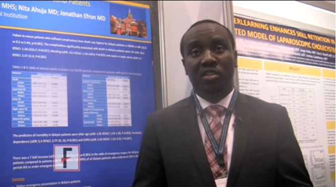
CHICAGO – The odds of emergency surgery were sevenfold higher in dialysis-dependent patients undergoing colorectal surgery than patients with renal insufficiency not on dialysis or those with normal renal function.
Dialysis patients were also far less likely to undergo laparoscopic surgery and to be rescued from death if they experienced a complication.
These are just some of the results of a retrospective study involving 156,645 elective colorectal surgery cases selected as a poster of exceptional merit here at the annual clinical congress of the American College of Surgeons.
Dialysis patients are known to be at high risk for postoperative complications, but few studies have evaluated outcomes after colorectal surgery in these patients or distinguished them from patients with non–dialysis dependent renal insufficiency (NDDRI) or normal renal function (NRF), observed study author Dr. Isibor Arhuidese of Johns Hopkins University in Baltimore.
Indeed, when the researchers compared these three groups, perioperative mortality and morbidity after elective colorectal surgery was the worst in dialysis patients.
Absolute perioperative mortality was highest for dialysis patients vs. NDDRI and NRF patients after open (13.4% vs. 4.8% vs. 2%; P less than .001) and laparoscopic (8% vs. 2% vs. 0.6%; P less than .001) surgery.
Three complications were significantly associated with death in dialysis patients: myocardial infarction (adjusted odds ratio, 48.6; P = .027), bleeding (aOR, 14.5; P = .025), and sepsis or septic shock (aOR, 8.7; P = .001).
It is not enough to simply identify dialysis dependence as a predictor of poor outcomes, but one must identify targets for improvement in surgical care, Dr. Arhuidese stressed.
Dr. Arhuidese reported having no relevant conflicts of interest.
The video associated with this article is no longer available on this site. Please view all of our videos on the MDedge YouTube channel
On Twitter @pwendl
AT THE ACS CLINICAL CONGRESS
Urinalysis Is Reliable Infection Predictor in Infants
Clinical question: In infants younger than three months of age with bacteremic urinary tract infection (UTI), how sensitive and specific are urinalysis (UA) findings?
Background: Infants are commonly hospitalized with UTIs. The gold standard for diagnosis is considered to be urine culture. When compared to this gold standard, the sensitivity of UA findings for the diagnosis of UTI has been previously reported to be around 75% to 85%; however, a positive urine culture alone in the setting of negative UA may not be reflective of a UTI due to asymptomatic bacteriuria or contamination. The 2011 American Academy of Pediatrics clinical guideline for UTIs suggests that the diagnosis should require positive urine culture in addition to abnormal UA. These guidelines do not include infants younger than two months of age, and positive cultures in this age group are generally regarded as a UTI and treated as such. Positive culture results with the same organism in the urine and blood indicates very low likelihood of contamination or asymptomatic bacteriuria, and patients with bacteremic UTI are likely to have a true infection.
Study design: Multicenter, retrospective, cross-sectional study.
Setting: Twenty hospitals in eleven hospital systems.
Synopsis: Researchers used a multicenter microbiology database to identify infants younger than three months of age with bacteremic UTI (same pathogenic organism in blood and urine). Data was collected on UA, including microscopy [white blood cells per high-power field (WBC/HPF), bacteria], dipstick [nitrites, leukocyte esterase (LE)], and urine culture in colony-forming units per mL (CFU/mL).
Exclusions included:
- Major comorbidities (defined in this study as neuromuscular conditions such as spina bifida, previous urologic surgery other than circumcision, or immunodeficiency);
- Patients managed in an ICU setting; and
- Patients with indwelling urinary or central venous catheters at the time of culture.
A total of 276 infants with bacteremic UTI were identified, with 31 exclusions (12 with no UA performed, 19 with cultures with <50,000 CFU/mL). The remaining 245 infants were included for analysis. The control group was a random sampling of 115 similarly aged infants who underwent evaluation for serious bacterial infection and had negative urine cultures.
Comparison between the study group (bacteremic UTI) and the controls showed:
- LE (including any “positive” LE) had a sensitivity of 97.6%, specificity of 93.9%;
- Considering “trace” LE as negative changed the sensitivity and specificity to 95.7% and 97.4%, respectively; and
- Positive nitrites had a specificity of 100%.
A definition of positive UA that includes pyuria (greater than 3 WBC/HPF) and/or any LE was highly sensitive (99.5%) and specific (87.8%). All but one of 203 infants with bacteremic UTI who had complete UA results were positive for LE and/or WBC/HPF. The one exception was a 64-day-old girl with Group B Streptococcus infection. Bacteria on microscopy showed poor specificity.
The authors discussed two possible explanations for the study’s finding of high sensitivity of the UA, including:
- The UA is in fact highly sensitive, and previous studies have been flawed by a faulty gold standard (positive cultures due to asymptomatic bacteriuria or contamination); or
- Screening tests are more sensitive in the setting of severe disease (in this case, UTI with bacteremia).
The second explanation is controversial, and the authors of this article cite previous studies showing minimal differences between UTI with or without bacteremia.
Bottom line: In infants younger than three months of age with bacteremic UTI, the findings of pyuria and/or any LE on UA are reliable predictors of infection, with higher sensitivity than previously reported.
Citation: Schroeder AR, Chang PW, Shen MW, Biondi EA, Greenhow TL. Diagnostic accuracy of the urinalysis for urinary tract infection in infants <3 months of age. Pediatrics. 2015;135(6):965-71.
A guide to topical options for easing vulvar discomfort
LAS VEGAS – For postmenopausal women who don’t want a hormonal topical treatment, there are plenty of other nonhormonal options to ease the symptoms of vulvovaginal atrophy, according to Susan Kellogg Spadt, Ph.D.
Dr. Kellogg Spadt, a certified sexual counselor and professor of obstetrics and gynecology at Drexel University, Philadelphia, said many of these topical products are available over the counter.
Lubricants – a good first-line choice to decrease pain and increase pleasure during intercourse – are primarily designed to minimize friction and irritation around the clitoral area, the labia, and the introitus and vagina. They are meant to be applied immediately before sex play, on an as-needed basis, Dr. Kellogg Spadt said at the annual meeting of the North American Menopause Society.

“The take-home message: Apply to both partner’s genitals,” she said.
However, some of these products have been associated with significant contact dermatitis, often because of additives such as camphor or menthol that are intended to provide a warming or tingling sensation. When women experience contact dermatitis from these or other causes in the vulvovaginal region, Dr. Kellogg Spadt said, clinicians should recommend that they avoid overzealous hygiene and treat the tissues gently. They can use a neutral emollient such as Aquaphor and use sleep aids or antihistamines for sedation and comfort at night as needed for a few days.
Lubricants are hyperosmotic, hypo-osmotic, or iso-osmotic. Hypo-osmotic lubricants such as FemGlide and Slippery Stuff can reduce mucus production. Hyperosmotic lubricants such as Replens lubricant, KY, and Astroglide can be irritating for some users, though some of these products have devoted long-time users who have never had difficulty, said Dr. Kellogg Spadt. Pre-seed is an iso-osmotic lubricant designed especially for couples who are trying to conceive.
Vaginal moisturizers have a different mode of action. These products use bioadhesive polymers that attach to mucin and the epithelial cells on the vaginal wall. These products can carry up to 60 times their weight in water, and will leave the water in place on the vulvovaginal epithelial surface until the product is eventually sloughed off, Dr. Kellogg Spadt said.
Vaginal moisturizers are meant to be used regularly – not necessarily every day, but at least two or three times weekly. They do not need reapplication before intercourse. Though studies on the efficacy of these products have generally been lacking, Replens is a vaginal moisturizer that, in one study, was found to improve vulvovaginal health with findings of normalized vaginal cytology and a return to premenopausal vaginal pH (Fertil Steril. 1994 Jan;61[1]:178-80.).
These changes in the vaginal environment, said Dr. Kellogg Spadt, won’t treat yeast or bacterial vaginal infections, but they may help prevent recurrent infections.
Some vaginal moisturizers use hyaluronic acid, and one in particular, Hyalogyn, is 100% hyaluronic acid. This product has been found to be as efficacious as local estrogen in a randomized clinical trial (J Sex Med. 2013 Jun;10[6]:1575-84).
Juvagyn is a hybrid lubricant-moisturizer with hyaluronic acid that can be applied with a fingertip. This allows more selective application to the external structures, where much of the discomfort of intercourse can occur, Dr. Kellogg Spadt said. Most other products use an intravaginal applicator to deliver a premeasured amount.
Another option is Neogyn, which is marketed as an external vulvar soothing cream and is also meant to be applied to the introitus and external vulvar tissue. It has been shown to reduce vulvar pain significantly, she said.
For patients who still have pain despite these measures and desire to have intercourse, over-the-counter lidocaine gel can be used. Patients should be warned that the lidocaine – 1% topical gel is available over the counter – will burn on application before numbing. It should be washed off after intercourse, she said.
Dr. Kellogg Stadt reported being a consultant or on the advisory board of Nuelle and of Neogyn, which also markets Juvagyn. She is also on the speakers bureau of Novo Nordisk and Shionogi.
On Twitter @karioakes
LAS VEGAS – For postmenopausal women who don’t want a hormonal topical treatment, there are plenty of other nonhormonal options to ease the symptoms of vulvovaginal atrophy, according to Susan Kellogg Spadt, Ph.D.
Dr. Kellogg Spadt, a certified sexual counselor and professor of obstetrics and gynecology at Drexel University, Philadelphia, said many of these topical products are available over the counter.
Lubricants – a good first-line choice to decrease pain and increase pleasure during intercourse – are primarily designed to minimize friction and irritation around the clitoral area, the labia, and the introitus and vagina. They are meant to be applied immediately before sex play, on an as-needed basis, Dr. Kellogg Spadt said at the annual meeting of the North American Menopause Society.
“The take-home message: Apply to both partner’s genitals,” she said.
However, some of these products have been associated with significant contact dermatitis, often because of additives such as camphor or menthol that are intended to provide a warming or tingling sensation. When women experience contact dermatitis from these or other causes in the vulvovaginal region, Dr. Kellogg Spadt said, clinicians should recommend that they avoid overzealous hygiene and treat the tissues gently. They can use a neutral emollient such as Aquaphor and use sleep aids or antihistamines for sedation and comfort at night as needed for a few days.
Lubricants are hyperosmotic, hypo-osmotic, or iso-osmotic. Hypo-osmotic lubricants such as FemGlide and Slippery Stuff can reduce mucus production. Hyperosmotic lubricants such as Replens lubricant, KY, and Astroglide can be irritating for some users, though some of these products have devoted long-time users who have never had difficulty, said Dr. Kellogg Spadt. Pre-seed is an iso-osmotic lubricant designed especially for couples who are trying to conceive.
Vaginal moisturizers have a different mode of action. These products use bioadhesive polymers that attach to mucin and the epithelial cells on the vaginal wall. These products can carry up to 60 times their weight in water, and will leave the water in place on the vulvovaginal epithelial surface until the product is eventually sloughed off, Dr. Kellogg Spadt said.
Vaginal moisturizers are meant to be used regularly – not necessarily every day, but at least two or three times weekly. They do not need reapplication before intercourse. Though studies on the efficacy of these products have generally been lacking, Replens is a vaginal moisturizer that, in one study, was found to improve vulvovaginal health with findings of normalized vaginal cytology and a return to premenopausal vaginal pH (Fertil Steril. 1994 Jan;61[1]:178-80.).
These changes in the vaginal environment, said Dr. Kellogg Spadt, won’t treat yeast or bacterial vaginal infections, but they may help prevent recurrent infections.
Some vaginal moisturizers use hyaluronic acid, and one in particular, Hyalogyn, is 100% hyaluronic acid. This product has been found to be as efficacious as local estrogen in a randomized clinical trial (J Sex Med. 2013 Jun;10[6]:1575-84).
Juvagyn is a hybrid lubricant-moisturizer with hyaluronic acid that can be applied with a fingertip. This allows more selective application to the external structures, where much of the discomfort of intercourse can occur, Dr. Kellogg Spadt said. Most other products use an intravaginal applicator to deliver a premeasured amount.
Another option is Neogyn, which is marketed as an external vulvar soothing cream and is also meant to be applied to the introitus and external vulvar tissue. It has been shown to reduce vulvar pain significantly, she said.
For patients who still have pain despite these measures and desire to have intercourse, over-the-counter lidocaine gel can be used. Patients should be warned that the lidocaine – 1% topical gel is available over the counter – will burn on application before numbing. It should be washed off after intercourse, she said.
Dr. Kellogg Stadt reported being a consultant or on the advisory board of Nuelle and of Neogyn, which also markets Juvagyn. She is also on the speakers bureau of Novo Nordisk and Shionogi.
On Twitter @karioakes
EXPERT ANALYSIS FROM THE NAMS 2015 ANNUAL MEETING

Everolimus makes splash in lung, GI neuroendocrine tumors
VIENNA – The mTOR inhibitor everolimus cut the risk of progression or death by 52% for patients with advanced, progressive, nonfunctional lung or gastrointestinal neuroendocrine tumors in RADIANT-4.
Median progression-free survival (PFS) by central review was 11.0 months with everolimus vs. 3.9 months with placebo (hazard ratio, 0.48; P less than .00001).
The robust benefit was confirmed in investigator assessment (median PFS, 14.0 months vs. 5.5 months; HR, 0.39; P less than .00001).

“Everolimus is the first targeted agent to show robust antitumor activity with acceptable toxicity across a broad spectrum of neuroendocrine tumors, including those arising from the pancreas, lung, and gastrointestinal tract,” Dr. James Yao, chair of gastrointestinal medical oncology at the University of Texas MD Anderson Cancer Center, Houston, said in a presidential session at the European Society for Medical Oncology congress.
The resultsare particularly important for patients with lung neuroendocrine tumors (NETs) because there is currently no approved treatment for such cases.
Everolimus, a mammalian target of rapamycin (mTOR) inhibitor, is already an approved standard of care in pancreatic NETs, based on the results of RADIANT-3.
Discussant Dr. Enrique Grande of Ram<scaps>ó</scaps>n y Cajal University Hospital in Madrid, said, “RADIANT-1, -2, -3, and now -4 results are all consistent and support the use of everolimus in grade 1 and 2 disseminated and progressive neuroendocrine regardless of primary tumor origin.”
That said, he observed that the median 11-month progression-free survival with everolimus is not superior to the 14.3-month PFS offered by the somatostatin analogue octreotide LAR in midgut NETs in PROMID, the 16.4-month PFS with everolimus plus octreotide LAR in RADIANT-2 in a cohort that included 15% lung NETs, or the 16.6-month PFS with bevacizumab (Avastin) plus octreotide LAR in a mixed cohort of NETs in SWOG S0518.

“Despite the encouraging activity favoring everolimus, we do not have the full picture yet,” Dr. Grande said. “Should everolimus be pushed after somatostatin analogue failure? If so, and this is something that most of the people here are doing, is single-agent everolimus better than maintenance of somatostatin analogues plus adding on everolimus?
“This is not like other tumor types, in which we are talking about first-line, second-line, third-line. We don’t have these things here. We have an unknown line of treatment.”
RADIANT-4 enrolled 302 patients with a median age of 63 years and randomized them in 2:1 ratio to everolimus 10 mg/day until disease progression or intolerable toxicity or to placebo. Patients were stratified by prior somatostatin analogue (SSA) treatment (yes vs. no), tumor origin (stratum A vs. stratum B), and WHO performance status (0 vs. 1). Stratum A (better prognosis) comprised appendix, caecum, jejunum, ileum, duodenum, and NET of unknown primary, while stratum B (worse prognosis) comprised lung, stomach, rectum, and colon except caecum. Crossover to open-label everolimus after progression in the placebo arm was not allowed prior to the primary analysis.
The most common primary tumor sites were lung and ileum in both the everolimus (31% and 23%) and placebo (28% and 25%) groups. In the everolimus group, 53% had prior SSA therapy, 59% surgery, 26% chemotherapy, and 22% radiotherapy. In the placebo group, the percentages were 56%, 72%, 24%, and 20%.
Overall, 64% of patients receiving everolimus had some tumor shrinkage vs. 26% receiving placebo, Dr. Yao said. The overall response rate, including complete and partial responses, was 2% vs. 1%. The disease control rate, which also included stable disease, was 82.4% vs. 65%.
The median duration of treatment was 40.4 weeks with everolimus and 19.6 weeks with placebo.
A consistent PFS benefit was seen with everolimus regardless of stratification factors, age, sex, race, tumor grade, prior therapy, and baseline chromogranin A or neuron-specific enolase status, he said.
Benefits also were seen across hepatic tumor load. This included an 82% reduction in progression with everolimus (HR, 0.18) among those with heavy liver tumor burden (greater than 25%), “a group of patients that really needs treatment,” Dr. Yao said.
The PFS benefit also was consistent across primary tumor site: lung (HR, 0.50), GI (HR, 0.56), and NET of unknown primary (HR, 0.60).
The first interim overall analysis showed a 36% reduction in the risk of death with everolimus (HR, 0.64; P = .037), but this was not statistically significant based on the prespecified P value boundary for significance of 0.0002, Dr. Yao said. There were 41 deaths in the everolimus arm and 28 in the placebo arm, of which 3.5% vs. 3.1% were on-treatment deaths. The next interim analysis is expected in 2016.
The safety profile was consistent with the known profile of everolimus, he said. In the everolimus arm, the most common drug-related adverse events of any grade were stomatitis (63%), diarrhea (31%), fatigue (31%), and infections (29%). Common grade 3/4 events were stomatitis (9%), diarrhea (7%), infections (7%), and fatigue (3%).
Dr. Peter Naredi, scientific cochair of the congress, commented that this year’s meeting is particularly exciting for clinicians treating patients with neuroendocrine tumors because two trials, RADIANT-4 and NETTER-1 both show positive results.
On Twitter @pwendl
VIENNA – The mTOR inhibitor everolimus cut the risk of progression or death by 52% for patients with advanced, progressive, nonfunctional lung or gastrointestinal neuroendocrine tumors in RADIANT-4.
Median progression-free survival (PFS) by central review was 11.0 months with everolimus vs. 3.9 months with placebo (hazard ratio, 0.48; P less than .00001).
The robust benefit was confirmed in investigator assessment (median PFS, 14.0 months vs. 5.5 months; HR, 0.39; P less than .00001).
“Everolimus is the first targeted agent to show robust antitumor activity with acceptable toxicity across a broad spectrum of neuroendocrine tumors, including those arising from the pancreas, lung, and gastrointestinal tract,” Dr. James Yao, chair of gastrointestinal medical oncology at the University of Texas MD Anderson Cancer Center, Houston, said in a presidential session at the European Society for Medical Oncology congress.
The resultsare particularly important for patients with lung neuroendocrine tumors (NETs) because there is currently no approved treatment for such cases.
Everolimus, a mammalian target of rapamycin (mTOR) inhibitor, is already an approved standard of care in pancreatic NETs, based on the results of RADIANT-3.
Discussant Dr. Enrique Grande of Ram<scaps>ó</scaps>n y Cajal University Hospital in Madrid, said, “RADIANT-1, -2, -3, and now -4 results are all consistent and support the use of everolimus in grade 1 and 2 disseminated and progressive neuroendocrine regardless of primary tumor origin.”
That said, he observed that the median 11-month progression-free survival with everolimus is not superior to the 14.3-month PFS offered by the somatostatin analogue octreotide LAR in midgut NETs in PROMID, the 16.4-month PFS with everolimus plus octreotide LAR in RADIANT-2 in a cohort that included 15% lung NETs, or the 16.6-month PFS with bevacizumab (Avastin) plus octreotide LAR in a mixed cohort of NETs in SWOG S0518.
“Despite the encouraging activity favoring everolimus, we do not have the full picture yet,” Dr. Grande said. “Should everolimus be pushed after somatostatin analogue failure? If so, and this is something that most of the people here are doing, is single-agent everolimus better than maintenance of somatostatin analogues plus adding on everolimus?
“This is not like other tumor types, in which we are talking about first-line, second-line, third-line. We don’t have these things here. We have an unknown line of treatment.”
RADIANT-4 enrolled 302 patients with a median age of 63 years and randomized them in 2:1 ratio to everolimus 10 mg/day until disease progression or intolerable toxicity or to placebo. Patients were stratified by prior somatostatin analogue (SSA) treatment (yes vs. no), tumor origin (stratum A vs. stratum B), and WHO performance status (0 vs. 1). Stratum A (better prognosis) comprised appendix, caecum, jejunum, ileum, duodenum, and NET of unknown primary, while stratum B (worse prognosis) comprised lung, stomach, rectum, and colon except caecum. Crossover to open-label everolimus after progression in the placebo arm was not allowed prior to the primary analysis.
The most common primary tumor sites were lung and ileum in both the everolimus (31% and 23%) and placebo (28% and 25%) groups. In the everolimus group, 53% had prior SSA therapy, 59% surgery, 26% chemotherapy, and 22% radiotherapy. In the placebo group, the percentages were 56%, 72%, 24%, and 20%.
Overall, 64% of patients receiving everolimus had some tumor shrinkage vs. 26% receiving placebo, Dr. Yao said. The overall response rate, including complete and partial responses, was 2% vs. 1%. The disease control rate, which also included stable disease, was 82.4% vs. 65%.
The median duration of treatment was 40.4 weeks with everolimus and 19.6 weeks with placebo.
A consistent PFS benefit was seen with everolimus regardless of stratification factors, age, sex, race, tumor grade, prior therapy, and baseline chromogranin A or neuron-specific enolase status, he said.
Benefits also were seen across hepatic tumor load. This included an 82% reduction in progression with everolimus (HR, 0.18) among those with heavy liver tumor burden (greater than 25%), “a group of patients that really needs treatment,” Dr. Yao said.
The PFS benefit also was consistent across primary tumor site: lung (HR, 0.50), GI (HR, 0.56), and NET of unknown primary (HR, 0.60).
The first interim overall analysis showed a 36% reduction in the risk of death with everolimus (HR, 0.64; P = .037), but this was not statistically significant based on the prespecified P value boundary for significance of 0.0002, Dr. Yao said. There were 41 deaths in the everolimus arm and 28 in the placebo arm, of which 3.5% vs. 3.1% were on-treatment deaths. The next interim analysis is expected in 2016.
The safety profile was consistent with the known profile of everolimus, he said. In the everolimus arm, the most common drug-related adverse events of any grade were stomatitis (63%), diarrhea (31%), fatigue (31%), and infections (29%). Common grade 3/4 events were stomatitis (9%), diarrhea (7%), infections (7%), and fatigue (3%).
Dr. Peter Naredi, scientific cochair of the congress, commented that this year’s meeting is particularly exciting for clinicians treating patients with neuroendocrine tumors because two trials, RADIANT-4 and NETTER-1 both show positive results.
On Twitter @pwendl
AT THE EUROPEAN CANCER CONGRESS 2015
Key clinical point: Everolimus significantly delays tumor growth in advanced lung and gastrointestinal neuroendocrine tumors.
Major finding: Median progression-free survival was 11 months for everolimus vs. 3.9 months for placebo (hazard ratio, 0.39; P < .00001).
Data source: Double-blind phase III study in 302 patients with advanced, progressive lung and GI neuroendocrine tumors.
Disclosures: Novartis sponsored the study. Dr. Yao reported serving as a consultant to and receiving research funding from Novartis.

Managing menopausal symptoms in women with a BRCA mutation
This audiocast was recorded at the North American Menopause Society Annual Meeting held September 30 to October 3, 2015, in Las Vegas, Nevada
For more on this topic, read Dr. Kaunitz's August 2015 Cases in Menopause article, Is menopausal hormone therapy safe when your patient carries a BRCA mutation?

Society of Hospital Medicine Opens Enrollment for Practice Administrators' Mentor Program
SHM’s Practice Administrators Committee piloted a mentor/mentee program in January 2014 as a way to assist those who were new to practice administration and/or to hospital medicine. SHM is continuing with the program and is now accepting enrollment for the fall of 2015.
The committee first learned of the interest among practice administrators in a mentorship program by monitoring communications within the Hospital Medicine Exchange (HMX) online community. The pilot program was developed in response to this significant interest.
Committee members volunteered to be mentors in the program’s first year, in an effort to provide a structured opportunity for hospitalist administrators seeking to strengthen their knowledge and skills. Today, the program consists of two models:
- Mentors/Mentees: Less experienced administrators will be paired with seasoned professionals to gain more experience and/or exposure.
- Buddy System: Administrators at any level of expertise or experience will be paired with a peer so that they can learn from one another.
How Does the Program Work?
Both mentors and mentees complete an online profile, which is used by the committee to match up appropriate individuals based on type of institution and skills that need to be improved. Once the matches are completed, mentors and mentees will be asked to attend an online webinar in order to understand the expectations for the mentor and the mentee, including the length of the commitment and the expected frequency of contact with each other.
A survey will be circulated to follow up with the mentors and mentees. Information gleaned from the survey will ensure that the mentor-mentee relationship is intact and will offer an opportunity to provide feedback on the program’s performance.
Tiffani Panek is manager of the division of hospital medicine and program director of the academic hospital medicine training program at Johns Hopkins Bayview Medical Center and Johns Hopkins University School of Medicine, Baltimore. She is a member of SHM’s Practice Administrators Committee.