User login
Azithromycin and doxycycline effective for urogenital chlamydia*
BRISBANE, AUSTRALIA – Azithromycin and doxycycline have been shown to still be highly effective treatments for urogenital chlamydia in a randomized clinical trial conducted in a youth correctional facility.*
The study, presented at the World STI & HIV Congress 2015, found a 7-day, twice-daily regimen of 100 mg of doxycycline achieved a 100% cure rate, compared with a 96.8% success rate with a single 1-g dose of azithromycin.
However, noninferiority of azithromycin to doxycycline was not established, said Dr. William Geisler of the division of infectious diseases at the University of Alabama at Birmingham.

“I think the big finding from the study was that both drugs still work quite well, and I think it puts to rest a lot of the concerns about azithromycin for urogenital chlamydia,” said Dr. Geisler.
The study enrolled 310 males and nonpregnant females (median age 17 years) who tested positive for chlamydia when screened upon arrival at the correctional facility.
They were then randomized to receive either doxycycline or azithromycin, and followed up with chlamydia testing 28 days after treatment, which included genotyping to distinguish between treatment failure and infection with a new chlamydia strain.
Of the five treatment failures, four were males and one female; the researchers noted that painful urination occurred more often in azithromycin-treated males who failed treatment versus those cured, although the difference was not statistically significant.
Commenting on this finding, a member of the audience asked whether Dr. Geisler would therefore reconsider treating a symptomatic man with azithromycin.
Dr. Geisler said that while he did have concerns about the efficacy of azithromycin in symptomatic men, adherence also was a factor to be taken into consideration.
“In general, most of the males I see, I don’t feel that great about their likelihood for full adherence [with doxycycline], so I would still feel very comfortable about giving azithromycin to a symptomatic male,” he said.
About three-quarters of the males who were positive for chlamydia at randomization were asymptomatic, compared with nearly 40% of the females.
Adherence was a nonissue for azithromycin, because the single dose could be taken under observation, Dr. Geisler noted. There were greater variations in adherence with the doxycycline, with participants taking between 11 and 16 doses, although about 80% took the prescribed 14 doses.
The study was conducted in correctional facilities in an attempt to limit the possibility of reinfection, and to help improve the likelihood of adherence – both issues that have plagued previous studies.
“These residents, once they get in the facilities, are not typically re-exposed to the untreated partner who’s outside of the facility, so whatever exposures they would have would be new partners in the facility,” Dr. Geisler said at the conference. “The facility that we used for this study, there were really minimal opportunities for re-exposure to chlamydia because everybody who entered the facility got routinely screened and treated for chlamydia.”
While it was challenging to get approval to conduct the study in the correctional facility, Dr. Geisler said it was worth considering.
“Maybe this will pave the way for other studies in these facilities, ones that have low-risk type of procedures [because] controlling for sex is a lot easier in these settings,” he said in an interview.
The National Institutes of Health funded the study. There were no conflicts of interest declared.
*Correction, 11/3/2015: An earlier version of this story incorrectly stated the study's major finding. Although the cure rates were high for both treatments, noninferiority of azithromycin to doxycycline was not established in the study.
BRISBANE, AUSTRALIA – Azithromycin and doxycycline have been shown to still be highly effective treatments for urogenital chlamydia in a randomized clinical trial conducted in a youth correctional facility.*
The study, presented at the World STI & HIV Congress 2015, found a 7-day, twice-daily regimen of 100 mg of doxycycline achieved a 100% cure rate, compared with a 96.8% success rate with a single 1-g dose of azithromycin.
However, noninferiority of azithromycin to doxycycline was not established, said Dr. William Geisler of the division of infectious diseases at the University of Alabama at Birmingham.
“I think the big finding from the study was that both drugs still work quite well, and I think it puts to rest a lot of the concerns about azithromycin for urogenital chlamydia,” said Dr. Geisler.
The study enrolled 310 males and nonpregnant females (median age 17 years) who tested positive for chlamydia when screened upon arrival at the correctional facility.
They were then randomized to receive either doxycycline or azithromycin, and followed up with chlamydia testing 28 days after treatment, which included genotyping to distinguish between treatment failure and infection with a new chlamydia strain.
Of the five treatment failures, four were males and one female; the researchers noted that painful urination occurred more often in azithromycin-treated males who failed treatment versus those cured, although the difference was not statistically significant.
Commenting on this finding, a member of the audience asked whether Dr. Geisler would therefore reconsider treating a symptomatic man with azithromycin.
Dr. Geisler said that while he did have concerns about the efficacy of azithromycin in symptomatic men, adherence also was a factor to be taken into consideration.
“In general, most of the males I see, I don’t feel that great about their likelihood for full adherence [with doxycycline], so I would still feel very comfortable about giving azithromycin to a symptomatic male,” he said.
About three-quarters of the males who were positive for chlamydia at randomization were asymptomatic, compared with nearly 40% of the females.
Adherence was a nonissue for azithromycin, because the single dose could be taken under observation, Dr. Geisler noted. There were greater variations in adherence with the doxycycline, with participants taking between 11 and 16 doses, although about 80% took the prescribed 14 doses.
The study was conducted in correctional facilities in an attempt to limit the possibility of reinfection, and to help improve the likelihood of adherence – both issues that have plagued previous studies.
“These residents, once they get in the facilities, are not typically re-exposed to the untreated partner who’s outside of the facility, so whatever exposures they would have would be new partners in the facility,” Dr. Geisler said at the conference. “The facility that we used for this study, there were really minimal opportunities for re-exposure to chlamydia because everybody who entered the facility got routinely screened and treated for chlamydia.”
While it was challenging to get approval to conduct the study in the correctional facility, Dr. Geisler said it was worth considering.
“Maybe this will pave the way for other studies in these facilities, ones that have low-risk type of procedures [because] controlling for sex is a lot easier in these settings,” he said in an interview.
The National Institutes of Health funded the study. There were no conflicts of interest declared.
*Correction, 11/3/2015: An earlier version of this story incorrectly stated the study's major finding. Although the cure rates were high for both treatments, noninferiority of azithromycin to doxycycline was not established in the study.
FROM THE WORLD STI & HIV CONGRESS 2015
Key clinical point: Azithromycin and doxycycline remain highly effective for the treatment of urogenital chlamydia with cure rates greater than 95%.
Major finding: Azithromycin achieved a 96.8% cure rate for chlamydia, compared with a 100% cure rate with doxycycline. Although the cure rates were high for both treatments, noninferiority of azithromycin to doxycycline was not established in the study.
Data source: Randomized clinical trial of 310 males and females in a youth correctional facility.
Disclosures: The National Institutes of Health funded the study. There were no conflicts of interest declared.

Intraperitoneal bupivacaine disappoints in postop pain relief
NEW YORK – While some evidence in the surgical literature suggests that intraperitoneal bupivacaine reduces postoperative pain scores and narcotic use, a new randomized controlled trial shows no significant improvement in patients undergoing robot-assisted total laparoscopic hysterectomy.
“Despite evidence of benefit in laparoscopic surgery, there does not appear to be a benefit for using intraperitoneal bupivacaine. We need a larger sample size to study this,” Dr. Allan Klapper said at the annual Minimally Invasive Surgery Week.

The uptake of robot-assisted total laparoscopic hysterectomy increased by 9.5% in the United States, and hospitals with robotic capabilities perform 22.4% of hysterectomies with this technique, according to Dr. Klapper, an ob.gyn. at West Pennsylvania Allegheny Health System in Pittsburgh. Minimally invasive surgery, however, does not completely eliminate postoperative pain, and between one-third and two-thirds of patients report pain following such surgery.
Intraperitoneal bupivacaine (IB) was selected for the study because of positive reports in small studies of patients undergoing laparoscopic cholecystectomy and gynecologic procedures, Dr. Klapper explained. He noted that the positive studies were of poor quality, but other studies – also of poor quality – had negative results.
To investigate the role of IB in reducing postoperative pain and narcotic requirements in patients undergoing robot-assisted total laparoscopic hysterectomy, Dr. Klapper and his colleagues launched a prospective, double-blind, randomized, placebo-controlled trial comparing IB to normal saline in 41 patients managed with a standardized postoperative analgesic regimen.
Baseline characteristics showed no differences between the two groups in surgical indications, body mass index, operating room time, number of previous surgeries, and the percentage of patients undergoing lymph node dissection. Patients were excluded from the analysis if they converted to laparotomy, were allergic to IB, or were currently on treatment for chronic pain.
Complete data on pain response, as assessed by the visual analog scale, was available for 29 of the 41 patients. No significant differences in postoperative pain scores were observed between the two groups at 1, 16, 18, and 24 hours. Further, there was no significant difference in morphine dose between the IB and normal saline groups.
“One finding was the opposite of what I would have expected. Patients who underwent lymph node dissection used significantly less narcotic analgesic [P = .03],” Dr. Klapper told the audience.
Study strengths were the prospective, randomized design, and its being adequately powered to detect a significant difference between the two groups. But the study was conducted at a single institution and did not include data beyond 24 hours. Also, Dr. Klapper said that perhaps results should have been stratified according to indications for surgery.
“There is more and more pressure on us to achieve higher patient satisfaction scores. Soon down the line, patient satisfaction will become a metric for compensation. We need to focus on what we can do to improve patient satisfaction scores and experience,” Dr. Klapper said. “A larger sample of gynecologic oncology patients is needed to further support these conclusions, and we need to fine tune to avoid the problems in this study.”
The meeting was presented by the Society of Laparoendoscopic Surgeons and affiliated societies. Dr. Klapper reported that he is on the speakers bureau for Astellas.
NEW YORK – While some evidence in the surgical literature suggests that intraperitoneal bupivacaine reduces postoperative pain scores and narcotic use, a new randomized controlled trial shows no significant improvement in patients undergoing robot-assisted total laparoscopic hysterectomy.
“Despite evidence of benefit in laparoscopic surgery, there does not appear to be a benefit for using intraperitoneal bupivacaine. We need a larger sample size to study this,” Dr. Allan Klapper said at the annual Minimally Invasive Surgery Week.
The uptake of robot-assisted total laparoscopic hysterectomy increased by 9.5% in the United States, and hospitals with robotic capabilities perform 22.4% of hysterectomies with this technique, according to Dr. Klapper, an ob.gyn. at West Pennsylvania Allegheny Health System in Pittsburgh. Minimally invasive surgery, however, does not completely eliminate postoperative pain, and between one-third and two-thirds of patients report pain following such surgery.
Intraperitoneal bupivacaine (IB) was selected for the study because of positive reports in small studies of patients undergoing laparoscopic cholecystectomy and gynecologic procedures, Dr. Klapper explained. He noted that the positive studies were of poor quality, but other studies – also of poor quality – had negative results.
To investigate the role of IB in reducing postoperative pain and narcotic requirements in patients undergoing robot-assisted total laparoscopic hysterectomy, Dr. Klapper and his colleagues launched a prospective, double-blind, randomized, placebo-controlled trial comparing IB to normal saline in 41 patients managed with a standardized postoperative analgesic regimen.
Baseline characteristics showed no differences between the two groups in surgical indications, body mass index, operating room time, number of previous surgeries, and the percentage of patients undergoing lymph node dissection. Patients were excluded from the analysis if they converted to laparotomy, were allergic to IB, or were currently on treatment for chronic pain.
Complete data on pain response, as assessed by the visual analog scale, was available for 29 of the 41 patients. No significant differences in postoperative pain scores were observed between the two groups at 1, 16, 18, and 24 hours. Further, there was no significant difference in morphine dose between the IB and normal saline groups.
“One finding was the opposite of what I would have expected. Patients who underwent lymph node dissection used significantly less narcotic analgesic [P = .03],” Dr. Klapper told the audience.
Study strengths were the prospective, randomized design, and its being adequately powered to detect a significant difference between the two groups. But the study was conducted at a single institution and did not include data beyond 24 hours. Also, Dr. Klapper said that perhaps results should have been stratified according to indications for surgery.
“There is more and more pressure on us to achieve higher patient satisfaction scores. Soon down the line, patient satisfaction will become a metric for compensation. We need to focus on what we can do to improve patient satisfaction scores and experience,” Dr. Klapper said. “A larger sample of gynecologic oncology patients is needed to further support these conclusions, and we need to fine tune to avoid the problems in this study.”
The meeting was presented by the Society of Laparoendoscopic Surgeons and affiliated societies. Dr. Klapper reported that he is on the speakers bureau for Astellas.
AT MINIMALLY INVASIVE SURGERY WEEK
Key clinical point: Intraperitoneal bupivacaine does not appear to achieve meaningful postoperative pain relief in total laparoscopic hysterectomy.
Major finding: No significant difference was observed between placebo and active treatment in pain scores or need for narcotics.
Data source: A prospective, randomized, placebo-controlled study of 41 patients.
Disclosures: Dr. Klapper reported that he is on the speakers bureau for Astellas.

Metronidazole and alcohol
A 32-year-old man develops diarrhea after receiving amoxicillin/clavulanate to treat an infection following a dog bite. He is diagnosed with Clostridium difficile and prescribed a 10-day course of metronidazole. He has no other medical problems. He will be the best man at his brother’s wedding tomorrow. What advice should you give him about alcohol use at the reception?
A. Do not take metronidazole the day of the wedding if you will be drinking alcohol.
B. Take metronidazole, do not drink alcohol.
C. It’s okay to drink alcohol.
For years, we have advised patients to not use alcohol if they are taking metronidazole because of concern for a disulfiram-like reaction between alcohol and metronidazole. This has been a standard warning given by physicians and appears as a contraindication in the prescribing information. It has been well accepted as a true, proven reaction.
Is it true?
As early as the 1960s, case reports and an uncontrolled study suggested that combining metronidazole with alcohol produced a disulfiram-like reaction, with case reports of severe reactions, including death.1, 2, 3 This was initially considered an area that might be therapeutic in the treatment of alcoholism, but several studies showed no benefit.4, 5
Caroline S. Williams and Dr. Kevin R. Woodcock reviewed the case reports for evidence of proof of a true interaction between metronidazole and ethanol.6 The case reports referenced textbooks to substantiate the interaction, but they did not present clear evidence of an interaction as the cause of elevated acetaldehyde levels.
Researchers have shown in a rat model that metronidazole can increase intracolonic, but not blood, acetaldehyde levels in rats that have received a combination of ethanol and metronidazole.7 Metronidazole did not have any inhibitory effect on hepatic or colonic alcohol dehydrogenase or aldehyde dehydrogenase. What was found was that rats treated with metronidazole had increased growth of Enterobacteriaceae, an alcohol dehydrogenase–containing aerobe, which could be the cause of the higher intracolonic acetaldehyde levels.
Jukka-Pekka Visapää and his colleagues studied the effect of coadministration of metronidazole and ethanol in young, healthy male volunteers.8 The study was a placebo-controlled, randomized trial. The study was small, with 12 participants. One-half of the study participants received metronidazole three times a day for 5 days; the other half received placebo. All participants then received ethanol 0.4g/kg, with blood testing being done every 20 minutes for the next 4 hours. Blood was tested for ethanol concentrations and for acetaldehyde levels. The study participants also had blood pressure, pulse, skin temperature, and symptoms monitored during the study.
There was no difference in blood acetaldehyde levels, vital signs, or symptoms between patients who received metronidazole or placebo. None of the subjects in the study had any measurable symptoms.
Metronidazole has many side effects, including nausea, vomiting, headache, dizziness, and seizures. These symptoms have a great deal of overlap with the symptoms of alcohol-disulfiram interaction. It has been assumed in early case reports that metronidazole caused a similar interaction with alcohol and raised acetaldehyde levels by interfering with aldehyde dehydrogenase.
Animal models and the human study do not show this to be the case. It is possible that metronidazole side effects alone were the cause of the symptoms in case reports. The one human study done was on healthy male volunteers, so projecting the results to a population with liver disease or other serious illness is a bit of a stretch. I think that if a problem exists with alcohol and metronidazole, it is uncommon and unlikely to occur in healthy individuals.
So, what would I advise the patient in the case about whether he can drink alcohol? I think that the risk would be minimal and that it would be safe for him to drink alcohol.
References
1. Br J Clin Pract. 1985 Jul;39(7):292-3.
2. Psychiatr Neurol. 1966;152:395-401.
3. Am J Forensic Med Pathol. 1996 Dec;17(4):343-6.
4. Q J Stud Alcohol. 1972 Sep;33: 734-40.
5. Q J Stud Ethanol. 1969 Mar;30: 140-51.
6. Ann Pharmacother. 2000 Feb;34(2):255-7.
7. Alcohol Clin Exp Res. 2000 Apr;24(4):570-5.
8. Ann Pharmacother. 2002 Jun;36(6):971-4.
Dr. Paauw is professor of medicine in the division of general internal medicine at the University of Washington, Seattle, and he serves as third-year medical student clerkship director at the University of Washington. Contact Dr. Paauw at dpaauw@uw.edu.

In-office cryoablation safe, effective in menorrhagia
NEW YORK – Cryoablation of the endometrium is a safe and effective office-based procedure for the treatment of menorrhagia, resulting in few operative complications, according to a chart review of 100 consecutive cases over a 3-year period.
“Abnormal uterine bleeding is the most common reason for referral to a gynecologist, and it is associated with an adverse impact on quality of life, health care use, and cost. Hysterectomy cures abnormal uterine bleeding, but surgery has risks,” study author Dr. Radha Syed said at the annual Minimally Invasive Surgery Week.
Between 2012 and 2015, Dr. Syed treated women aged 37-51 years with cryoablation of the endometrium under ultrasound guidance in her office. Anesthesia was provided by intravenous conscious sedation and paracervical blocks. Manufacturer’s guidelines were followed for the procedure, with voice prompts from the generator device, said Dr. Syed of the North Shore LIJ Health System, Staten Island, N.Y.
Indications for cryoablation included refractory menorrhagia or menorrhagia affecting quality of life with benign etiology; patients who did not want hysterectomy were not operative candidates.
Patient-based outcome measures were used to assess results of cryoablation. In the recovery room, pain scores were between 2 and 3, as assessed by a visual analog scale ranging from 0 to 10, with 10 signifying the most severe pain. Patients were able to return to work on the first or second postoperative day.
There were no intraoperative or immediate postoperative complications among the 100 consecutive cases reviewed. The maximum follow-up time was 36 months.
The most pressing postoperative symptom was excess watery discharge lasting 2-3 weeks, which was sometimes bloody, Dr. Syed said at the meeting, which was presented by the Society of Laparoendoscopic Surgeons and affiliated societies.
Delayed complications included two hematometra due to cervical cicatrix 4-6 weeks from surgery, which was managed by dilation under ultrasound guidance. One to two years after surgery, two patients underwent hysterectomy for recurrence of menorrhagia; both were associated with fibroids.
Patient satisfaction was 90%, as assessed over the phone using patient-based outcome measures. Most patients achieved hypomenorrhea or eumenorrhea. The rate of amenorrhea was less than 30%.
“Other minimally invasive procedures are available, but it is difficult to compare these procedures due to the subjective nature of complaints and variable symptoms,” Dr. Syed said. “I find cryoablation useful. There is less pain than with hysterectomy, and patient satisfaction is high. Even though the equipment is expensive, cryoablation avoids hysterectomy and all its attendant risks.”
Dr. Syed reported having no financial disclosures.
AT MINIMALLY INVASIVE SURGERY WEEK
Low-volume surgeons have most complications with mesh slings
The 10-year incidence of serious complications after mesh-sling surgery for stress urinary incontinence is a relatively low 3.29, but patients treated by surgeons who perform a low volume of the procedures have a 37% higher relative risk of requiring further surgery for complications, compared with patients of experienced surgeons, according to a report published online Sept. 9 in JAMA Surgery.
“These findings support the regulatory statements that suggest that patients should be counseled regarding serious complications that can occur with mesh-based procedures for SUI and that surgeons should achieve expertise in their chosen procedure,” wrote Dr. Blayne Welk of the department of surgery and the department of epidemiology and biostatistics at Western University and St. Joseph’s Health Care, London (Ont.) and his associates.
The investigators performed a population-based retrospective cohort study to determine the incidence of surgical removal or revision after a mesh-sling procedure for SUI and to examine whether there are specific risk factors for mesh-related complications. They analyzed data for 59,887 women who underwent the procedure across Ontario during a 10-year period. Median follow-up was 4.4 years.
The procedures were performed by 1,068 surgeons: 293 urologists, 625 gynecologists, and 150 unspecified clinicians. Cases were classified according to whether the surgeon performed a high or low volume of mesh-sling procedures specifically for SUI. High volume was defined as a number at or above the 75th percentile for yearly volume in the province, or more than 16 procedures per year.
Overall, 1,307 women (2.2%) required mesh removal or revision a median of 1 year after the initial surgery. The cumulative incidence of the composite outcome of removal/revision of vaginal or urethral mesh, removal of a foreign body, endoscopic treatment of a urethral foreign body or mesh encrustation, uretrolysis, or repair of a urethrovaginal fistula was 3.29 at 10 years, Dr. Welk and his associates reported. Surgical specialty had no significant effect on complication risk.
This incidence is consistent with previous report from HMOs in the United States and a meta-analysis of clinical trial results, the investigators noted (JAMA Surg. 2015 Sept. 9. doi: 10.1001/jamasurg.2015.2590). Patients of low-volume surgeons had a 37% higher relative risk of complications requiring reoperation than did patients of high-volume surgeons. In a further analysis of the data, patients of low-volume surgeons were significantly more likely to experience the composite outcome than were patients of high-volume surgeons (hazard ratio, 1.37), and, again, the difference between surgical specialties was nonsignificant.
Urologists and gynecologists have very different surgical training and day-to-day practices, the researchers noted, and as a result they hypothesized that complication rates might differ between the two groups of clinicians. But that was not proven in the findings.
“This suggests that procedure-specific knowledge and experience is important for surgery to treat SUI, rather than general operative training,” Dr. Welk and his associates wrote.
Women who underwent multiple mesh-based procedures for SUI were at highest risk for the composite endpoint. Their absolute risk for later mesh removal or revision was 4.87%.
“This novel finding should temper the enthusiasm of case series that suggest that the use of multiple synthetic slings is safe and efficacious,” the researchers wrote.
This finding is also important in light of the fact that 1,307 of the study participants underwent more than one mesh implant procedure, presumably for recurrent SUI.
This study was supported by the Ontario Ministry of Health and Long-term Care and the Academic Medical Organization of Southwestern Ontario. Dr. Welk reported receiving grant funding from Astellas Canada.
The call to centralize complex, high-risk surgical procedures at expert centers is well known, but what about commonly performed, same-day procedures with low risks of complications, such as mesh-sling surgery for stress urinary incontinence? Must patients be referred only to high-volume surgeons?
This likely would be impractical if not impossible for a procedure that is performed so frequently. A more reasonable approach would be for low-volume surgeons to use structured proctoring and/or coaching models to develop expertise and mandatory outcomes reporting to assure high-quality care. Even though clinicians will not welcome this approach, it probably will become required in the near future and tied to reimbursements. Ultimately, surgeons should be the drivers for change and should not wait for payers or regulators to impose punitive measures.
Dr. Christian P. Meyer and Dr. Quoc-Dien Trinh are with the Center for Surgery and Public Health and the division of urologic surgery at Brigham and Women’s Hospital and Harvard Medical School, Boston. They reported having no relevant financial disclosures. These remarks are adapted from an invited commentary (JAMA Surg. 2015 Sept. 9. doi: 10.1001/jamasurg.2015.2596) accompanying Dr. Welk’s report.
The 10-year incidence of serious complications after mesh-sling surgery for stress urinary incontinence is a relatively low 3.29, but patients treated by surgeons who perform a low volume of the procedures have a 37% higher relative risk of requiring further surgery for complications, compared with patients of experienced surgeons, according to a report published online Sept. 9 in JAMA Surgery.
“These findings support the regulatory statements that suggest that patients should be counseled regarding serious complications that can occur with mesh-based procedures for SUI and that surgeons should achieve expertise in their chosen procedure,” wrote Dr. Blayne Welk of the department of surgery and the department of epidemiology and biostatistics at Western University and St. Joseph’s Health Care, London (Ont.) and his associates.
The investigators performed a population-based retrospective cohort study to determine the incidence of surgical removal or revision after a mesh-sling procedure for SUI and to examine whether there are specific risk factors for mesh-related complications. They analyzed data for 59,887 women who underwent the procedure across Ontario during a 10-year period. Median follow-up was 4.4 years.
The procedures were performed by 1,068 surgeons: 293 urologists, 625 gynecologists, and 150 unspecified clinicians. Cases were classified according to whether the surgeon performed a high or low volume of mesh-sling procedures specifically for SUI. High volume was defined as a number at or above the 75th percentile for yearly volume in the province, or more than 16 procedures per year.
Overall, 1,307 women (2.2%) required mesh removal or revision a median of 1 year after the initial surgery. The cumulative incidence of the composite outcome of removal/revision of vaginal or urethral mesh, removal of a foreign body, endoscopic treatment of a urethral foreign body or mesh encrustation, uretrolysis, or repair of a urethrovaginal fistula was 3.29 at 10 years, Dr. Welk and his associates reported. Surgical specialty had no significant effect on complication risk.
This incidence is consistent with previous report from HMOs in the United States and a meta-analysis of clinical trial results, the investigators noted (JAMA Surg. 2015 Sept. 9. doi: 10.1001/jamasurg.2015.2590). Patients of low-volume surgeons had a 37% higher relative risk of complications requiring reoperation than did patients of high-volume surgeons. In a further analysis of the data, patients of low-volume surgeons were significantly more likely to experience the composite outcome than were patients of high-volume surgeons (hazard ratio, 1.37), and, again, the difference between surgical specialties was nonsignificant.
Urologists and gynecologists have very different surgical training and day-to-day practices, the researchers noted, and as a result they hypothesized that complication rates might differ between the two groups of clinicians. But that was not proven in the findings.
“This suggests that procedure-specific knowledge and experience is important for surgery to treat SUI, rather than general operative training,” Dr. Welk and his associates wrote.
Women who underwent multiple mesh-based procedures for SUI were at highest risk for the composite endpoint. Their absolute risk for later mesh removal or revision was 4.87%.
“This novel finding should temper the enthusiasm of case series that suggest that the use of multiple synthetic slings is safe and efficacious,” the researchers wrote.
This finding is also important in light of the fact that 1,307 of the study participants underwent more than one mesh implant procedure, presumably for recurrent SUI.
This study was supported by the Ontario Ministry of Health and Long-term Care and the Academic Medical Organization of Southwestern Ontario. Dr. Welk reported receiving grant funding from Astellas Canada.
FROM JAMA SURGERY
Key clinical point: The complication rate after mesh sling surgery for SUI is relatively low and highly correlated with the surgeon’s experience with the procedure.
Major finding: Patients of low-volume surgeons had a 37% higher relative risk of complications requiring reoperation than did patients of high-volume surgeons.
Data source: A population-based retrospective cohort study of 59,887 women who had SUI mesh surgery across Ontario during a 10-year period.
Disclosures: This study was supported by the Ontario Ministry of Health and Long-term Care and the Academic Medical Organization of Southwestern Ontario. Dr. Welk reported receiving grant funding from Astellas Canada.

Early results encouraging for nivolumab in ovarian cancer patients
Nivolumab demonstrated encouraging clinical efficacy and tolerability in patients with platinum-resistant ovarian cancer in a small phase II trial, according to a study published online Sept. 7 in the Journal of Clinical Oncology.

The safety and antitumor efficacy of nivolumab, a fully human immunoglobulin G4 anti–PD-1 receptor–blocking monoclonal antibody, was evaluated in 20 patients with platinum-resistant, recurrent, or advanced ovarian cancer, reported Dr. Junzo Hamanishi of Kyoto of University Graduate School of Medicine, Kyoto, Japan, and his colleagues. They noted that to their knowledge, this is the first investigator-initiated, phase II clinical trial to be done in this setting.
The patients were treated with an intravenous infusion of nivolumab every 2 weeks at a dose of either 1 or 3 mg/kg (two 10-patient cohorts), and the primary end point of this study was best overall response.
Patients received up to six cycles (four doses per cycle) of nivolumab treatment or remained on therapy until their disease progressed.
The best overall response rate across both groups was 15% (3 patients; 95% CI, 3.2% to 37.9%) and the disease control rate was 45% (9 patients; 95%CI, 23.1% to 68.5%). Among patients who received 1 mg/kg, there was one partial response and four had stable disease. The objective response rate was 10% (95% CI, 0.3% to 44.5%), while disease control rate was 50% (95% CI, 18.7% to 81.3%).
In the 3-mg/kg cohort, two patients achieved a complete response, and two had stable disease, with a total objective response rate of 20% (95% CI, 2.5% to 55.6%), and a disease control rate was 40% (95% CI, 12.2% to 73.8%).
For both groups, the median progression-free survival was 3.5 months and median overall survival was 20.0 months.
The most common adverse events were increased serum AST, hypothyroidism, lymphocytopenia, decreased serum albumin, fever, increased serum ALT, maculopapular rash, arthralgia, arrhythmia, fatigue, and anemia. Grade 3 or 4 treatment-related adverse events occurred in eight (40%) patients, although the majority of the cohort was able to continue on the regimen (n = 19, 95%). The authors pointed out that the frequencies of arrhythmias and elevated AST were relatively higher in this study than in larger previous studies that assessed nivolumab in other types of solid tumors, but all of these events were grade 1 or 2 and manageable.
“This study indicates the merit of a large-scale investigation to evaluate the priority of nivolumab for platinum-resistant ovarian cancer,” the researchers wrote, adding that they plan to conduct such a study in the future (J Clin Oncol. 2015 Sep 7. doi:10.1200/JCO.2015.62.3397).
Nivolumab demonstrated encouraging clinical efficacy and tolerability in patients with platinum-resistant ovarian cancer in a small phase II trial, according to a study published online Sept. 7 in the Journal of Clinical Oncology.
The safety and antitumor efficacy of nivolumab, a fully human immunoglobulin G4 anti–PD-1 receptor–blocking monoclonal antibody, was evaluated in 20 patients with platinum-resistant, recurrent, or advanced ovarian cancer, reported Dr. Junzo Hamanishi of Kyoto of University Graduate School of Medicine, Kyoto, Japan, and his colleagues. They noted that to their knowledge, this is the first investigator-initiated, phase II clinical trial to be done in this setting.
The patients were treated with an intravenous infusion of nivolumab every 2 weeks at a dose of either 1 or 3 mg/kg (two 10-patient cohorts), and the primary end point of this study was best overall response.
Patients received up to six cycles (four doses per cycle) of nivolumab treatment or remained on therapy until their disease progressed.
The best overall response rate across both groups was 15% (3 patients; 95% CI, 3.2% to 37.9%) and the disease control rate was 45% (9 patients; 95%CI, 23.1% to 68.5%). Among patients who received 1 mg/kg, there was one partial response and four had stable disease. The objective response rate was 10% (95% CI, 0.3% to 44.5%), while disease control rate was 50% (95% CI, 18.7% to 81.3%).
In the 3-mg/kg cohort, two patients achieved a complete response, and two had stable disease, with a total objective response rate of 20% (95% CI, 2.5% to 55.6%), and a disease control rate was 40% (95% CI, 12.2% to 73.8%).
For both groups, the median progression-free survival was 3.5 months and median overall survival was 20.0 months.
The most common adverse events were increased serum AST, hypothyroidism, lymphocytopenia, decreased serum albumin, fever, increased serum ALT, maculopapular rash, arthralgia, arrhythmia, fatigue, and anemia. Grade 3 or 4 treatment-related adverse events occurred in eight (40%) patients, although the majority of the cohort was able to continue on the regimen (n = 19, 95%). The authors pointed out that the frequencies of arrhythmias and elevated AST were relatively higher in this study than in larger previous studies that assessed nivolumab in other types of solid tumors, but all of these events were grade 1 or 2 and manageable.
“This study indicates the merit of a large-scale investigation to evaluate the priority of nivolumab for platinum-resistant ovarian cancer,” the researchers wrote, adding that they plan to conduct such a study in the future (J Clin Oncol. 2015 Sep 7. doi:10.1200/JCO.2015.62.3397).
FROM JOURNAL OF CLINICAL ONCOLOGY
Key clinical point: Nivolumab demonstrated encouraging clinical efficacy and tolerability in patients with platinum-resistant ovarian cancer.
Major finding: The best overall response was 15%, including 2 patients with a durable complete response, and the disease control rate in all 20 patients was 45%.
Data source: A phase II trial of 20 patients with platinum-resistant ovarian cancer who received an intravenous infusion of nivolumab every 2 weeks at a dose of 1 or 3 mg/kg.
Disclosures: The study was supported by a Health and Labour Sciences Research grant, by a grant from the Translational Research Network Program of the Ministry of Education, Culture, Sports, Science and Technology of Japan, and a grant from Kyoto University. Several of the coauthors reported financial relationships with industry.
Repeat uterine preservation procedures likely for women with fibroids
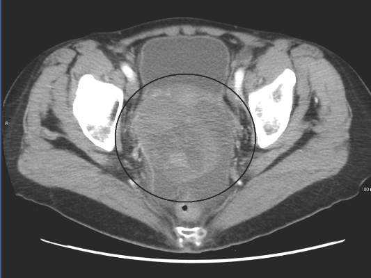
Women who undergo a uterus-preserving procedure to manage uterine fibroids often have to have further procedures, according to Dr. Elisa Martin-Merino of the Spanish Centre for Pharmacoepidemiologic Research in Madrid and her associates.
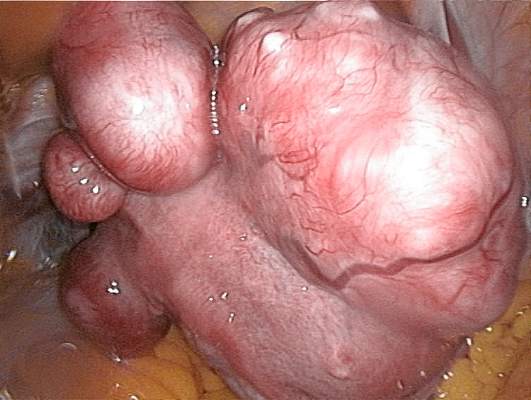
After 1 year, 23.6% of women aged 15-54 with uterine fibroids had a hysterectomy or uterus-preserving procedure (UPP), with the rate increasing to 40.9% at the end of the follow-up period, which was a median of 3.6 years. About one-third of all study participants underwent a hysterectomy during the study period. Myomectomy, endometrial ablation, and uterine artery embolization were much less common, performed in 3.9%, 6.4%, and 1.9% of study participants respectively.
The incidence of a repeat UPP after being initially treated with a UPP was 11.5% at 1 year, and 26.1% at the end of the study period. The cumulative incidences of women undergoing hysterectomy, myomectomy, endometrial ablation, and uterine artery embolization were 19.0%, 4.3%, 3.4%, and 1.4%, respectively.
“Women considering UPPs for the management of [uterine fibroids] should be made aware that the incidence of further treatments is high, with hysterectomy being the most frequent procedure undergone,” the investigators wrote.
Find the full study in the European Journal of Obstetrics & Gynecology and Reproductive Biology (doi: 10.1016/j.ejogrb.2015.08.034).
Women who undergo a uterus-preserving procedure to manage uterine fibroids often have to have further procedures, according to Dr. Elisa Martin-Merino of the Spanish Centre for Pharmacoepidemiologic Research in Madrid and her associates.
After 1 year, 23.6% of women aged 15-54 with uterine fibroids had a hysterectomy or uterus-preserving procedure (UPP), with the rate increasing to 40.9% at the end of the follow-up period, which was a median of 3.6 years. About one-third of all study participants underwent a hysterectomy during the study period. Myomectomy, endometrial ablation, and uterine artery embolization were much less common, performed in 3.9%, 6.4%, and 1.9% of study participants respectively.
The incidence of a repeat UPP after being initially treated with a UPP was 11.5% at 1 year, and 26.1% at the end of the study period. The cumulative incidences of women undergoing hysterectomy, myomectomy, endometrial ablation, and uterine artery embolization were 19.0%, 4.3%, 3.4%, and 1.4%, respectively.
“Women considering UPPs for the management of [uterine fibroids] should be made aware that the incidence of further treatments is high, with hysterectomy being the most frequent procedure undergone,” the investigators wrote.
Find the full study in the European Journal of Obstetrics & Gynecology and Reproductive Biology (doi: 10.1016/j.ejogrb.2015.08.034).

Sexual well-being impaired in women with vulvar disease
Women with vulvar disease experience impaired sexual well-being compared with their healthy counterparts, report Dr. Sophie Wylomanski and coauthors from the department of gynecology and obstetrics at Nantes (France) University Hospital.
In a study of 72 women with vulvar disease and 72 controls who completed the Female Sexual Function Index (FSFI) assessment, the median FSFI score was 21.1 in the vulvar disease patients, compared with 28.1 in the control group. The sexual function scores were significantly lower in the vulvar disease group – by an average of 4.5 points – when multivariate analysis was performed (P = .003), Dr. Wylomanski and associates said.
FSFI subscores showed that vulvar disease had a significant effect on arousal, pain, lubrication, satisfaction, and desire. Scores “seemed lower” for premalignant vulvar disease when compared with inflammatory, infectious, and other disease types, the investigators reported.
Read the full article in the European Journal of Obstetrics & Gynecology and Reproductive Biology (doi: 10.1016/j.ejogrb.2015.08.011).
Vulvar lichen sclerosus often overlooked in women of reproductive age
NEW YORK – A delayed diagnosis of vulvar lichen sclerosus is common, and the risk of this delay is permanent scarring and structural genital changes, cautioned an expert who holds teaching appointments in both dermatology and obstetrics/gynecology.
Vulvar lichen sclerosus (VLS) should be considered in females of any age complaining of vulvar itching, including sexually active women for whom other diseases may be more likely, emphasized Dr. Bethanee J. Schlosser, director of the Vulvar Mucosal Specialty Clinic at Northwestern University, Chicago. She spoke at the American Academy of Dermatology summer meeting in a session jointly sponsored by the European Academy of Dermatology and Venereology.

“We need to recognize that when women in their 20s and 30s complain of vulvar pruritus, it is not always vulvovaginal candidiasis,” Dr. Schlosser emphasized. She said some clinicians do not even consider VLS in this age group, because they have been mistakenly informed that this disease has a bimodal distribution that restricts most cases to preadolescent girls and postmenopausal women. According to Dr. Schlosser, up to 40% of cases occur in women of reproductive age.
VLS is a variation on lichen sclerosus, which is a chronic inflammatory condition associated with epithelial thinning that can occur anywhere on the body. The disease is progressive. It is not an erosive process initially, but Dr. Schlosser presented several cases that demonstrated secondary erosions and fissures can eventually result in permanent structural damage to the anatomy.
The etiology remains incompletely understood, but Dr. Schlosser said that VLS is now considered an autoimmune condition that is commonly associated with other autoimmune diseases, particularly thyroiditis. Dr. Schlosser advised that screening VLS patients for additional autoimmune disorders is appropriate.
The key issue, however, is making the diagnosis in the first place. The waxy plaques and epidermal wrinkling that characterize this disease may not be immediately distinguishable from other dermatologic lesions, particularly as the severity varies. Pruritus is the most common symptom, but up to 30% of women are asymptomatic, according to Dr. Schlosser. Histologic evidence of hyperkeratosis on biopsy in the context of characteristic clinical signs confirms the diagnosis.
Whether in girls, women of reproductive age, or postmenopausal women, the substantial gap between the median age of onset and the median age of diagnosis is a source of concern. In peri- and postmenopausal women, Dr. Schlosser cited data suggesting that the mean delay to a diagnosis can be 5 or more years. According to Dr. Schlosser, one source of delay may be the well-known reluctance of many patients to disclose genital symptoms, but she also maintained that VLS, which has an incidence of 0.1%-0.2%, is not often considered in the initial evaluation of vulvar dermatologic complaints.
The standard first-line therapy for VLS is highly potent corticosteroids. In contrast, topical testosterone, once widely used when VLS was thought to be a product of hormonal imbalance, “has no role in this disease,” according to Dr. Schlosser. Second-line treatments for those who need an alternative to steroids include topical calcineurin inhibitors, such as tacrolimus or pimecrolimus, the synthetic vitamin D cream calcipotriene, and topical retinoids. Oral steroids can be used in difficult cases, but Dr. Schlosser said this is uncommon, estimating that she may have placed only 4 of the 150 VLS cases she has accumulated in her clinic on a systemic therapy.
Generally, topical therapies, which must be maintained indefinitely, suppress symptoms and slow or halt the progressive disease process, according to Dr. Schlosser, who cited a large study published more than 10 years ago. In this study, 66% of 255 VLS patients followed for a median of 66 months on topical steroids became symptom free (Arch Dermatol. 2004;140:702-6). All but 4% improved. Normal skin texture was achieved in 23%, and 68% showed partial improvement. Scarring was less common in children than adults. Squamous cell carcinoma occurred in 2.4%.
The small but clinically significant risk of squamous cell carcinoma has been documented previously, but Dr. Schlosser said there is new evidence that topical therapy may reduce the risk. In a study of 507 women with a median follow-up of 4.7 years, the risk of squamous cell carcinoma was inversely related to compliance with therapy.
“Among the women who used the assigned therapy most or all of the time, zero developed squamous cell carcinoma as opposed to seven cases in the partially compliant therapy group,” she reported. Although this does not prove that treatment reduces risk of cancer, Dr. Schlosser indicated that it is strongly suggestive.
Several questions about VLS remain unanswered, according to Dr. Schlosser. More data, for example, are needed to determine whether the current order of therapies for first-line treatment as well as maintenance regimens is optimal, particularly for reducing cancer risk. However, Dr. Schlosser emphasized that the most important challenge to better outcomes is early recognition of this entity.
Dr. Schlosser reported financial disclosures with Allergan and Galderma Laboratories.
NEW YORK – A delayed diagnosis of vulvar lichen sclerosus is common, and the risk of this delay is permanent scarring and structural genital changes, cautioned an expert who holds teaching appointments in both dermatology and obstetrics/gynecology.
Vulvar lichen sclerosus (VLS) should be considered in females of any age complaining of vulvar itching, including sexually active women for whom other diseases may be more likely, emphasized Dr. Bethanee J. Schlosser, director of the Vulvar Mucosal Specialty Clinic at Northwestern University, Chicago. She spoke at the American Academy of Dermatology summer meeting in a session jointly sponsored by the European Academy of Dermatology and Venereology.
“We need to recognize that when women in their 20s and 30s complain of vulvar pruritus, it is not always vulvovaginal candidiasis,” Dr. Schlosser emphasized. She said some clinicians do not even consider VLS in this age group, because they have been mistakenly informed that this disease has a bimodal distribution that restricts most cases to preadolescent girls and postmenopausal women. According to Dr. Schlosser, up to 40% of cases occur in women of reproductive age.
VLS is a variation on lichen sclerosus, which is a chronic inflammatory condition associated with epithelial thinning that can occur anywhere on the body. The disease is progressive. It is not an erosive process initially, but Dr. Schlosser presented several cases that demonstrated secondary erosions and fissures can eventually result in permanent structural damage to the anatomy.
The etiology remains incompletely understood, but Dr. Schlosser said that VLS is now considered an autoimmune condition that is commonly associated with other autoimmune diseases, particularly thyroiditis. Dr. Schlosser advised that screening VLS patients for additional autoimmune disorders is appropriate.
The key issue, however, is making the diagnosis in the first place. The waxy plaques and epidermal wrinkling that characterize this disease may not be immediately distinguishable from other dermatologic lesions, particularly as the severity varies. Pruritus is the most common symptom, but up to 30% of women are asymptomatic, according to Dr. Schlosser. Histologic evidence of hyperkeratosis on biopsy in the context of characteristic clinical signs confirms the diagnosis.
Whether in girls, women of reproductive age, or postmenopausal women, the substantial gap between the median age of onset and the median age of diagnosis is a source of concern. In peri- and postmenopausal women, Dr. Schlosser cited data suggesting that the mean delay to a diagnosis can be 5 or more years. According to Dr. Schlosser, one source of delay may be the well-known reluctance of many patients to disclose genital symptoms, but she also maintained that VLS, which has an incidence of 0.1%-0.2%, is not often considered in the initial evaluation of vulvar dermatologic complaints.
The standard first-line therapy for VLS is highly potent corticosteroids. In contrast, topical testosterone, once widely used when VLS was thought to be a product of hormonal imbalance, “has no role in this disease,” according to Dr. Schlosser. Second-line treatments for those who need an alternative to steroids include topical calcineurin inhibitors, such as tacrolimus or pimecrolimus, the synthetic vitamin D cream calcipotriene, and topical retinoids. Oral steroids can be used in difficult cases, but Dr. Schlosser said this is uncommon, estimating that she may have placed only 4 of the 150 VLS cases she has accumulated in her clinic on a systemic therapy.
Generally, topical therapies, which must be maintained indefinitely, suppress symptoms and slow or halt the progressive disease process, according to Dr. Schlosser, who cited a large study published more than 10 years ago. In this study, 66% of 255 VLS patients followed for a median of 66 months on topical steroids became symptom free (Arch Dermatol. 2004;140:702-6). All but 4% improved. Normal skin texture was achieved in 23%, and 68% showed partial improvement. Scarring was less common in children than adults. Squamous cell carcinoma occurred in 2.4%.
The small but clinically significant risk of squamous cell carcinoma has been documented previously, but Dr. Schlosser said there is new evidence that topical therapy may reduce the risk. In a study of 507 women with a median follow-up of 4.7 years, the risk of squamous cell carcinoma was inversely related to compliance with therapy.
“Among the women who used the assigned therapy most or all of the time, zero developed squamous cell carcinoma as opposed to seven cases in the partially compliant therapy group,” she reported. Although this does not prove that treatment reduces risk of cancer, Dr. Schlosser indicated that it is strongly suggestive.
Several questions about VLS remain unanswered, according to Dr. Schlosser. More data, for example, are needed to determine whether the current order of therapies for first-line treatment as well as maintenance regimens is optimal, particularly for reducing cancer risk. However, Dr. Schlosser emphasized that the most important challenge to better outcomes is early recognition of this entity.
Dr. Schlosser reported financial disclosures with Allergan and Galderma Laboratories.
EXPERT ANALYSIS FROM AAD SUMMER ACADEMY 2015

Surgeons tout Essure fix without hysterectomy
On Sept. 24, the Food and Drug Administration’s Obstetrics and Gynecology Devices Panel will meet in Silver Spring, Md., to consider the future of the Essure permanent contraceptive device.
Essure consists of an inner stainless steel coil wrapped in polyethylene terephthalate fibers and encased in an outer nickel-titanium coil. It’s delivered hysteroscopically for partial placement into the fallopian tube.
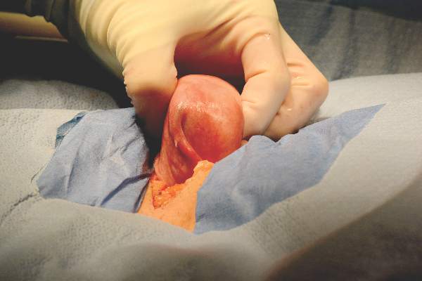
Once in place, the fibers trigger scar formation to block the tube.
When approved in 2002, “it finally looked like we had the answer that we were looking for,” an effective, quick, and inexpensive sterilization procedure that could be done in-office without anesthesia, said Dr. James Presthus, an early adopter at Minnesota Gynecology & Surgery in Edina, Minn., and a former Essure adviser.
But through May 2015, the FDA has received 5,093 reports of sometimes-severe problems with Essure, including chronic pelvic pain, dysmenorrhea, constitutional symptoms, and a handful of deaths that may, or may not, be associated with the device.

The reports and the upcoming FDA meeting are being driven by women who say they have been hurt by Essure. They’ve organized online to share their stories; the Facebook page Essure Problems is probably their largest meeting space, with almost 20,000 members.
Patients on the page have been encouraging each other to report problems to the FDA, and some are involved in legal action against manufacturer Bayer HealthCare.
Complaints ignored
There’s anger behind those efforts, and part of it is due to how women say they have been treated by their physicians. Social media is full of stories from patients who say their concerns were dismissed by physicians who told them that Essure couldn’t be the cause of their problems, or that hysterectomy was the only way to get the coils out.
Both of those assertions are false, said Dr. Edio Zampaglione, vice president of Bayer’s Women’s Health Division and an ob.gyn.

According to company estimates, Essure causes chronic pain in about 4% of women, especially if it’s not placed correctly or migrates and punctures surrounding tissue. Constitutional symptoms – for example, rashes, joint pain, hair loss, and weight changes – could be related to the nickel in Essure’s outer coil, if patients are allergic.
“We do believe these women” and take their reports seriously, Dr. Zampaglione said.
Bayer is reaching out to them for more information, and beefing up the physician-training programs it inherited when it bought Essure’s original maker, Conceptus, in 2013, he said.
Current labeling recommends salpingotomy or salpingectomy as first-line options for removal, and after a request from the FDA, Bayer is updating Essure’s label to give more information on how to use those procedures to remove the device.
Doctors who recommend hysterectomy first are probably just staying in their comfort zone, Dr, Zampaglione said.
It’s difficult to pinpoint the exact adverse event rate for Essure, but Bayer asserts – and physicians with no connections to the company said – that complications are rare.
The cases reported to the FDA represent less than 1% of the 750,000 women Bayer estimates have received the device, most of them in the U.S. Even so, adverse event reporting to the agency is notoriously low for drugs and devices, 10% or less by most estimates.
“I’ve implanted hundreds of these over the years. A lot of those patients come back to me for annual examinations, and they are not reporting problems,” Dr. Presthus said, but “after almost 13 years, we are finding out that Essure is not perfect; nothing is.”

It’s also difficult to separate out coincidence from true problems with the device. Outside of uterine anomalies, nickel allergy seems to be the only definitive indicator that patients will have problems with Essure. Until more is known, timing is the key to figuring out if Essure needs to come out, Dr. Presthus said.
“If somebody has it put in and then has all kinds of pelvic pain, it’s pretty easy to make the connection that it could be the device. Certainly, when patients have allergic reactions that started after they had it implanted,” Essure needs to be addressed, he said.
Removal without hysterectomy
His first step in such cases is to remove the device to see if it helps. Over the past year, Dr. Presthus has done two or more cases a week laparoscopically and said he hasn’t run into problems. The cases take about a half-hour each, and 80% or more of women report symptom relief.
“I do a linear salpingotomy over the proximal portion of the fallopian tube where the device is, and use fine-tip forceps to tease the tissue apart and identify the device. You tease it out, regrasp it, tease it out, and regrasp it. It’s really important to pull in a straight line” so Essure doesn’t break. “It’s not necessary to cut deep into the cornea of the uterus. I get an x-ray on the table to ensure there’s no metal left,” Dr. Presthus said.
Women have to know that there are alternatives before they opt for Essure, and that it can be taken out if needed. “They don’t have to [just] live with their problems or have a hysterectomy. The discussion you have before the procedure is important,” he said.
Dr. Charles Monteith in Raleigh, N.C., and Dr. Mark Sanchez in Clearwater, Fla., prefer an open approach, operating through a small incision above the bikini line.

Both are long-time experts in reversing tubal ligations, and are in high demand lately to take out Essure, and sometimes even restore fertility. Fertility can be restored in about a third of patients, they said.
“If the device is removed within 3 months, it’ll come out fairly easily with traction,” Dr. Monteith said. After that, scarring locks it into place, “so we start dissection over the device and follow it down into the uterine cavity, and remove it intact” – without traction – “with the surrounding tubal muscularis. That’s worked well for us,” he said.
Dr. Monteith said he has removed or reversed Essure in about 200 women.
“Avoid electrocautery because it causes the nickel in the outer coil to burn, spark, and fracture. Also, have an appreciation for variable insertions of the device. We’ve seen the coils pushed too deeply” into the fallopian tube, and “we’ve seen them barely going in. You have to palpate them to know where they are” before taking them out, so as not to cut into them by accident, he said.
Dr. Sanchez, also a high-volume Essure surgeon, cuts down into the corneal segment of the uterus and slowly dissects out the coil. “You have to pay close attention to make sure you get all the bits of the coil out as much as possible.” From there, restoring fertility usually means reanastomosing the fallopian tubes and corneal reimplantation, he said.
Other surgeons have developed similar techniques (Contraception. 2013 Sep;88[3]:334-6.).
Dr. Presthus, Dr. Monteith, and Dr. Sanchez reported having no current connections to Bayer, and are not involved in legal action against the company.
On Sept. 24, the Food and Drug Administration’s Obstetrics and Gynecology Devices Panel will meet in Silver Spring, Md., to consider the future of the Essure permanent contraceptive device.
Essure consists of an inner stainless steel coil wrapped in polyethylene terephthalate fibers and encased in an outer nickel-titanium coil. It’s delivered hysteroscopically for partial placement into the fallopian tube.
Once in place, the fibers trigger scar formation to block the tube.
When approved in 2002, “it finally looked like we had the answer that we were looking for,” an effective, quick, and inexpensive sterilization procedure that could be done in-office without anesthesia, said Dr. James Presthus, an early adopter at Minnesota Gynecology & Surgery in Edina, Minn., and a former Essure adviser.
But through May 2015, the FDA has received 5,093 reports of sometimes-severe problems with Essure, including chronic pelvic pain, dysmenorrhea, constitutional symptoms, and a handful of deaths that may, or may not, be associated with the device.
The reports and the upcoming FDA meeting are being driven by women who say they have been hurt by Essure. They’ve organized online to share their stories; the Facebook page Essure Problems is probably their largest meeting space, with almost 20,000 members.
Patients on the page have been encouraging each other to report problems to the FDA, and some are involved in legal action against manufacturer Bayer HealthCare.
Complaints ignored
There’s anger behind those efforts, and part of it is due to how women say they have been treated by their physicians. Social media is full of stories from patients who say their concerns were dismissed by physicians who told them that Essure couldn’t be the cause of their problems, or that hysterectomy was the only way to get the coils out.
Both of those assertions are false, said Dr. Edio Zampaglione, vice president of Bayer’s Women’s Health Division and an ob.gyn.
According to company estimates, Essure causes chronic pain in about 4% of women, especially if it’s not placed correctly or migrates and punctures surrounding tissue. Constitutional symptoms – for example, rashes, joint pain, hair loss, and weight changes – could be related to the nickel in Essure’s outer coil, if patients are allergic.
“We do believe these women” and take their reports seriously, Dr. Zampaglione said.
Bayer is reaching out to them for more information, and beefing up the physician-training programs it inherited when it bought Essure’s original maker, Conceptus, in 2013, he said.
Current labeling recommends salpingotomy or salpingectomy as first-line options for removal, and after a request from the FDA, Bayer is updating Essure’s label to give more information on how to use those procedures to remove the device.
Doctors who recommend hysterectomy first are probably just staying in their comfort zone, Dr, Zampaglione said.
It’s difficult to pinpoint the exact adverse event rate for Essure, but Bayer asserts – and physicians with no connections to the company said – that complications are rare.
The cases reported to the FDA represent less than 1% of the 750,000 women Bayer estimates have received the device, most of them in the U.S. Even so, adverse event reporting to the agency is notoriously low for drugs and devices, 10% or less by most estimates.
“I’ve implanted hundreds of these over the years. A lot of those patients come back to me for annual examinations, and they are not reporting problems,” Dr. Presthus said, but “after almost 13 years, we are finding out that Essure is not perfect; nothing is.”
It’s also difficult to separate out coincidence from true problems with the device. Outside of uterine anomalies, nickel allergy seems to be the only definitive indicator that patients will have problems with Essure. Until more is known, timing is the key to figuring out if Essure needs to come out, Dr. Presthus said.
“If somebody has it put in and then has all kinds of pelvic pain, it’s pretty easy to make the connection that it could be the device. Certainly, when patients have allergic reactions that started after they had it implanted,” Essure needs to be addressed, he said.
Removal without hysterectomy
His first step in such cases is to remove the device to see if it helps. Over the past year, Dr. Presthus has done two or more cases a week laparoscopically and said he hasn’t run into problems. The cases take about a half-hour each, and 80% or more of women report symptom relief.
“I do a linear salpingotomy over the proximal portion of the fallopian tube where the device is, and use fine-tip forceps to tease the tissue apart and identify the device. You tease it out, regrasp it, tease it out, and regrasp it. It’s really important to pull in a straight line” so Essure doesn’t break. “It’s not necessary to cut deep into the cornea of the uterus. I get an x-ray on the table to ensure there’s no metal left,” Dr. Presthus said.
Women have to know that there are alternatives before they opt for Essure, and that it can be taken out if needed. “They don’t have to [just] live with their problems or have a hysterectomy. The discussion you have before the procedure is important,” he said.
Dr. Charles Monteith in Raleigh, N.C., and Dr. Mark Sanchez in Clearwater, Fla., prefer an open approach, operating through a small incision above the bikini line.
Both are long-time experts in reversing tubal ligations, and are in high demand lately to take out Essure, and sometimes even restore fertility. Fertility can be restored in about a third of patients, they said.
“If the device is removed within 3 months, it’ll come out fairly easily with traction,” Dr. Monteith said. After that, scarring locks it into place, “so we start dissection over the device and follow it down into the uterine cavity, and remove it intact” – without traction – “with the surrounding tubal muscularis. That’s worked well for us,” he said.
Dr. Monteith said he has removed or reversed Essure in about 200 women.
“Avoid electrocautery because it causes the nickel in the outer coil to burn, spark, and fracture. Also, have an appreciation for variable insertions of the device. We’ve seen the coils pushed too deeply” into the fallopian tube, and “we’ve seen them barely going in. You have to palpate them to know where they are” before taking them out, so as not to cut into them by accident, he said.
Dr. Sanchez, also a high-volume Essure surgeon, cuts down into the corneal segment of the uterus and slowly dissects out the coil. “You have to pay close attention to make sure you get all the bits of the coil out as much as possible.” From there, restoring fertility usually means reanastomosing the fallopian tubes and corneal reimplantation, he said.
Other surgeons have developed similar techniques (Contraception. 2013 Sep;88[3]:334-6.).
Dr. Presthus, Dr. Monteith, and Dr. Sanchez reported having no current connections to Bayer, and are not involved in legal action against the company.
