User login
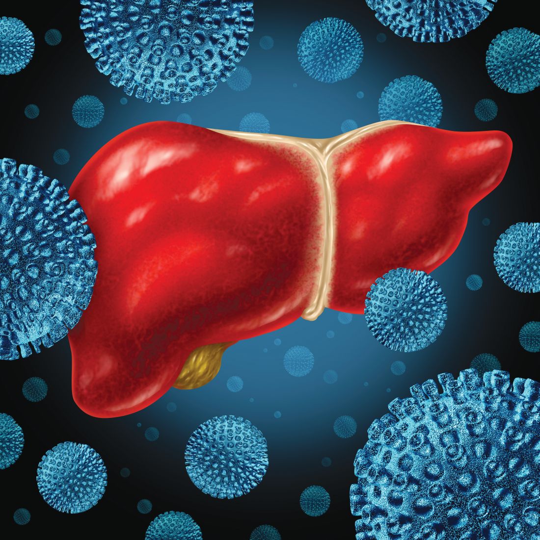
New antibody cuts the fat in NAFLD
BOSTON – A new bispecific antibody, BFKB8488A, may be able to reduce hepatic fat fraction and improve liver health in patients with nonalcoholic fatty liver disease (NAFLD), according to investigators.
In a phase 1b trial, treatment with tolerable doses of the antibody reduced hepatic fat fraction by a mean of 38%, reported lead author Rebecca Kunder, MD, PhD, medical director at Genentech in San Francisco, and colleagues.
According to the investigators, BFKB8488A, which is also being tested in patients with type 2 diabetes mellitus, binds two adipocyte proteins: fibroblast growth factor receptor type 1c and Klotho beta, thereby mimicking metabolic hormone FGF21.
The present trial involved 63 patients with NAFLD who had at least 10% hepatic fat fraction based on MRI. Patients were randomized and for 12 weeks received placebo, one of four doses ranging from 50 to 130 mg given every 2 weeks, a dose of 250 mg given every 4 weeks, or an escalating dose regimen (the results of which were not reported). Treatments were blinded and delivered subcutaneously.
BFKB8488A was generally safe; the trial finished without life-threatening adverse events or deaths. Still, gastrointestinal issues became more common with higher doses, leading the investigators to identify well-tolerated doses as those of 100 mg or less, given every 2 weeks.
The investigators reported efficacy results for patients who received these lower doses, with outcomes presented as mean percentage changes in biomarkers from baseline to 12 weeks.
Adipose-specific pharmacodynamic effect was demonstrated by a mean increase in adiponectin of up to 17%. Positive cardiometabolic effects were also reported, with HDL cholesterol increasing 14% and triglyceride decreasing 24%. In addition, several other markers of liver health improved. Patients with baseline elevations of ALT had decreases in this marker of 10%-30%; plasma type 3 collagen propeptide, which is a measure of fibrosis, fell by 37%; and hepatic fat fraction, as previously stated, decreased by 38%, with a standard deviation of 25%. In contrast, treatment with placebo was associated with a mean 0% change in fat fraction, with a standard deviation of 28%.
“In patients with NAFLD, well-tolerated doses of BFKB8488A were highly effective at decreasing hepatic fat fraction and improving liver health,” the investigators concluded in an abstract that will be presented at the annual meeting of the American Association for the Study of Liver Diseases.
According to the investigators, clinical efficacy of the antibody will be assessed in a phase 2 trial involving patients with nonalcoholic steatohepatitis.
The investigators reported financial relationships with Genentech and Gilead.
Share AGA’s patient education content on NAFLD to help your patients understand the condition. Visit https://www.gastro.org/practice-guidance/gi-patient-center/topic/nonalcoholic-steatohepatitis-nash to learn more.
SOURCE: Kunder R et al. The Liver Meeting 2019, Abstract LP8.
REPORTING FROM THE LIVER MEETING 2019
Key clinical point: The bispecific antibody BFKB8488A may be able to reduce hepatic fat fraction and improve liver health in patients with nonalcoholic fatty liver disease.
Major finding: Among patients given well-tolerated doses, treatment with BFKB8488A reduced hepatic fat fraction by a mean of 38%, compared with 0% for placebo.
Study details: A blinded, randomized, placebo-controlled, phase 1b trial involving 62 patients with nonalcoholic fatty liver disease.
Disclosures: The investigators reported financial relationships with Genentech and Gilead.
Source: Kunder R et al. The Liver Meeting 2019, Abstract LP8.
HDV combo therapy reduces viral loads
BOSTON – For most patients with chronic hepatitis D virus (HDV) infection, combination therapy with lonafarnib, ritonavir, and peginterferon may significantly decrease viral loads, based on interim results from the phase IIa LIFT trial.
After 6 months of therapy, more than one-third of evaluable patients (37%) achieved undetectable levels of HDV RNA in serum, according to lead author Christopher Koh, MD, of the National Institute of Diabetes, Digestive and Kidney Diseases at the National Institutes of Health and colleagues.
The open-label LIFT trial, which is ongoing, initially recruited 26 patients with HDV RNA who had serum levels of at least 40 IU/mL (lower limit of quantification). After starting tenofovir or entecavir, patients began a combination regimen of twice-daily oral lonafarnib (50 mg) and ritonavir (100 mg) plus weekly subcutaneous injections of Peginterferon Lambda-1a (180 mcg).
The median patient age was 40 years, with a slightly higher proportion of male participants (60%). Approximately half of the patients were of Asian descent (52%), followed by patients who were white (32%), or African (16%). The investigators reported median baseline measurements of modified histology activity index (9) and Ishak fibrosis stage (3), as well as serum levels of alanine aminotransferase (64 IU/mL), aspartate aminotransferase (47 IU/mL), hepatitis B virus DNA (less than 21 IU/mL), and log HDV RNA (4.74 IU/mL), with this latter measurement serving as a key determinant of efficacy.
After 12 weeks of therapy, the median decrease in HDV RNA among 21 evaluable patients was 3.6 log IU/mL with an interquartile range from 2.6 to 4.2 (P less than .0001). Of these patients, 5 (24%) achieved undetectable levels of HDV RNA, while another 5 tested below the lower limit of quantification.
Following an additional 12 weeks of therapy, 19 patients remained evaluable, among whom the median decrease in HDV RNA was 3.4 log IU/mL with an interquartile range from 2.9 to 4.5 (P less than .0001). Seven of these patients (37%) achieved undetectable HDV RNA, whereas 3 others fell below the lower limit of quantification. Furthermore, 18 out of 19 of these patients (95%) experienced a decline in HDV RNA of more than 2 log IU/mL.
According to the investigators, the trial regimen was safe and well tolerated. Adverse events were mild to moderate; most common were anemia, hyperbilirubinemia, weight loss, and gastrointestinal issues. Doses were reduced in three patients while four others discontinued therapy prematurely.
“These interim results support continued exploration of this therapeutic combination in HDV,” the investigators concluded.
The above findings will be presented in an oral abstract session at the annual meeting of the American Association for the Study of Liver Diseases.
The investigators disclosed relationships with I-Cubed Therapeutics, Eiger BioPharmaceuticals, Riboscience, and others.
SOURCE: Koh C et al. The Liver Meeting 2019. Abstract LO8.
REPORTING FROM THE LIVER MEETING 2019
Key clinical point: For most patients with chronic hepatitis D virus infection, combination therapy with lonafarnib, ritonavir, and peginterferon may significantly decrease viral loads.
Major finding: After 6 months of therapy, 37% of evaluable patients achieved undetectable levels of hepatitis D virus RNA.
Study details: The phase IIa open-label LIFT trial involving 26 patients with chronic hepatitis delta virus (HDV).
Disclosures: The investigators disclosed relationships with I-Cubed Therapeutics, Eiger BioPharmaceuticals, Riboscience, and others.
Source: Koh C et al. The Liver Meeting 2019. Abstract LO8.
DUR-928 shows promise for alcoholic hepatitis
BOSTON – Treatment with novel agent DUR-928 may be able to reduce mortality rates among patients with severe alcoholic hepatitis, investigators predicted.
In an open-label, phase IIa trial, 89% of patients with alcoholic hepatitis responded to treatment with the new therapy, reported lead author Tarek Hassanein, MD, of Southern California Research Center in Coronado, Calif., and colleagues.
In an abstract that will be presented at the annual meeting of the American Association for the Study of Liver Diseases, the investigators explained the urgent need for an agent such as DUR-928: “The mortality of severe alcoholic hepatitis remains high in the absence of effective treatment,” they wrote, noting that corticosteroids are only suitable for select patients. According to the investigators, DUR-928 is an endogenous sulfated oxysterol that has been shown to control lipotoxicity and inflammation while increasing hepatic regeneration and cell survival.
The agent was tested among 19 patients with alcoholic hepatitis, many of whom had severe disease; at baseline, 15 had Maddrey’s discriminant function (DF) scores of 32 or less, 12 had Model for End-stage Liver Disease (MELD) scores between 12 and 30, and 11 had serum bilirubin levels higher than 8 mg/dL.
Via intravenous infusion, three dose levels were given: 30 mg, 90 mg, or 150 mg. All patients received at least one dose on day 1, and if still hospitalized, a second dose on day 4, with a total follow-up of 28 days. Treatment response was defined by a Lille score of less than 0.45.
DUR-928 was well tolerated; no serious drug-related adverse events occurred and all patients survived the 28-day follow-up period. Across the population, the response rate was 89%. This figure fell slightly to 87% when considering only patients with severe disease (DF scores of 32 or less), and marginally further still to 83% for those with MELD scores between 21 and 30. Among patients with severe disease, MELD scores decreased by a median of 17.5% (P = .01) over the 28-day period, and in cases with bilirubin starting higher than 8 mg/dL, levels dropped by a median of 25.1% (P = .02) within the first week.
A comparison of these results with historical data revealed that treatment with DUR-928 led to significantly better Lille scores than previously reported (P less than .0001).
“These initial findings are encouraging for further development of DUR-928 in patients with alcoholic hepatitis, including severe alcoholic hepatitis,” the investigators concluded.
The investigators disclosed relationships with DURECT Corporation, Assembly Biosciences, Gilead, and others.
SOURCE: Hassanein T et al. The Liver Meeting 2019, Abstract LO9.
REPORTING FROM THE LIVER MEETING 2019
Key clinical point: For patients with alcoholic hepatitis, treatment with novel agent DUR-928 could offer better outcomes than those of existing therapies.
Major finding: Among 15 patients with severe alcoholic hepatitis, 87% responded to treatment (Lille score less than 0.45).
Study details: A phase IIa open-label trial involving 19 patients with alcoholic hepatitis.
Disclosures: The investigators disclosed relationships with DURECT Corporation, Assembly Biosciences, Gilead, and others.
Source: Hassanein T et al. The Liver Meeting 2019, Abstract LO9.
Glecaprevir/pibrentasvir highly effective in HCV genotype 3, among others
BOSTON – Specifically in patients with hepatitis C virus (HCV) genotype 3 infection and compensated cirrhosis, glecaprevir/pibrentasvir (Mavyret) was safe and had high efficacy in a phase 3 clinical trial, echoing an earlier report describing clinical results for the fixed-dose combination in multiple other genotypes.
Treatment with glecaprevir/pibrentasvir was safe and produced high rates of sustained virologic response 12 weeks after treatment (SVR12) for the genotype 3 patients and compensated cirrhosis in the recent results from the EXPEDITION-8 study, which will be presented at the annual meeting of the American Association for the Study of Liver Diseases.
In a previous report from EXPEDITION-8, investigators said that the treatment was well tolerated and effective in patients with HCV genotypes 1, 2, 4, 5, and 6.
“The availability of an 8-week, pangenotypic regimen for all treatment-naive HCV-infected patients regardless of cirrhosis status may simplify the HCV care pathway, furthering progress towards HCV elimination,” said investigator Robert S. Brown Jr., MD, MPH, and coinvestigators in a late-breaking abstract of the latest study results.
The nonrandomized, multicenter, phase 3b study included 343 adults with HCV genotypes 1-6 who received glecaprevir 300 mg and pibrentasvir 120 mg once daily for 8 weeks. A total of 63% of patients were male, 83% were white; 18% had HCV genotype 3, while the majority (67%) had HCV genotype 1.
For the genotype 3 patients, SVR12 rates were 95.2% in the intention-to-treat population, and 98.4% in the per-protocol population, Dr. Brown and coauthors said in their report on the study. For genotype 1, 2, 4, 5, and 6 patients, the intention-to-treat and per-protocol SVR12 rates were 98.2% and 100%.
Taken together, the SVR12 rates for all genotypes were 97.7% and 99.7%, respectively, for the intention-to-treat and per-protocol populations, according to the investigators.
There were no virologic failures on treatment, and one patients with genotype 3 relapsed in week 4 posttreatment, while one genotype 1 patient discontinued treatment though not because of adverse events, they said.
Most adverse events were grade 1 in severity, and included fatigue, pruritus, headache, and nausea. There were no liver-related toxicities, and no serious adverse events that were related to the study treatment, according to the investigators.
Glecaprevir/pibrentasvir is indicated for patients aged 12 years and older with treatment-naive HCV genotype 1-6 infection without cirrhosis or with compensated cirrhosis, and in patients with HCV genotype 1 infection previously treated with an HCV NS5A inhibitor or an NS3/4A protease inhibitor.
Dr. Brown reported disclosures related to pharmaceutical companies including AbbVie, which markets Mavyret.
SOURCE: Brown RS et al. The Liver Meeting 2019. Abstract LP9.
REPORTING FROM THE LIVER MEETING 2019
Key clinical point: In patients with hepatitis C virus (HCV) genotype 3 infection and compensated cirrhosis, glecaprevir/pibrentasvir was safe and had high efficacy, echoing an earlier result for the fixed-dose combination in genotypes 1, 2, and 4-6.
Major finding: For the genotype 3 patients, the rate of sustained virologic response 12 weeks after treatment (SVR12) was 95.2% in the intention-to-treat population, and 98.4% in the per-protocol population.
Study details: Further results from EXPEDITION-8, a single-arm phase 3b study including 343 adult patients with HCV genotypes 1-6.
Disclosures: Dr. Brown reported disclosures related to AbbVie, Gilead, Intercept, Dova, Shionogi, Merck, and Bristol-Myers Squibb.
Source: Brown RS et al. The Liver Meeting 2019. Abstract LP9.
Hepatitis C vaccine alters viral trajectory, but fails in chronic infection protection
BOSTON – A prime-boost hepatitis C virus (HCV) vaccine regimen did not protect against chronic infection, but it did evoke immune responses and differences in viral trajectory, according to investigators in what is believed to be the first randomized, placebo-controlled efficacy trial in this setting.
There were no apparent safety concerns with the vaccine according to investigators, led by Kimberly Page, PhD, MPH, of the University of New Mexico, Albuquerque.
“A safe and effective vaccine to prevent chronic hepatitis C virus infection is essential to reduce transmission,” Dr. Page and coauthors said in a late-breaking abstract of the study results, which will be presented at the annual meeting of the American Association for the Study of Liver Diseases.
The phase 1/2 trial described by Dr. Page and colleagues included 455 adults at risk of HCV infection because of injection drug use. They were randomized to vaccine, which consisted of a recombinant chimpanzee adenovirus-3 vectored vaccine prime plus a recombinant Modified Vaccinia virus Ankara boost, or to two doses of placebo at days 0 and 56 of the study.
There was no difference in chronic HCV infection at 6 months, the primary endpoint of the study. There were 14 chronically infected participants in the vaccine group, as well as 14 in the placebo group, for an overall incidence of infection of 13.0/100 person-years, Dr. Page and coauthors reported in the abstract.
However, there were significant differences in HCV RNA geometric mean peak at 1 month, which was 193,795 IU/L in the vaccine group and 1,078,092 IU/L in the placebo group, according to investigators. Similarly, geometric mean fold rise after infection was 0.2 in the vaccine group and 13.5 in the placebo group.
A total of 78% of vaccinated individuals had T-cell responses to at least one vaccine antigen pool, investigators said, adding that the vaccine was safe, well tolerated, and not associated with any serious adverse events.
Dr. Page had no disclosures related to the abstract.
SOURCE: Page K et al. The Liver Meeting 2019. Abstract LP17.
REPORTING FROM THE LIVER MEETING 2019
Key clinical point: A prime-boost HCV vaccine altered viral trajectory but did not protect against chronic infection.
Major finding: At 6 months after vaccination, there were 14 chronically infected participants in the vaccine group, and 14 in the placebo group.
Study details: A randomized, placebo controlled phase 1/2 trial including 455 adults at risk of HCV infection.
Disclosures: The first author reported no disclosures.
Source: Page K et al. The Liver Meeting 2019. Abstract LP17.
Fenofibrate fights increased triglycerides in NASH
BOSTON – Fenofibrate is safe and effective for limiting triglyceride increases in patients with nonalcoholic steatohepatitis and advanced fibrosis, based on data from 31 adults.
Treatment of NASH with acetyl-CoA carboxylase inhibitors has been shown to improve liver fat and other liver conditions but may be associated with hyperlipidemia, according to Eric J. Lawitz, MD, of the University of Texas Health, San Antonio, and colleagues. The researchers examined the safety and effectiveness of fenofibrate to mitigate serum triglyceride increases in a study be presented in a late-breaking session at the annual meeting of the American Association for the Study of Liver Diseases.
The researchers randomized 15 patients to treatment with 48 mg of fenofibrate or 145 mg of fenofibrate once daily for 2 weeks, followed by a combination of the fenofibrate doses plus 20 mg of ACC inhibitor firsocostat daily for 24 weeks.
The median fasting triglycerides (TG) in the 48-mg and 145-mg fenofibrate groups were 218 mg/dL and 202 mg/dL, respectively. After 2 weeks, the median change in TG was +2 mg/dL in the 48-mg group and –42 mg/dL in the 145-mg group. After 24 weeks of combination therapy, TG levels were not significantly different from baseline in either group (+19 mg/dL in 48-mg group and +6 mg/dL in the 145-mg group). Significant reductions in serum alanine aminotransferase from baseline to week 24 occurred in the combined groups (median of 39 U/L vs. 27 U/L, respectively). In addition, 43% of patients overall showed at least a 30% reduction in protein density fat fraction.
Both firsocostat and fenofibrate were well tolerated, the researchers said. One treatment-emergent grade 3 TG elevation (defined as greater than 500 mg/dL) occurred in the 48-mg group at week 24. No hepatotoxicity was noted, no patients discontinued therapy because of adverse events, and no other grade 3 or 4 adverse events were reported.
“The combination of firsocostat and fenofibrate led to improvements in hepatic fat, liver biochemistry, and markers of fibrosis,” the researchers concluded in their abstract.
Lead author Dr. Lawitz disclosed financial relationships with Allergan, Akcea Therapeutics, Bristol-Myers Squibb, Boehringer Ingelheim, Gilead Sciences, Madrigal Pharmaceuticals, and Novartis.
The AGA GI Patient Center provides education to help your patients understand NASH at https://www.gastro.org/practice-guidance/gi-patient-center/topic/nonalcoholic-steatohepatitis-nash.
SOURCE: Lawitz E et al. The Liver Meeting 2019. Presentation LP5.
REPORTING FROM THE LIVER MEETING 2019
Key clinical point: Fenofibrate mitigated serum triglyceride increases in patients with NASH.
Major finding: After 24 weeks of fenofibrate treatment, 43% of patients showed a relative reduction of at least 30% in protein density fat fraction with an average of 40% at a 48 mg-dose and 47% at a 145-mg dose.
Study details: The data come from a 24-week study of 31 adults with advanced fibrosis caused by NASH.
Disclosures: Lead author Dr. Lawitz disclosed financial relationships with Allergan, Akcea Therapeutics, Bristol-Myers Squibb, Boehringer Ingelheim, Gilead Sciences, Madrigal Pharmaceuticals, and Novartis.
Source: Lawitz E et al. The Liver Meeting 2019. Presentation LP5.
High doses of FXR agonist tropifexor reduce hepatic fat, serum ALT in patients with NASH
BOSTON – Alanine aminotransferase, hepatic fat, and body weight all decreased in patients with nonalcoholic steatohepatitis (NASH) treated for 12 weeks with higher doses of tropifexor, an investigational farnesoid X receptor agonist, according to interim results of a randomized trial.
Mild pruritus and minor decreases in LDL cholesterol were seen with tropifexor, similar to what has been seen with other farnesoid X receptor agonists, according to investigators, including senior study author Arun Sanyal, MD, professor in the gastroenterology division of the department of internal medicine at Virgina Commonwealth University in Richmond.
“Changes in liver histology resulting from this trial, along with trials of tropifexor in combination with drugs with other mechanisms of action, will define future therapeutic options in fibrotic NASH,” Dr. Sanyal and coauthors said in a late-breaking abstract for the study, which will be presented at the annual meeting of the American Association for the Study of Liver Diseases.
That randomized, double-blind, placebo-controlled, phase 2 study, called FLIGHT-FXR, is designed to evaluate tropifexor (LJN452) in patients with NASH at several different doses, according to Dr. Sanyal and coinvestigators.
Previous reports on the trial demonstrated that lower doses of tropifexor (60 and 90 mcg) had favorable safety with anti-inflammatory and antisteatotic efficacy based on biomarker analysis, according to investigators.
This latest report from FLIGHT-FXR includes 152 patients randomly allocated to receive placebo or tropifexor at one of two higher doses (140 or 200 mcg).
At the highest tropifexor dose of 200 mcg, decreases in alanine aminotransferase (ALT), hepatic fat fraction (HFF), gamma-glutamyl transferase (GGT), and body weight were all significant, compared with the decreases in the placebo arm, according to reported data, while in the 140-mcg arm, the findings were significant versus placebo for GGT and body weight.
Relative HFF reduction by at least 30% was seen in 64% of patients in the tropifexor 200-mcg arm, 32% in the tropifexor 140-mcg arm, and 20% in the placebo arm, the investigators reported.
Pruritus was mild among tropifexor-treated patients in more than 60% of cases, investigators said, and pruritus-related discontinuation rates were low, at 2% (1 patient) for the 140-mcg dose and 6% (3 patients) for 200 mcg.
Investigators noted a dose-related increase in LDL cholesterol with tropifexor treatment, but those lipid changes didn’t lead to reduced doses or stopped treatment, they said.
Serious adverse events were infrequent, with a comparable incidence across treatment and placebo groups, investigators added.
Dr. Sanyal provided disclosures related to Sanyal Bio, Exhalenz, Akarna, Fractyl Laboratories, Genfit, Durect, Tiziana, Novartis, Merck, Galectin, and Janssen, among others.
REPORTING FROM THE LIVER MEETING 2019
Key clinical point: Alanine aminotransferase, hepatic fat fraction, gamma-glutamyl transferase, and body weight decreased in patients with nonalcoholic steatohepatitis (NASH) treated for 12 weeks with higher doses of tropifexor, an investigational farnesoid X receptor agonist.
Major finding: ALT, HFF, GGT, and body weight were all significantly decreased versus placebo in patients treated with tropifexor at 200-mcg dose.
Study details: Interim results from the randomized FLIGHT-FXR trial, including 152 patients with NASH.
Disclosures: Dr. Sanyal provided disclosures related to Sanyal Bio, Exhalenz, Akarna, Fractyl Laboratories, Genfit, Durect, Tiziana, Novartis, Merck, Galectin, and Janssen, among others.
Source: Lucas KJ et al. The Liver Meeting 2019, Presentation LO4.
Duodenal mucosal resurfacing has metabolic effects in type 2 diabetes
BOSTON – An ablative procedure intended to promote regrowth of duodenal mucosa was safe and had disease-modifying metabolic effects in a randomized study including patients with type 2 diabetes, according to investigators.
A single duodenal mucosal resurfacing (DMR) procedure improved glycemic, hepatic, and body-weight measures at 24 weeks in the multicenter study, investigators will report at the annual meeting of the American Association for the Study of Liver Diseases.
The novel and minimally invasive endoscopic procedure treats the duodenum, which is increasingly recognized as a key metabolic signaling center, according to the study authors, including senior author Arun Sanyal, MD, professor in the gastroenterology division of the department of internal medicine at Virginia Commonwealth University, Richmond.
“Duodenal mucosal hyperplasia is a potential therapeutic target for insulin-resistance–related metabolic diseases,” Dr. Sanyal and coauthors said in a late-breaking abstract for the study published in the AASLD meeting proceedings.
In a previous international open-label, prospective, multicenter study, published in July in Gut, DMR was feasible and safe, producing durable glycemic improvement in patients with type 2 diabetes with suboptimal control on oral glucose-lowering mediation, according to investigators.
The present study, conducted at nine sites in the European Union and two in Brazil, is the first sham-controlled, double-blind, prospective study of the modality in patients with suboptimally controlled type 2 diabetes, according to Dr. Sanyal and coauthors.
A total of 39 patients in the study underwent DMR, while 36 underwent a sham procedure, according to the published abstract. The mean hemoglobin A1c for those patients was 8.3, the mean body mass index was 31.1 kg/m2, and most (77%) were male.
Median change in hemoglobin A1c from baseline to 24 weeks, one of two primary endpoints in the study, was –0.6% for DMR and –0.3% for the sham procedure (P less than 0.05), according to the study abstract.
Likewise, the primary efficacy endpoint of change in a nonalcoholic steatohepatitis biomarker favored the DMR arm. The median change in liver MRI–proton density fat fraction (MRI-PDFF) from baseline to 12 weeks was –5.4% for DMR and –2.4% for the sham procedure (P less than 0.05), according to the reported data.
Hypoglycemia rates were similar in the DMR and sham arms, and over 24 weeks of study, there were no unanticipated adverse effects attributable to the device and no serious adverse events, Dr. Sanyal and colleagues reported.
Dr. Sanyal reported disclosures related to Fractyl Laboratories, Sanyal Biotechnology, Exalenz Bioscience, Akarna Therapeutics, Genfit, Durect, Indalo, Tiziana, Novartis, Merck, Galectin Therapeutics, Janssen, and others.
REPORTING FROM THE LIVER MEETING 2019
Key clinical point: Duodenal mucosal resurfacing was safe and had disease-modifying metabolic effects in patients with type 2 diabetes.
Major finding: Results favored duodenal mucosal resurfacing over sham procedure for changes in median HbA1c (–0.6% vs. –0.3%; P less than .05) and liver MRI–proton density fat fraction (–5.4% vs. –2.4%; P less than 0.05).
Study details: A report on 75 patients treated in a randomized, sham-controlled, double-blind, prospective study.
Disclosures: Dr. Sanyal reported disclosures related to Fractyl Laboratories, Sanyal Biotechnology, Exalenz Bioscience, Akarna Therapeutics, Genfit, Durect, Indalo, Tiziana, Novartis, Merck, Galectin Therapeutics, Janssen, and others.
Source: Sanyal A et al. Liver Meeting 2019, Presentation LO2.
Terlipressin reversed hepatorenal syndrome in large prospective study
BOSTON – Terlipressin, an investigational vasopressin analogue, improved renal function and reversed hepatorenal syndrome type 1 (HRS-1) among patients with progressive advanced liver disease in a randomized trial, according to investigators.
Compared with albumin alone, terlipressin plus albumin significantly reversed worsening of renal function in cirrhotic patients, including those meeting systemic inflammatory response syndrome (SIRS) criteria, said investigators, led by Florence Wong, MD, from the University of Toronto.
“This response was durable and associated with less need for early renal replacement therapy,” said Dr. Wong and coauthors of an abstract describing the study, which will be presented in a late-breaking study session here at the annual meeting of the American Association for the Study of Liver Diseases.
The North American randomized, controlled trial, known as CONFIRM, was designed to confirm the safety and efficacy of terlipressin/albumin as a treatment for HRS-1, a serious but potentially reversible type of acute kidney injury seen in patients with cirrhosis and ascites, investigators said in their communication.
Patients in CONFIRM had “well-defined” HRS-1, based on diagnostic criteria as outlined by the International Club of Ascites, investigators said.
A total of 300 participants were randomized, 199 to terlipressin 1 mg IV every 6 hours and 101 to placebo, with both groups also receiving albumin, for up to 14 days of treatment. According to the report, 132 subjects (44%) met SIRS criteria.
Verified HRS reversal was documented in 29.1% of terlipressin-treated patients versus just 15.8% of the placebo group (P less than .012), investigators reported in their abstract. Among the SIRS patients, verified HRS reversal was seen in 33.3% and 6.3% of the terlipressin- and placebo-treated groups, respectively (P less than .001).
As the primary endpoint of the study, verified HRS reversal is an outcome that combines improvement in renal function, short-term survival following improvement, and avoidance of renal replacement therapy, investigators explained in their report.
Liver transplantation occurred in 23.1% of the terlipressin group and 28.7% of the placebo group, investigators also reported in their abstract.
Ischemia-associated adverse events were seen in 4.5% of the terlipressin arm and 0% for placebo, though in general the rate of adverse events were similar for the treatment and control arms, Dr. Wong and colleagues noted in their report.
The CONFIRM study is supported by Mallinckrodt. Dr. Wong provided disclosures related to Mallinckrodt, Ferring, and Sequana.
REPORTING FROM THE LIVER MEETING 2019
Key clinical point: Terlipressin, an investigational vasopressin analogue, improved renal function and reversed hepatorenal syndrome type 1 (HRS-1) among patients with progressive advanced liver disease.
Major finding: Verified HRS reversal was documented in 29.1% of patients treated with terlipressin/albumin versus 15.8% with placebo/albumin (P less than .012).
Study details: CONFIRM, a randomized, controlled trial including 300 patients with HRS-1.
Disclosures: The CONFIRM study is supported by Mallinckrodt. Dr. Wong provided disclosures related to Mallinckrodt, Ferring, and Sequana.
Source: Wong F et al. The Liver Meeting 2019, Presentation LO5.
Blocking TLR9 may halt brain edema in acute liver failure
A toll-like receptor 9 (TLR9) antagonist may eventually be used to combat brain edema in acute liver failure, according to investigators.
This prediction is based on results of a recent study involving mouse models, which showed that ODN2088, a TLR9 antagonist, could stop ammonia-induced colocalization of DNA with TLR9 in innate immune cells, thereby blocking cytokine production and ensuant brain edema, reported lead author Godhev Kumar Manakkat Vijay of King’s College London and colleagues.
“Ammonia plays a pivotal role in the development of hepatic encephalopathy and brain edema in acute liver failure,” the investigators explained in Cellular and Molecular Gastroenterology and Hepatology. “A robust systemic inflammatory response and susceptibility to developing infection are common in acute liver failure, exacerbate the development of ammonia-induced brain edema and are major prognosticators. Experimental models have unequivocally associated ammonia exposure with astrocyte swelling and brain edema, potentiated by proinflammatory cytokines.”
The investigators added that, “although the evidence base supporting the relationship between ammonia, inflammation, and brain edema is robust in acute liver failure, there is a paucity of data characterizing the specific pathogenic mechanisms entailed.” Previous research suggested that TLR9 plays a key role in acetaminophen-induced liver inflammation, they noted, and that ammonia, in combination with DNA, triggers TLR9 expression in neutrophils, which brought TLR9 into focus for the present study.
Along with wild-type mice, the investigators relied upon two knockout models: TLR9–/– mice, in which TLR9 is entirely absent, and LysM-Cre TLR9fl/fl mice, in which TLR9 is absent from lysozyme-expressing cells (predominantly neutrophils and macrophages). Comparing against controls, the investigators assessed cytokine production and brain edema in each type of mouse when intraperitoneally injected with ammonium acetate (4 mmol/kg). Specifically, 6 hours after injection, they measured intracellular cytokines in splenic macrophages, CD8+ T cells, and CD4+ T cells. In addition, they recorded total plasma DNA and brain water, a measure of brain edema.
Following ammonium acetate injection, wild-type mice developed brain edema and liver enlargement, while TLR9–/– mice and control-injected mice did not. After injection, total plasma DNA levels rose by comparable magnitudes in both wild-type mice and TLR9–/– mice, but did not change in control-injected mice, suggesting that ammonium-acetate injection was causing a release of DNA, which was binding with TLR9, resulting in activation of the innate immune system.
This hypothesis was supported by measurements of cytokines in T cells and splenic macrophages, which showed that wild-type mice had elevations of cytokines, whereas knockout mice did not. Further experiments showed that LysM-Cre TLR9fl/fl mice had similar outcomes as TLR9–/– mice, highlighting that macrophages and neutrophils are the key immune cells linking TLR9 activation with cytokine release, and therefore brain edema.
To ensure that brain edema was not directly caused by the acetate component of ammonium acetate, or acetate’s potential to increase pH, a different set of wild-type mice were injected with sodium acetate adjusted to the same pH as ammonium acetate. This had no impact on cytokine production, brain-water content, or liver-to-body weight ratio, confirming that acetate was not responsible for brain edema while providing further support for the role of TLR9.
Finally, the investigators treated wild-type mice immediately after ammonium acetate injection with the TLR9 antagonist ODN2088 (50 mcg/mouse). This treatment halted cytokine production, inflammation, and brain edema, strongly supporting the link between these ammonia-induced processes and TLR9 activation.
“These data are well supported by the findings of Imaeda et al. (J Clin Invest. 2009 Feb 2. doi: 10.1172/JCI35958), who in an acetaminophen-induced hepatotoxicity model established that inhibition of TLR9 using ODN2088 and IRS954, a TLR7/9 antagonist, down-regulated proinflammatory cytokine release and reduced mortality,” the investigators wrote. “The amelioration of brain edema and cytokine production by ODN2088 supports exploration of TLR9 antagonism as a therapeutic modality in early acute liver failure to prevent the development of brain edema and intracranial hypertension.”
The study was funded by the U.K. Institute of Liver Studies Charitable Fund and the National Institutes of Health. The investigators reported no conflicts of interest.
SOURCE: Vijay GKM et al. Cell Mol Gastroenterol Hepatol. 2019 Aug 8. doi: 10.1016/j.jcmgh.2019.08.002.
Acute liver failure is a devastating disease, which has a high mortality burden and often requires liver transplant. One of the major complications is cerebral edema that leads to encephalopathy and could be fatal. These brain changes are accompanied by inflammation, immune activation, and hyperammonemia, but further mechanistic approaches are needed.

This data adds to the growing literature about the interaction between immune dysfunction and brain diseases such as schizophrenia, autism, depression, and multiple sclerosis. However, further studies in models of brain edema with concomitant liver failure, which are closer to the human disease process, are needed. This exciting investigation of neuroimmune regulation of brain edema could set the basis for new therapeutic options for the prevention and treatment of this feared complication of acute liver failure.
Jasmohan S. Bajaj, MD, AGAF, is professor in the division of gastroenterology, hepatology, and nutrition at Virginia Commonwealth University, Richmond. He reported no conflicts of interest.
Acute liver failure is a devastating disease, which has a high mortality burden and often requires liver transplant. One of the major complications is cerebral edema that leads to encephalopathy and could be fatal. These brain changes are accompanied by inflammation, immune activation, and hyperammonemia, but further mechanistic approaches are needed.
This data adds to the growing literature about the interaction between immune dysfunction and brain diseases such as schizophrenia, autism, depression, and multiple sclerosis. However, further studies in models of brain edema with concomitant liver failure, which are closer to the human disease process, are needed. This exciting investigation of neuroimmune regulation of brain edema could set the basis for new therapeutic options for the prevention and treatment of this feared complication of acute liver failure.
Jasmohan S. Bajaj, MD, AGAF, is professor in the division of gastroenterology, hepatology, and nutrition at Virginia Commonwealth University, Richmond. He reported no conflicts of interest.
A toll-like receptor 9 (TLR9) antagonist may eventually be used to combat brain edema in acute liver failure, according to investigators.
This prediction is based on results of a recent study involving mouse models, which showed that ODN2088, a TLR9 antagonist, could stop ammonia-induced colocalization of DNA with TLR9 in innate immune cells, thereby blocking cytokine production and ensuant brain edema, reported lead author Godhev Kumar Manakkat Vijay of King’s College London and colleagues.
“Ammonia plays a pivotal role in the development of hepatic encephalopathy and brain edema in acute liver failure,” the investigators explained in Cellular and Molecular Gastroenterology and Hepatology. “A robust systemic inflammatory response and susceptibility to developing infection are common in acute liver failure, exacerbate the development of ammonia-induced brain edema and are major prognosticators. Experimental models have unequivocally associated ammonia exposure with astrocyte swelling and brain edema, potentiated by proinflammatory cytokines.”
The investigators added that, “although the evidence base supporting the relationship between ammonia, inflammation, and brain edema is robust in acute liver failure, there is a paucity of data characterizing the specific pathogenic mechanisms entailed.” Previous research suggested that TLR9 plays a key role in acetaminophen-induced liver inflammation, they noted, and that ammonia, in combination with DNA, triggers TLR9 expression in neutrophils, which brought TLR9 into focus for the present study.
Along with wild-type mice, the investigators relied upon two knockout models: TLR9–/– mice, in which TLR9 is entirely absent, and LysM-Cre TLR9fl/fl mice, in which TLR9 is absent from lysozyme-expressing cells (predominantly neutrophils and macrophages). Comparing against controls, the investigators assessed cytokine production and brain edema in each type of mouse when intraperitoneally injected with ammonium acetate (4 mmol/kg). Specifically, 6 hours after injection, they measured intracellular cytokines in splenic macrophages, CD8+ T cells, and CD4+ T cells. In addition, they recorded total plasma DNA and brain water, a measure of brain edema.
Following ammonium acetate injection, wild-type mice developed brain edema and liver enlargement, while TLR9–/– mice and control-injected mice did not. After injection, total plasma DNA levels rose by comparable magnitudes in both wild-type mice and TLR9–/– mice, but did not change in control-injected mice, suggesting that ammonium-acetate injection was causing a release of DNA, which was binding with TLR9, resulting in activation of the innate immune system.
This hypothesis was supported by measurements of cytokines in T cells and splenic macrophages, which showed that wild-type mice had elevations of cytokines, whereas knockout mice did not. Further experiments showed that LysM-Cre TLR9fl/fl mice had similar outcomes as TLR9–/– mice, highlighting that macrophages and neutrophils are the key immune cells linking TLR9 activation with cytokine release, and therefore brain edema.
To ensure that brain edema was not directly caused by the acetate component of ammonium acetate, or acetate’s potential to increase pH, a different set of wild-type mice were injected with sodium acetate adjusted to the same pH as ammonium acetate. This had no impact on cytokine production, brain-water content, or liver-to-body weight ratio, confirming that acetate was not responsible for brain edema while providing further support for the role of TLR9.
Finally, the investigators treated wild-type mice immediately after ammonium acetate injection with the TLR9 antagonist ODN2088 (50 mcg/mouse). This treatment halted cytokine production, inflammation, and brain edema, strongly supporting the link between these ammonia-induced processes and TLR9 activation.
“These data are well supported by the findings of Imaeda et al. (J Clin Invest. 2009 Feb 2. doi: 10.1172/JCI35958), who in an acetaminophen-induced hepatotoxicity model established that inhibition of TLR9 using ODN2088 and IRS954, a TLR7/9 antagonist, down-regulated proinflammatory cytokine release and reduced mortality,” the investigators wrote. “The amelioration of brain edema and cytokine production by ODN2088 supports exploration of TLR9 antagonism as a therapeutic modality in early acute liver failure to prevent the development of brain edema and intracranial hypertension.”
The study was funded by the U.K. Institute of Liver Studies Charitable Fund and the National Institutes of Health. The investigators reported no conflicts of interest.
SOURCE: Vijay GKM et al. Cell Mol Gastroenterol Hepatol. 2019 Aug 8. doi: 10.1016/j.jcmgh.2019.08.002.
FROM CELLULAR AND MOLECULAR GASTROENTEROLOGY AND HEPATOLOGY