User login
Bringing respiratory care to asthma clinics in Guyana
How it all started
The study abroad project was truly a goal and vision that came about after returning to Guyana after approximately 46 years. I was born in Guyana but left as a child and returned later and joined a mission group. In 2015, I began a personal journey of missionary service with the team of Bridge Global Medical Missions (BGMM) in Georgetown, Guyana. I was the first respiratory therapist to join the team.
I remember during the first few days in the hospitals I was told that there was “a lot of wheezing” in the EDs. Treating patients consisted of just administrating short-acting nebulizer treatments, but I remember being very impressed with the ICU at the main public hospital, Georgetown Public Hospital Corporation (GPHC), because they had the ventilators I could use. However, physicians only managed the patients while the nurses were left to monitor the ventilators and equipment, which they did not understand.
At the Linden Hospital in Guyana, the ED was constantly full of the “wheezers,” and the ICU only had ventilators that were basically nonfunctioning due to language barriers or a lack of biomed professionals. One of my fondest memories was fixing two ventilators from China. I could get the ventilators to work and explain the basic modes because in my mind, it was just a ventilator, and they could see the modes. The problem was the language was all in Chinese! So, we all got together: a Cuban doctor, a Cuban biomed, and a nurse with a translation program and, finally, changed the language to English. It was an interesting day!
When we were on our study abroad trip this past January, I was able to place an intubated patient on that same ventilator. After my first visit to Linden Hospital, I addressed a few of my observations with the medical director, and I will never forget his comment. He said, “I thought respiratory would just come do some nebulizer treatments and show us oxygen.”
Study abroad and respiratory care
Then the vision of my project began, because I needed to show him the scope of the practice of a respiratory therapist. I asked Dr. Heyliger-Thomas of BGMM if she could assist me in promoting a study abroad program in Guyana with the Ministry of Health. It was very important for me to bring my students to Guyana for many reasons, the most important being the profession was needed there, and our students would be excellent representatives.
In 2015, the study “Introduction of spirometry into clinical practice in Georgetown, Guyana: quality and diagnostic outcomes” highlighted increased physician referral to the country’s only COPD/asthma clinic. I wanted to promote the importance of study abroad and international mission work, especially when promoting the care of asthma and the pulmonary patient, which I believe we did. The main project during study abroad was to test the school-aged children in Linden, thereby showing that there was undiagnosed asthma.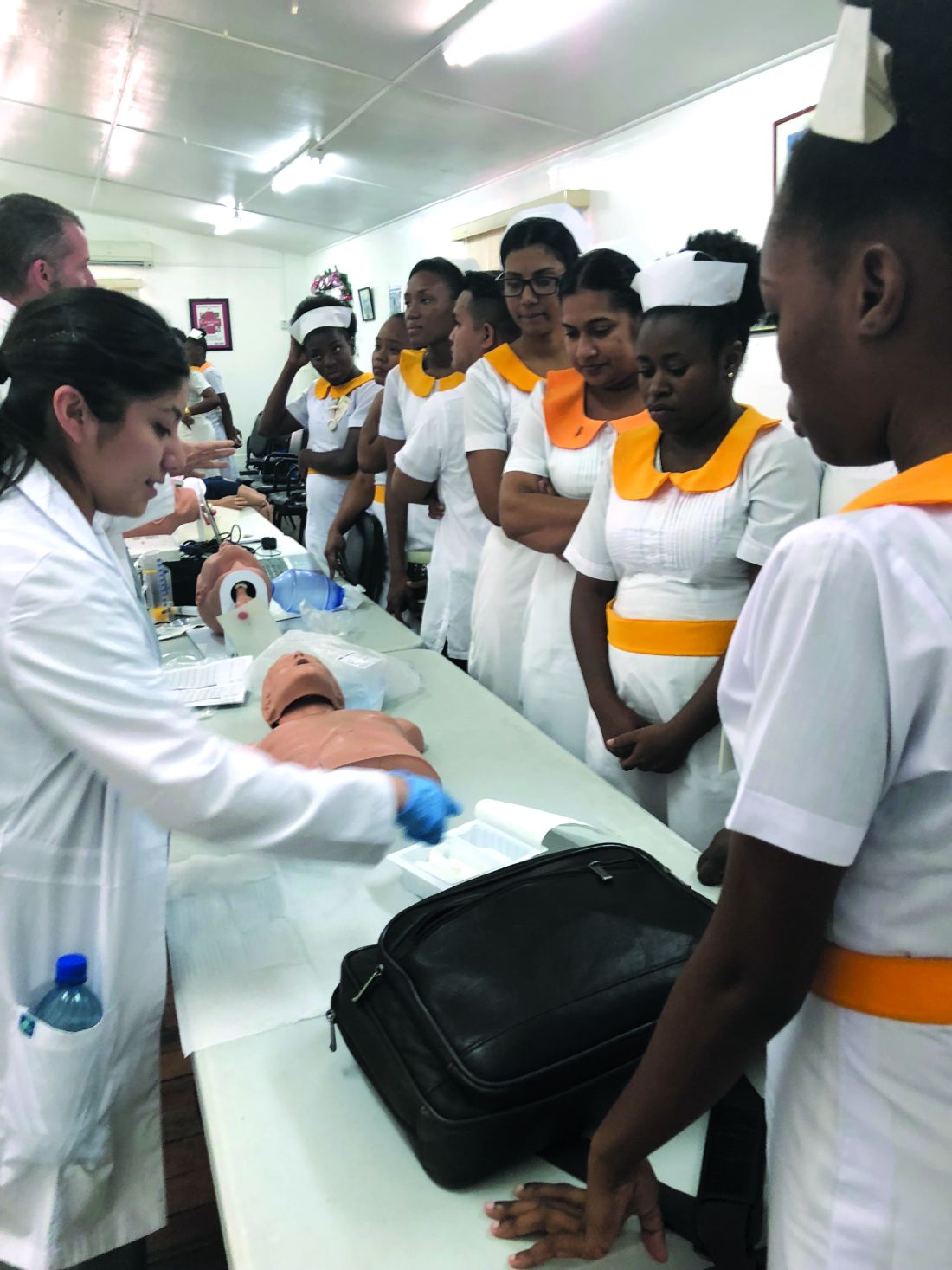
The 2 days that we were in Linden brought the largest sign-up for their clinic. When we did our screening at Mackenzie High School, we were able to utilize the portable spirometers and printer purchased by the CHEST Foundation community service grant. We are still collecting data, but the one thing that was revealed was the difficulty in obtaining medication for the treatment of asthma and COPD in some areas.
This project was also a learning experience for our students in many ways: in how they performed their interviews, how the culture affected the way their patients answered their questionnaires, and even how they performed on the tests. The value to the student and the individual of working within a different culture, far away from the norms of North America, allows them to appreciate their patients, the work they do, and their interprofessional team in a whole new light.
I want this experience to have an impact on each student’s life. You are a teacher, an instructor, a mentor, professor, and much more when traveling with 10 students. The most satisfying moment is the transformation you see in them. They are no longer timid and unsure of themselves; they have greater confidence in their abilities and a deeper understanding of the needs of a patient. They finally understand the importance of culture as it pertains to health care.
The effect of the CHEST Foundation grant
Applying for the CHEST Foundation community service grant was the largest grant I had ever attempted. Having a support system behind you is the most important piece of advice I can give to future grant applicants. I could not have completed my grant without our grant team at Texas State University. They truly had my back; and close to the deadline when it seemed insurmountable, they helped push me through it. The other piece of advice is to have a true vision and stick to that vision. The most difficult part of my project was the budget, prioritizing the things or people that I needed. Honestly, I needed help here, because for me, I needed everything. I had to make choices and leave some things out. I focused on what the actual need was for the many.
My ultimate goal for Guyana is to promote and show the need for respiratory care professionals to have that education offered at the University of Guyana as part of its allied health program and assist those in the application to the International Fellowship Program of the American Association of Respiratory Care—there has never been a fellow from Guyana. I believe that Guyana will have the resources, and with assistance, could achieve the goal. My vision and goal started in 2016, and I want to achieve it in the next 10 years.
I would like to thank all the CHEST Foundation donors from the bottom of my heart. This project was real and, as a CHEST member myself, it encourages me to be a better donor. Thank you—for it was and is much appreciated. Finally, I would like to express my thanks to my Co-Assistant Program Director, Holly Wise (Mass Communications) and Amber Hazelett, RRT (RC assistant), and the BGMM team for their entire support throughout the study abroad journey.
(This article was previous published in CHEST Thought Leaders.)
This grant is supported in full by the CHEST Foundation. Donors like you make grants like this possible. Thank you for your generosity and passion for community service and moving the needle forward on improving patient outcomes. To support community service initiatives, and the next generation of lung health champions, please go to foundation.chestnet.org/donate
How it all started
The study abroad project was truly a goal and vision that came about after returning to Guyana after approximately 46 years. I was born in Guyana but left as a child and returned later and joined a mission group. In 2015, I began a personal journey of missionary service with the team of Bridge Global Medical Missions (BGMM) in Georgetown, Guyana. I was the first respiratory therapist to join the team.
I remember during the first few days in the hospitals I was told that there was “a lot of wheezing” in the EDs. Treating patients consisted of just administrating short-acting nebulizer treatments, but I remember being very impressed with the ICU at the main public hospital, Georgetown Public Hospital Corporation (GPHC), because they had the ventilators I could use. However, physicians only managed the patients while the nurses were left to monitor the ventilators and equipment, which they did not understand.
At the Linden Hospital in Guyana, the ED was constantly full of the “wheezers,” and the ICU only had ventilators that were basically nonfunctioning due to language barriers or a lack of biomed professionals. One of my fondest memories was fixing two ventilators from China. I could get the ventilators to work and explain the basic modes because in my mind, it was just a ventilator, and they could see the modes. The problem was the language was all in Chinese! So, we all got together: a Cuban doctor, a Cuban biomed, and a nurse with a translation program and, finally, changed the language to English. It was an interesting day!
When we were on our study abroad trip this past January, I was able to place an intubated patient on that same ventilator. After my first visit to Linden Hospital, I addressed a few of my observations with the medical director, and I will never forget his comment. He said, “I thought respiratory would just come do some nebulizer treatments and show us oxygen.”
Study abroad and respiratory care
Then the vision of my project began, because I needed to show him the scope of the practice of a respiratory therapist. I asked Dr. Heyliger-Thomas of BGMM if she could assist me in promoting a study abroad program in Guyana with the Ministry of Health. It was very important for me to bring my students to Guyana for many reasons, the most important being the profession was needed there, and our students would be excellent representatives.
In 2015, the study “Introduction of spirometry into clinical practice in Georgetown, Guyana: quality and diagnostic outcomes” highlighted increased physician referral to the country’s only COPD/asthma clinic. I wanted to promote the importance of study abroad and international mission work, especially when promoting the care of asthma and the pulmonary patient, which I believe we did. The main project during study abroad was to test the school-aged children in Linden, thereby showing that there was undiagnosed asthma.
The 2 days that we were in Linden brought the largest sign-up for their clinic. When we did our screening at Mackenzie High School, we were able to utilize the portable spirometers and printer purchased by the CHEST Foundation community service grant. We are still collecting data, but the one thing that was revealed was the difficulty in obtaining medication for the treatment of asthma and COPD in some areas.
This project was also a learning experience for our students in many ways: in how they performed their interviews, how the culture affected the way their patients answered their questionnaires, and even how they performed on the tests. The value to the student and the individual of working within a different culture, far away from the norms of North America, allows them to appreciate their patients, the work they do, and their interprofessional team in a whole new light.
I want this experience to have an impact on each student’s life. You are a teacher, an instructor, a mentor, professor, and much more when traveling with 10 students. The most satisfying moment is the transformation you see in them. They are no longer timid and unsure of themselves; they have greater confidence in their abilities and a deeper understanding of the needs of a patient. They finally understand the importance of culture as it pertains to health care.
The effect of the CHEST Foundation grant
Applying for the CHEST Foundation community service grant was the largest grant I had ever attempted. Having a support system behind you is the most important piece of advice I can give to future grant applicants. I could not have completed my grant without our grant team at Texas State University. They truly had my back; and close to the deadline when it seemed insurmountable, they helped push me through it. The other piece of advice is to have a true vision and stick to that vision. The most difficult part of my project was the budget, prioritizing the things or people that I needed. Honestly, I needed help here, because for me, I needed everything. I had to make choices and leave some things out. I focused on what the actual need was for the many.
My ultimate goal for Guyana is to promote and show the need for respiratory care professionals to have that education offered at the University of Guyana as part of its allied health program and assist those in the application to the International Fellowship Program of the American Association of Respiratory Care—there has never been a fellow from Guyana. I believe that Guyana will have the resources, and with assistance, could achieve the goal. My vision and goal started in 2016, and I want to achieve it in the next 10 years.
I would like to thank all the CHEST Foundation donors from the bottom of my heart. This project was real and, as a CHEST member myself, it encourages me to be a better donor. Thank you—for it was and is much appreciated. Finally, I would like to express my thanks to my Co-Assistant Program Director, Holly Wise (Mass Communications) and Amber Hazelett, RRT (RC assistant), and the BGMM team for their entire support throughout the study abroad journey.
(This article was previous published in CHEST Thought Leaders.)
This grant is supported in full by the CHEST Foundation. Donors like you make grants like this possible. Thank you for your generosity and passion for community service and moving the needle forward on improving patient outcomes. To support community service initiatives, and the next generation of lung health champions, please go to foundation.chestnet.org/donate
How it all started
The study abroad project was truly a goal and vision that came about after returning to Guyana after approximately 46 years. I was born in Guyana but left as a child and returned later and joined a mission group. In 2015, I began a personal journey of missionary service with the team of Bridge Global Medical Missions (BGMM) in Georgetown, Guyana. I was the first respiratory therapist to join the team.
I remember during the first few days in the hospitals I was told that there was “a lot of wheezing” in the EDs. Treating patients consisted of just administrating short-acting nebulizer treatments, but I remember being very impressed with the ICU at the main public hospital, Georgetown Public Hospital Corporation (GPHC), because they had the ventilators I could use. However, physicians only managed the patients while the nurses were left to monitor the ventilators and equipment, which they did not understand.
At the Linden Hospital in Guyana, the ED was constantly full of the “wheezers,” and the ICU only had ventilators that were basically nonfunctioning due to language barriers or a lack of biomed professionals. One of my fondest memories was fixing two ventilators from China. I could get the ventilators to work and explain the basic modes because in my mind, it was just a ventilator, and they could see the modes. The problem was the language was all in Chinese! So, we all got together: a Cuban doctor, a Cuban biomed, and a nurse with a translation program and, finally, changed the language to English. It was an interesting day!
When we were on our study abroad trip this past January, I was able to place an intubated patient on that same ventilator. After my first visit to Linden Hospital, I addressed a few of my observations with the medical director, and I will never forget his comment. He said, “I thought respiratory would just come do some nebulizer treatments and show us oxygen.”
Study abroad and respiratory care
Then the vision of my project began, because I needed to show him the scope of the practice of a respiratory therapist. I asked Dr. Heyliger-Thomas of BGMM if she could assist me in promoting a study abroad program in Guyana with the Ministry of Health. It was very important for me to bring my students to Guyana for many reasons, the most important being the profession was needed there, and our students would be excellent representatives.
In 2015, the study “Introduction of spirometry into clinical practice in Georgetown, Guyana: quality and diagnostic outcomes” highlighted increased physician referral to the country’s only COPD/asthma clinic. I wanted to promote the importance of study abroad and international mission work, especially when promoting the care of asthma and the pulmonary patient, which I believe we did. The main project during study abroad was to test the school-aged children in Linden, thereby showing that there was undiagnosed asthma.
The 2 days that we were in Linden brought the largest sign-up for their clinic. When we did our screening at Mackenzie High School, we were able to utilize the portable spirometers and printer purchased by the CHEST Foundation community service grant. We are still collecting data, but the one thing that was revealed was the difficulty in obtaining medication for the treatment of asthma and COPD in some areas.
This project was also a learning experience for our students in many ways: in how they performed their interviews, how the culture affected the way their patients answered their questionnaires, and even how they performed on the tests. The value to the student and the individual of working within a different culture, far away from the norms of North America, allows them to appreciate their patients, the work they do, and their interprofessional team in a whole new light.
I want this experience to have an impact on each student’s life. You are a teacher, an instructor, a mentor, professor, and much more when traveling with 10 students. The most satisfying moment is the transformation you see in them. They are no longer timid and unsure of themselves; they have greater confidence in their abilities and a deeper understanding of the needs of a patient. They finally understand the importance of culture as it pertains to health care.
The effect of the CHEST Foundation grant
Applying for the CHEST Foundation community service grant was the largest grant I had ever attempted. Having a support system behind you is the most important piece of advice I can give to future grant applicants. I could not have completed my grant without our grant team at Texas State University. They truly had my back; and close to the deadline when it seemed insurmountable, they helped push me through it. The other piece of advice is to have a true vision and stick to that vision. The most difficult part of my project was the budget, prioritizing the things or people that I needed. Honestly, I needed help here, because for me, I needed everything. I had to make choices and leave some things out. I focused on what the actual need was for the many.
My ultimate goal for Guyana is to promote and show the need for respiratory care professionals to have that education offered at the University of Guyana as part of its allied health program and assist those in the application to the International Fellowship Program of the American Association of Respiratory Care—there has never been a fellow from Guyana. I believe that Guyana will have the resources, and with assistance, could achieve the goal. My vision and goal started in 2016, and I want to achieve it in the next 10 years.
I would like to thank all the CHEST Foundation donors from the bottom of my heart. This project was real and, as a CHEST member myself, it encourages me to be a better donor. Thank you—for it was and is much appreciated. Finally, I would like to express my thanks to my Co-Assistant Program Director, Holly Wise (Mass Communications) and Amber Hazelett, RRT (RC assistant), and the BGMM team for their entire support throughout the study abroad journey.
(This article was previous published in CHEST Thought Leaders.)
This grant is supported in full by the CHEST Foundation. Donors like you make grants like this possible. Thank you for your generosity and passion for community service and moving the needle forward on improving patient outcomes. To support community service initiatives, and the next generation of lung health champions, please go to foundation.chestnet.org/donate

FDA to host meeting about sleep apnea devices
You are invited to attend this open meeting on April 16, held at the FDA White Oak Campus in Silver Spring, Md. (https://www.fda.gov/MedicalDevices/NewsEvents/WorkshopsConferences/ucm596147.htm). The FDA is soliciting ideas or opinions about criteria or processes for FDA review of medical devices to diagnose or treat sleep apnea. CHEST is represented by Dr. Neil Freedman (neilfreedman@comcast.net) and Dr. Barbara Phillips (bphil020@gmail.com) who also welcome your input by email prior to the meeting. Home testing, “apps,” and the criteria to diagnose sleep apnea and/or its resolution are among the topics to be discussed.
You are invited to attend this open meeting on April 16, held at the FDA White Oak Campus in Silver Spring, Md. (https://www.fda.gov/MedicalDevices/NewsEvents/WorkshopsConferences/ucm596147.htm). The FDA is soliciting ideas or opinions about criteria or processes for FDA review of medical devices to diagnose or treat sleep apnea. CHEST is represented by Dr. Neil Freedman (neilfreedman@comcast.net) and Dr. Barbara Phillips (bphil020@gmail.com) who also welcome your input by email prior to the meeting. Home testing, “apps,” and the criteria to diagnose sleep apnea and/or its resolution are among the topics to be discussed.
You are invited to attend this open meeting on April 16, held at the FDA White Oak Campus in Silver Spring, Md. (https://www.fda.gov/MedicalDevices/NewsEvents/WorkshopsConferences/ucm596147.htm). The FDA is soliciting ideas or opinions about criteria or processes for FDA review of medical devices to diagnose or treat sleep apnea. CHEST is represented by Dr. Neil Freedman (neilfreedman@comcast.net) and Dr. Barbara Phillips (bphil020@gmail.com) who also welcome your input by email prior to the meeting. Home testing, “apps,” and the criteria to diagnose sleep apnea and/or its resolution are among the topics to be discussed.
“No consequence” Knowledge Check-In expands
In 2018, ABIM is introducing the new Knowledge Check-In assessment option, an every-2-year assessment option serving as an alternative to the 10-year assessment model. Initially, for 2018, this option will be piloted for both Internal Medicine and Nephrology. In 2019, the Knowledge Check-In will expand to several additional specialties, including Pulmonary Disease. The remaining specialties, including Critical Care Medicine, will become available in 2020.
Previously, ABIM announced that physicians taking the Knowledge Check-In in 2018—the initial year it is offered in Internal Medicine or Nephrology—would have another chance to take it again 2 years later if they were unsuccessful, even if they were due to pass the exam that year. Based on feedback ABIM received from the physician community, this feature is now being extended to include all other Internal Medicine subspecialties in the future. Therefore, if a physician opts to take the Knowledge Check-In the first year it is offered in their subspecialty and is unsuccessful, they will get at least one additional opportunity to take it 2 years later.
For more information visit www.abim.org/checkin.
In 2018, ABIM is introducing the new Knowledge Check-In assessment option, an every-2-year assessment option serving as an alternative to the 10-year assessment model. Initially, for 2018, this option will be piloted for both Internal Medicine and Nephrology. In 2019, the Knowledge Check-In will expand to several additional specialties, including Pulmonary Disease. The remaining specialties, including Critical Care Medicine, will become available in 2020.
Previously, ABIM announced that physicians taking the Knowledge Check-In in 2018—the initial year it is offered in Internal Medicine or Nephrology—would have another chance to take it again 2 years later if they were unsuccessful, even if they were due to pass the exam that year. Based on feedback ABIM received from the physician community, this feature is now being extended to include all other Internal Medicine subspecialties in the future. Therefore, if a physician opts to take the Knowledge Check-In the first year it is offered in their subspecialty and is unsuccessful, they will get at least one additional opportunity to take it 2 years later.
For more information visit www.abim.org/checkin.
In 2018, ABIM is introducing the new Knowledge Check-In assessment option, an every-2-year assessment option serving as an alternative to the 10-year assessment model. Initially, for 2018, this option will be piloted for both Internal Medicine and Nephrology. In 2019, the Knowledge Check-In will expand to several additional specialties, including Pulmonary Disease. The remaining specialties, including Critical Care Medicine, will become available in 2020.
Previously, ABIM announced that physicians taking the Knowledge Check-In in 2018—the initial year it is offered in Internal Medicine or Nephrology—would have another chance to take it again 2 years later if they were unsuccessful, even if they were due to pass the exam that year. Based on feedback ABIM received from the physician community, this feature is now being extended to include all other Internal Medicine subspecialties in the future. Therefore, if a physician opts to take the Knowledge Check-In the first year it is offered in their subspecialty and is unsuccessful, they will get at least one additional opportunity to take it 2 years later.
For more information visit www.abim.org/checkin.
New strategic plan for CHEST
We are pleased to announce the completion of a new, multiyear strategic plan for CHEST. Over the past few years, key stakeholders have provided essential input, resulting in a plan that identifies a very focused set of priorities we’ll pursue to help achieve our overarching strategy. Having selected these priorities, which leverage our strengths and strategic advantages, we are committed to dedicating sufficient resources toward their accomplishment over the next several years.
Each year, the plan will be reviewed and modified to reflect changes to CHEST priorities.
A strategic plan is an important tool for our organization because it truly does focus and direct our efforts and resources. Guided by our 2013-2017 strategic plan, we were able to accomplish the following:
- Developed events, products, and services that produced meaningful education for the CHEST community and generated positive financial margins;
- Optimized our membership model to increase engagement of all clinicians on the health-care team;
- Enhanced our global presence through guideline development and increased educational offerings;
- Launched a new Association Management System (AMS) and made strides to becoming a data-driven organization;
- Built and moved into a new building that enhanced our ability to develop and host courses in the CHEST Innovation, Simulation, and Training Center;
- Increased our visibility through our rebrand as “CHEST”;
- Fostered relationships and collaborated with other organizations to promote lung health through the CHEST Foundation; and
- Met our budget goals and financial covenants with our bank, and increased the CHEST Foundation’s corpus for grants and awards.
This new strategic plan can be found on chestnet.org under the “About” section. As members of CHEST, we invite you to review what’s outlined and become familiar with what the plan encompasses. This plan provides details to help you understand the future direction of CHEST, and we know you’ll support us in these important endeavors.
We are pleased to announce the completion of a new, multiyear strategic plan for CHEST. Over the past few years, key stakeholders have provided essential input, resulting in a plan that identifies a very focused set of priorities we’ll pursue to help achieve our overarching strategy. Having selected these priorities, which leverage our strengths and strategic advantages, we are committed to dedicating sufficient resources toward their accomplishment over the next several years.
Each year, the plan will be reviewed and modified to reflect changes to CHEST priorities.
A strategic plan is an important tool for our organization because it truly does focus and direct our efforts and resources. Guided by our 2013-2017 strategic plan, we were able to accomplish the following:
- Developed events, products, and services that produced meaningful education for the CHEST community and generated positive financial margins;
- Optimized our membership model to increase engagement of all clinicians on the health-care team;
- Enhanced our global presence through guideline development and increased educational offerings;
- Launched a new Association Management System (AMS) and made strides to becoming a data-driven organization;
- Built and moved into a new building that enhanced our ability to develop and host courses in the CHEST Innovation, Simulation, and Training Center;
- Increased our visibility through our rebrand as “CHEST”;
- Fostered relationships and collaborated with other organizations to promote lung health through the CHEST Foundation; and
- Met our budget goals and financial covenants with our bank, and increased the CHEST Foundation’s corpus for grants and awards.
This new strategic plan can be found on chestnet.org under the “About” section. As members of CHEST, we invite you to review what’s outlined and become familiar with what the plan encompasses. This plan provides details to help you understand the future direction of CHEST, and we know you’ll support us in these important endeavors.
We are pleased to announce the completion of a new, multiyear strategic plan for CHEST. Over the past few years, key stakeholders have provided essential input, resulting in a plan that identifies a very focused set of priorities we’ll pursue to help achieve our overarching strategy. Having selected these priorities, which leverage our strengths and strategic advantages, we are committed to dedicating sufficient resources toward their accomplishment over the next several years.
Each year, the plan will be reviewed and modified to reflect changes to CHEST priorities.
A strategic plan is an important tool for our organization because it truly does focus and direct our efforts and resources. Guided by our 2013-2017 strategic plan, we were able to accomplish the following:
- Developed events, products, and services that produced meaningful education for the CHEST community and generated positive financial margins;
- Optimized our membership model to increase engagement of all clinicians on the health-care team;
- Enhanced our global presence through guideline development and increased educational offerings;
- Launched a new Association Management System (AMS) and made strides to becoming a data-driven organization;
- Built and moved into a new building that enhanced our ability to develop and host courses in the CHEST Innovation, Simulation, and Training Center;
- Increased our visibility through our rebrand as “CHEST”;
- Fostered relationships and collaborated with other organizations to promote lung health through the CHEST Foundation; and
- Met our budget goals and financial covenants with our bank, and increased the CHEST Foundation’s corpus for grants and awards.
This new strategic plan can be found on chestnet.org under the “About” section. As members of CHEST, we invite you to review what’s outlined and become familiar with what the plan encompasses. This plan provides details to help you understand the future direction of CHEST, and we know you’ll support us in these important endeavors.
Study: Preop EKGs have little utility for benign hysterectomy
ORLANDO – Preoperative electrocardiograms (EKGs) had no effect on management or perioperative complications in women undergoing benign hysterectomy over a 12-month period at a single medical center, according to a review of records.
Of 587 patients included in the review, 182 (31%) underwent EKG as part of their preoperative evaluation, and the majority of those were indicated according to institutional criteria (166; 28%) or National Institute for Health Care Excellence (NICE) guidelines (177; 30%), Nemi M. Shah, MD, reported at the annual scientific meeting of the Society of Gynecologic Surgeons.
“EKG was indicated in 91% of these patients according to institutional criteria, and in 97% of patients per NICE criteria,” said Dr. Shah, a third-year resident at the University of Texas Southwestern Medical Center, Dallas.
By institutional criteria, hypertension was the most common indication (68% of cases), and by NICE criteria, American Society of Anesthesia class 2 physical status was the most common indication (80% of cases), she noted, adding that obesity, which was present in 70% of patients, was the most common comorbidity classifying patients with American Society of Anesthesia class 2 or above.
Of the 182 EKGs performed, findings were abnormal in 89, but further workup was pursued in only 16 patients, and included repeat EKG, echocardiogram, and/or stress testing and cardiology consultation. Surgical delays of 1 and 4 months occurred in 2 patients as a result of the additional workup, and ultimately, all planned hysterectomies were completed by the primary surgical team without changes in management, she said.
Perioperative complications occurred in two patients, and included nonspecific postinduction EKG changes that led to surgery being aborted in one patient who had left ventricular hypertrophy on the preoperative EKG, and failed extubation in a patient with airway edema whose preoperative EKG showed a nonacute inferior infarct.
For the first, cardiology was consulted and determined the findings to be benign; the patient underwent hysterectomy at a later date without complications. The second patient was taken to the surgical intensive care unit for management, Dr. Shah said.
“Preoperative testing for benign hysterectomy is variable as there is no single standard of care,” she explained. “Though tests such as EKG are commonly ordered, there are no data linking study results to surgical outcomes.”
The current study was conducted to evaluate the rate of preoperative EKG performed in concordance with institutional and NICE guidelines, and to assess implications for management and perioperative complications.
Patients included in the review were adult women who underwent scheduled benign hysterectomy during 2016. Women who underwent emergency surgery or whose surgery was performed by gynecologic oncologists were excluded.
Subjects were primarily Hispanic, and had a mean age of 45 years, Dr. Shah noted.
Though limited by the single-center design and retrospective nature of the study, the findings suggest that preoperative EKG has little clinical utility.
“We found that practice patterns were highly concordant with institutional and NICE guidelines. However, EKG resulted in minimal impact on perioperative management, and no association between abnormal EKG and perioperative complications was found,” she said. “EKG may not accurately stratify perioperative cardiopulmonary risk, and alternative methods for preoperative evaluation should be considered.”
Dr. Shah reported having no disclosures.
SOURCE: Shah N et al. SGS 2018 Oral Poster 3.
ORLANDO – Preoperative electrocardiograms (EKGs) had no effect on management or perioperative complications in women undergoing benign hysterectomy over a 12-month period at a single medical center, according to a review of records.
Of 587 patients included in the review, 182 (31%) underwent EKG as part of their preoperative evaluation, and the majority of those were indicated according to institutional criteria (166; 28%) or National Institute for Health Care Excellence (NICE) guidelines (177; 30%), Nemi M. Shah, MD, reported at the annual scientific meeting of the Society of Gynecologic Surgeons.
“EKG was indicated in 91% of these patients according to institutional criteria, and in 97% of patients per NICE criteria,” said Dr. Shah, a third-year resident at the University of Texas Southwestern Medical Center, Dallas.
By institutional criteria, hypertension was the most common indication (68% of cases), and by NICE criteria, American Society of Anesthesia class 2 physical status was the most common indication (80% of cases), she noted, adding that obesity, which was present in 70% of patients, was the most common comorbidity classifying patients with American Society of Anesthesia class 2 or above.
Of the 182 EKGs performed, findings were abnormal in 89, but further workup was pursued in only 16 patients, and included repeat EKG, echocardiogram, and/or stress testing and cardiology consultation. Surgical delays of 1 and 4 months occurred in 2 patients as a result of the additional workup, and ultimately, all planned hysterectomies were completed by the primary surgical team without changes in management, she said.
Perioperative complications occurred in two patients, and included nonspecific postinduction EKG changes that led to surgery being aborted in one patient who had left ventricular hypertrophy on the preoperative EKG, and failed extubation in a patient with airway edema whose preoperative EKG showed a nonacute inferior infarct.
For the first, cardiology was consulted and determined the findings to be benign; the patient underwent hysterectomy at a later date without complications. The second patient was taken to the surgical intensive care unit for management, Dr. Shah said.
“Preoperative testing for benign hysterectomy is variable as there is no single standard of care,” she explained. “Though tests such as EKG are commonly ordered, there are no data linking study results to surgical outcomes.”
The current study was conducted to evaluate the rate of preoperative EKG performed in concordance with institutional and NICE guidelines, and to assess implications for management and perioperative complications.
Patients included in the review were adult women who underwent scheduled benign hysterectomy during 2016. Women who underwent emergency surgery or whose surgery was performed by gynecologic oncologists were excluded.
Subjects were primarily Hispanic, and had a mean age of 45 years, Dr. Shah noted.
Though limited by the single-center design and retrospective nature of the study, the findings suggest that preoperative EKG has little clinical utility.
“We found that practice patterns were highly concordant with institutional and NICE guidelines. However, EKG resulted in minimal impact on perioperative management, and no association between abnormal EKG and perioperative complications was found,” she said. “EKG may not accurately stratify perioperative cardiopulmonary risk, and alternative methods for preoperative evaluation should be considered.”
Dr. Shah reported having no disclosures.
SOURCE: Shah N et al. SGS 2018 Oral Poster 3.
ORLANDO – Preoperative electrocardiograms (EKGs) had no effect on management or perioperative complications in women undergoing benign hysterectomy over a 12-month period at a single medical center, according to a review of records.
Of 587 patients included in the review, 182 (31%) underwent EKG as part of their preoperative evaluation, and the majority of those were indicated according to institutional criteria (166; 28%) or National Institute for Health Care Excellence (NICE) guidelines (177; 30%), Nemi M. Shah, MD, reported at the annual scientific meeting of the Society of Gynecologic Surgeons.
“EKG was indicated in 91% of these patients according to institutional criteria, and in 97% of patients per NICE criteria,” said Dr. Shah, a third-year resident at the University of Texas Southwestern Medical Center, Dallas.
By institutional criteria, hypertension was the most common indication (68% of cases), and by NICE criteria, American Society of Anesthesia class 2 physical status was the most common indication (80% of cases), she noted, adding that obesity, which was present in 70% of patients, was the most common comorbidity classifying patients with American Society of Anesthesia class 2 or above.
Of the 182 EKGs performed, findings were abnormal in 89, but further workup was pursued in only 16 patients, and included repeat EKG, echocardiogram, and/or stress testing and cardiology consultation. Surgical delays of 1 and 4 months occurred in 2 patients as a result of the additional workup, and ultimately, all planned hysterectomies were completed by the primary surgical team without changes in management, she said.
Perioperative complications occurred in two patients, and included nonspecific postinduction EKG changes that led to surgery being aborted in one patient who had left ventricular hypertrophy on the preoperative EKG, and failed extubation in a patient with airway edema whose preoperative EKG showed a nonacute inferior infarct.
For the first, cardiology was consulted and determined the findings to be benign; the patient underwent hysterectomy at a later date without complications. The second patient was taken to the surgical intensive care unit for management, Dr. Shah said.
“Preoperative testing for benign hysterectomy is variable as there is no single standard of care,” she explained. “Though tests such as EKG are commonly ordered, there are no data linking study results to surgical outcomes.”
The current study was conducted to evaluate the rate of preoperative EKG performed in concordance with institutional and NICE guidelines, and to assess implications for management and perioperative complications.
Patients included in the review were adult women who underwent scheduled benign hysterectomy during 2016. Women who underwent emergency surgery or whose surgery was performed by gynecologic oncologists were excluded.
Subjects were primarily Hispanic, and had a mean age of 45 years, Dr. Shah noted.
Though limited by the single-center design and retrospective nature of the study, the findings suggest that preoperative EKG has little clinical utility.
“We found that practice patterns were highly concordant with institutional and NICE guidelines. However, EKG resulted in minimal impact on perioperative management, and no association between abnormal EKG and perioperative complications was found,” she said. “EKG may not accurately stratify perioperative cardiopulmonary risk, and alternative methods for preoperative evaluation should be considered.”
Dr. Shah reported having no disclosures.
SOURCE: Shah N et al. SGS 2018 Oral Poster 3.
REPORTING FROM SGS 2018
Key clinical point: Preoperative EKG appears to have little utility in patients undergoing benign hysterectomy.
Major finding: Only 16 of 89 patients with abnormal preoperative EKG underwent further workup, and surgery was delayed in only two cases.
Study details: A retrospective review of 587 cases.
Disclosures: Dr. Shah reported having no disclosures.
Source: Shah N et al. SGS 2018 Oral Poster 3.
Trastuzumab plus chemo shows efficacy for high HER2 endometrial cancer
NEW ORLEANS – Adding the anti-HER2 antibody trastuzumab to a standard chemotherapy regimen produced markedly improved responses among patients with an advanced stage or recurrent uterine serous carcinoma that also overexpressed the HER2/neu cell receptor protein in a controlled, multicenter, phase 2 study with 58 evaluable patients.
The 30 patients who received trastuzumab along with carboplatin plus paclitaxel had a median progression-free survival of 12.6 months compared with 8.0 months among the 28 control patients who received only carboplatin plus paclitaxel, a hazard reduction of 56% with trastuzumab that was statistically significant (P = .0052), Alessandro D. Santin, MD, said at the annual meeting of the Society of Gynecologic Oncology.
In the subgroup of 41 patients with advanced (not recurrent) uterine serous carcinoma (USC) the incremental difference in median progression-free survival among patients treated with trastuzumab was 8.6 months, based on a 17.9-month duration in the 20 patients who received trastuzumab and 9.3 months in 20 control patients on chemotherapy only (P = .013).
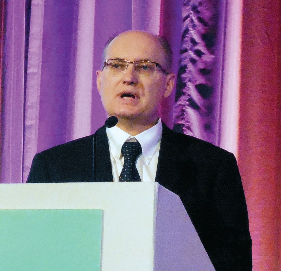
“We’ll push to have the trastuzumab, carboplatin, and paclitaxel regimen in the guidelines” for treating patients with USC that overexpresses HER2/neu, agreed Amanda N. Fader, MD, director of gynecologic oncology at Johns Hopkins Medicine in Baltimore and lead author on the study reported by Dr. Santin.
Shortly after Dr. Santin’s report at the meeting, the results appeared in an article published online March 27 in Journal of Clinical Oncology.
The phase 2 trial ran at 11 U.S. centers during 2011-2016, and randomized 61 patients with stage III or IV or recurrent USC and high HER2 protein expression measured by immunohistochemistry and amplification of the HER2 gene shown by fluorescence in situ hybridization. The researchers collected data from 58 treated patients. Among the 17 patients with recurrent disease, those who received trastuzumab had a median progression-free survival of 9.2 months compared with 6.0 months in the controls, a hazard reduction of 86% with trastuzumab that was statistically significant (P = .0029). The addition of trastuzumab to carboplatin and paclitaxel was well tolerated.
The greatest benefit from the trastuzumab plus chemotherapy regimen might occur when used as first-line treatment, Dr. Santin suggested. He expressed interest in running a second study to validate the current findings, and a new study that would combine a second antibody directed against HER2, pertuzumab (Perjeta) with trastuzumab, carboplatin, and paclitaxel.
A key factor in making the trastuzumab plus chemotherapy regimen more widely available to USC patients would be routinely screening these patients for high levels of HER2/neu expression immediately after USC is diagnosed. “At Yale and Johns Hopkins this testing is done on all USC patients. That’s not standard care everywhere, but it should be based on these data,” Dr. Santin said in an interview.
SOURCE: Fader A et al. Abstract 22. J Clin Oncol. 2018 Mar 27. doi: 10.1200/JCO.2017.76.5966.
NEW ORLEANS – Adding the anti-HER2 antibody trastuzumab to a standard chemotherapy regimen produced markedly improved responses among patients with an advanced stage or recurrent uterine serous carcinoma that also overexpressed the HER2/neu cell receptor protein in a controlled, multicenter, phase 2 study with 58 evaluable patients.
The 30 patients who received trastuzumab along with carboplatin plus paclitaxel had a median progression-free survival of 12.6 months compared with 8.0 months among the 28 control patients who received only carboplatin plus paclitaxel, a hazard reduction of 56% with trastuzumab that was statistically significant (P = .0052), Alessandro D. Santin, MD, said at the annual meeting of the Society of Gynecologic Oncology.
In the subgroup of 41 patients with advanced (not recurrent) uterine serous carcinoma (USC) the incremental difference in median progression-free survival among patients treated with trastuzumab was 8.6 months, based on a 17.9-month duration in the 20 patients who received trastuzumab and 9.3 months in 20 control patients on chemotherapy only (P = .013).
“We’ll push to have the trastuzumab, carboplatin, and paclitaxel regimen in the guidelines” for treating patients with USC that overexpresses HER2/neu, agreed Amanda N. Fader, MD, director of gynecologic oncology at Johns Hopkins Medicine in Baltimore and lead author on the study reported by Dr. Santin.
Shortly after Dr. Santin’s report at the meeting, the results appeared in an article published online March 27 in Journal of Clinical Oncology.
The phase 2 trial ran at 11 U.S. centers during 2011-2016, and randomized 61 patients with stage III or IV or recurrent USC and high HER2 protein expression measured by immunohistochemistry and amplification of the HER2 gene shown by fluorescence in situ hybridization. The researchers collected data from 58 treated patients. Among the 17 patients with recurrent disease, those who received trastuzumab had a median progression-free survival of 9.2 months compared with 6.0 months in the controls, a hazard reduction of 86% with trastuzumab that was statistically significant (P = .0029). The addition of trastuzumab to carboplatin and paclitaxel was well tolerated.
The greatest benefit from the trastuzumab plus chemotherapy regimen might occur when used as first-line treatment, Dr. Santin suggested. He expressed interest in running a second study to validate the current findings, and a new study that would combine a second antibody directed against HER2, pertuzumab (Perjeta) with trastuzumab, carboplatin, and paclitaxel.
A key factor in making the trastuzumab plus chemotherapy regimen more widely available to USC patients would be routinely screening these patients for high levels of HER2/neu expression immediately after USC is diagnosed. “At Yale and Johns Hopkins this testing is done on all USC patients. That’s not standard care everywhere, but it should be based on these data,” Dr. Santin said in an interview.
SOURCE: Fader A et al. Abstract 22. J Clin Oncol. 2018 Mar 27. doi: 10.1200/JCO.2017.76.5966.
NEW ORLEANS – Adding the anti-HER2 antibody trastuzumab to a standard chemotherapy regimen produced markedly improved responses among patients with an advanced stage or recurrent uterine serous carcinoma that also overexpressed the HER2/neu cell receptor protein in a controlled, multicenter, phase 2 study with 58 evaluable patients.
The 30 patients who received trastuzumab along with carboplatin plus paclitaxel had a median progression-free survival of 12.6 months compared with 8.0 months among the 28 control patients who received only carboplatin plus paclitaxel, a hazard reduction of 56% with trastuzumab that was statistically significant (P = .0052), Alessandro D. Santin, MD, said at the annual meeting of the Society of Gynecologic Oncology.
In the subgroup of 41 patients with advanced (not recurrent) uterine serous carcinoma (USC) the incremental difference in median progression-free survival among patients treated with trastuzumab was 8.6 months, based on a 17.9-month duration in the 20 patients who received trastuzumab and 9.3 months in 20 control patients on chemotherapy only (P = .013).
“We’ll push to have the trastuzumab, carboplatin, and paclitaxel regimen in the guidelines” for treating patients with USC that overexpresses HER2/neu, agreed Amanda N. Fader, MD, director of gynecologic oncology at Johns Hopkins Medicine in Baltimore and lead author on the study reported by Dr. Santin.
Shortly after Dr. Santin’s report at the meeting, the results appeared in an article published online March 27 in Journal of Clinical Oncology.
The phase 2 trial ran at 11 U.S. centers during 2011-2016, and randomized 61 patients with stage III or IV or recurrent USC and high HER2 protein expression measured by immunohistochemistry and amplification of the HER2 gene shown by fluorescence in situ hybridization. The researchers collected data from 58 treated patients. Among the 17 patients with recurrent disease, those who received trastuzumab had a median progression-free survival of 9.2 months compared with 6.0 months in the controls, a hazard reduction of 86% with trastuzumab that was statistically significant (P = .0029). The addition of trastuzumab to carboplatin and paclitaxel was well tolerated.
The greatest benefit from the trastuzumab plus chemotherapy regimen might occur when used as first-line treatment, Dr. Santin suggested. He expressed interest in running a second study to validate the current findings, and a new study that would combine a second antibody directed against HER2, pertuzumab (Perjeta) with trastuzumab, carboplatin, and paclitaxel.
A key factor in making the trastuzumab plus chemotherapy regimen more widely available to USC patients would be routinely screening these patients for high levels of HER2/neu expression immediately after USC is diagnosed. “At Yale and Johns Hopkins this testing is done on all USC patients. That’s not standard care everywhere, but it should be based on these data,” Dr. Santin said in an interview.
SOURCE: Fader A et al. Abstract 22. J Clin Oncol. 2018 Mar 27. doi: 10.1200/JCO.2017.76.5966.
REPORTING FROM SGO 2018
Key clinical point: Trastuzumab plus chemotherapy improved progression-free survival in a phase 2 study.
Major finding: Median progression-free survival was 12.6 months with trastuzumab and 8.0 months in controls.
Study details: A multicenter, phase 2 study that enrolled 58 evaluable patients.
Disclosures: Trastuzumab was provided at no charge by Genentech. Dr. Santin and Dr. Fader had no relevant disclosures.
Source: Fader A et al. Abstract 22. J Clin Oncol. 2018 Mar 27. doi: 10.1200/JCO.2017.76.5966)
views
The biological relevance of HER2 amplification has been clearly established in the results from this and other studies. But we must keep in kind that the results reported by Dr. Santin have applicability to a subset of a subset of patients. Specifically, about 10% of endometrial cancers are uterine serous carcinoma (USC) (although they account for roughly 40% of deaths among endometrial cancer patients), about half of USC patients show a high level of HER2/neu expression by immunohistochemistry, and about 60% of these high-expressing USC also have HER2 gene amplification. The study presented by Dr. Santin ran at 11 centers for 5.5 years and enrolled just 61 patients, fewer than the planned enrollment target of 100 patients.
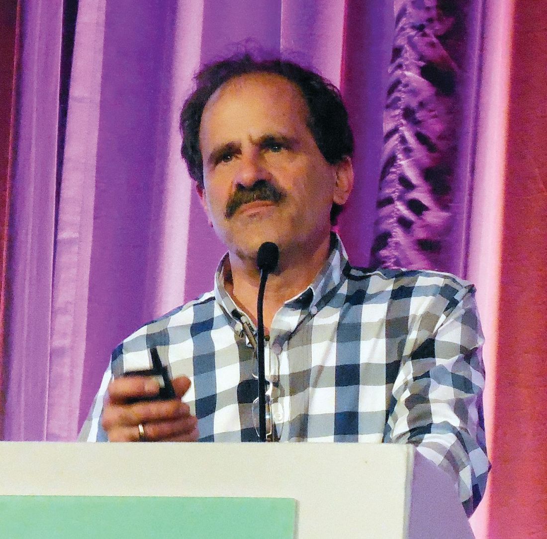
Michael A. Bookman, MD , is director of gynecologic oncology therapeutics at the Permanente Medical Group in San Francisco. He has been an adviser to AstraZeneca, Bayer, Clovis, Merck, Pfizer, and Tesaro, and he has participated in trials funded by Abbvie, Genentech, Immunogen, Mateon, and Roche. He made these comments as designated discussant for the study.
Myelodysplastic syndromes: etiologies, evaluation, and therapy
In this interview, Dr David Henry, MD, the Editor-in-Chief of JCSO, and David Steensma, MD, of the Dana-Farber Cancer Institute in Boston, talk about myelodysplasic syndromes, from diagnosis, evaluation, and etiologies, to therapy options and molecular sequencing.
Listen to the podcast below, or click on the PDF icon at the top of this introduction to read a transcript of the interview.
In this interview, Dr David Henry, MD, the Editor-in-Chief of JCSO, and David Steensma, MD, of the Dana-Farber Cancer Institute in Boston, talk about myelodysplasic syndromes, from diagnosis, evaluation, and etiologies, to therapy options and molecular sequencing.
Listen to the podcast below, or click on the PDF icon at the top of this introduction to read a transcript of the interview.
In this interview, Dr David Henry, MD, the Editor-in-Chief of JCSO, and David Steensma, MD, of the Dana-Farber Cancer Institute in Boston, talk about myelodysplasic syndromes, from diagnosis, evaluation, and etiologies, to therapy options and molecular sequencing.
Listen to the podcast below, or click on the PDF icon at the top of this introduction to read a transcript of the interview.
Estrogen patch counters eating disorders in women athletes
CHICAGO – It’s a good idea to look for eating disorders in young, athletic women who present with oligomenorrhea; they are at high risk for them and can be helped with estrogen replacement, especially with a patch, according to investigators from Massachusetts General Hospital, Boston.
Estrogen replacement will also help with stress fractures and other bone problems, but young, athletic women might resist the idea. They worry that it might hurt their performance and are often proud that they work out hard enough to stop their periods, according to investigator Vibha Singhal, MD, a pediatric endocrinologist and eating disorder specialist at the hospital.
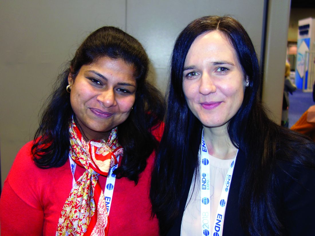
Earlier research done at Massachusetts General had shown that replacing estrogen to physiological levels helped reduce anxiety and body dissatisfaction in anorexia nervosa. Both are key drivers of the condition, along with the drive for thinness, lead investigator Franziska Plessow, PhD, a neuroendocrine researcher at the hospital, said at the Endocrine Society’s annual meeting.
But the researchers wondered whether estrogen also helps healthy weight women with nascent eating disorders, so the investigators turned to female athletes aged 14-25 years who had stopped menstruating or were about to.
The 117 oligomenorrheic athletes (OA) they investigated – none of whom had formally diagnosed eating disorders – scored significantly higher on measures of body dissatisfaction, drive for thinness, perfectionism, and “cognitive restraint of eating,” compared with 50 female athletes and 41 nonathletic women, both with normal periods. For instance, OA women scored a mean of 4.21 on the drive for thinness scale of the Eating Disorder Inventory–2, a low score, but still significantly higher than the 1.66 points in eumenorrheic athletes and 1.61 points in nonathletes (P = .0005).
Oligomenorrheic female athletes “show more disordered eating behavior and psychopathology at the subclinical level,” Dr. Plessow said.
Seventy OA women were then randomized to three groups: 25 to physiological estrogen replacement with an estradiol patch and cyclic progesterone for 12 days/month; 19 to replacement with contraceptive pills on the standard monthly schedule; and 26 to no replacement.
Over 12 months, women on the patch dropped their drive for thinness, body dissatisfaction, and uncontrolled eating scores. Body dissatisfaction scores on the Eating Disorder Inventory–2, for example, fell about two points, compared with staying about the same in the pill group and increasing by about two points in the no-estrogen group.
The pill seemed to help a bit on some measures, too, but the benefit of the pill versus that of no estrogen wasn’t generally statistically significant. Meanwhile, symptoms worsened in women who didn’t get estrogen. “The practice right now is the pill. We are shifting our practice with these data to the patch,” Dr. Singhal said.
Overall, “these findings emphasize the importance of normalizing estrogen levels in this population,” she and her colleagues concluded.
It’s a mystery why estrogen helps with eating disorders. Maybe it has something to do with the estrogen receptors in the appetite centers of the brain. Maybe the patch works better than the pill because there’s no first-pass through the liver, Dr. Singhal said.
Subjects were aged about 20 years, on average. OA women had a mean body mass index of 20.6 kg/m2 and a mean estradiol level of 45 pg/mL. Subjects with regular periods had mean BMIs of about 22 kg/m2; mean estradiol levels were 70.2 pg/mL in eumenorrheic athletes and 83.6 pg/mL in nonathletic women.
The work was funded by the National Institutes of Health. The investigators didn’t have any relevant disclosures.
SOURCE: Plessow F et al. ENDO 2018, Abstract SAT-290.
CHICAGO – It’s a good idea to look for eating disorders in young, athletic women who present with oligomenorrhea; they are at high risk for them and can be helped with estrogen replacement, especially with a patch, according to investigators from Massachusetts General Hospital, Boston.
Estrogen replacement will also help with stress fractures and other bone problems, but young, athletic women might resist the idea. They worry that it might hurt their performance and are often proud that they work out hard enough to stop their periods, according to investigator Vibha Singhal, MD, a pediatric endocrinologist and eating disorder specialist at the hospital.
Earlier research done at Massachusetts General had shown that replacing estrogen to physiological levels helped reduce anxiety and body dissatisfaction in anorexia nervosa. Both are key drivers of the condition, along with the drive for thinness, lead investigator Franziska Plessow, PhD, a neuroendocrine researcher at the hospital, said at the Endocrine Society’s annual meeting.
But the researchers wondered whether estrogen also helps healthy weight women with nascent eating disorders, so the investigators turned to female athletes aged 14-25 years who had stopped menstruating or were about to.
The 117 oligomenorrheic athletes (OA) they investigated – none of whom had formally diagnosed eating disorders – scored significantly higher on measures of body dissatisfaction, drive for thinness, perfectionism, and “cognitive restraint of eating,” compared with 50 female athletes and 41 nonathletic women, both with normal periods. For instance, OA women scored a mean of 4.21 on the drive for thinness scale of the Eating Disorder Inventory–2, a low score, but still significantly higher than the 1.66 points in eumenorrheic athletes and 1.61 points in nonathletes (P = .0005).
Oligomenorrheic female athletes “show more disordered eating behavior and psychopathology at the subclinical level,” Dr. Plessow said.
Seventy OA women were then randomized to three groups: 25 to physiological estrogen replacement with an estradiol patch and cyclic progesterone for 12 days/month; 19 to replacement with contraceptive pills on the standard monthly schedule; and 26 to no replacement.
Over 12 months, women on the patch dropped their drive for thinness, body dissatisfaction, and uncontrolled eating scores. Body dissatisfaction scores on the Eating Disorder Inventory–2, for example, fell about two points, compared with staying about the same in the pill group and increasing by about two points in the no-estrogen group.
The pill seemed to help a bit on some measures, too, but the benefit of the pill versus that of no estrogen wasn’t generally statistically significant. Meanwhile, symptoms worsened in women who didn’t get estrogen. “The practice right now is the pill. We are shifting our practice with these data to the patch,” Dr. Singhal said.
Overall, “these findings emphasize the importance of normalizing estrogen levels in this population,” she and her colleagues concluded.
It’s a mystery why estrogen helps with eating disorders. Maybe it has something to do with the estrogen receptors in the appetite centers of the brain. Maybe the patch works better than the pill because there’s no first-pass through the liver, Dr. Singhal said.
Subjects were aged about 20 years, on average. OA women had a mean body mass index of 20.6 kg/m2 and a mean estradiol level of 45 pg/mL. Subjects with regular periods had mean BMIs of about 22 kg/m2; mean estradiol levels were 70.2 pg/mL in eumenorrheic athletes and 83.6 pg/mL in nonathletic women.
The work was funded by the National Institutes of Health. The investigators didn’t have any relevant disclosures.
SOURCE: Plessow F et al. ENDO 2018, Abstract SAT-290.
CHICAGO – It’s a good idea to look for eating disorders in young, athletic women who present with oligomenorrhea; they are at high risk for them and can be helped with estrogen replacement, especially with a patch, according to investigators from Massachusetts General Hospital, Boston.
Estrogen replacement will also help with stress fractures and other bone problems, but young, athletic women might resist the idea. They worry that it might hurt their performance and are often proud that they work out hard enough to stop their periods, according to investigator Vibha Singhal, MD, a pediatric endocrinologist and eating disorder specialist at the hospital.
Earlier research done at Massachusetts General had shown that replacing estrogen to physiological levels helped reduce anxiety and body dissatisfaction in anorexia nervosa. Both are key drivers of the condition, along with the drive for thinness, lead investigator Franziska Plessow, PhD, a neuroendocrine researcher at the hospital, said at the Endocrine Society’s annual meeting.
But the researchers wondered whether estrogen also helps healthy weight women with nascent eating disorders, so the investigators turned to female athletes aged 14-25 years who had stopped menstruating or were about to.
The 117 oligomenorrheic athletes (OA) they investigated – none of whom had formally diagnosed eating disorders – scored significantly higher on measures of body dissatisfaction, drive for thinness, perfectionism, and “cognitive restraint of eating,” compared with 50 female athletes and 41 nonathletic women, both with normal periods. For instance, OA women scored a mean of 4.21 on the drive for thinness scale of the Eating Disorder Inventory–2, a low score, but still significantly higher than the 1.66 points in eumenorrheic athletes and 1.61 points in nonathletes (P = .0005).
Oligomenorrheic female athletes “show more disordered eating behavior and psychopathology at the subclinical level,” Dr. Plessow said.
Seventy OA women were then randomized to three groups: 25 to physiological estrogen replacement with an estradiol patch and cyclic progesterone for 12 days/month; 19 to replacement with contraceptive pills on the standard monthly schedule; and 26 to no replacement.
Over 12 months, women on the patch dropped their drive for thinness, body dissatisfaction, and uncontrolled eating scores. Body dissatisfaction scores on the Eating Disorder Inventory–2, for example, fell about two points, compared with staying about the same in the pill group and increasing by about two points in the no-estrogen group.
The pill seemed to help a bit on some measures, too, but the benefit of the pill versus that of no estrogen wasn’t generally statistically significant. Meanwhile, symptoms worsened in women who didn’t get estrogen. “The practice right now is the pill. We are shifting our practice with these data to the patch,” Dr. Singhal said.
Overall, “these findings emphasize the importance of normalizing estrogen levels in this population,” she and her colleagues concluded.
It’s a mystery why estrogen helps with eating disorders. Maybe it has something to do with the estrogen receptors in the appetite centers of the brain. Maybe the patch works better than the pill because there’s no first-pass through the liver, Dr. Singhal said.
Subjects were aged about 20 years, on average. OA women had a mean body mass index of 20.6 kg/m2 and a mean estradiol level of 45 pg/mL. Subjects with regular periods had mean BMIs of about 22 kg/m2; mean estradiol levels were 70.2 pg/mL in eumenorrheic athletes and 83.6 pg/mL in nonathletic women.
The work was funded by the National Institutes of Health. The investigators didn’t have any relevant disclosures.
SOURCE: Plessow F et al. ENDO 2018, Abstract SAT-290.
REPORTING FROM ENDO 2018
Key clinical point: It’s a good idea to look for eating disorders in young, athletic women who present with oligo-amenorrhea; they’re at high risk for them and can be helped with estrogen replacement, especially with a patch.
Major finding: Over 12 months, body dissatisfaction scores fell about 2 points among women on the patch, versus staying about the same in the pill group, and increasing about 2 points in the no-estrogen group.
Study details: Combined cross-sectional and randomized investigation
Disclosures: The work was supported by the National Institutes of Health. The investigators didn’t have any disclosures.
Source: Plessow F et al. ENDO 2018 abstract SAT-290
Complication rates rise after decline in uterine fibroid morcellation
The rate of major and minor 30-day complications from the treatment of uterine fibroids has increased significantly since the Food and Drug Administration’s black-box warning against the use of power morcellation, data suggest.
Researchers examined the incidence of 30-day posthysterectomy complications in 75,487 women who underwent treatment for benign gynecologic indications before and after November 2014, when the FDA’s edict was issued over concerns about the risk of disseminating benign or malignant disease. Of these women, 25,571 had uterine fibroids as the indication for hysterectomy.
The retrospective cohort study, published online April 11 in JAMA Surgery, showed that while the overall rate of complications in the cohort was relatively unchanged before and after the FDA’s warning, complication rates increased significantly in women undergoing treatment for uterine fibroids.
Before the FDA’s announcement, the 30-day major complication rate in women undergoing hysterectomy for uterine fibroids was 1.9%, which increased to 2.4% after the FDA’s warning (odds ratio, 1.23; 95% confidence interval, 1.04-1.47; P = .02). Similarly, the rate of minor 30-day complications increased from 2.7% before the warning to 3.3% after the warning (OR, 1.21; 95% CI, 1.04-1.40; P = .01), after adjustment for factors such as age, body mass index, comorbidities, and other associated procedures.
“This 20% increase in the odds of major and minor complications could translate into a large number of additional complications among the 200,000 hysterectomies performed annually for uterine fibroids in the United States,” wrote Francesco Multinu, MD, of the department of obstetrics and gynecology at the Mayo Clinic, Rochester, Minn., and his coauthors.
Overall, the researchers saw a much higher rate of major complications in women undergoing open abdominal surgery, compared with women who underwent minimally invasive surgery or vaginal hysterectomy (3.5% vs. 1.7% vs. 1.7%). A similar pattern was seen in the subgroup of women who underwent hysterectomy for uterine fibroids (2.8% vs. 1.8% vs. 1.8%).
However, minor 30-day complication rates were higher in women who underwent vaginal hysterectomy (4.5%), compared with open abdominal surgery (4.1%) and minimally invasive surgery (3.2%). In women with uterine fibroids, the minor complication rates were slightly higher in those who underwent open hysterectomy or vaginal hysterectomy than in those who had minimally invasive surgery, but this was not statistically significant.
The type of surgery in women with uterine fibroids changed significantly after the FDA announcement. Before the black-box warning against power morcellation was issued, 37.2% of hysterectomy procedures for uterine fibroids were open, 56.1% were minimally invasive, and 6.7% were vaginal. After the announcement, the percentage that were open procedures increased to 43%, minimally invasive procedures decreased to 49.7%, and vaginal hysterectomies increased to 7.3% (P less than .001).
A similar but less pronounced trend was seen across all hysterectomies for benign gynecological indications.
The authors noted that the study analyzed data on 30-day complications, so they weren’t able to draw conclusions about longer-term outcomes.
“Although caution is required to avoid morcellation of unexpected uterine malignant neoplasms, our results should be considered by women and clinicians during the process of shared decision making and by medical societies and regulatory bodies when issuing safety communications,” the authors wrote.
The study was supported by a grant from the National Center for Advancing Translational Sciences, and one author was supported by the University of Insubria, and by Fondo Miglierina, Varese, Italy. No conflicts of interest were declared.
SOURCE: Multinu F et al. JAMA Surg. 2018 Apr 11. doi: 10.1001/jamasurg.2018.0141.
The rate of major and minor 30-day complications from the treatment of uterine fibroids has increased significantly since the Food and Drug Administration’s black-box warning against the use of power morcellation, data suggest.
Researchers examined the incidence of 30-day posthysterectomy complications in 75,487 women who underwent treatment for benign gynecologic indications before and after November 2014, when the FDA’s edict was issued over concerns about the risk of disseminating benign or malignant disease. Of these women, 25,571 had uterine fibroids as the indication for hysterectomy.
The retrospective cohort study, published online April 11 in JAMA Surgery, showed that while the overall rate of complications in the cohort was relatively unchanged before and after the FDA’s warning, complication rates increased significantly in women undergoing treatment for uterine fibroids.
Before the FDA’s announcement, the 30-day major complication rate in women undergoing hysterectomy for uterine fibroids was 1.9%, which increased to 2.4% after the FDA’s warning (odds ratio, 1.23; 95% confidence interval, 1.04-1.47; P = .02). Similarly, the rate of minor 30-day complications increased from 2.7% before the warning to 3.3% after the warning (OR, 1.21; 95% CI, 1.04-1.40; P = .01), after adjustment for factors such as age, body mass index, comorbidities, and other associated procedures.
“This 20% increase in the odds of major and minor complications could translate into a large number of additional complications among the 200,000 hysterectomies performed annually for uterine fibroids in the United States,” wrote Francesco Multinu, MD, of the department of obstetrics and gynecology at the Mayo Clinic, Rochester, Minn., and his coauthors.
Overall, the researchers saw a much higher rate of major complications in women undergoing open abdominal surgery, compared with women who underwent minimally invasive surgery or vaginal hysterectomy (3.5% vs. 1.7% vs. 1.7%). A similar pattern was seen in the subgroup of women who underwent hysterectomy for uterine fibroids (2.8% vs. 1.8% vs. 1.8%).
However, minor 30-day complication rates were higher in women who underwent vaginal hysterectomy (4.5%), compared with open abdominal surgery (4.1%) and minimally invasive surgery (3.2%). In women with uterine fibroids, the minor complication rates were slightly higher in those who underwent open hysterectomy or vaginal hysterectomy than in those who had minimally invasive surgery, but this was not statistically significant.
The type of surgery in women with uterine fibroids changed significantly after the FDA announcement. Before the black-box warning against power morcellation was issued, 37.2% of hysterectomy procedures for uterine fibroids were open, 56.1% were minimally invasive, and 6.7% were vaginal. After the announcement, the percentage that were open procedures increased to 43%, minimally invasive procedures decreased to 49.7%, and vaginal hysterectomies increased to 7.3% (P less than .001).
A similar but less pronounced trend was seen across all hysterectomies for benign gynecological indications.
The authors noted that the study analyzed data on 30-day complications, so they weren’t able to draw conclusions about longer-term outcomes.
“Although caution is required to avoid morcellation of unexpected uterine malignant neoplasms, our results should be considered by women and clinicians during the process of shared decision making and by medical societies and regulatory bodies when issuing safety communications,” the authors wrote.
The study was supported by a grant from the National Center for Advancing Translational Sciences, and one author was supported by the University of Insubria, and by Fondo Miglierina, Varese, Italy. No conflicts of interest were declared.
SOURCE: Multinu F et al. JAMA Surg. 2018 Apr 11. doi: 10.1001/jamasurg.2018.0141.
The rate of major and minor 30-day complications from the treatment of uterine fibroids has increased significantly since the Food and Drug Administration’s black-box warning against the use of power morcellation, data suggest.
Researchers examined the incidence of 30-day posthysterectomy complications in 75,487 women who underwent treatment for benign gynecologic indications before and after November 2014, when the FDA’s edict was issued over concerns about the risk of disseminating benign or malignant disease. Of these women, 25,571 had uterine fibroids as the indication for hysterectomy.
The retrospective cohort study, published online April 11 in JAMA Surgery, showed that while the overall rate of complications in the cohort was relatively unchanged before and after the FDA’s warning, complication rates increased significantly in women undergoing treatment for uterine fibroids.
Before the FDA’s announcement, the 30-day major complication rate in women undergoing hysterectomy for uterine fibroids was 1.9%, which increased to 2.4% after the FDA’s warning (odds ratio, 1.23; 95% confidence interval, 1.04-1.47; P = .02). Similarly, the rate of minor 30-day complications increased from 2.7% before the warning to 3.3% after the warning (OR, 1.21; 95% CI, 1.04-1.40; P = .01), after adjustment for factors such as age, body mass index, comorbidities, and other associated procedures.
“This 20% increase in the odds of major and minor complications could translate into a large number of additional complications among the 200,000 hysterectomies performed annually for uterine fibroids in the United States,” wrote Francesco Multinu, MD, of the department of obstetrics and gynecology at the Mayo Clinic, Rochester, Minn., and his coauthors.
Overall, the researchers saw a much higher rate of major complications in women undergoing open abdominal surgery, compared with women who underwent minimally invasive surgery or vaginal hysterectomy (3.5% vs. 1.7% vs. 1.7%). A similar pattern was seen in the subgroup of women who underwent hysterectomy for uterine fibroids (2.8% vs. 1.8% vs. 1.8%).
However, minor 30-day complication rates were higher in women who underwent vaginal hysterectomy (4.5%), compared with open abdominal surgery (4.1%) and minimally invasive surgery (3.2%). In women with uterine fibroids, the minor complication rates were slightly higher in those who underwent open hysterectomy or vaginal hysterectomy than in those who had minimally invasive surgery, but this was not statistically significant.
The type of surgery in women with uterine fibroids changed significantly after the FDA announcement. Before the black-box warning against power morcellation was issued, 37.2% of hysterectomy procedures for uterine fibroids were open, 56.1% were minimally invasive, and 6.7% were vaginal. After the announcement, the percentage that were open procedures increased to 43%, minimally invasive procedures decreased to 49.7%, and vaginal hysterectomies increased to 7.3% (P less than .001).
A similar but less pronounced trend was seen across all hysterectomies for benign gynecological indications.
The authors noted that the study analyzed data on 30-day complications, so they weren’t able to draw conclusions about longer-term outcomes.
“Although caution is required to avoid morcellation of unexpected uterine malignant neoplasms, our results should be considered by women and clinicians during the process of shared decision making and by medical societies and regulatory bodies when issuing safety communications,” the authors wrote.
The study was supported by a grant from the National Center for Advancing Translational Sciences, and one author was supported by the University of Insubria, and by Fondo Miglierina, Varese, Italy. No conflicts of interest were declared.
SOURCE: Multinu F et al. JAMA Surg. 2018 Apr 11. doi: 10.1001/jamasurg.2018.0141.
FROM JAMA SURGERY
Key clinical point: Major complications from uterine fibroid hysterectomies have increased with more open surgeries.
Major finding: The rate of 30-day complications after uterine fibroid hysterectomy has increased by 20%.
Study details: A retrospective cohort study of 75,487 women.
Disclosures: The study was supported by a grant from the National Center for Advancing Translational Sciences, a component of the National Institutes of Health, and one author was supported by the University of Insubria, and by Fondo Miglierina, Varese, Italy. No conflicts of interest were declared.
Source: Multinu F et al. JAMA Surg. 2018 Apr 11. doi: 10.1001/jamasurg.2018.0141.
Risk of ED visit/hospitalization increases when brand-name angiotensin receptor blockers (ARB) are switched to generic versions
Clinical question: Does switching from a brand name ARB to its generic lead to more ED visits and hospitalizations?
Background: Once a brand name drug’s patent expires, its generic form is commercialized and patients may be switched to the generic version. The drug equivalence of the generic vs. the brand name product may be substantial enough to affect clinically what is happening to the patient. Very few studies exist on the impact of the differences between brand-name and generic ARBs; those that do exist show conflicting results on clinical outcomes for the patient.
Study design: Observational retrospective interrupted time–series analysis.

Setting: Quebec Integrated Chronic Disease Surveillance System in Quebec.
Synopsis: The study analyzed 136,177 patients older than 66 years old with multiple comorbidities during the transition from brand-name to generic versions of losartan, valsartan, and candesartan. The authors compared ER visits or hospitalization of the brand-name users for 24 months before and 12 months after being transitioned from a brand-name ARB to a generic. All three groups were found to have higher rates of adverse events after switching to generics (8% for losartan, 11.7% for valsartan, and 16.6% for candesartan). The study was limited as the authors did not have access to the reason for the ER visits/admissions or the ability to determine which generic version was used (e.g., losartan has eight generic versions). The study highlights the need for further evaluation by risk and survival analysis to control confounders when switching to a generic formulation.
Bottom line: Switching patients from a brand-name to a generic ARB may lead to more ED consultations and hospital admissions.
Citation: Leclerc J et al. Impact of the commercialization of three generic angiotensin II receptor blockers on adverse events in Quebec Canada. Circ Cardiovasc Qual Outcomes. 2017 Oct 3. pii 10e003891. doi: 10.1161/circoutcomes.117.003891.
Dr. Smith is assistant professor of medicine in the Division of Hospital Medicine, Emory University, Atlanta.
Clinical question: Does switching from a brand name ARB to its generic lead to more ED visits and hospitalizations?
Background: Once a brand name drug’s patent expires, its generic form is commercialized and patients may be switched to the generic version. The drug equivalence of the generic vs. the brand name product may be substantial enough to affect clinically what is happening to the patient. Very few studies exist on the impact of the differences between brand-name and generic ARBs; those that do exist show conflicting results on clinical outcomes for the patient.
Study design: Observational retrospective interrupted time–series analysis.
Setting: Quebec Integrated Chronic Disease Surveillance System in Quebec.
Synopsis: The study analyzed 136,177 patients older than 66 years old with multiple comorbidities during the transition from brand-name to generic versions of losartan, valsartan, and candesartan. The authors compared ER visits or hospitalization of the brand-name users for 24 months before and 12 months after being transitioned from a brand-name ARB to a generic. All three groups were found to have higher rates of adverse events after switching to generics (8% for losartan, 11.7% for valsartan, and 16.6% for candesartan). The study was limited as the authors did not have access to the reason for the ER visits/admissions or the ability to determine which generic version was used (e.g., losartan has eight generic versions). The study highlights the need for further evaluation by risk and survival analysis to control confounders when switching to a generic formulation.
Bottom line: Switching patients from a brand-name to a generic ARB may lead to more ED consultations and hospital admissions.
Citation: Leclerc J et al. Impact of the commercialization of three generic angiotensin II receptor blockers on adverse events in Quebec Canada. Circ Cardiovasc Qual Outcomes. 2017 Oct 3. pii 10e003891. doi: 10.1161/circoutcomes.117.003891.
Dr. Smith is assistant professor of medicine in the Division of Hospital Medicine, Emory University, Atlanta.
Clinical question: Does switching from a brand name ARB to its generic lead to more ED visits and hospitalizations?
Background: Once a brand name drug’s patent expires, its generic form is commercialized and patients may be switched to the generic version. The drug equivalence of the generic vs. the brand name product may be substantial enough to affect clinically what is happening to the patient. Very few studies exist on the impact of the differences between brand-name and generic ARBs; those that do exist show conflicting results on clinical outcomes for the patient.
Study design: Observational retrospective interrupted time–series analysis.
Setting: Quebec Integrated Chronic Disease Surveillance System in Quebec.
Synopsis: The study analyzed 136,177 patients older than 66 years old with multiple comorbidities during the transition from brand-name to generic versions of losartan, valsartan, and candesartan. The authors compared ER visits or hospitalization of the brand-name users for 24 months before and 12 months after being transitioned from a brand-name ARB to a generic. All three groups were found to have higher rates of adverse events after switching to generics (8% for losartan, 11.7% for valsartan, and 16.6% for candesartan). The study was limited as the authors did not have access to the reason for the ER visits/admissions or the ability to determine which generic version was used (e.g., losartan has eight generic versions). The study highlights the need for further evaluation by risk and survival analysis to control confounders when switching to a generic formulation.
Bottom line: Switching patients from a brand-name to a generic ARB may lead to more ED consultations and hospital admissions.
Citation: Leclerc J et al. Impact of the commercialization of three generic angiotensin II receptor blockers on adverse events in Quebec Canada. Circ Cardiovasc Qual Outcomes. 2017 Oct 3. pii 10e003891. doi: 10.1161/circoutcomes.117.003891.
Dr. Smith is assistant professor of medicine in the Division of Hospital Medicine, Emory University, Atlanta.