User login
ERV expression may predict response to immune checkpoint blockade in ccRCC, other solid tumors
SAN FRANCISCO – The (ccRCC), according to findings from an analysis of nearly 5,000 tumor samples.
Similar associations may exist in other solid tumors, Shridar Ganesan, MD, reported at the ASCO-SITC Clinical Immuno-Oncology Symposium.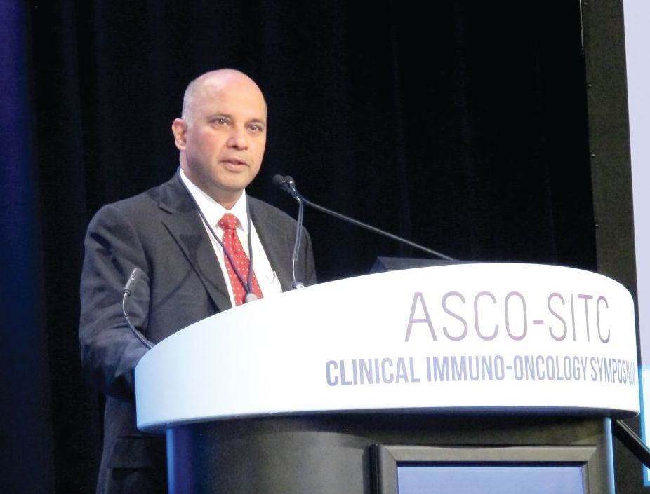
Merkel cell carcinoma, for example, has a high response rate despite its relatively low mutation burden. This is explained by the presence of infection with the Merkel cell polyomavirus in a low mutation burden subset, he said, noting that expression of the virus likely leads to expression of antigens that makes the disease both highly immunogenic and responsive to immune checkpoint therapy.
Expression of exogenous viruses is associated with response to immune checkpoint therapy in other cancers with low mutation burden, such as Epstein-Barr virus-positive gastric cancer, natural killer lymphoma, and Hodgkin disease.
“Intriguingly, renal cell carcinoma also has a relatively higher response rate to immune checkpoint therapy than would be anticipated by its relatively low mutation burden. However, this has no evidence of exogenous virus infection. Therefore, we turned our attention to endogenous retroviruses, which are an abundant potential stimulant of innate immunity in cancer,” he said.
Endogenous retroviruses (ERVs) now comprise about 5-8% of the human genome, and almost all of them have mutations that disable key coding genes of the retrovirus, meaning most cannot produce all the proteins necessary for viral replication.
“They’re essentially genomic fossils,” Dr. Ganesan said, adding that they all are silenced epigenetically in most normal adult tissues, both by DNA methylation and histone methylation. “However, inappropriate expression of endogenous retroviruses has been reported by multiple groups in some cancers and has been associated with evidence of immune activation. One can imagine that endogenous retroviral expression can be immunogenic both by expression of some [open reading frames] that are not disabled ... or just the viral RNA itself, which can be detected by cytoplasmic sensors ... and activate innate immunity,” he said.
To assess whether ERV expression corresponds with evidence of immune checkpoint activation in cancer, he and his colleagues conducted a pan-cancer analysis of more than 4,900 tumors across 21 cancer types using data from the Cancer Genome Atlas (TCGA).
“When we did this for about 66 annotated endogenous retroviruses in the TCGA database, we could see that different cancers had different amounts of retrovirus associated with immune activation ... in fact the strongest signal seen in this analysis was in clear cell renal cell carcinoma,” he said.
Signals were also seen in estrogen receptor–positive/human epidermal growth factor receptor 2-negative breast cancer, head and neck squamous cell carcinoma, and colon cancer.
A variety of ERVs were upregulated in RCC; different panels were associated with different cancer types. But two – ERVK.2 and ERV3.2 – were consistently upregulated across all the tumor types.
A closer look at ERV expression showed three clear clusters: extremely-high, intermediate, and low expression of ERV, and the very-high expression cluster had increased expression of numerous checkpoint genes, including CD8A, PD1, CTLA4, and LAG3 among others, compared with the low expression cluster.
This leads to the question of why a subset of RCCs have ERV expression.
In an attempt to answer that question, a transcript analysis was conducted to determine which transcripts are differentially regulated between the high and low ERV-expressing groups, and gene ontology analysis was performed.
“The results were quite striking. If you look at the modules that are differentially expressed between the high-ERV and the low-ERV groups, what pops up is really a lot of histone methyltransferase modules and chromatin regulation modules, ” he said, noting that this makes sense because of the silencing of ERV expression by histone methylation and DNA methylation, and suggests that “there is some deep abnormality in chromatin modulation in this subset of RCCs.
“This is intriguing, because RCCs are known to have mutations in chromatin modifying genes,” he said.
A look into whether the high-ERV–expressing group was enriched in any of these mutations showed some enrichment of BAP1 in the high- vs. low-expressing group, and that is currently being looked at further, he noted.
The next question was whether ERV expression correlated with response to immune checkpoint blockade in ccRCC, and this was looked at in a nonrandomized group of 15 patients with metastatic ccRCC who were treated with single-agent immune checkpoint therapy who had either clearly documented partial response or progressive disease. In 13 patient samples for which RNA expression of ERV3.2 was successfully measured by quantitative real-time polymerase chain reaction using two primer sets, ERV3.2 expression was significantly higher in responders vs. nonresponders in both primer sets (P less than .05 and .005).
“In summary, we have shown that expression of ERVs correlates with immune activation and increased expression of immune checkpoint genes in a subset of ccRCC, and perhaps several other solid tumor classes, and the expression of ERV3.2 is perhaps associated with response to PD1 blockade in this small preliminary cohort of ccRCC patients,” he said, noting that abnormal expression of ERV may be a biomarker of immune checkpoint therapy response in some cancers with a low mutation burden. “Mechanisms underlying ERV expression need to be investigated and may reflect underlying chromatin alterations or epigenetic abnormalities.”
Dr. Ganesan reported that his spouse is employed by Merck and that he is a consultant and/or advisory board member for Novartis, Roche, and Inspirata. He also holds patents with Inspirata.
sworcester@frontlinemedcom.com
SOURCE: Panda A et al., ASCO-SITC, Abstract #104.
REPORTING FROM THE CLINICAL IMMUNO-ONCOLOGY SYMPOSIUM
Key clinical point: ERV3.2 expression is associated with immune checkpoint blockade response in ccRCC.
Major finding: ERV3.2 expression was significantly higher in responders vs. nonresponders in two primer sets (P less than .05 and.005).
Study details: A pan-cancer analysis of more 4,900 tumors.
Disclosures: Dr. Ganesan reported that his spouse is employed by Merck and that he is a consultant and/or advisory board member for Novartis, Roche, and Inspirata. He also holds patents with Inspirata
Source: Panda A et al. ASCO-SITC, Abstract #104.
Prenatal betamethasone could save millions in care costs for women at risk of late preterm delivery
DALLAS – If betamethasone became a routine part of managing women at risk for late preterm birth, the U.S. health care system could save up to $200 million in direct costs every year.
A significant decrease in the cost of managing newborn respiratory morbidity would account for most of the savings, Cynthia Gyamfi-Bannerman, MD, said at the meeting sponsored by the Society for Maternal-Fetal Medicine. Not only was betamethasone treatment cost effective at its base price, but treatment retained economic dominance over no treatment even when its cost was inflated up to 500%.
She presented a cost analysis of the 2016 PARENT trial. PARENT randomized 2,831 women at risk of late preterm delivery to two injections of betamethasone or matching placebo 24 hours apart. Infants exposed to betamethasone were 20% less likely to experience the primary endpoint – a composite of the need for continuous positive airway pressure or high-flow nasal cannula, supplemental oxygen, extracorporeal membrane oxygenation, or mechanical ventilation. They were also significantly less likely to be stillborn or to die within 72 hours of delivery.
“We found that antenatal betamethasone significantly decreased perinatal morbidity and mortality,” she said. “However, the costs of this intervention were unknown. Therefore, we compared the cost-effectiveness of treatment with no treatment in these women.”
Dr. Gyamfi-Bannerman described the study as a cost-minimization analysis. “The analysis took a third-party payer approach with a time horizon to first hospital discharge to home.”
Maternal costs were based on Medicaid rates. These included the cost of the betamethasone, and any out- or inpatient visits necessary to administer the drug. The neonatal costs included all direct medical costs for the newborn, including neonatal ICU admissions and any respiratory therapy the infant required.
The analysis included all mother-infant pairs in PARENT who had at least one dose of their assigned study drug and full follow-up. This comprised 1,425 pairs who received betamethasone and 1,396 who received placebo. The total mean cost of maternal/newborn care in the betamethasone group was $4,774, compared with $5,473 in the placebo group – a significant difference.
The cost-effectiveness analysis, with effectiveness defined as the proportion not reaching the primary outcome, significantly favored betamethasone as well (88.4% vs. 85.5%).
Dr. Gyamfi-Bannerman also examined cost-effectiveness in a variety of pricing scenarios, from looking at betamethasone at its base cost with hospital and physician services priced at 50% of the current level, to betamethasone inflated by 500% and other costs by 200%.
“In almost every estimate, betamethasone remained the cost-effective management option,” she said. “ and was cost effective across most of our variable estimate.”
She had no financial disclosures.
SOURCE: Gyamfi-Bannerman C et al. Am J Obstet Gynecol. 2018;218:S14.
REPORTING FROM THE PREGNANCY MEETING
Key clinical point: If widely adopted, prenatal betamethasone could save up to $200 million per year by preventing neonatal respiratory morbidity.
Major finding: The total mean maternal/newborn care cost was $4,774 in the betamethasone group and $5,473 in the placebo group.
Study details: The cost analysis of the PARENT trial comprised 2,821 mother/infant pairs.
Disclosures: Dr. Gyamfi-Bannerman had no financial disclosures.
Source: Gyamfi-Bannerman C et al. Am J Obstet Gynecol. 2018;218:S14.
Hypertension Risk Found in Women with Migraine
Women with migraine have a higher relative risk of developing hypertension compared to women without migraine, according to a recent study. Researchers performed a prospective cohort study among 29,040 women without hypertension at baseline. Women were classified as having active migraine with aura, active migraine without aura, a past history of migraine, or no history of migraine. Incident hypertension was defined as new physician diagnosis or newly self-reported systolic or diastolic blood pressure ≥140 mmHg or ≥90 mmHg, respectively. They found:
- During a mean follow-up of 12.2 years, 15,176 incident hypertension cases occurred.
- Compared to those with no history of migraine, women who experience migraine with aura had a 9% increase in their risk of developing hypertension; women who experience migraine without aura had a 21% increase in their risk of developing hypertension; and women with a past history of migraine had a 15% increase in their risk of developing hypertension.
Migraine and the risk of incident hypertension among women. [Published online ahead of print February 1, 2018]. Cephalalgia. doi:10.1177/0333102418756865.
Promising outcomes of thrombolysis for caval extension of iliofemoral DVT
CHICAGO – Caval extension of an acute iliofemoral deep vein thrombosis paradoxically portends better treatment outcomes than does thrombolysis of a DVT without involvement of the inferior vena cava, according to Rabih A. Chaer, MD, professor of surgery at the University of Pittsburgh.
This finding from a retrospective analysis of the University of Pittsburgh experience might seem counterintuitive. After all, caval extension clearly indicates a greater clot burden. One possible explanation: Clearing a thrombus from a large vessel, such as the inferior vena cava (IVC), provides an added protective effect. Also, since the caval segments don’t have valves – their flow is based upon negative pressure in the chest – they may not contribute as much to postthrombotic morbidity to the same extent as do thrombosed iliofemoral segments, Dr. Chaer speculated at a symposium on vascular surgery sponsored by Northwestern University.
The impetus for Dr. Chaer and coinvestigators to review the Pittsburgh experience was a lack of clarity in the literature as to the effect IVC thrombosis has on thrombolysis outcomes in patients with acute iliofemoral DVT. Even though caval thrombus extension is present in up to 22% of patients with iliofemoral DVT, current guidelines issued by the American College of Chest Physicians, the American Heart Association, and the Society for Vascular Surgery don’t address the distinction between iliofemoral DVT with and without IVC extension in regard to the occurrence of postthrombotic syndrome (PTS), the most common complication of DVT.
The incidence of PTS in patients whose iliofemoral DVT is treated by anticoagulation and compression alone is up to 50%. Mounting evidence indicates that catheter-directed thrombolysis and pharmacomechanical thrombolysis aimed at achieving early thrombus removal and symptom relief help maintain valvular competence and reduce the risk of PTS, the surgeon noted.
PTS is diagnosed using the validated Villalta scale, which incorporates clinical signs including pain on calf compression, skin edema and redness, and ulcers, as well as symptoms such as leg cramping, heaviness, itching, and paresthesia.
The Pittsburgh series included 102 consecutive patients treated with various combinations of catheter-directed or pharmacomechanical thrombolysis in 127 limbs with acute iliofemoral thrombosis. In 46 patients, the thrombus extended into the IVC, all the way up to the renal veins in most cases.
The groups with and without caval extension were similar in terms of age and prevalence of malignancy, hypercoagulable state, and clot age. However, a history of previous DVT was significantly more common in the group with IVC thrombus. Also, more than 60% of patients with caval extension got an IVC filter, a rate more than 10-fold greater than that in patients without caval extension.
In this series, caval thrombosis had no effect on the technical success of thrombolysis. The technical success rate –defined as at least 50% clot lysis – was 89% in both groups. Rates of recurrent DVT within 30 days were similar in the two groups as well: 11% in the caval thrombosis group and 14% in the noncaval group. At 2 years postintervention, 77%-78% of patients in both groups remained free of DVT recurrence. The rate of PTS – defined by a Villalta score of 5 or more – at 2 years was 34% in the noncaval group, which was significantly higher than the 11% rate in patients with IVC thrombus extension. Ultrasound-identified valve reflux was present in 51% of the noncaval group at 2 years, compared with 51% of the noncaval group.
On multivariate analysis, incomplete clot lysis was associated with nearly a 23-fold increased risk of recurrent DVT and a 5.6-fold increased risk of PTS. Caval involvement was independently associated with a 78% reduction in PTS risk.
The Society for Vascular Surgery’s guidelines recommend pharmacomechanical thrombolysis over catheter-directed thrombolysis if the expertise is available. The Pittsburgh experience speaks to the worth of that recommendation.
“Pharmacomechanical techniques can be advantageous. They can expedite the lysis process by clearing most of the clot. In our series, 20 patients were treated with pharmacomechanical techniques in a single session,” Dr. Chaer noted.
The use of IVC filters in the setting of caval extension of iliofemoral DVT is controversial, according to the surgeon: A thrombus that gets trapped in the filter is tough to remove, precluding successful recanalization.
“One-third of the patients in our series got a filter, but we’ve become more conservative nowadays. We don’t use filters anymore. But I think those patients who might benefit from an IVC filter are those who present with a PE [pulmonary embolism], because that’s telling you they might develop another PE, as well as those patients in whom pharmacomechanical thrombolysis is anticipated because we’ve seen that those patients are also more likely to develop a PE,” he said.
The University of Pittsburgh study on the effect of IVC thrombus extension has been published (J Vasc Surg Venous Lymphat Disord. 2016 Oct;4[4]:385-91).
Dr. Chaer reported serving as a paid speaker for Boston Scientific.
SOURCE: Chaer RA. Northwestern Vascular Symposium 2017.
REPORTING FROM THE NORTHWESTERN VASCULAR SYMPOSIUM
Group Education Is Effective Migraine Intervention
Group education on headache evaluation and lifestyle management has potential as an effective, low-cost intervention for treatment of chronic headaches among adolescents, according to a recent study. This retrospective chart review study evaluated a group education program for 155 adolescents, aged 12-17 years, enrolled in the US military medical system with at least 3 months of chronic headaches and who were referred to a headache evaluation clinic. The primary outcome of the study was self-reported number of days with a headache in the previous 30 days based on patient recall. Researchers found:
- Most participants in the program were female (114/155 [73.5%]) and suffered from migraine headaches (108/155 [69.8%]).
- Severe headache-related disability was reported by 40.6% of subjects (63/155).
- Subjects reported an average of 19 days with headache during the previous 30 days.
- Participation in the group education was associated with an 11.5-day decrease in the frequency of headaches during the previous 30 days at follow-up at least 6 months after the class, with largest decline seen in patients with the highest level of migraine-related disability at baseline.
Group education and multidisciplinary management for chronic headaches among adolescents in a military treatment facility: A retrospective chart review. [Published online ahead of print February 7, 2018]. Headache. doi:10.1111/head.13274.
Splenic artery embolization increases risk of complications
LAKE BUENA VISTA, FLA. – are at higher risk of infectious complications and readmissions in the long term, according to a study presented at the annual scientific assembly of the Eastern Association for the Surgery of Trauma.
As nonoperative treatments are becoming more common for managing blunt splenic injury (BSI), it is important to understand the risks associated with splenic artery embolization (SAE) and how this treatment may be impacting a larger trend of posttrauma readmissions, according to presenter Rishi Rattan, MD, an acute care surgeon at the University of Miami.
The retrospective study included 37,986 BSI patients admitted into the National Readmissions Database from 2010 to 2014, treated with either nonoperative management (NOM), SAE, or operative management (OM).
Readmission rates for infection after 30 days were significantly higher among SAE (15.4%) and OM (21.9%) patients, compared with NOM patients (6.7%), according to Dr. Rattan. Patients who underwent SAE also had a 17.2% rate of infection after 1 year; significantly higher than the 8.1% of patients who underwent NOM, although less than the 23.2% of those who underwent OM.
For readmission due to organ surgical site infection, patients with SAE had a higher frequency at 30-day (2.9%) and 1-year (3.9%) readmission, compared with both NOM (1.3%, 1.7%) and OM (2.0%, 2.2%).
This can be particularly problematic as these organ surgical site infections, deep in the abdominal cavity around the splenic bed, are usually more complicated to manage, compared with a superficial infection, explained Dr. Rattan. Physiologically, it makes sense that having dead tissue left in the splenic bed could lead to a rise in infection, although more data are necessary to confirm that hypothesis.
SAE was a significant predictive factor for complications after BSI, increasing the odds of 30-day and 1-year readmission by 76% and 99%, respectively, from organ surgical site infection, compared with NOM (P less than .01). Other predictive factors included hospital stays longer than 4 days, not being discharged to home, and a Charlson Comorbidity index score greater than 1.
With an incidence rate of readmission among embolization patients at 30 days and 1 year double that of NOM, Dr. Rattan and fellow investigators suggest surgeons should be conscious of the risks of SAE and OM, especially as infection is a major case of morbidity after trauma in splenectomy patients.
The investigators reported no relevant financial disclosures.
REPORTING FROM EAST 2018
Key clinical point: Splenic artery embolization can increase risk of infectious complications in patients with blunt splenic injury.
Major finding: Patients who underwent splenic artery embolization had an infectious complication rate of 20% after 1 year.
Data source: Study of 37,986 blunt splenic injury patients gathered from the Nationwide Readmissions Database during 2010-2014.
Disclosures: Investigators reported no relevant financial disclosures.
Nearly half of MS patients treated by primary docs miss out on meds
SAN DIEGO – than are those treated by neurologists, even though they have more symptoms.
Nearly 85% of those treated at MS centers by neurologists received the drugs, compared with just 51% of those treated at primary care offices, according to results of a study reported at ACTRIMS Forum 2018, held by the Americas Committee for Treatment and Research in Multiple Sclerosis.
Dr. Halpern spoke in an interview at the ACTRIMS Forum.
The researchers analyzed data from the Sonya Slifka Longitudinal Multiple Sclerosis Study and focused on MS patients who received care at MS centers (376 patients, all treated by neurologists), neurology practices (552 patients), and primary care practices (55 patients).
In the three groups, most of the patients were female (77%-82%). To a statistically significant degree, those who were treated at primary care practices, compared with those at MS centers, were more likely to be white (98% vs. 82%), to have less than a college education (69% vs. 42%), and to have Medicaid/veteran coverage or be uninsured (22% vs. 11%).
In terms of rates of patients receiving disease-modifying therapies, there was a small difference between MS centers (84%) and neurology practices (79%); P less than .05.
However, the gap between these patients and those treated by primary care doctors was wide: Only 51% in the latter group received disease-modifying therapies, even though they reported more symptoms in areas such as vision, walking, bowel, speech, and numbness, compared with those in the other groups (P less than .05).
There was no statistically significant difference among the groups in reported symptoms of tremor, headache, pain, fatigue, cognition, swallowing, depression, mood, and anxiety.
The study doesn’t explain why the MS patients treated by primary care physicians are missing out on care, and it is not known whether the absence of treatment makes their conditions worse.
However, “it’s been well documented that the disease-modifying therapies can reduce the disease progression and the likelihood of experiencing relapses,” Dr. Halpern said. “Individuals with MS who are not being appropriately treated are more likely to experience symptoms, relapses, and faster disability.”
He speculated that primary care doctors may be falling behind on the treatment front because they lack the training and expertise to properly prescribe the MS medications, which are “difficult and complex drugs.”
Whatever the case, he said, there’s a clear need for more collaboration between MS subspecialists and primary care doctors.
The study was funded by the National MS Society. Dr. Halpern reported no relevant disclosures.
SOURCE: Halpern MT et al. ACTRIMS Forum 2018 Abstract P226.
REPORTING FROM ACTRIMS FORUM 2018
OSA may provide cardioprotection
, according to researchers.
In a study of 127 patients presenting with acute coronary syndromes (ACS), median peak cardiac troponin-I (cTn-I) values were significantly higher in patients without obstructive sleep apnea, compared with OSA patients (10.7; interquartile range: 1.78-40.1, vs. 3.79; IQR: 0.37-24.3, respectively; P = .04 ). The findings were published Feb. 5 in CHEST.
The study comprised 89 OSA patients and 38 non-OSA patients who were admitted to a hospital for acute coronary syndromes. The OSA group had a median apnea-hypopnea index (AHI) of 32, while the non-OSA group had a median AHI of 4.8. There was no significant difference between the two groups in gender, age, or cardiovascular risk factors such as hypertension, diabetes mellitus, body mass index, dyslipidemia, and smoking.
The cohort was part of the Continuous Positive Airway Pressure (CPAP) in Patients With Acute Coronary Syndrome and Obstructive Sleep Apnea (ISAACC) study, a prior randomized, controlled trial that evaluated the effect of CPAP treatment on new cardiovascular events in patients with an episode of ACS and OSA, reported Alicia Sánchez-de-la-Torre, PhD, of the respiratory department at Hospital Universitari Arnau de Vilanova and Santa Maria in Catalonia, Spain, and her coauthors.
Respiratory polygraphy was performed in the first 24-72 hours after hospital admission, and patients with an AHI of at least 15 events per hour were considered to have OSA. Those with an AHI less than 15 events per hour were included in the non-OSA group.
Blood samples were collected from patients every 6 hours until two consecutive cTn-I measurements showed a decrease, with the highest measurement considered the peak cTn-I value.
Peak cTn-I value was significantly higher in non-OSA patients than in OSA patients. Median infarct size, measured by calculating the area under the cTn-I curve, was significantly different between the two groups (451 for non-OSA patients vs. 143 in OSA patients; P = .049), wrote Dr. Sánchez-de-la-Torre and her colleagues.
As cTn-I levels decreased, there was a trend toward increased OSA severity (P = .058). In the multivariable linear regression model used to assess OSA severity, patients with severe OSA had 61% lower cTn-I levels than non-OSA patients, the authors noted.
“These results suggest that patients with higher AHI are significantly more likely to have low cTn-I levels than patients without evidence of OSA, which could imply that patients with elevated AHI, particularly those with severe OSA, may experience less severe myocardial injury,” the authors said in the report. The findings “suggest that OSA has a protective effect in the context of MI,” they added.
Limitations of the study include exclusion of patients with severe ACS, exclusion of sleepy subjects, and assessment of myocardial injury using cTn-I as a biomarker, without further data to determine infarct size.
“The possible role of OSA in cardioprotection should be explored in future studies,” the authors concluded.
The authors disclosed relationships with ResMed Inc., Spanish Ministry of Health, Spanish Respiratory Society, Catalonian Cardiology Society, and ALLER. No other disclosures were reported.
SOURCE: Chest. 2018 Feb 5;153[2]:329-38. doi: 10.1016/j.chest.2017.06.046
Although this study cannot definitively establish a clinically meaningful protective effect, it does provide important “preliminary evidence supporting the concept of OSA-induced cardioprotection” and challenges existing research, according to an editorial by Doron Aronson, MD, of the department of cardiology at Rambam Medical Center, Haifa, Israel, and coauthors (CHEST. 2018 Feb 153[2]:295-7. doi: 10.1016/j.chest.2017.07.036).
The results should be interpreted with caution, especially since accurate assessment of infarct size poses a challenge, they wrote.
“Myocardial infarct size is highly variable and is influenced by the duration of coronary occlusion, ST-segment elevation or non–ST elevation myocardial infarction, infarct location, residual antegrade infarct-related artery flow, collateral flow, the presence of non–culprit vessel coronary artery disease and myocardial metabolic demand,” they wrote. “Without accounting for these variables in a small study, results may be affected by variation in the characteristics of the patients.”
Though further study is needed, the findings may have “profound clinical implications regarding our therapeutic approach to patients with sleep apnea” if confirmed, the authors concluded.
, according to researchers.
In a study of 127 patients presenting with acute coronary syndromes (ACS), median peak cardiac troponin-I (cTn-I) values were significantly higher in patients without obstructive sleep apnea, compared with OSA patients (10.7; interquartile range: 1.78-40.1, vs. 3.79; IQR: 0.37-24.3, respectively; P = .04 ). The findings were published Feb. 5 in CHEST.
The study comprised 89 OSA patients and 38 non-OSA patients who were admitted to a hospital for acute coronary syndromes. The OSA group had a median apnea-hypopnea index (AHI) of 32, while the non-OSA group had a median AHI of 4.8. There was no significant difference between the two groups in gender, age, or cardiovascular risk factors such as hypertension, diabetes mellitus, body mass index, dyslipidemia, and smoking.
The cohort was part of the Continuous Positive Airway Pressure (CPAP) in Patients With Acute Coronary Syndrome and Obstructive Sleep Apnea (ISAACC) study, a prior randomized, controlled trial that evaluated the effect of CPAP treatment on new cardiovascular events in patients with an episode of ACS and OSA, reported Alicia Sánchez-de-la-Torre, PhD, of the respiratory department at Hospital Universitari Arnau de Vilanova and Santa Maria in Catalonia, Spain, and her coauthors.
Respiratory polygraphy was performed in the first 24-72 hours after hospital admission, and patients with an AHI of at least 15 events per hour were considered to have OSA. Those with an AHI less than 15 events per hour were included in the non-OSA group.
Blood samples were collected from patients every 6 hours until two consecutive cTn-I measurements showed a decrease, with the highest measurement considered the peak cTn-I value.
Peak cTn-I value was significantly higher in non-OSA patients than in OSA patients. Median infarct size, measured by calculating the area under the cTn-I curve, was significantly different between the two groups (451 for non-OSA patients vs. 143 in OSA patients; P = .049), wrote Dr. Sánchez-de-la-Torre and her colleagues.
As cTn-I levels decreased, there was a trend toward increased OSA severity (P = .058). In the multivariable linear regression model used to assess OSA severity, patients with severe OSA had 61% lower cTn-I levels than non-OSA patients, the authors noted.
“These results suggest that patients with higher AHI are significantly more likely to have low cTn-I levels than patients without evidence of OSA, which could imply that patients with elevated AHI, particularly those with severe OSA, may experience less severe myocardial injury,” the authors said in the report. The findings “suggest that OSA has a protective effect in the context of MI,” they added.
Limitations of the study include exclusion of patients with severe ACS, exclusion of sleepy subjects, and assessment of myocardial injury using cTn-I as a biomarker, without further data to determine infarct size.
“The possible role of OSA in cardioprotection should be explored in future studies,” the authors concluded.
The authors disclosed relationships with ResMed Inc., Spanish Ministry of Health, Spanish Respiratory Society, Catalonian Cardiology Society, and ALLER. No other disclosures were reported.
SOURCE: Chest. 2018 Feb 5;153[2]:329-38. doi: 10.1016/j.chest.2017.06.046
FROM CHEST
Key clinical point: Obstructive sleep apnea may have a protective effect in acute coronary syndromes.
Major finding: Median peak cTn-I value was significantly higher in patients without obstructive sleep apnea than in OSA patients (10.7, interquartile range: 1.78-40.1 vs. 3.79; IQR: 0.37-24.3 respectively; P = .04).
Data source: An observational study of 89 OSA and 38 non-OSA patients admitted for acute coronary syndromes.
Disclosures: The authors disclosed relationships with ResMed Inc., Spanish Ministry of Health, Spanish Respiratory Society, Catalonian Cardiology Society, and ALLER.
Source: Sánchez-de-la-Torre, A et al. CHEST. 2018 Feb 5;153[2]:329-38.
Multisite, same-day cryolipolysis treatments don’t skew lipids, liver enzymes
Multiple, same-day cryolipolysis treatments don’t adversely affect serum lipids or liver enzymes, either acutely or after 12 weeks.
Among all the lipids measured, only triglycerides showed a significant increase, jumping from a mean 77 mg/dL to 83.4 mg/dL. The just over 6 mg/dL increase was “clinically trivial” and driven by a single patient in the 35-subject study, Kenneth B. Klein, MD, and his colleagues reported in Lasers in Surgery and Medicine.
Results of the small, prospective study are reassuring, if not surprising, the authors wrote. Even four flank treatments, as performed in this cohort, release only about 160 g of fat, at a rate of less than 2 g/day. “To put this figure in perspective, the typical American diet contains at least 75 g of fat per day. Moreover, it has been shown that consuming as much as 261 grams of fat per day causes no ill effects or laboratory abnormalities.”
The study included 35 men and women with a mean age of 45 years but a range of 20-67 years. The mean body mass index was 24.7 kg/m2, although the range was quite wide, at 18-29.7 kg/m2. All subjects underwent cryolipolysis of the lower abdomen and both flanks. Blood was drawn for analysis at baseline and at weeks 1, 4, and 14 after treatment. One patient didn’t complete the treatment because it was impossible to draw enough flank tissue into the applicator.
Patients experienced the expected procedural side effects of erythema, numbness, and edema, with a few reports of tingling and bruising. All of these resolved spontaneously. Immediately after the procedure, the mean pain score was 4 on a 1-10 scale; by week 12, there were no reports of pain.
Other than the triglyceride change in the single patient, there were no statistically or clinically significant changes from baseline to week 12 in total cholesterol (mean 186.5 mg/dL vs. 189.2 mg/dL), HDL cholesterol (71 mg/dL vs. 73.8 mg/dL), LDL cholesterol (100 mg/dL vs. 102.5 mg/dL), or very low-density cholesterol (15.7 mg/dL vs. 16.8 mg/dL). Although the mean triglyceride change was statistically significant, the final measurement was still well below the upper limit of reference (150 mg/dL).
The only liver enzyme change of note occurred in one patient, who had one sharp increase in aminotransferase. That was related to alcohol consumption the night before the week 12 blood draw.
The study was sponsored by Zeltiq Aesthetics, which manufactures the device used in the trial. Dr. Klein is a consultant to Zeltiq. All three coauthors were paid investigators; in addition, one of them, Eric P. Bachelor, MD, is on the Zeltiq speakers’ bureau.
SOURCE: Klein KB et al. Lasers Surg Med. 2017 Sep;49(7):640-4.
FROM LASERS IN SURGERY AND MEDICINE
Key clinical point:
Major finding: There were no statistically or clinically significant changes from baseline to week 12 in total cholesterol (mean 186.5 mg/dL vs. 189.2 mg/dL), HDL cholesterol (71 mg/dL vs. 73.8 mg/dL), LDL cholesterol (100 mg/dL vs. 102.5 mg/dL), or VLDL cholesterol (15.7 mg/dL vs. 16.8 mg/dL).
Data source: A small prospective study of 35 men and women who underwent cryolipolysis of the lower abdomen and both flanks.
Disclosures: The study was sponsored by Zeltiq Aesthetics, which manufactures the device used in the trial. Dr. Klein is a consultant for Zeltiq. All three coauthors were paid investigators; in addition, one of them, Eric Bachelor, MD, is on the Zeltiq speakers’ bureau.
Source: Klein KB et al. Lasers Surg Med. 2017 Sep;49(7):640-4.
VIDEO: With tinea capitis, don’t be fooled by a Wood’s lamp
KAUAI, HAWAII – When it comes to diagnosing tinea capitis, you really can’t rely on a Wood’s lamp, according to pediatric dermatologist Robert Silverman, MD.
The Wood’s lamp generally misses the most common cause of tinea capitis in urban and suburban environments today, Trichophyton tonsurans, said Dr. Silverman, a pediatric dermatologist and clinical associate professor in the department of pediatrics at Georgetown University, Washington.
The dermatoscope, which “will allow you to see what is called the black dots of tinea capitis very, very closely,” is far better, he said in an interview at the Hawaii Dermatology Seminar provided by the Global Academy for Medical Education/Skin Disease Education Foundation
He explained the limitations of the Wood’s lamp in this setting, and what to look for with a dermatoscope. He had other tips to share, too, from years of experience treating the condition, including how to differentiate tinea capitis on exam from its most common mimics, why it’s best to include a counter stain when using potassium hydroxide, and how to motivate parents to wash their child’s hair more frequently to reduce spores on the hair and scalp.
Dr. Silverman disclosed relationships with Pierre Fabre, Pfizer, and Regeneron.
SDEF/Global Academy for Medical Education and this news organization are owned by the same parent company.
REPORTING FROM SDEF HAWAII DERMATOLOGY SEMINAR