User login
VIDEO: NOACs cut intracranial bleeds in real-world atrial fib patients
ROME – The new oral anticoagulants performed as advertised in a real-world, Danish registry of more than 40,000 patients with atrial fibrillation.
During the first year on anticoagulant treatment, patients who received a new oral anticoagulant (NOAC) had an ischemic stroke rate similar to that of patients who received the traditional oral anticoagulant, warfarin, but a significantly reduced rate of intracranial hemorrhage, Laila Stærk, MD, reported at the annual congress of the European Society of Cardiology.

These results “reinforce what we have seen in the clinical trials, but with the strength of looking in the entire Danish population,” said Dan Atar, MD, a cardiologist and professor of medicine at the University of Oslo.
“It is enlightening and very reassuring to have these real-world, unselected, registry data. They provide reassurance about safety and efficacy” when prescribing a NOAC, Dr. Atar said in an interview.
The study reported by Dr. Stærk and her associates included 43,299 Danish patients who were recently diagnosed with nonvalvular atrial fibrillation and started on treatment with an oral anticoagulant during the period August 2011 (when the first NOAC, dabigatran, became available for routine use in Denmark) through December 2015. During this period, 42% of these patients received warfarin, 29% received dabigatran (Pradaxa), 16% received apixaban (Eliquis) and 13% received rivaroxaban (Xarelto).

In a propensity-score type of analysis that controlled for baseline differences in clinical and demographic parameters, the results showed that the rate of ischemic stroke during the first year on treatment ranged from 2.0% to 2.5% in the four subgroups based on the anticoagulant received with no statistically-significant differences among the four subgroups. In other words, all three NOACs had efficacy profiles similar to those of warfarin, said Dr. Stærk, a cardiology researcher at Herlev and Gentofte University Hospitals in Hellerup, Denmark.
But on the safety side, all three NOACs were linked with lower rates of intracranial hemorrhages during the 1-year follow-up compared with the patients who received warfarin. In the cases of dabigatran and apixaban, the reduced intracranial hemorrhage rates were statistically significant, with a 0.6% rate among the patients on warfarin and rates that were reduced by a relative 34% for patients who received dabigatran and by a relative 20% among those on apixaban. Rivaroxaban linked with a 13% relative risk reduction in intracranial hemorrhage that was not statistically significant.
Dr. Atar said he would not make comparisons among the three NOACs based on these data, but rather interpreted the finding as showing that collectively the three NOACs assessed had comparable efficacy but better safety compared with warfarin.
He also noted that the Danish registry data document the transition that occurred during 2011 to 2015 in anticoagulant prescribing that shifted from warfarin to NOACs, with 57% of atrial fibrillation patients receiving a NOAC. In Norway, NOAC prescriptions for atrial fibrillation patients recently pulled ahead of warfarin prescription rates, Dr. Atar said. Reassuring data such as those in this report will help to further drive the shift from warfarin to NOACs, and he predicted that soon NOACs will be the anticoagulants used to treat the overwhelming majority of patients with nonvalvular atrial fibrillation.
Dr. Stærk has received research funding from Boehringer Ingelheim, the company that markets dabigatran (Pradaxa). Dr. Atar said that he has been a consultant to and has received research funding from several drug companies.
The video associated with this article is no longer available on this site. Please view all of our videos on the MDedge YouTube channel
On Twitter @mitchelzoler
ROME – The new oral anticoagulants performed as advertised in a real-world, Danish registry of more than 40,000 patients with atrial fibrillation.
During the first year on anticoagulant treatment, patients who received a new oral anticoagulant (NOAC) had an ischemic stroke rate similar to that of patients who received the traditional oral anticoagulant, warfarin, but a significantly reduced rate of intracranial hemorrhage, Laila Stærk, MD, reported at the annual congress of the European Society of Cardiology.
These results “reinforce what we have seen in the clinical trials, but with the strength of looking in the entire Danish population,” said Dan Atar, MD, a cardiologist and professor of medicine at the University of Oslo.
“It is enlightening and very reassuring to have these real-world, unselected, registry data. They provide reassurance about safety and efficacy” when prescribing a NOAC, Dr. Atar said in an interview.
The study reported by Dr. Stærk and her associates included 43,299 Danish patients who were recently diagnosed with nonvalvular atrial fibrillation and started on treatment with an oral anticoagulant during the period August 2011 (when the first NOAC, dabigatran, became available for routine use in Denmark) through December 2015. During this period, 42% of these patients received warfarin, 29% received dabigatran (Pradaxa), 16% received apixaban (Eliquis) and 13% received rivaroxaban (Xarelto).
In a propensity-score type of analysis that controlled for baseline differences in clinical and demographic parameters, the results showed that the rate of ischemic stroke during the first year on treatment ranged from 2.0% to 2.5% in the four subgroups based on the anticoagulant received with no statistically-significant differences among the four subgroups. In other words, all three NOACs had efficacy profiles similar to those of warfarin, said Dr. Stærk, a cardiology researcher at Herlev and Gentofte University Hospitals in Hellerup, Denmark.
But on the safety side, all three NOACs were linked with lower rates of intracranial hemorrhages during the 1-year follow-up compared with the patients who received warfarin. In the cases of dabigatran and apixaban, the reduced intracranial hemorrhage rates were statistically significant, with a 0.6% rate among the patients on warfarin and rates that were reduced by a relative 34% for patients who received dabigatran and by a relative 20% among those on apixaban. Rivaroxaban linked with a 13% relative risk reduction in intracranial hemorrhage that was not statistically significant.
Dr. Atar said he would not make comparisons among the three NOACs based on these data, but rather interpreted the finding as showing that collectively the three NOACs assessed had comparable efficacy but better safety compared with warfarin.
He also noted that the Danish registry data document the transition that occurred during 2011 to 2015 in anticoagulant prescribing that shifted from warfarin to NOACs, with 57% of atrial fibrillation patients receiving a NOAC. In Norway, NOAC prescriptions for atrial fibrillation patients recently pulled ahead of warfarin prescription rates, Dr. Atar said. Reassuring data such as those in this report will help to further drive the shift from warfarin to NOACs, and he predicted that soon NOACs will be the anticoagulants used to treat the overwhelming majority of patients with nonvalvular atrial fibrillation.
Dr. Stærk has received research funding from Boehringer Ingelheim, the company that markets dabigatran (Pradaxa). Dr. Atar said that he has been a consultant to and has received research funding from several drug companies.
The video associated with this article is no longer available on this site. Please view all of our videos on the MDedge YouTube channel
On Twitter @mitchelzoler
AT THE ESC CONGRESS 2016
Medical Student Guides Aspiring Physicians into Hospital Medicine
Editor’s note: As SHM celebrates the “Year of the Hospitalist,” we’re putting the spotlight on some of our most active members who are making substantial contributions to hospital medicine. You can get involved, too! Log on to www.hospitalmedicine.org/yoth for more information on how you can join the yearlong celebration and help SHM improve the care of hospitalized patients.

This month, The Hospitalist spotlights Aram Namavar, MS, a recipient of SHM’s Student Hospitalist Scholar Grant and a second-year medical student at Loyola University Chicago’s Stritch School of Medicine. Namavar is one of the first students to sit on an SHM committee as part of the Physicians in Training (PIT) Committee, which is focused on ensuring the successful transition of medical students and residents into the practice of hospital medicine. He recently started a hospital medicine special interest group at his medical school and is playing an active role in the launch of SHM’s new Students Community on the Hospital Medicine Exchange (HMX).
What piqued your interest in hospital medicine as you were choosing a specialty?

The laundry room was located right next to the training room, so I had the opportunity to meet the orthopedic surgeons and training staff. After spending some time in this program, I felt that there were aspects to a career in orthopedics that did not fit with my personality, such as what I felt was limited patient interaction and ability to impact multiple domains of their care outside of surgery. I pivoted and began volunteering at UCLA Health and was exposed to a plethora of medical specialties and patient populations. Through the self-discovery of my potential career path, I also discovered myself and my values. I realized that I desired flexibility and versatility in my career to engage in clinical, leadership, mentorship, research, education, and advocacy roles, all of which are possible in hospital medicine. I am now certain that my career in medicine will lead me to become an academic hospitalist.
Can you tell us about your quality improvement project that you initiated as a result of winning the SHM Student Hospitalist Scholar Grant?
This summer has been a very enriching experience for my personal and professional development. My main project is focused on patient-centered readmissions and has three components. I am promoting a patient-centered approach to readmissions through examination of the role that decisional conflict plays in hospital readmissions.
In shared decision-making models, decisional conflict is a measure of uncertainty, readiness, and comfort level in making a decision. Aware of the plight of diverse populations in accessing healthcare and having higher readmission rates, I have widened the scope of my project. A second arm of my study is investigating which social determinants of health may be a root cause for why Hispanic patients are being readmitted at an increased rate compared to their non-readmitted counterparts. The third arm of my study is elucidating patient-centered views of the cause and preventability of readmission for Hispanic versus non-Hispanic patients.
What inspired you to become involved with the PIT Committee?
Working with undergraduate and medical students and resident physicians, I have always had a passion for inspiring the future of medicine. Even in hospital medicine’s 20 years of existence, I knew there were many opportunities for me to engage trainees. For this reason, I applied for a seat on the PIT Committee and was accepted as the only medical student to sit on an SHM committee.
Having founded the first hospital medicine interest group at a medical school in the U.S. at Loyola, my work on the PIT Committee is focused on enhancing our ability to engage medical students and residents. I am a member of the Student Interest Group Task Force that is creating a toolkit for medical schools from across the nation to gain recognition from SHM for creating a hospital medicine interest group. The blueprint we used at Loyola is being adopted and incorporated in our toolkit. Another avenue of engagement is through the new Students Community on HMX, SHM’s online member engagement platform. This new community, which I helped to launch, will serve as an important channel for us to connect with trainees nationally and encourage them to consider hospital medicine as a specialty.
What opportunities do you see for hospitalists as the medical landscape continues to evolve?
I see hospitalists playing important roles in value-based care and population health. Hospitalists have carved their niche in quality improvement within health systems, and I believe they will be best suited for spearheading projects to enhance the value of medical care. Hospitalists will also no longer have an impact within the confines of a hospital as the landscape is shifting toward population health; therefore, hospitalists will be charged with helping to devise methods to proactively identify care gaps in their patients to promote preventive care and chronic disease management.
What would you tell medical students about choosing hospital medicine as a career?
I believe that a career in hospital medicine is one of the most versatile. If you are someone who must be continually stimulated by various work settings, then hospital medicine is right for you. I enjoy multidimensional work, and as a future academic hospitalist, I know that my scope of practice will include mentorship, education, research, leadership, and clinical duties. The ability to have such a versatile career will be extremely fulfilling.
What’s next for you in your medical career?
As a second-year medical student, my primary focus is on succeeding academically and adequately preparing for my USMLE Step 1 exam to match at my desired internal medicine residency program. Outside of my studies, I am continuing to develop my analytical skills and leadership acumen so that I can become a major player in hospital medicine once I am an academic hospitalist. After residency, I will be pursuing a fully employed MBA program where I will be equipped with the necessary skills to realize my professional goals. TH
Editor’s note: As SHM celebrates the “Year of the Hospitalist,” we’re putting the spotlight on some of our most active members who are making substantial contributions to hospital medicine. You can get involved, too! Log on to www.hospitalmedicine.org/yoth for more information on how you can join the yearlong celebration and help SHM improve the care of hospitalized patients.
This month, The Hospitalist spotlights Aram Namavar, MS, a recipient of SHM’s Student Hospitalist Scholar Grant and a second-year medical student at Loyola University Chicago’s Stritch School of Medicine. Namavar is one of the first students to sit on an SHM committee as part of the Physicians in Training (PIT) Committee, which is focused on ensuring the successful transition of medical students and residents into the practice of hospital medicine. He recently started a hospital medicine special interest group at his medical school and is playing an active role in the launch of SHM’s new Students Community on the Hospital Medicine Exchange (HMX).
What piqued your interest in hospital medicine as you were choosing a specialty?
The laundry room was located right next to the training room, so I had the opportunity to meet the orthopedic surgeons and training staff. After spending some time in this program, I felt that there were aspects to a career in orthopedics that did not fit with my personality, such as what I felt was limited patient interaction and ability to impact multiple domains of their care outside of surgery. I pivoted and began volunteering at UCLA Health and was exposed to a plethora of medical specialties and patient populations. Through the self-discovery of my potential career path, I also discovered myself and my values. I realized that I desired flexibility and versatility in my career to engage in clinical, leadership, mentorship, research, education, and advocacy roles, all of which are possible in hospital medicine. I am now certain that my career in medicine will lead me to become an academic hospitalist.
Can you tell us about your quality improvement project that you initiated as a result of winning the SHM Student Hospitalist Scholar Grant?
This summer has been a very enriching experience for my personal and professional development. My main project is focused on patient-centered readmissions and has three components. I am promoting a patient-centered approach to readmissions through examination of the role that decisional conflict plays in hospital readmissions.
In shared decision-making models, decisional conflict is a measure of uncertainty, readiness, and comfort level in making a decision. Aware of the plight of diverse populations in accessing healthcare and having higher readmission rates, I have widened the scope of my project. A second arm of my study is investigating which social determinants of health may be a root cause for why Hispanic patients are being readmitted at an increased rate compared to their non-readmitted counterparts. The third arm of my study is elucidating patient-centered views of the cause and preventability of readmission for Hispanic versus non-Hispanic patients.
What inspired you to become involved with the PIT Committee?
Working with undergraduate and medical students and resident physicians, I have always had a passion for inspiring the future of medicine. Even in hospital medicine’s 20 years of existence, I knew there were many opportunities for me to engage trainees. For this reason, I applied for a seat on the PIT Committee and was accepted as the only medical student to sit on an SHM committee.
Having founded the first hospital medicine interest group at a medical school in the U.S. at Loyola, my work on the PIT Committee is focused on enhancing our ability to engage medical students and residents. I am a member of the Student Interest Group Task Force that is creating a toolkit for medical schools from across the nation to gain recognition from SHM for creating a hospital medicine interest group. The blueprint we used at Loyola is being adopted and incorporated in our toolkit. Another avenue of engagement is through the new Students Community on HMX, SHM’s online member engagement platform. This new community, which I helped to launch, will serve as an important channel for us to connect with trainees nationally and encourage them to consider hospital medicine as a specialty.
What opportunities do you see for hospitalists as the medical landscape continues to evolve?
I see hospitalists playing important roles in value-based care and population health. Hospitalists have carved their niche in quality improvement within health systems, and I believe they will be best suited for spearheading projects to enhance the value of medical care. Hospitalists will also no longer have an impact within the confines of a hospital as the landscape is shifting toward population health; therefore, hospitalists will be charged with helping to devise methods to proactively identify care gaps in their patients to promote preventive care and chronic disease management.
What would you tell medical students about choosing hospital medicine as a career?
I believe that a career in hospital medicine is one of the most versatile. If you are someone who must be continually stimulated by various work settings, then hospital medicine is right for you. I enjoy multidimensional work, and as a future academic hospitalist, I know that my scope of practice will include mentorship, education, research, leadership, and clinical duties. The ability to have such a versatile career will be extremely fulfilling.
What’s next for you in your medical career?
As a second-year medical student, my primary focus is on succeeding academically and adequately preparing for my USMLE Step 1 exam to match at my desired internal medicine residency program. Outside of my studies, I am continuing to develop my analytical skills and leadership acumen so that I can become a major player in hospital medicine once I am an academic hospitalist. After residency, I will be pursuing a fully employed MBA program where I will be equipped with the necessary skills to realize my professional goals. TH

Discovery in mice may have implications for HSCT

Preclinical research has shown how a cell surface molecule, Lymphotoxin β receptor, controls the entry of T cells into the thymus.
Researchers believe this might represent a pathway that could be targeted to reboot the immune system after hematopoietic stem cell transplant (HSCT).
Graham Anderson, PhD, of the University of Birmingham in the UK, and his colleagues conducted this research and reported their findings in the Journal of Immunology.
“The thymus is often something of an ignored organ, but it plays a crucial role in maintaining an effective immune system,” Dr Anderson said.
“Post-transplantation, T-cell progenitors derived from the bone marrow transplant can struggle to enter the thymus, as if the doorway to the thymus is closed. Identifying molecular regulators that can ‘prop open’ the door and allow these cells to enter and mature could well be a means to help reboot the immune system.”
Conducting experiments in mice, Dr Anderson and his colleagues found that Lymphotoxin β receptor was required to allow the entry of T-cell progenitors to the thymus both in a healthy state and during immune recovery after HSCT.
When the researchers used an antibody to stimulate Lymphotoxin β receptor after HSCT, they observed enhanced thymus recovery and an increase in the number of transplant-derived T cells, which correlated with increased adhesion molecule expression by thymic stroma.
The team said this study has revealed a novel link between Lymphotoxin β receptor and thymic stromal cells in thymus colonization and highlights its potential as an immunotherapeutic target to boost T-cell reconstitution after HSCT.
“This is just one piece of the puzzle,” said study author Beth Lucas, PhD, also of the University of Birmingham.
“It may be that there are adverse effects to opening the door to the thymus, but identifying a pathway that regulates this process is a significant step.”
For their next step, the researchers plan to study in vitro samples of the human thymus to examine the role Lymphotoxin β receptor might play in regulating thymus function in humans. ![]()
Host RNA biosignatures distinguish bacterial from viral fever
RNA-expression biosignatures derived from the patient’s peripheral blood distinguish bacterial from viral causes of fever in young children, according to two separate preliminary studies published online Aug. 23 in JAMA.
Several studies have suggested that the source of infection in febrile children might be identified by examining the pattern of host genes that are either activated or suppressed during the body’s inflammatory response. Distinguishing the relatively few but potentially life-threatening bacterial infections from the more common but milder, self-resolving viral infections is difficult, and current practice is to admit “ill-appearing” febrile children to the hospital and administer parenteral antibiotics while awaiting the results of blood and tissue cultures. Those results are often ambiguous, and the whole process represents a large burden on health care resources as well as contributing to inappropriate antibiotic treatment.

Two multinational research groups developed different techniques for detecting RNA biosignatures in patients’ blood samples, then assessed the accuracy of those tests in validation cohorts. One group focused on ruling out bacterial infection as the source of fever in young children (median age, 19 months), while the other investigated whether the host responses of the youngest children (aged 60 days and younger), who have immature immune systems, are robust enough to allow detection of RNA biosignatures.
In the discovery phase of the first study, analysis of RNA gene expression was performed on blood samples obtained from 240 children at admission to hospitals in the United Kingdom, Spain, and the United States during a 4-year period. A total of 8,565 RNA transcript signatures were identified as potential biomarkers to discriminate between viral and bacterial infection. This was narrowed down to 38 transcript signatures, and then to only 2 – IFI44L and FAM89A – that were used to devise a Disease Risk Score (DRS) for each patient, said Jethro A. Herberg, PhD, of the division of infectious diseases, Imperial College London, and his associates.
IFI44L expression was increased in patients who had viral infection, while FAM89A expression was increased in those who had bacterial infection, as compared with healthy children. (In previous studies, IFI44L was reported to be up-regulated in interferon-mediated antiviral responses and FAM89A was reported to be elevated among children with septic shock.)
The DRS showed 90% sensitivity in distinguishing viral from bacterial infection in the discovery cohort. It then showed 96.4% sensitivity in a validation cohort of 130 febrile children (mean age, 17 months). The DRS also identified bacterial infection in a validation cohort of 24 children with meningococcal infection (91.7% sensitivity and 96.0% specificity), and distinguished it from inflammatory conditions in another cohort of 30 children with juvenile idiopathic arthritis and 18 with Henoch-Schönlein purpura (90.0% sensitivity and 95.8% specificity).
The DRS discriminated among viral, bacterial, and inflammatory diseases including systemic lupus erythematosus in a further validation cohort, a published dataset from children and adults who had all three types of illness. It was accurate regardless of the severity of infection and regardless of the duration of infection, as well as in cases where patients were coinfected with both virus and bacteria, the investigators said (JAMA. 2016 Aug 23. doi:10.1001/jama.2016.11236).
“The DRS signature, distinguishing viral from bacterial infections with only two transcripts, has potential to be translated into a clinically applicable test using current technology. Furthermore, new methods for rapid detection of nucleic acids, including nanoparticles and electrical impedance, have potential for low-cost, rapid analysis of multitranscript signatures,” Dr. Herberg and his associates noted.
Further research is needed to assess the accuracy and clinical utility of this technique in different settings, they added.
In the second study, RNA gene expression was analyzed from blood samples from 1,883 febrile infants (median age, 37 days) “who posed diagnostic quandaries” at admission to 22 emergency departments during a 2-year period. The discovery phase involved 89 of these infants who ultimately were found to have bacterial infections (bacteremia or UTIs), 190 who didn’t have bacterial infections (enterovirus, influenza, or other viruses), and 19 healthy control infants, said Prashant Mahajan, MD, division chief and research director, pediatric emergency medicine, Children’s Hospital of Michigan, Detroit, and his associates.

The investigators identified 3,753 RNA transcript signatures that could potentially identify or rule out bacterial sources of infection, which they then narrowed down to 66. This set of 66 signatures showed 82% sensitivity and 88% specificity in the discovery cohort and 87% sensitivity and 89% specificity in a validation cohort.
“The bacterial RNA biosignature was notably more predictive of bacterial infection than clinical examination” and use of the Yale Observation Score, and it “added significantly to prediction beyond the YOS alone,” Dr. Mahajan and his associates said (JAMA. 2016 Aug 23. doi: 10.1001/jama.2016.9207).
“Despite the young age of the febrile infants evaluated, they carried robust RNA biosignatures and demonstrated that regardless of the etiology of the infections, their immune systems are programed to respond not only with shared elements induced by common microbes but also with specific patterns that allow discrimination by class of pathogen,” they noted.
Further research is needed to confirm and refine these preliminary results. “As technology advances, RNA biosignatures may prove to be an alternative and accurate method to identify infants with bacterial infections. This would help clinicians target evaluation and therapy when they are needed and avoid invasive procedures, antibiotics, and hospitalizations when they are not,” Dr. Mahajan and his associates said.
Dr. Herberg’s study was supported by the Imperial College Comprehensive Biomedical Research Center, the National Institutes of Health, the European Union’s Seventh Framework Program, and numerous other groups. Dr. Herberg reported having no relevant financial disclosures. Dr. Mahajan’s study was supported by the Health Resources and Services Administration, Emergency Services for Children, the Eunice Kennedy Shriver National Institute of Child Health and Human Development, and the National Institutes of Health. Dr. Mahajan reported having no relevant financial disclosures.
The work by Herberg et al. and Mahajan et al. represents an important advance: the potential of genetics to help in the evaluation of febrile children.
 |
Dr. Howard Bauchner |
Clearly RNA sequencing and other methods for RNA quantification are in the early days, and clinical applications must await further replication and refinement of these results in rigorous studies. But the day may soon arise when a parent of a febrile child may do a laboratory test at home, call a physician, and mutually decide whether the child should be seen for further evaluation.
Howard Bauchner, MD, is JAMA Editor in Chief. He reported having no relevant financial disclosures. Dr. Bauchner made these remarks in an editorial accompanying the two reports on RNA biosignatures (JAMA 2016;316:824-5).
The work by Herberg et al. and Mahajan et al. represents an important advance: the potential of genetics to help in the evaluation of febrile children.
|
Dr. Howard Bauchner |
Clearly RNA sequencing and other methods for RNA quantification are in the early days, and clinical applications must await further replication and refinement of these results in rigorous studies. But the day may soon arise when a parent of a febrile child may do a laboratory test at home, call a physician, and mutually decide whether the child should be seen for further evaluation.
Howard Bauchner, MD, is JAMA Editor in Chief. He reported having no relevant financial disclosures. Dr. Bauchner made these remarks in an editorial accompanying the two reports on RNA biosignatures (JAMA 2016;316:824-5).
RNA-expression biosignatures derived from the patient’s peripheral blood distinguish bacterial from viral causes of fever in young children, according to two separate preliminary studies published online Aug. 23 in JAMA.
Several studies have suggested that the source of infection in febrile children might be identified by examining the pattern of host genes that are either activated or suppressed during the body’s inflammatory response. Distinguishing the relatively few but potentially life-threatening bacterial infections from the more common but milder, self-resolving viral infections is difficult, and current practice is to admit “ill-appearing” febrile children to the hospital and administer parenteral antibiotics while awaiting the results of blood and tissue cultures. Those results are often ambiguous, and the whole process represents a large burden on health care resources as well as contributing to inappropriate antibiotic treatment.
Two multinational research groups developed different techniques for detecting RNA biosignatures in patients’ blood samples, then assessed the accuracy of those tests in validation cohorts. One group focused on ruling out bacterial infection as the source of fever in young children (median age, 19 months), while the other investigated whether the host responses of the youngest children (aged 60 days and younger), who have immature immune systems, are robust enough to allow detection of RNA biosignatures.
In the discovery phase of the first study, analysis of RNA gene expression was performed on blood samples obtained from 240 children at admission to hospitals in the United Kingdom, Spain, and the United States during a 4-year period. A total of 8,565 RNA transcript signatures were identified as potential biomarkers to discriminate between viral and bacterial infection. This was narrowed down to 38 transcript signatures, and then to only 2 – IFI44L and FAM89A – that were used to devise a Disease Risk Score (DRS) for each patient, said Jethro A. Herberg, PhD, of the division of infectious diseases, Imperial College London, and his associates.
IFI44L expression was increased in patients who had viral infection, while FAM89A expression was increased in those who had bacterial infection, as compared with healthy children. (In previous studies, IFI44L was reported to be up-regulated in interferon-mediated antiviral responses and FAM89A was reported to be elevated among children with septic shock.)
The DRS showed 90% sensitivity in distinguishing viral from bacterial infection in the discovery cohort. It then showed 96.4% sensitivity in a validation cohort of 130 febrile children (mean age, 17 months). The DRS also identified bacterial infection in a validation cohort of 24 children with meningococcal infection (91.7% sensitivity and 96.0% specificity), and distinguished it from inflammatory conditions in another cohort of 30 children with juvenile idiopathic arthritis and 18 with Henoch-Schönlein purpura (90.0% sensitivity and 95.8% specificity).
The DRS discriminated among viral, bacterial, and inflammatory diseases including systemic lupus erythematosus in a further validation cohort, a published dataset from children and adults who had all three types of illness. It was accurate regardless of the severity of infection and regardless of the duration of infection, as well as in cases where patients were coinfected with both virus and bacteria, the investigators said (JAMA. 2016 Aug 23. doi:10.1001/jama.2016.11236).
“The DRS signature, distinguishing viral from bacterial infections with only two transcripts, has potential to be translated into a clinically applicable test using current technology. Furthermore, new methods for rapid detection of nucleic acids, including nanoparticles and electrical impedance, have potential for low-cost, rapid analysis of multitranscript signatures,” Dr. Herberg and his associates noted.
Further research is needed to assess the accuracy and clinical utility of this technique in different settings, they added.
In the second study, RNA gene expression was analyzed from blood samples from 1,883 febrile infants (median age, 37 days) “who posed diagnostic quandaries” at admission to 22 emergency departments during a 2-year period. The discovery phase involved 89 of these infants who ultimately were found to have bacterial infections (bacteremia or UTIs), 190 who didn’t have bacterial infections (enterovirus, influenza, or other viruses), and 19 healthy control infants, said Prashant Mahajan, MD, division chief and research director, pediatric emergency medicine, Children’s Hospital of Michigan, Detroit, and his associates.
The investigators identified 3,753 RNA transcript signatures that could potentially identify or rule out bacterial sources of infection, which they then narrowed down to 66. This set of 66 signatures showed 82% sensitivity and 88% specificity in the discovery cohort and 87% sensitivity and 89% specificity in a validation cohort.
“The bacterial RNA biosignature was notably more predictive of bacterial infection than clinical examination” and use of the Yale Observation Score, and it “added significantly to prediction beyond the YOS alone,” Dr. Mahajan and his associates said (JAMA. 2016 Aug 23. doi: 10.1001/jama.2016.9207).
“Despite the young age of the febrile infants evaluated, they carried robust RNA biosignatures and demonstrated that regardless of the etiology of the infections, their immune systems are programed to respond not only with shared elements induced by common microbes but also with specific patterns that allow discrimination by class of pathogen,” they noted.
Further research is needed to confirm and refine these preliminary results. “As technology advances, RNA biosignatures may prove to be an alternative and accurate method to identify infants with bacterial infections. This would help clinicians target evaluation and therapy when they are needed and avoid invasive procedures, antibiotics, and hospitalizations when they are not,” Dr. Mahajan and his associates said.
Dr. Herberg’s study was supported by the Imperial College Comprehensive Biomedical Research Center, the National Institutes of Health, the European Union’s Seventh Framework Program, and numerous other groups. Dr. Herberg reported having no relevant financial disclosures. Dr. Mahajan’s study was supported by the Health Resources and Services Administration, Emergency Services for Children, the Eunice Kennedy Shriver National Institute of Child Health and Human Development, and the National Institutes of Health. Dr. Mahajan reported having no relevant financial disclosures.
FROM JAMA
Key clinical point: RNA biosignatures derived from the patient’s peripheral blood distinguish bacterial from viral causes of fever in young children.
Major finding: Study 1. The DRS showed 96.4% sensitivity in a validation cohort of 130 febrile children. Study 2. A set of 66 RNA transcript signatures showed 82% sensitivity and 88% specificity in the discovery cohort and 87% sensitivity and 89% specificity in a validation cohort.
Data source: Two separate preliminary studies developing and validating tests of host responses to infection, involving 240 and 279 patients, respectively.
Disclosures: Dr. Herberg’s study was supported by the Imperial College Comprehensive Biomedical Research Center, the National Institutes of Health, the European Union’s Seventh Framework Program, and numerous other groups. Dr. Herberg reported having no relevant financial disclosures. Dr. Mahajan’s study was supported by the Health Resources and Services Administration, Emergency Services for Children, the Eunice Kennedy Shriver National Institute of Child Health and Human Development, and the National Institutes of Health. Dr. Mahajan reported having no relevant financial disclosures.

Treatment may allow HSCT without radiation, chemotherapy
A new therapy combining an anti-c-Kit monoclonal antibody with a CD47 blocker allowed hematopoietic stem cell engraftment in immunocompetent mice without the need for toxic preconditioning using radiation or chemotherapy, according to a report published in Science Translational Medicine.
Until now, hematopoietic stem cell transplantation has required rigorous conditioning regimens to clear out the host’s bone marrow, which can cause lifelong complications. So the procedure has been reserved for patients whose life-threatening disorders justified such toxicity. “Safer and more targeted conditioning protocols could both improve the safety of transplantation and extend the existing clinical utility of this powerful form of cell therapy,” said Akanksha Chhabra, PhD, of the department of blood and marrow transplantation, Stanford (Calif.) University, and her associates.
They assessed the new combined treatment in a series of laboratory and mouse studies. The opsonizing anti-c-Kit monoclonal antibodies induced robust depletion of functional hematopoietic stem cells in immunocompetent mice, which allowed donor stem cells to engraft in these hosts. Adding the T-cell–depleting CD47-antagonists further facilitated immune ablation of host stem cells and progenitor cells. Combined, the two agents eliminated more than 99% of host hematopoietic stem cells in the bone marrow and enabled strong engraftment of the donor stem cells, while avoiding radiation- and chemotherapy-related adverse effects.
The main toxicities that occurred in treated mice were, as expected, reductions in hematologic parameters, especially red blood cell indices. This may be related to a factor in mouse physiology that is not present in humans. But if such toxicities do develop in human subjects, they can be mitigated by careful monitoring and occasional supportive transfusions, Dr. Chhabra and her associates said (Sci Transl Med. 2016;8:351ra105).
These two types of antibodies are already being investigated separately in early-phase clinical trials. If the combined treatment proves effective and safe in humans – a question that awaits further clinical studies – hematopoietic stem cell transplantation might be extended to nonmalignant conditions such as inherited immunodeficiency, inborn errors of metabolism, and hemoglobinopathies. It might also be adapted for use in solid-organ transplants, the researchers added.
This work was supported by the Virginia and D.K. Ludwig Fund for Cancer Research and several other nonprofit organizations, the California Institute for Regenerative Medicine, and the National Institutes of Health. Dr. Chhabra is a coinventor on a patent described in this article, and her associates are cofounders of Forty Seven, the company that licensed the technology for radiation- and chemotherapy-free stem-cell transplantation. Two associates also serve as advisors for Alexo Therapeutics, which develops CD47-based treatments.
FROM SCIENCE TRANSLATIONAL MEDICINE
Key clinical point: A new treatment allowed hematopoietic stem cell engraftment in immunocompetent mice without the need for toxic preconditioning using radiation or chemotherapy.
Major finding: The combined therapy eliminated more than 99% of host hematopoietic stem cells.
Data source: A series of laboratory and mouse studies of combined treatment with anti-c-Kit monoclonal antibodies plus CD47 blockers.
Disclosures: This work was supported by the Virginia and D.K. Ludwig Fund for Cancer Research and several other nonprofit organizations, the California Institute for Regenerative Medicine, and the National Institutes of Health. Dr. Chhabra is a coinventor on a patent described in this article, and her associates are cofounders of Forty Seven, the company that licensed the technology for radiation- and chemotherapy-free stem-cell transplantation. Two associates also serve as advisors for Alexo Therapeutics, which develops CD47-based treatments.
Hemophilia carriers are at risk for abnormal bleeding
ORLANDO – The traditional view that women who are hemophilia carriers are unaffected by the disease may not be accurate, because many hemophilia carriers experience abnormal bleeding, a hemophilia specialist contends.
“We do know that hemophilia carriers have increased bleeding scores compared to controls,” said Michelle Sholzberg, MD, a hematologist and medical director of the coagulation laboratory at St. Michael’s Hospital in Toronto.

Bleeding in carriers is predominantly mucocutaneous bleeding, and may include epistaxis, heavy menstrual bleeding, bleeding with interventional procedures, and postpartum hemorrhage, she said at the World Federation of Hemophilia World Congress.
The majority of women who carry a factor VIII or factor IX mutation on one X chromosome have factor levels ranging from 0.40 to 0.60 IU/mL, which are generally considered to be adequate for hemostasis.
Yet, “we know that many carriers bleed, and in fact there are carriers who bleed with higher factor levels that are truly in the normal range,” Dr. Sholzberg said.
Normal coagulation factor levels in the general population range from about 0.50 to 1.50 IU/mL. By this standard, approximately 30% of hemophilia carriers have low factor levels, she said.
Joint damage
In addition, 14%-19% of hemophilia A carriers report hemarthrosis, and there is an association in factor VIII or IX deficiencies among hemophilia carriers and reduced joint range of motion. In one study, this decreased range of motion was evident as early as the preteen years, and was suggestive of subclinical musculoskeletal bleeding among carriers. In a separate study, investigators found evidence that hemophilia A carriers have pathologic and radiologic evidence of structural joint damage.
Variability in factor levels among carriers may be caused by lyonization (the inactivation of one of the X-chromosomes), ABO blood type, the presence of mutations in genes encoding for von Willebrand factor, compound heterozygosity or homozygosity, or Turner’s mosaicism.
Clotting factor levels can also change with pregnancy, with hemophilia A carriers experiencing an increase in mean factor VIII levels from 0.46 IU/mL prepregnancy to 1.21 IU/mL in the third trimester, and hemophilia B carriers having a corresponding rise in factor IX levels from 0.31 IU/mL to 0.48 IU/mL, and many carriers still have suboptimal factor levels at pregnancy, Dr. Sholzberg said.
Postpartum hemorrhagic complications among carriers can lead to iron-deficiency anemia. The Centers for Disease Control and Prevention recommends testing all nonpregnant women for anemia every 5-10 years throughout their childbearing years, and annual objective testing for women with risk factors, she noted.
“I think we can all appreciate now that the multidisciplinary approach is important for women with bleeding disorders, and carriers of hemophilia can be safely cared for at HTCs [hemophilia treatment centers],” she said.
Carriers should be treated with a multimodal approach that enhances patient education and awareness, with an emphasis on self-report of symptoms and communication with health care providers.
“It’s also critical never to start a conversation with a woman who has a bleeding disorder with ‘Do you have heavy menstrual bleeding?’ I think we all know that the answer is almost always ‘No, I don’t,’ and that’s because if she has bled heavily for her entire life, she doesn’t know what normal is,” Dr. Sholzberg said.
She described a typical comprehensive care plan for a pregnant hemophilia A carrier. The plan will include information about the diagnosis, recommendations to the obstetricians to avoid the use of invasive instrumentation, anesthesia recommendations, recommendations for care of the mother regarding hemostatic agents, postpartum tranexamic acid, and factor levels, and hematology recommendations for the newborn.
Dr. Sholzberg disclosed research support and/or honoraria from Shire (previously Baxter, Baxalta), Octapharma, CSL Behring, Pfizer, and Novo Nordisk, and advisory committee activity for Shire.
ORLANDO – The traditional view that women who are hemophilia carriers are unaffected by the disease may not be accurate, because many hemophilia carriers experience abnormal bleeding, a hemophilia specialist contends.
“We do know that hemophilia carriers have increased bleeding scores compared to controls,” said Michelle Sholzberg, MD, a hematologist and medical director of the coagulation laboratory at St. Michael’s Hospital in Toronto.
Bleeding in carriers is predominantly mucocutaneous bleeding, and may include epistaxis, heavy menstrual bleeding, bleeding with interventional procedures, and postpartum hemorrhage, she said at the World Federation of Hemophilia World Congress.
The majority of women who carry a factor VIII or factor IX mutation on one X chromosome have factor levels ranging from 0.40 to 0.60 IU/mL, which are generally considered to be adequate for hemostasis.
Yet, “we know that many carriers bleed, and in fact there are carriers who bleed with higher factor levels that are truly in the normal range,” Dr. Sholzberg said.
Normal coagulation factor levels in the general population range from about 0.50 to 1.50 IU/mL. By this standard, approximately 30% of hemophilia carriers have low factor levels, she said.
Joint damage
In addition, 14%-19% of hemophilia A carriers report hemarthrosis, and there is an association in factor VIII or IX deficiencies among hemophilia carriers and reduced joint range of motion. In one study, this decreased range of motion was evident as early as the preteen years, and was suggestive of subclinical musculoskeletal bleeding among carriers. In a separate study, investigators found evidence that hemophilia A carriers have pathologic and radiologic evidence of structural joint damage.
Variability in factor levels among carriers may be caused by lyonization (the inactivation of one of the X-chromosomes), ABO blood type, the presence of mutations in genes encoding for von Willebrand factor, compound heterozygosity or homozygosity, or Turner’s mosaicism.
Clotting factor levels can also change with pregnancy, with hemophilia A carriers experiencing an increase in mean factor VIII levels from 0.46 IU/mL prepregnancy to 1.21 IU/mL in the third trimester, and hemophilia B carriers having a corresponding rise in factor IX levels from 0.31 IU/mL to 0.48 IU/mL, and many carriers still have suboptimal factor levels at pregnancy, Dr. Sholzberg said.
Postpartum hemorrhagic complications among carriers can lead to iron-deficiency anemia. The Centers for Disease Control and Prevention recommends testing all nonpregnant women for anemia every 5-10 years throughout their childbearing years, and annual objective testing for women with risk factors, she noted.
“I think we can all appreciate now that the multidisciplinary approach is important for women with bleeding disorders, and carriers of hemophilia can be safely cared for at HTCs [hemophilia treatment centers],” she said.
Carriers should be treated with a multimodal approach that enhances patient education and awareness, with an emphasis on self-report of symptoms and communication with health care providers.
“It’s also critical never to start a conversation with a woman who has a bleeding disorder with ‘Do you have heavy menstrual bleeding?’ I think we all know that the answer is almost always ‘No, I don’t,’ and that’s because if she has bled heavily for her entire life, she doesn’t know what normal is,” Dr. Sholzberg said.
She described a typical comprehensive care plan for a pregnant hemophilia A carrier. The plan will include information about the diagnosis, recommendations to the obstetricians to avoid the use of invasive instrumentation, anesthesia recommendations, recommendations for care of the mother regarding hemostatic agents, postpartum tranexamic acid, and factor levels, and hematology recommendations for the newborn.
Dr. Sholzberg disclosed research support and/or honoraria from Shire (previously Baxter, Baxalta), Octapharma, CSL Behring, Pfizer, and Novo Nordisk, and advisory committee activity for Shire.
EXPERT ANALYSIS FROM WFH 2016 WORLD CONGRESS

NAF1 gene mutations predispose to pulmonary fibrosis, emphysema
Rare frameshift mutations in the NAF1 gene were discovered to cause a telomere-shortening syndrome which, among other adverse effects, predisposes carriers to develop pulmonary fibrosis (PF) and emphysema, according to a report published in Science Translational Medicine.
“Our findings here ... highlight how telomere shortening is a relevant mechanism for PF-emphysema susceptibility in a subset of patients beyond those with mutations in the telomerase core components. It is thus possible that efforts to reverse the telomere defect, or other regenerative approaches, will influence the natural history of these progressive pathologies in patients with telomere-mediated lung disease,” said Susan E. Stanley, an MD-PhD candidate in the department of oncology, Johns Hopkins University, Baltimore, and her associates.

Pulmonary fibrosis and emphysema cluster in some families, but the genetic basis of such cases is poorly understood. Both PF and emphysema have been linked to premature aging of lung tissue and to abnormalities in the maintenance of telomere length. In addition, at least half of patients with familial and sporadic PF, and many with emphysema, have the clinical features of a short-telomere syndrome, including bone marrow failure/myelodysplastic syndrome, liver disease, and infertility.
The diagnosis of a short-telomere syndrome, as opposed to isolated PF-emphysema, is essential for appropriate treatment because if the defect is systemic, patients will “show exquisite sensitivity to otherwise tolerated medications and procedures, especially in the setting of lung transplantation,” the investigators said (Sci Transl Med. 2016;8:351ra107).
To explore the genetic basis of familial PF-emphysema, the researchers performed a series of studies, beginning with whole-genome sequencing on peripheral blood samples from five unrelated probands in familial PF-emphysema pedigrees. These participants had abnormally short telomeres and extrapulmonary features of short-telomere syndrome. Three of them who had low levels of the telomerase RNA component TR were selected for a candidate gene search, which revealed the NAF1 mutations.
The mutations were then found to be present in 2 of 30 (7%) affected members of a prevalence cohort but in none of 134 unaffected control subjects (0%), and in none of 9,006 samples from a public database of unaffected people (0%). Further genetic laboratory and mouse studies were performed to link the mutations with specific pathologies and to trace their functional effects. Their results led the researchers to conclude that these rare NAF1 variants interfere with RNA biogenesis, causing short telomeres resulting in lung disease and other abnormalities.
This work was supported by the National Institutes of Health, the Commonwealth Foundation, and the American Cancer Society. Ms. Stanley and her associates reported having no relevant financial disclosures.
Rare frameshift mutations in the NAF1 gene were discovered to cause a telomere-shortening syndrome which, among other adverse effects, predisposes carriers to develop pulmonary fibrosis (PF) and emphysema, according to a report published in Science Translational Medicine.
“Our findings here ... highlight how telomere shortening is a relevant mechanism for PF-emphysema susceptibility in a subset of patients beyond those with mutations in the telomerase core components. It is thus possible that efforts to reverse the telomere defect, or other regenerative approaches, will influence the natural history of these progressive pathologies in patients with telomere-mediated lung disease,” said Susan E. Stanley, an MD-PhD candidate in the department of oncology, Johns Hopkins University, Baltimore, and her associates.
Pulmonary fibrosis and emphysema cluster in some families, but the genetic basis of such cases is poorly understood. Both PF and emphysema have been linked to premature aging of lung tissue and to abnormalities in the maintenance of telomere length. In addition, at least half of patients with familial and sporadic PF, and many with emphysema, have the clinical features of a short-telomere syndrome, including bone marrow failure/myelodysplastic syndrome, liver disease, and infertility.
The diagnosis of a short-telomere syndrome, as opposed to isolated PF-emphysema, is essential for appropriate treatment because if the defect is systemic, patients will “show exquisite sensitivity to otherwise tolerated medications and procedures, especially in the setting of lung transplantation,” the investigators said (Sci Transl Med. 2016;8:351ra107).
To explore the genetic basis of familial PF-emphysema, the researchers performed a series of studies, beginning with whole-genome sequencing on peripheral blood samples from five unrelated probands in familial PF-emphysema pedigrees. These participants had abnormally short telomeres and extrapulmonary features of short-telomere syndrome. Three of them who had low levels of the telomerase RNA component TR were selected for a candidate gene search, which revealed the NAF1 mutations.
The mutations were then found to be present in 2 of 30 (7%) affected members of a prevalence cohort but in none of 134 unaffected control subjects (0%), and in none of 9,006 samples from a public database of unaffected people (0%). Further genetic laboratory and mouse studies were performed to link the mutations with specific pathologies and to trace their functional effects. Their results led the researchers to conclude that these rare NAF1 variants interfere with RNA biogenesis, causing short telomeres resulting in lung disease and other abnormalities.
This work was supported by the National Institutes of Health, the Commonwealth Foundation, and the American Cancer Society. Ms. Stanley and her associates reported having no relevant financial disclosures.
FROM SCIENCE TRANSLATIONAL MEDICINE
Key clinical point: Certain rare mutations in the NAF1 gene were discovered to predispose carriers to develop pulmonary fibrosis and emphysema.
Major finding: The rare NAF1 mutations were detected in 2 of 30 (7%) family members in an affected pedigree but in 0 of 134 controls.
Data source: A series of genetic sequencing and other studies involving five affected probands, 30 unrelated but affected patients, and 134 control subjects.
Disclosures: This work was supported by the National Institutes of Health, the Commonwealth Foundation, and the American Cancer Society. Ms. Stanley and her associates reported having no relevant financial disclosures.
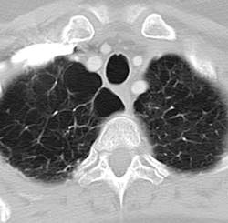
No VTE prophylaxis needed after joint surgery in patients with hemophilia
ORLANDO – In patients with hemophilia who have therapeutic factor levels at the time of joint replacement surgery, prophylaxis against venous thromboembolism (VTE) may be unnecessary.
In a cohort study of patients with hemophilia A or B who underwent total joint replacement surgery while being in proper hemostasis with therapeutic factor levels, there were no clinically evident episodes of venous thromboembolism, even though none of the patients had received perioperative anticoagulant prophylaxis, reported investigators from the National Hemophilia Center and Institute of Thrombosis and Hemostasis at the Sheba Medical Center in Tel Hashomer, Israel.
The data should be reassuring to clinicians whose patients with hemophilia require major orthopedic procedures, said lead author Dr. Anna Seltser, an orthopedic resident at Sheba Medical Center, in an interview.
“We have a lot of hemophilia patients who are not well treated because they live in the desert or distant communities, and we also sometimes treat patients from the Palestinian side of the Gaza Strip who don’t have access to care and need this type of surgery,” she said.
“We collected what I think is the biggest series of patients until now, we didn’t give any of them VTE prophylaxis, and none of them had any DVT [deep vein thrombosis], PE [pulmonary embolism], or similar complication,” she said.
Skip the heparin?
VTE prophylaxis with low-molecular-weight heparin, warfarin, or other anticoagulant agents is a common practice following orthopedic surgery in patients without bleeding disorders. But for patients with severe hemophilia, who often require major joint replacement surgery following years of bleeding-induced arthropathy, it’s unclear whether perioperative anticoagulation is beneficial, the investigators noted in a scientific poster at the World Federation of Hemophilia World Congress.
Dr. Seltser and colleagues therefore conducted a prospective cohort study of 50 patients with hemophilia A or B treated with major joint surgery and subsequent revisions from 1988 through 2015 at their center. In all, 47 patients had severe hemophilia A, 2 had mild hemophilia A, and 1 had hemophilia B.
The authors analyzed data on demographics, comorbidities, type of surgery, use of factor concentrates therapy around the time of surgery, and complications during follow-up, including massive hemorrhage, infections, implant loosening, DVT, and PE.
The patients underwent a total of 74 primary joint replacements (16 hips, 52 knees, and 6 ankles) and 23 revision surgeries.
As noted, there were no episodes of either DVT or PE among any of the patients. All but one complication occurred among patients undergoing total knee replacement. These included three cases of hemarthrosis, three limited-range-of-motion cases requiring closed manipulations, four soft-tissue hematomas, and one case each of superficial wound infection, urinary tract infection, pneumonia, and Candida infection of the tongue.
The only other complication was a case of disseminated intravascular coagulation, sepsis, and hemorrhagic shock in a patient who had undergone a revision (original procedure unspecified).
“Despite the concern that proper replacement factor therapy, applied before and after the surgery, may increase the risk for thromboembolic complications in patients with hemophilia undergoing joint replacement, our data show that prophylactic anticoagulation in this group of patients is not necessary,” the investigators concluded.
The study was internally funded. The investigators reported no conflicts of interest.
AT WFH 2016 WORLD CONGRESS
Key clinical point: Prophylaxis against thromboembolic events after orthopedic surgery in patients with hemophilia may not be necessary.
Major finding: There were no thromboembolic events after joint surgery without anticoagulant prophylaxis in patients with hemophilia A or B.
Data source: Cohort study of 50 patients with hemophilia A or B undergoing major joint replacement surgery.
Disclosures: The study was internally funded. The investigators reported no conflicts of interest.

CANDLE syndrome case highlights key features of this type 1 interferonopathy
BOSTON – Annular erythematous plaques in a child with fever and dense, atypical, mixed mononuclear and neutrophilic dermal infiltrate on biopsy could signal the recently described autoinflammatory disorder known as CANDLE Syndrome, according to Raegan Hunt, MD.
CANDLE, which stands for chronic atypical neutrophilic dermatosis with lipodystrophy and elevated temperature, is a proteasome-associated autoinflammatory syndrome characterized by dysregulation of type 1 interferon signaling, Dr. Hunt of Baylor College of Medicine and Texas Children’s Hospital said during a presentation at the American Academy of Dermatology summer meeting.
She described a case involving a 12-year-old girl with erythematous, warm, tender, nonpruritic papules and plaques on the superior chest, neck, and upper back. Most had an annular configuration with central clearing and occasionally central duskiness and ulceration.
The child also sometimes had violaceous swelling of the eyelid, nodules on the ear, and figurate erythematous plaques on the upper arm.
“She’s been having these since she was an infant in periodic bursts, and carried a diagnosis of annular erythema of infancy,” Dr. Hunt said.
Other symptoms included recurrent fevers, myalgias, transient and migratory arthritis, elevated C-reactive protein and erythrocyte sedimentation rate, and aseptic meningitis requiring hospitalization.
She had no family history of autoimmune disease, immune deficiency, or other genetic diseases, Dr. Hunt said.
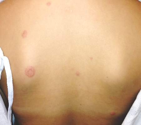
“She was treated with methotrexate and prednisone, as well as IVIG [intravenous immunoglobulin] every 4 weeks. Her prednisone was never weaned successfully below 0.8 mg/kg. She had complications of chronic corticosteroid disease, including many fractures,” she said.
A biopsy showed a dense infiltrate of atypical, mixed mononuclear and neutrophilic dermal infiltrate.
“These taken together are suggestive of ... CANDLE,” she said.
Genetic testing confirmed the presence of a compound heterozygous mutation in the proteasome subunit beta type 9, or PSMB8 gene.
Key features of CANDLE syndrome, first described in a 2010 article and further described by Liu, et al in 2012, include early disease onset, recurrent febrile episodes, skin lesions – including recurrent attacks of erythematous annular plaques and violaceous swelling of the eyelids, delayed physical development, progressive lipodystrophy, arthralgias, systemic inflammation, and aseptic meningitis episodes.
“Our patient did have very, very subtle lipodystrophy, but overall not as quick onset as some other [cases in children] that have been described,” Dr. Hunt said.
The type 1 interferonopathy is of autosomal recessive inheritance, and involves either homozygous or compound heterozygous mutations in PSMB8, as found in this patient. PSMB8 is an immunoproteasome unit involved in proteolysis and maintenance of cell homeostasis, she explained.
Treatment of the syndrome involves high-dose corticosteroids (usually 1-2 mg/kg day), which helps improve cutaneous eruptions, joint pain, and fever, but it is important to remember that disease flares can occur with tapering, Dr. Hunt noted.
Response is typically poor in patients treated with steroid-sparing agents, such as methotrexate, cyclosporine, azathioprine, or IVIG, as well as with tumor necrosis factor–alpha inhibitors and interleukin-1 receptor antagonists.
“But there is an interesting molecule on the horizon for possibly treating this more specifically and that’s JAK inhibitors,” she said, noting that a National Institutes of Health compassionate use clinical research trial is evaluating the JAK 1/2 inhibitor, baricitinib.
Dr. Hunt reported having no relevant disclosures.
BOSTON – Annular erythematous plaques in a child with fever and dense, atypical, mixed mononuclear and neutrophilic dermal infiltrate on biopsy could signal the recently described autoinflammatory disorder known as CANDLE Syndrome, according to Raegan Hunt, MD.
CANDLE, which stands for chronic atypical neutrophilic dermatosis with lipodystrophy and elevated temperature, is a proteasome-associated autoinflammatory syndrome characterized by dysregulation of type 1 interferon signaling, Dr. Hunt of Baylor College of Medicine and Texas Children’s Hospital said during a presentation at the American Academy of Dermatology summer meeting.
She described a case involving a 12-year-old girl with erythematous, warm, tender, nonpruritic papules and plaques on the superior chest, neck, and upper back. Most had an annular configuration with central clearing and occasionally central duskiness and ulceration.
The child also sometimes had violaceous swelling of the eyelid, nodules on the ear, and figurate erythematous plaques on the upper arm.
“She’s been having these since she was an infant in periodic bursts, and carried a diagnosis of annular erythema of infancy,” Dr. Hunt said.
Other symptoms included recurrent fevers, myalgias, transient and migratory arthritis, elevated C-reactive protein and erythrocyte sedimentation rate, and aseptic meningitis requiring hospitalization.
She had no family history of autoimmune disease, immune deficiency, or other genetic diseases, Dr. Hunt said.
“She was treated with methotrexate and prednisone, as well as IVIG [intravenous immunoglobulin] every 4 weeks. Her prednisone was never weaned successfully below 0.8 mg/kg. She had complications of chronic corticosteroid disease, including many fractures,” she said.
A biopsy showed a dense infiltrate of atypical, mixed mononuclear and neutrophilic dermal infiltrate.
“These taken together are suggestive of ... CANDLE,” she said.
Genetic testing confirmed the presence of a compound heterozygous mutation in the proteasome subunit beta type 9, or PSMB8 gene.
Key features of CANDLE syndrome, first described in a 2010 article and further described by Liu, et al in 2012, include early disease onset, recurrent febrile episodes, skin lesions – including recurrent attacks of erythematous annular plaques and violaceous swelling of the eyelids, delayed physical development, progressive lipodystrophy, arthralgias, systemic inflammation, and aseptic meningitis episodes.
“Our patient did have very, very subtle lipodystrophy, but overall not as quick onset as some other [cases in children] that have been described,” Dr. Hunt said.
The type 1 interferonopathy is of autosomal recessive inheritance, and involves either homozygous or compound heterozygous mutations in PSMB8, as found in this patient. PSMB8 is an immunoproteasome unit involved in proteolysis and maintenance of cell homeostasis, she explained.
Treatment of the syndrome involves high-dose corticosteroids (usually 1-2 mg/kg day), which helps improve cutaneous eruptions, joint pain, and fever, but it is important to remember that disease flares can occur with tapering, Dr. Hunt noted.
Response is typically poor in patients treated with steroid-sparing agents, such as methotrexate, cyclosporine, azathioprine, or IVIG, as well as with tumor necrosis factor–alpha inhibitors and interleukin-1 receptor antagonists.
“But there is an interesting molecule on the horizon for possibly treating this more specifically and that’s JAK inhibitors,” she said, noting that a National Institutes of Health compassionate use clinical research trial is evaluating the JAK 1/2 inhibitor, baricitinib.
Dr. Hunt reported having no relevant disclosures.
EXPERT ANALYSIS FROM THE AAD SUMMER ACADEMY 2016
Key clinical point: Annular erythematous plaques in a child with fever and dense, atypical, mixed mononuclear and neutrophilic dermal infiltrate on biopsy could signal the recently described autoinflammatory disorder known as CANDLE syndrome.
Major finding: Genetic testing confirmed the presence of a compound heterozygous mutation in the proteasome subunit beta type 9, or PSMB8 gene.
Data source: Expert analysis – case description.
Disclosures: Dr. Hunt reported having no disclosures.

Flu vaccine prevented hospitalizations in patients 50 and older
The seasonal influenza vaccination reduced flu-related hospitalizations by 56.8% among people aged 50 and older during a recent flu season, according to a report published in Clinical Infectious Diseases.
Even in the oldest age group – the population with the highest risk of developing flu complications and perhaps the weakest immune response – influenza vaccination prevented serious complications, said Fiona P. Havers, MD, of the influenza division, Centers for Disease Control and Prevention, Atlanta, and her associates.

Data on vaccine efficacy in older adults are sparse, and randomized, placebo-controlled trials to gather evidence would be unethical. Dr. Havers and her colleagues studied the issue using a case-control design, focusing on community-dwelling adults aged 50 years and older during the 2010-2011 flu season. They identified 368 patients across 10 states who were hospitalized for polymerase chain reaction–confirmed influenza and matched them for age and county of residence with 773 control subjects.
Hospitalized case-patients were less likely to have been vaccinated (55%) than were control subjects (63%). Thus, the flu vaccine reduced the risk of hospitalization for influenza by 56.8% overall.

Vaccination reduced hospitalization for influenza by 63.9% in the youngest age group (50-64 years), by 61.0% in the intermediate age group (65-74 years), and by 57.3% in the oldest age group (75 years and older).
These results are similar to those reported in other studies assessing the same time period, including one that evaluated vaccine efficacy in ambulatory adults in the United States and Europe. They also are consistent with the results of observational studies performed during different flu seasons, the investigators said (Clin Infect Dis. 2016 Aug 2. doi: 10.1093/cid/ciw512).
Compared with control subjects, case-patients were more likely to be of nonwhite race, to be of Hispanic ethnicity, to have a lower income, to have had fewer years of education, to have two or more chronic health conditions, to have required recent hospitalization for respiratory problems, to have impaired mobility, and to have lower functional status.
“These findings support current U.S. recommendations for annual influenza vaccination in older adults, especially in adults aged 65 and older who are at higher risk of influenza-associated complications,” Dr. Havers and her associates said.
The Centers for Disease Control and Prevention supported the study. Dr. Havers reported having no relevant financial disclosures; one of her associates reported ties to Genentech, Merck, Novavax, and Pfizer.
The seasonal influenza vaccination reduced flu-related hospitalizations by 56.8% among people aged 50 and older during a recent flu season, according to a report published in Clinical Infectious Diseases.
Even in the oldest age group – the population with the highest risk of developing flu complications and perhaps the weakest immune response – influenza vaccination prevented serious complications, said Fiona P. Havers, MD, of the influenza division, Centers for Disease Control and Prevention, Atlanta, and her associates.
Data on vaccine efficacy in older adults are sparse, and randomized, placebo-controlled trials to gather evidence would be unethical. Dr. Havers and her colleagues studied the issue using a case-control design, focusing on community-dwelling adults aged 50 years and older during the 2010-2011 flu season. They identified 368 patients across 10 states who were hospitalized for polymerase chain reaction–confirmed influenza and matched them for age and county of residence with 773 control subjects.
Hospitalized case-patients were less likely to have been vaccinated (55%) than were control subjects (63%). Thus, the flu vaccine reduced the risk of hospitalization for influenza by 56.8% overall.
Vaccination reduced hospitalization for influenza by 63.9% in the youngest age group (50-64 years), by 61.0% in the intermediate age group (65-74 years), and by 57.3% in the oldest age group (75 years and older).
These results are similar to those reported in other studies assessing the same time period, including one that evaluated vaccine efficacy in ambulatory adults in the United States and Europe. They also are consistent with the results of observational studies performed during different flu seasons, the investigators said (Clin Infect Dis. 2016 Aug 2. doi: 10.1093/cid/ciw512).
Compared with control subjects, case-patients were more likely to be of nonwhite race, to be of Hispanic ethnicity, to have a lower income, to have had fewer years of education, to have two or more chronic health conditions, to have required recent hospitalization for respiratory problems, to have impaired mobility, and to have lower functional status.
“These findings support current U.S. recommendations for annual influenza vaccination in older adults, especially in adults aged 65 and older who are at higher risk of influenza-associated complications,” Dr. Havers and her associates said.
The Centers for Disease Control and Prevention supported the study. Dr. Havers reported having no relevant financial disclosures; one of her associates reported ties to Genentech, Merck, Novavax, and Pfizer.
FROM CLINICAL INFECTIOUS DISEASES
Key clinical point: Seasonal influenza vaccination reduced flu-related hospitalizations by 56.8% in people aged 50 years and older.
Major finding: Vaccination reduced hospitalization for influenza by 63.9% in people aged 50-64 years, by 61.0% in those aged 65-74 years, and by 57.3% in those aged 75 years and older.
Data source: A retrospective case-control study involving 368 cases and 773 matched controls assessed during a single recent flu season.
Disclosures: The Centers for Disease Control and Prevention supported the study. Dr. Havers reported having no relevant financial disclosures; one of her associates reported ties to Genentech, Merck, Novavax, and Pfizer.